Standing Frontonasal Flap and Maxillary Sinus Trephination … PAPERS/JTAS Vol. 39 (1) Feb... ·...
10
Pertanika J. Trop. Agric. Sci. 39 (1): 117 - 126 (2016) ISSN: 1511-3701 © Universiti Putra Malaysia Press TROPICAL AGRICULTURAL SCIENCE Journal homepage: http://www.pertanika.upm.edu.my/ Article history: Received: 6 May 2014 Accepted: 28 September 2015 ARTICLE INFO E-mail addresses: [email protected], [email protected] (Khairuddin, N. H.), [email protected], [email protected] (Mimi Armiladiana, M.) * Corresponding author Case Report Standing Frontonasal Flap and Maxillary Sinus Trephination in a Horse with Sinusitis Khairuddin, N. H. 1 * and Mimi Armiladiana, M. 2 1 Department of Farm and Exotic Animal Medicine and Surgery, Faculty of Veterinary Medicine, Universiti Putra Malaysia, 43400 UPM Serdang, Selangor, Malaysia 2 Large Animal Unit, University Veterinary Hospital, Universiti Putra Malaysia, 43400 UPM Serdang, Selangor, Malaysia ABSTRACT This report describes use of a standing frontonasal bone flap (SFF) technique to treat a case of primary sinusitis affecting the right frontal and maxillary sinuses in a horse. The diagnosis was made based on a history of prolonged unilateral nasal discharge, endoscopy and radiographic findings. A frontonasal bone flap was created and maxillary sinus trephination was performed while the horse was standing and sedated. Standing sedation throughout the whole procedure was achieved through constant rate infusion (CRI) technique with detomidine hydrochloride. The outcome of this case confirmed that standing frontonasal flap surgery provides an effective method to specifically treat primary sinusitis in a horse and eliminates the risk of general anaesthesia in order to perform such invasive surgery. Keywords: Horse, sinusitis, frontonasal flap, standing surgery INTRODUCTION Paranasal sinuses of the horse are air-filled spaces within the skull. There are six pairs of paranasal sinuses: the dorsal, middle and ventral conchal sinus, the sphenopalatine sinus, the frontal sinus and the caudal and rostral maxillary sinuses. The caudal and rostral maxillary sinuses are separated by the maxillary septum and the rostral sinus opens into the ventral conchal sinus. The caudal maxillary sinus has communication with the frontal sinus and the sphenopalatine sinuses. Diseases that could affect the paranasal sinuses are sinusitis, empyema, paranasal sinus cyst, neoplasia and sinus
Transcript of Standing Frontonasal Flap and Maxillary Sinus Trephination … PAPERS/JTAS Vol. 39 (1) Feb... ·...

Pertanika J. Trop. Agric. Sci. 39 (1): 117 - 126 (2016)
ISSN: 1511-3701 © Universiti Putra Malaysia Press
TROPICAL AGRICULTURAL SCIENCEJournal homepage: http://www.pertanika.upm.edu.my/
Article history:Received: 6 May 2014Accepted: 28 September 2015
ARTICLE INFO
E-mail addresses: [email protected], [email protected] (Khairuddin, N. H.),[email protected], [email protected] (Mimi Armiladiana, M.)* Corresponding author
Case Report
Standing Frontonasal Flap and Maxillary Sinus Trephination in a Horse with Sinusitis
Khairuddin, N. H.1* and Mimi Armiladiana, M.2
1Department of Farm and Exotic Animal Medicine and Surgery, Faculty of Veterinary Medicine, Universiti Putra Malaysia, 43400 UPM Serdang, Selangor, Malaysia2Large Animal Unit, University Veterinary Hospital, Universiti Putra Malaysia, 43400 UPM Serdang, Selangor, Malaysia
ABSTRACT
This report describes use of a standing frontonasal bone flap (SFF) technique to treat a case of primary sinusitis affecting the right frontal and maxillary sinuses in a horse. The diagnosis was made based on a history of prolonged unilateral nasal discharge, endoscopy and radiographic findings. A frontonasal bone flap was created and maxillary sinus trephination was performed while the horse was standing and sedated. Standing sedation throughout the whole procedure was achieved through constant rate infusion (CRI) technique with detomidine hydrochloride. The outcome of this case confirmed that standing frontonasal flap surgery provides an effective method to specifically treat primary sinusitis in a horse and eliminates the risk of general anaesthesia in order to perform such invasive surgery.
Keywords: Horse, sinusitis, frontonasal flap, standing surgery
INTRODUCTION
Paranasal sinuses of the horse are air-filled spaces within the skull. There are six pairs of paranasal sinuses: the dorsal, middle and
ventral conchal sinus, the sphenopalatine sinus, the frontal sinus and the caudal and rostral maxillary sinuses. The caudal and rostral maxillary sinuses are separated by the maxillary septum and the rostral sinus opens into the ventral conchal sinus. The caudal maxillary sinus has communication with the frontal sinus and the sphenopalatine sinuses. Diseases that could affect the paranasal sinuses are sinusitis, empyema, paranasal sinus cyst, neoplasia and sinus

Khairuddin, N. H. and Mimi Armiladiana, M.
118 Pertanika J. Trop. Agric. Sci. 39 (1) 118 - 126 (2016)
trauma (Nickels, 2006). Clinically, the paranasal sinus becomes important due to its susceptibility to infections extending from the nasal cavity or from the upper root tooth infections leading to primary and secondary sinusitis. Primary sinusitis is usually caused by complications caused by Streptococcus species in acute or chronic respiratory disease (Ruggles et al., 1991). In a simple acute case of sinusitis, a course of antimicrobial therapy with or without sinus trephination and flushing of the paranasal sinuses may be sufficient to resolve the infection. Where there is little drainage from the sinuses with trephination and flushing and no response to treatment, with presence of inspissated purulent material in any sinus compartments, it is very unlikely the sinusitis is able to resolve with only medical management, and surgical treatment becomes necessary (Schumacher & Crossland, 1994; Nickels, 2006; Dixon et al., 2012).
In the past, exploratory surgery of the paranasal sinuses was performed through trephinated openings (Ruggles et al., 1993). Through this sinus portal, endoscopic examinations of the paranasal sinuses, as well as collection of sinus contents for bacteriologic, cytologic and histological examinations are able to be performed. However, adequate exposure for surgical manipulations through this small opening is limited (Ruggles et al., 1991), and examination of the rostral and ventral conchal sinus can be difficult. Hence, surgery of the paranasal sinuses through a large frontonasal bone flap technique is indicated to allow for optimum exposure of most parts of
the paranasal sinuses, including the rostral maxillary and ventral conchal sinuses and to provide a larger entrance for surgical manipulation as well as direct assessment of the sinuses.
Frontonasal bone flap surgery and bone trephination can be performed while the horse is under general anaesthesia (Freeman et al., 1990) or performed while the horse is standing and sedated (Schumacher et al., 2000; Schumacher & Perkins, 2005). Advances in techniques and improved agents for equine sedation and analgesia have allowed many procedures to be performed while the horse is standing, including surgery of the paranasal sinuses. The purpose of this report is to share our experience of Standing Frontonasal Flap (SFF) surgery to further investigate and treat primary sinusitis in a horse.
CASE DETAILS
History
Ishmael, a six-year old thoroughbred, gelding, weighing 450 kg, used for pleasure riding, was presented to the Equine Unit, Veterinary Hospital, Universiti Putra Malaysia, for treatment of prolonged unilateral nasal discharge. According to the caretaker of the horse, the nasal discharge was noted two to three weeks before a veterinarian was summoned.
Clinical Findings
On physical examination, the temperature, heart rate and respiratory rate were found to be normal. Purulent malodorous nasal

Standing Sinus Surgery in a Horse with Sinusitis
119Pertanika J. Trop. Agric. Sci. 39 (1): 119 - 126 (2016)
discharges could be seen draining from the right nostril (Fig.1). Auscultation of the lung and trachea revealed no abnormalities. A facial swelling at the region of the right side of the frontal bone was noted. Palpation of the facial protuberance did not elicit signs of discomfort or pain. However, percussion of the area over the right maxillary and frontal sinus revealed changes in resonance compared to the same region on the left side. Examination of the teeth and soft tissue structures in the oral cavity revealed no abnormal findings. Right-sided nasal endoscopy was then performed while the horse was standing and sedated (Detomidine 10 mg/ml). Purulent exudates draining from the nasomaxillary ostia of the maxillary sinus were observed. The ethmoids were
normal. Images from the endoscopic view were not able to be photographed due to technical limitations.
Radiography of the paranasal sinuses was performed. Right lateral, lateral oblique and dorsoventral radiographs of the paranasal sinuses were obtained while the horse was standing and sedated (Detomidine 0.01 mg/kg). On the lateral view (Fig.2), the ethmoid was found to be normal, but there were multiple fluid lines in both the frontal and maxillary sinuses. Thus, this finding supports the diagnosis of primary sinusitis in both sinus compartments. On the dorsoventral view (Fig.3), the fluid lines noted on lateral view were confined to the right side and a sign of soft tissue opacification was observed on the region
Fig.1: Purulent malodorous nasal discharge can be seen draining from the right nostril.

Khairuddin, N. H. and Mimi Armiladiana, M.
120 Pertanika J. Trop. Agric. Sci. 39 (1) 120 - 126 (2016)
Fig.2: Right lateral view of the paranasal sinuses. The ethmoid region was normal in radiographic appearance, but there are multiple fluid lines in both the frontal and maxillary sinuses.
Fig.3: Dorsoventral view of the paranasal sinuses. The fluid lines noted on lateral view were confined to the right side.

Standing Sinus Surgery in a Horse with Sinusitis
121Pertanika J. Trop. Agric. Sci. 39 (1): 121 - 126 (2016)
of the right maxillary sinus. A diagnosis of primary sinusitis affecting the right frontal and maxillary sinuses was made based on the endoscopy and radiographic findings. All haematological values were within normal limits.
Surgical Protocol
Standing Frontonasal Flap (SFF) surgery was the choice of treatment for this case. The horse was given analgesic drug of flunixin meglumin (2.2 mg/kg, intravenously), broad spectrum antibiotic of penicillin-streptomycin (22,000 U/kg of body weight, intramuscularly) and pethidine hydrochloride (0.5 mg/kg, intramuscularly) preoperatively. The horse was restrained in stocks for the surgical procedure. A 14-gauge intravascular jugular catheter was placed, and the horse was sedated using detomidine infusion. A detomidine constant rate infusion (CRI) was delivered using a 60 drop/ml microdrip administration set. Five ml of NaCl 0.9% was removed from the 500 ml bag and 5 ml of 10 mg/ml detomidine was added into the saline bag to produce 100 µg/ml of detomidine infusion. After intravenous administration of a bolus of detomidine (8.4 µg/kg), the drip rates were adjusted at the following rates: 150 drops/minute for the first 15 minutes, 90 drops/minute for the next 15 minutes and 45 drops/minute thereafter throughout the surgery.
The dorsal aspect of the affected side of the head was clipped and prepared for aseptic surgery. Local anaesthetic (Lidocaine 2%) was infused subcutaneously along the proposed incision line. The dorsal
midline of the face served as the base of the flap. The caudal margin of the flap was a perpendicular line from the dorsal midline to a point between supraorbital foramen and medial canthus of the eye. The lateral margin of the flap began from the caudal margin 2.0 to 2.5 cm medial to the medial canthus of the eye, and extended approximately 2/3 the distance from the medial canthus to the infraorbital foramen. For the rostral margin of the flap, the incision line was a perpendicular line from the dorsal midline to the rostral extension of lateral margin (Nickels, 2006) (Fig.4).
Once the boundaries of the surgical incisions were determined, a skin incision, extending through subcutaneous tissue and the periosteum, was made along the rostral, lateral and caudal margin. A small strip (2 mm) of periosteum was reflected from the bone along the incision line. Osteotomies were then performed using a chisel and a mallet along the incised margins. The cut on the bone was bevelled to ensure secure closure when the bone flap was replaced. Once the osteotomy was performed on all the three margins, the flap was elevated using a chisel until the surgeon’s fingers could be placed underneath the flap to grab the flap steadily. The bone flap was slowly elevated with a steady, even pressure until it fractured along the dorsal side (base) of the flap that is beneath the intact tissue (Fig.5). Upon opening up of the flap, the frontal sinus and maxillary sinus compartments were exposed. Purulent material and necrotic tissues were observed lining the sinuses mucous membrane. Swab and

Khairuddin, N. H. and Mimi Armiladiana, M.
122 Pertanika J. Trop. Agric. Sci. 39 (1) 122 - 126 (2016)
samples of the necrotic materials from the sinus compartments were taken for further diagnostic testing. Samples were submitted for bacterial culture and sensitivity testing and cytological examination. Cytology was requested to rule out the presence of neoplastic cells. The paranasal sinus compartments were then explored and all necrotic tissues and purulent material were removed using fingers, currette and haemostat. Copious lavage using a hose with tap water was directed into the sinus opening to thoroughly dislodge any inspissated pus, necrotic tissue and haemorrhage. This procedure was tolerated well by the horse and provided us with an opportunity to thoroughly clean all the sinus compartments. For closure, the bone flap was replaced, and
the subcutaneous tissue was closed with a simple continuous pattern using 2-0 Vicryl. The skin was closed with horizontal mattress pattern using 2-0 Ethilon (Fig.6).
It was noted that during lavage of the sinus compartments, there was still some purulent material left in the base of the caudal maxillary sinus despite attempts to curette it out. Due to its deeper anatomical location (more ventrally), we were not able to properly curette out purulent material from this compartment, and fenestration of the ventral conchal bulla was not performed. With that consideration, trephination of the caudal maxillary sinus was performed using a 5-cm trephine to create a portal for daily flushing of the sinus. A male Foley catheter size 8Fr was placed and secured into the
Fig.4: Skin incisions, extending through subcutaneous tissue and the periosteum were made along the rostral, lateral and caudal margin for the boundaries of the frontonasal bone flap.

Standing Sinus Surgery in a Horse with Sinusitis
123Pertanika J. Trop. Agric. Sci. 39 (1): 123 - 126 (2016)
trephinated hole for this purpose. Then both operative sites were covered
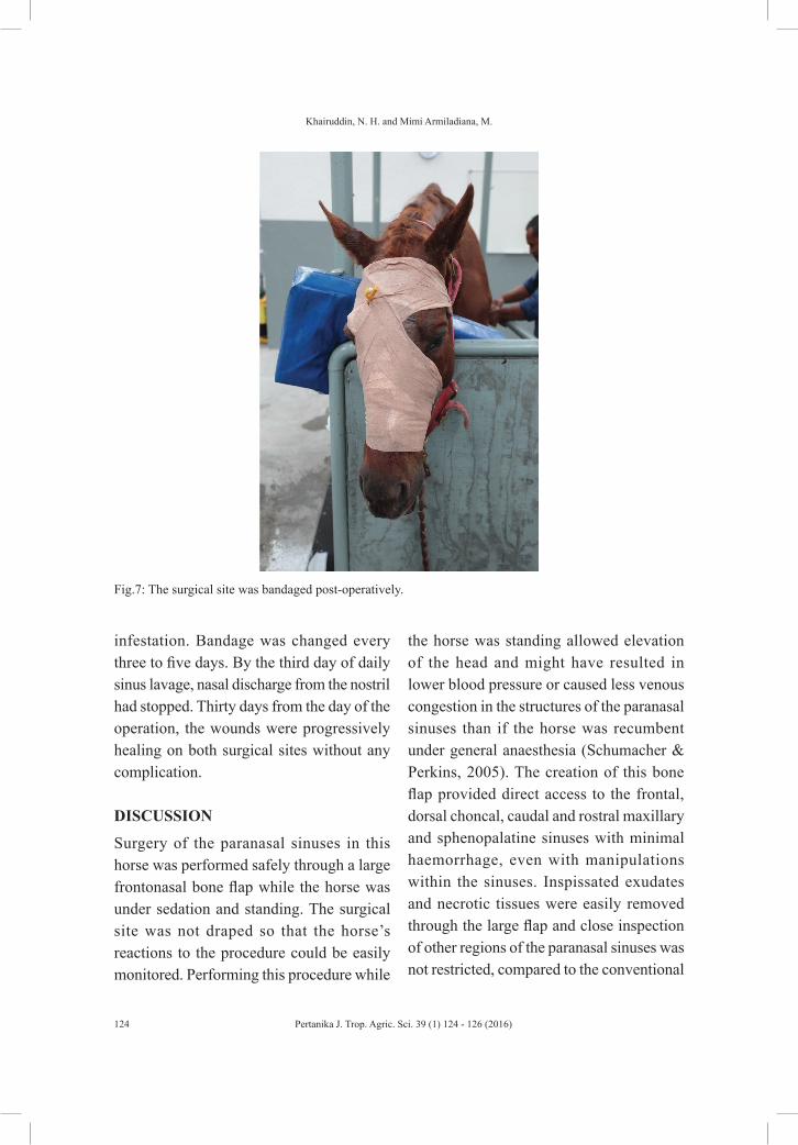
with a layer of Op-site that was spread with Solcoceryl gel to create a moist environment for the wound to heal. A second layer of gauze was lined on top of the Op-site, followed by an elastic adhesive bandage roll around the operative site and the horse head (Fig.7). The free end of the Foley catheter was left hanging out for the purpose of daily flushing.
Post-Operative Care and Outcome
The right maxillary and frontal sinus compartments were lavaged twice daily via the Foley catheter for four days using physiologic saline solution (0.9% NaCl).
Anti-inflammatory, flunixin (1.1 mg/ml, IV, once daily) and oral phenylbutazone (2 sachet of 2 gram phenylbutazone, once daily) were given for the first three days post-operatively. Penicillin-Streptomycin (22,000 U/kg, IM, every 12 hours) was administered for five days. The cutaneous portion of the bone flap and the trephine site were bandaged and left to heal by first intention. However, on day 5, it was noticed that fly larvae had infested the trephinated site due to movement of the bandage that had exposed this wound. The wound was then properly debrided and all maggots were removed. Due to continuous presence of flies, the wound was kept in bandage the whole time to prevent further maggot
Fig.5: The frontonasal bone flap was created, exposing the frontal sinus and maxillary sinus compartments.
Khairuddin, N. H. and Mimi Armiladiana, M.
124 Pertanika J. Trop. Agric. Sci. 39 (1) 124 - 126 (2016)
infestation. Bandage was changed every three to five days. By the third day of daily sinus lavage, nasal discharge from the nostril had stopped. Thirty days from the day of the operation, the wounds were progressively healing on both surgical sites without any complication.
DISCUSSION
Surgery of the paranasal sinuses in this horse was performed safely through a large frontonasal bone flap while the horse was under sedation and standing. The surgical site was not draped so that the horse’s reactions to the procedure could be easily monitored. Performing this procedure while
the horse was standing allowed elevation of the head and might have resulted in lower blood pressure or caused less venous congestion in the structures of the paranasal sinuses than if the horse was recumbent under general anaesthesia (Schumacher & Perkins, 2005). The creation of this bone flap provided direct access to the frontal, dorsal choncal, caudal and rostral maxillary and sphenopalatine sinuses with minimal haemorrhage, even with manipulations within the sinuses. Inspissated exudates and necrotic tissues were easily removed through the large flap and close inspection of other regions of the paranasal sinuses was not restricted, compared to the conventional
Fig.7: The surgical site was bandaged post-operatively.

Standing Sinus Surgery in a Horse with Sinusitis
125Pertanika J. Trop. Agric. Sci. 39 (1): 125 - 126 (2016)
method of using trephination hole of sinus compartment (Schumacher & Crossland, 1994). Combination of frontanasal bone flap and fenestration of the ventral conchal bulla would be beneficial to gain further access to the rostral maxillary and the ventral conchal sinuses (Perkins et al., 2009) in order to remove inspissated material within these regions. Standing frontonasal sinus flap (SFF) is recommended as a diagnostic and therapeutic measure where clinical signs fail to respond to non-surgical treatment with antibiotics, mucolytics, steam and volatile inhalation or minor surgery through a trephine portal created in a sinus compartment (Lane, 1993).
Throughout the frontonasal bone flap surgery, the detomidine infusion resulted in an excellent level of sedation, in which the surgical manipulation, removal of diffuse and localised lesions in the affected paranasal sinuses and copious lavage of the sinus opening were all well tolerated by the horse. For this surgery, the horse’s head needs to be supported by a stand and padding or alternatively, the horse’s head needs to be cross-tied from the halter to the stocks to raise the head for the surgeon to perform the surgery and to avoid iatrogenic facial nerve paralysis. A constant rate infusion technique using detomidine hydrochloride provides a safe and effective method to achieve a consistent standing sedation for prolonged standing surgical procedures for horses. Despite the excellent sedation and analgesic effects, a sedated horse is still conscious and must be handled with caution as it may still be able to kick accurately and unpredictably
(England et al., 1992). The outcome of this case confirmed that
standing frontonasal flap surgery provides an effective method to specifically treat primary sinusitis in a horse, and also as a treatment of many other cases of paranasal disease. This procedure could be performed safely while the horse is standing, thus it is more convenient and it eliminates the risk of general anaesthesia. In order to perform this procedure, a sound knowledge of the anatomy of the area is required. The most important part is the post-operative care of the bone flap. In a tropical country, the presence of flies is unavoidable, unless the horse is placed in an indoor stable. Thus, intact bandaging at all time is mandatory to prevent maggot infestation until the wound has healed. In this case, it took about 30 days of bandaging.
REFERENCESDixon, P. M., Parkin, T. D., Collins, N., Hawkes, C.,
Townsend, N., Tremaine, W. H., ... & Barakzai, S. Z. (2012). Equine paranasal sinus disease: A long term study of 200 cases (1997-2009): Ancillary diagnostic findings and involvement of the various sinus compartments. Equine Veterinary Journal, 44(3), 267-271.
England, G. C., Clarke, K. W., & Goosens, L. (1992). A comparison of the sedative effects of three alpha 2-adrenoceptor agonists (romifidine, detomodine and xylazine) in the horse. Journal of Veterinary Pharmacology and Therapeutics, 15(2), 194-201.
Freeman, D. E., Orsini, P. G., & Ross, M. W. (1990). A large frontonasal flap for sinus surgery in the horse. Veterinary Surgery, 19(2), 122-130. .

Khairuddin, N. H. and Mimi Armiladiana, M.
126 Pertanika J. Trop. Agric. Sci. 39 (1) 126 - 126 (2016)
Lane, J. G. (1993). The management of sinus disorders of horses- part 2. Equine Veterinary Education, 5(2), 69-73.
Nickels, F. A. (2006). Nasal passages and paranasal sinuses. In J. A. Auer, & J. A. Stick (Eds.), Equine surgery (3rd ed.). Missouri: Elsevier.
Perkins, D. J., Bennet, C., Windley, Z., & Schumacher, J. (2009). Comparison of sinoscopic techniques for examining the rostral maxillary and ventral conchal sinuses of horses. Veterinary Surgery, 38(5), 607-612.
Ruggles, A. J., Ross, M. W., & Freeman, D. E. (1991). Endoscopic examination of normal paranasal sinuses in horses. Veterinary Surgery, 20(6), 418-423.
Ruggles, A. J., Ross, M. W., & Freeman, D. E. (1993). Endoscopic examination and treatment of paranasal sinus disease in 16 horses. Veterinary Surgery, 22(6), 508-514.
Schumacher, J. & Crossland, L. (1994). Removal of inspissated purulent exudate from the ventral conchal sinus of three standing horses. Journal of American Veterinary Medicine Association, 205(9), 1312-1314.
Schumacher, J., Dutton, D. M., Murphy, D. J., Hague, B. A., & Taylor, T. S. (2000). Paranasal sinus surgery through a frontonasal flap in sedated, standing horses. Veterinary Surgery, 29(2), 173-177.
Schumacher, J., & Perkins, J. (2005). Surgery of the paranasal sinuses performed with the horse standing. Clinical Techniques in Equine Practice, 4(2), 188-194.
Schumacher, J., & Perkins, J. (2005). Surgery of the paranasal sinuses performed with the horse standing. Clinical Techniques in Equine Practice, 4(2), 188-194.