Standards of Eye Banking in India 2009 for eye bankig.pdf · module on “Standards of Eye Banking...
133
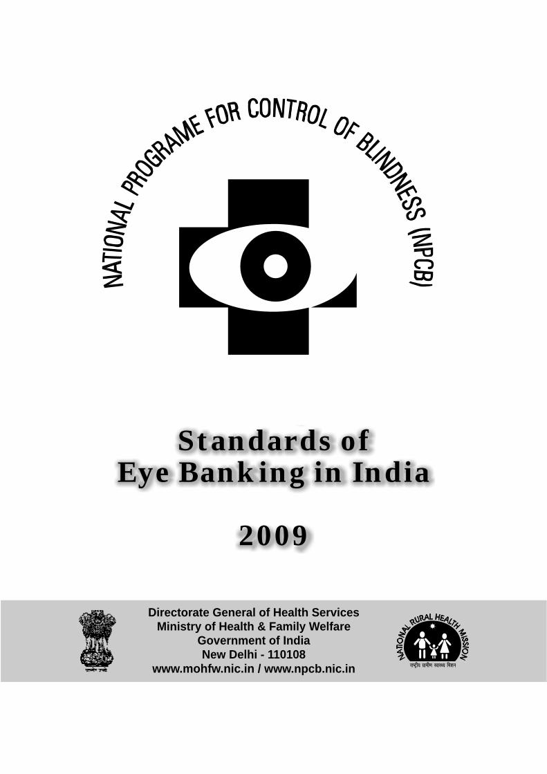
Standards of Eye Banking in India 2009 Directorate General of Health Services Ministry of Health & Family Welfare Government of India New Delhi - 110108 www.mohfw.nic.in / www.npcb.nic.in
Transcript of Standards of Eye Banking in India 2009 for eye bankig.pdf · module on “Standards of Eye Banking...

Standards ofEye Banking in India
2009
Directorate General of Health ServicesMinistry of Health & Family Welfare
Government of IndiaNew Delhi - 110108
www.mohfw.nic.in / www.npcb.nic.in
Standards ofEye Banking in India
2009
Directorate General of Health ServicesMinistry of Health & Family Welfare
Government of IndiaNew Delhi - 110108
www.mohfw.nic.in / www.npcb.nic.in
Hkkjr ljdkjLokLF; lsok egkfuns'kky;
fuekZ.k Hkou] ubZ fnYyh&110 108
GOVERNMENT OF INDIADIRECTORATE GENERAL OF HEALTH SERVICES
NIRMAN BHAVAN, NEW DELHI-110 108TEL. NO. 91-11-23061438, 23061063
FAX NO. : 91-11-23061924E-mail : [email protected]
Date : 01 October 2009
Dr. R.K. SRIVASTAVAM.S. (Ortho) D.N.B. (PMR)
DIRECTOR GENERAL
FOREWARD
I am happy to announce that National Programme for Control of Blindness [NPCB], Directorate General of Health Services, Ministry of Health and Family Welfare, New Delhi has conceptualized and produced this module on “Standards of Eye Banking in India, 2009” so as to establish & communicate quality standards to all stakeholders involved at various level of eye care services.
With the implementation of Eleventh five year plan period of NPCB, a new thrust and vigor has been infused for improvement of eye donation, collection, processing, maintenance of quality standards, equitable distribution of scarce tissue, strengthening of institutional capacity for undertaking corneal transplantation, community awareness and training of health personnel.
I hope that the valuable information given in the module will guide the programme managers, eye surgeons, and health personnel in establishing, strengthening, maintaining and delivering highest level of quality services in eye banking activities.
I appreciate the efforts & contributions of the entire team from governmental and non-governmental sector efficiently led by Dr. [Mrs.] R Jose, Additional DG in producing this visionary document.
[Dr. R.K. Srivastava]

DR. (MRS.) R. JOSEAddl. Director General
LokLF; lsok egkfuns'kky;fuekZ.k Hkou]
ubZ fnYyh&110 108
DIRECTORATE GENERAL OF HEALTH SERVICESNIRMAN BHAWAN,
NEW DELHI - 110 108TEL. NO.: 011-23061594, FAX : 011-23062702
E-mail : [email protected]
PREFACE
Over the last three decades, country has seen a phenomenal rise in socio-economical, industrial, manufacturing, information-technology, infrastructural development, human resource management, service sector, so on and so forth with far reaching consequences on health. Today, we have reached a very commendable stage of Cataract Surgical Rate and the momentum thus generated would continue in future also. In this context, National Programme for Control of Blindness [NPCB] has taken a lead in addressing other issues of blindness as well in a comprehensive manner.
Eleventh plan [2007-12] of NPCB has attempted to cover and address emerging eye diseases other than cataract like Diabetic Retinopathy, Glaucoma, Childhood blindness, Low Vision and ocular injuries in a mission mode through successful Public Private Partnership. The endeavor of -the programme is to eliminate all causes of avoidable blindness and to reach a sustainable level where people have access to level appropriate eye care service.
Corneal blindness is one of the eye diseases that is being covered under the global initiative of VISION 2020: The Right to Sight Initiative to which India is also signatory. Eye banking activities becomes a critical component and pillar for managing this disease wherein infrastructure, human resource, logistics & service delivery should match international quality standards and the needs of patients in effective and efficient manner.
Eye Banking Standard manual would not have been possible without constant supervision, encouragement and direction from Dr. R.K. Srivastava DGHS and Ms. Shalini Prasad, Joint Secretary, to Government of India. I would also like to place on record my appreciation to my team members especially Dr. A.S. Rathore, Additional Director General [0], Dr. V. Rajshekhar, Dy. Assistant Director General [0], Dr. V.K. Tewari,. Education Health Officer and Dr. Sandeep Sachdeva, National Consultant in supporting and contributing immensely in program activities.
The outcome was spearheaded by various consultative processes and deliberation with stakeholders from Government and Non-Governmental Organization working closely with NPCB since inception. I extend my gratitude to representative from All India Institute of Medical Sciences [New Delhi], Guru Nanak Eye Centre [New Delhi] Eye Bank Association of India [Hyderabad], Arvind Eye Hospital [Madurai], LV Prasad Eye Institute [Hyderabad], Disha Eye Hospital [Barrackpore], HV Desai Hospital [Pune], International Eye Bank [Bangalore], Venu Eye Institute Research Centre [New Delhi], All India Ophthalmic Society [New Delhi], ORBIS [New Delhi] in supporting the larger platform of critical discussion, deliberation and producing the template of Eye Banking Standard.
I strongly believe that implementation of these standards in letter and spirit will pave way to amelioration of corneal blindness from our country in near future.
[Dr. (Mrs.) R. Jose]Additional Director General
Directorate General of Health Services
Date : 01 October 2009

DR. A.S. RATHOREAsstt. Director General
Hkkjr ljdkjLokLF; ,oa ifjokj dY;k.k ea=kky;
National Programme for Control of BlindnessMinistry of Health & Family Welfare
Government of India
755-A, Nirman Bhavan,New Delhi - 110 108
ACKNOWLEDGEMENTS
The production of the module “Standards of Eye Banking in India” has been a collective effort of all stakeholders including non-governmental organization especially Eye Bank Association of India [EBAI]. Nevertheless, constant support and sustained guidance has been made available from Dr. R.K Srivastava, DG, HS and Dr. [Mrs.] R. Jose, Additional DG.
The standard for eye banking though existed in the past however it was urgently felt that it required updation. The outcome in the present format was realized over a period of time through intense consultative processes, deliberations and meetings. Every attempt has been made while compiling keeping in view international eye banking protocols & standards and suitably adapted to meet local regulations so as to be applicable and relevant in the Indian context. We look forward to suggestions and feedback for improvement by our learned colleagues and readers.
The language used in the module has been kept simple, lucid and at the same time comprehensive to cover all aspect on Eye Banking activities. Module has been divided into chapters and sub-section for easy reference, clarity and understanding. It covers aspects relating to type of facilities, infrastructure, human resource, equipment, instruments, infection control practices; standard operating procedures [SOP]; cornea tissue/preservation/evaluation standards; retrieval procedures, screening of donors, contraindications, quality control, procedures for storage, labeling and equitable distribution of cornea for Keratoplasty.
The exercise was made possible by active contribution of experts especially Dr. Ritu Arora [New Delhi], Dr. Radhika Tandon [New Delhi], Dr. M. Srinivasan [Madurai], Dr. Usha Gopinathan [Hyderabad], Dr. Samar Basak [Barrackpore], Dr. Ayan Mohanta [Barrackpore], Dr. Gobinda Mukherjee [New Delhi], Dr. AVN Chetty [Vishakhapatnam], Dr. Bageshri Gogate [Pune], Dr. Prashant Garg [Hyderabad], Dr. Rekha Gyanchand [Bangalore], Ms. Tanuja Joshi [New Delhi], Mr. G Ganesh [Hyderabad], Dr. GV Rao [New Delhi], Dr. Preeti Singh [New Delhi] and Ms. Deepti Bajaj [New Delhi].
I extend my heartiest appreciation to all the people who contributed directly of indirectly in our journey of achieving this endeavour.
[Dr. A.S. Rathore]
Date : 08 October 2009

Standards of Eye Banking in IndiaContents
CHAPTER-1Page No.
1.1. Facilities, Equipment & Maintenance
1.1.1 Facilities (Organization and Infrastructure) 1
1.1.2 Procedures Manual 1
1.1.3 Infrastructure – Physical Space 1
1.1.4 Infrastructure - Equipment 2
1.1.5 Eye Bank Maintenance 3
1.1.6 Equipment Maintenance & Cleaning 3
1.1.7 Instruments & Reagents 3
1.1.8 Infection Control & Safety 3
1.1.9 Waste Disposal 3
1.2. Donor Cornea / Eye Retrieval Related Standards
1.2.1 Donor Eye / Cornea Retrieval 4
1.2.2 Personnel Authorized to Retrieve Eyes / Corneas 4
1.2.3 Pre-Recovery Procedure 4
1.2.4 Retrieval Procedure 4
1.2.5 Screening of Donors 4
1.2.6 Contraindications 5
1.2.7 Donor Age 7
1.2.8 Interval Between Death, Enucleation and Preservation 7
1.2.9 Recovery Procedure 7
1.2.10 Donor Blood Sample 7
1.3. Donor Tissue Preservation Standards
1.3.1 In Situ and Laboratory Removal of Corneoscleral Rim 8
1.3.2 Short Term Preservation 8
1.3.3 Long Term Preservation 8
1.3.4 Whole Globe Preservation 8
1.3.5 Sclera Preservation 8
1.4. Tissue Evaluation Standards
1.4.1 Gross Examination 8
1.4.2 Slit-Lamp Examination 8
1.4.3 Specular Microscopy 9

1.5. Donor Blood Screening
1.5.1 HIV Screening 9
1.5.2 Hepatitis B & C Screening 9
1.5.3 HTLV-I and HTLV-II Screening 9
1.5.4 Syphilis Screening 9
1.5.5 Non-Required Laboratory Results 9
1.6. Quality Assurance
1.6.1 Quality Assurance (QA) 10
1.7. Quality Control
1.7.1 Testing 10
1.7.2 Microbiologic Culturing 10
1.8. Non Surgical Donor Tissue
1.8.1 Non-Surgical Donor Tissue 11
1.9. Storage
1.9.1 Storage 11
1.10. Labelling
1.10.1 Labeling 11
1.11. Distribution of Tissue
1.11.1 Review of Donor Medical History 12
1.11.2 Receivers of Tissue 12
1.11.3 Fair and Equitable System 12
1.11.4 Returned Tissue 12
1.11.5 Tissue Recalls 12

CHAPTER - 2
Standards for Non-Technical Activities of Eye Banks
2.1. Eye Banking System2.1.1 Eye Donation Center (EDC) 172.1.2 Eye Bank (EB) 172.1.3 Eye Bank Training Centre (EBTC): 18
2.2. Awareness2.2.1 General Awareness: 182.2.2 Focussed Awareness Campaigns: 182.2.3 Choice of Hospitals 182.2.4 Link Between the Hospital and the Eye Bank 192.2.5 Attributes of An Eye Donation Counsellor (EDC) 192.2.6 Grief Counselling Techniques 202.2.7 Alerting the Eye Bank Team 202.2.8 Expression of Gratitude 202.2.9 Documentation of Case Reports 21
2.3. Manpower Requirements2.3.1 Responsibilities of Various Eye Banking Personnel 212.3.2 Board of Directors 212.3.3 Medical Director 212.3.4 Executive Director 212.3.5 Eye Bank Manager 222.3.6 Eye Bank Technicians 222.3.7 Eye Donation Counselor 222.3.8 Administrative Secretary cum Telephone Operator 222.3.9 Training & Human Resource Development 222.3.10 Medical Director 222.3.11 Executive Director & Eye Bank Manager: 222.3.12 Eye Bank Technician 232.3.13 Eye Donation Counselor 23
2.4. Documentation2.4.1 Length of Storage 232.4.2 Confidentiality 232.4.3 Documents & Logbooks 23
2.5. Registration & Accreditation2.5.1 Registration 24
2.5.2 Accreditation 24
2.5.3 Accreditation Authority 25
2.6. Eye Bank Inspection2.6.1 Procurement of Eye Bank Essentials 25

CHAPTER - 3
Standard Operating Procedures of Eye Banks3.1. Scope of the Manual
Personnel
3.1.1 Recruitment 33
3.1.2 Personnel & Responsibilities 33
3.1.3 Medical Director 33
3.1.4 Executive Director 34
3.1.5 Technical Staff 34
3.1.6 Eye Donation Counselors 34
3.1.7 Secretary 35
3.2. Appraisal & PromotionSkill EnhancementTraining & Certification (In case of an Eye Bank Training Center)
3.2.1 Training Centre 35
3.3. Eye Bank Facilities
3.3.1 Eye Bank Laboratories & Operations 35
3.3.2 Laboratory 1 – Handling Contaminated Materials 36
3.3.3 Laboratory 2 - Laboratory for Corneal Excision 36
3.3.4 Laboratory 3 – Cornea Evaluation 36
3.4. Cleaning, Maintenance & Calibration
3.4.1 Cleaning of Laboratories 36
3.4.2 Cleaning, Packing and Sterilization of Instruments 36
3.4.3 Cleaning, Maintenance and Calibration of Equipment 37
3.4.4 Equipment Cleaning 37
3.4.5 Equipment Maintenance & Calibration 37
3.5. Complaints 383.5. New Work 383.5. Waste Disposal 393.5. Infection Control & Safety 39
3.5.1 Infection 393.5.2 Accidents 403.5.3 Immunisation 40
3.5.4 Training 41
3.6. Donor Tissue Recovery & Screening of Donor Blood
3.6.1 Introduction 41
3.6.2 Objectives 41

3.7. Pre Recovery Procedures
3.7.1 Pre Recovery Review and Donor Preparation 423.7.2 Consent for Ocular Tissue Recovery 423.7.3 Identify the Donor 423.7.4 Worksite Preparation 433.7.5 Gross Inspection of the Donor 43
3.8. Donor Preparation
3.8.1 Donor Face Cleaning 433.8.2 5% Povidone-Iodine Solution Preparation 433.8.3 Lid Margin and Lash Prep 433.8.4 Conjunctiva Prep 443.8.5 Globe Prep 443.8.6 Lids and Face Prep 443.8.7 Sterile Field Preparation 453.8.8 Attire of Field Personnel 45
3.9. In Situ Corneal ExcisionMaterials
3.9.1 Corneo Scleral Rim Excision 463.9.2 Lab Corneal Excision 483.9.3 Donor Cornea Evaluation 493.9.4 Slit Lamp Examination 503.9.5 Specular Examination 54
3.10. Sclera Processing & PreservationSerological Testing of Donor Blood Sample
3.10.1 Blood Sample for Serological Testing 573.10.2 Drawing of Donor Blood Sample 583.10.3 Transferring Blood from Syringe to Vacutainer Tubes 593.10.4 Blood Tube Labeling and Handling 593.10.5 Screening of Human Donor Blood Sample for HIV, 59
Hepatitis B, Hepatitis C & Syphilis
Appendix - 1 60Donor Call- Initial Information
Appendix - 2 61Eye Donor Medical Particulars
Appendix - 3 62Familiy Pledge Form For Eye Donation
Appendix - 4 63Donor Information Sheet

Appendix - 5 65Immunology Laboratory Serology Report
Appendix - 6 66Tissue Evaluation Report
Appendix - 7 68Hemodilution Assessment
Appendix - 8 69Tissue Distribution InformationDuplicate form of Appendix - 8 71
Appendix - 9 72Donor Tissue Tentative Utility Data & Adverse Report Form
Appendix - 10 74Instrument Cleaning Log
Appendix - 11 75Instrument Sterilization Log
Appendix - 12 76Laboratory Cleaning Log
Appendix - 13 77Human Organs Transplantation Act (HOTA)
Appendix - 14 97Laboratory Equipment Cleaning Log
Appendix - 15 98Ferquently asked questions About Eye Donation
Appendix - 16 100Facts and Myths About Donating Eyes
Appendix - 17 101Eye Donation Counsellor (EDC)Daily Report Form
Appendix - 18 102Eye Donation Counsellor (EDC) ReportCase Summary
Appendix - 19 103Eye Donation Counsellor (EDC)Monthly Report Form
Appendix - 20 106Financial Assistance available under NPCB
Appendix - 21 109Eye Banks* In India

FACILITIES, EQUIPMENT &MAINTENANCE
Chapter-1Chapter-1Chapter-1Chapter-1Chapter-1


Standards of Eye Banking in IndiaContents
CHAPTER-1Page No.
1.1. Facilities, Equipment & Maintenance
1.1.1 Facilities (Organization and Infrastructure) 1
1.1.2 Procedures Manual 1
1.1.3 Infrastructure – Physical Space 1
1.1.4 Infrastructure - Equipment 2
1.1.5 Eye Bank Maintenance 3
1.1.6 Equipment Maintenance & Cleaning 3
1.1.7 Instruments & Reagents 3
1.1.8 Infection Control & Safety 3
1.1.9 Waste Disposal 3
1.2. Donor Cornea / Eye Retrieval Related Standards
1.2.1 Donor Eye / Cornea Retrieval 4
1.2.2 Personnel Authorized to Retrieve Eyes / Corneas 4
1.2.3 Pre-Recovery Procedure 4
1.2.4 Retrieval Procedure 4
1.2.5 Screening of Donors 4
1.2.6 Contraindications 5
1.2.7 Donor Age 7
1.2.8 Interval Between Death, Enucleation and Preservation 7
1.2.9 Recovery Procedure 7
1.2.10 Donor Blood Sample 7
1.3. Donor Tissue Preservation Standards
1.3.1 In Situ and Laboratory Removal of Corneoscleral Rim 8
1.3.2 Short Term Preservation 8
1.3.3 Long Term Preservation 8
1.3.4 Whole Globe Preservation 8
1.3.5 Sclera Preservation 8
1.4. Tissue Evaluation Standards
1.4.1 Gross Examination 8
1.4.2 Slit-Lamp Examination 8
1.4.3 Specular Microscopy 9

1.5. Donor Blood Screening
1.5.1 HIV Screening 9
1.5.2 Hepatitis B & C Screening 9
1.5.3 HTLV-I and HTLV-II Screening 9
1.5.4 Syphilis Screening 9
1.5.5 Non-Required Laboratory Results 9
1.6. Quality Assurance
1.6.1 Quality Assurance (QA) 10
1.7. Quality Control
1.7.1 Testing 10
1.7.2 Microbiologic Culturing 10
1.8. Non Surgical Donor Tissue
1.8.1 Non-Surgical Donor Tissue 11
1.9. Storage
1.9.1 Storage 11
1.10. Labelling
1.10.1 Labeling 11
1.11. Distribution of Tissue
1.11.1 Review of Donor Medical History 12
1.11.2 Receivers of Tissue 12
1.11.3 Fair and Equitable System 12
1.11.4 Returned Tissue 12
1.11.5 Tissue Recalls 12

1
1.1 Facilities, Equipment & Maintenance
1.1.1. Facilities (Organization and Infrastructure)
For an efficient eye banking system, a three tier organization structure has been recommended. At thetop of the pyramid is the Eye Bank Training Center followed by Eye Banks and at the base of the pyramidis the eye donation center. Activities, responsibilities, manpower required for each of the above viz., EyeBank Training Center (EBTC), Eye Bank (EB) and Eye Donation Center (EDC) has been dealt in detailunder section A1.100 to section A1.300
1.2.2. Procedures manualEach eye bank shall maintain its own procedures manual (SOP) that details all aspects of its specific retrieval,processing, testing, storage, distribution and quality assurance practices. Each procedure must be initiallyapproved signed and dated by the Medical director or Officer-in-charge of the eye bank. An annual reviewof each eye bank’s procedures manual with signing and dating by the Medical director or Officer-in-chargeis required. Each eye bank must maintain copies of each procedure it uses and the length of time theprocedure was in use. The current standards of eye banking document can be used as the proceduresmanual with a document detailing any deviations or modifications with justification as required.
The following facilities and infrastructure is required
1.1.3. Physical Space
A minimum area of 600 Sq.Ft is required which accommodates a serology lab, tissue processing lab andevaluation, storage & shipping lab.
Instrument Cleaning Lab:
1. Sink – For washing instruments
2. Autoclave for sterilizing
3. Counter top & storage space for storing instruments & supplies
Serology Lab
1.Sink for washing
2.Refrigerator – For storing blood samples& kits
3.Counter tops, cabinets & drawers forworkspace and storing supplies
4.Centrifuge & Serum testing equipment likeELISA Reader, Rapid test etc.
Tissue Processing Lab
1.Sterile counter / table top for processing(Laminar Flow Hood / Bio-Hazard Cabinet)
2.Counter tops, drawers and cabinets forstorage
Evaluation, Storage & Shipping Lab1.Slit Lamp and Specular Microscope for
Tissue Evaluation.
2.Counter Tops and Cabinets for Storage ofSupplies Packing and Shipping.
3.Refrigerator for Storing Donor Tissue
Required
Required
Required
Required
Access
Required
Access to accreditedtesting lab or all facilitiesare required.Accredited lab shouldhave all the mentionedfacilities and would beinspected beforeaccreditation is given toeye bank.
Required
Required
Required, but access to aspecular microscope isalso acceptable.
Required
Required
Required
Access
Required
Serology lab is notrequired. However,sink for washingand facility forstoring bloodsamples inrefrigerator shouldbe available.
Not required
Required
Required
INFRASTRUCTURE EBTC E B EDC
Required
Required
Required
Required
Required
Required
Required
Required
Required

2
EQUIPMENT EYE BANK TRAINING EYE BANK EYE DONATIONCENTRE CENTRE
1.1.4. Equipment & Other Facilities
Each eye bank must have the following equipment and facilities to perform the volume of laboratoryservices with optimal accuracy, efficiency, sterility, timelines, and safety.
Slit Lamp Required Required Not required
Refrigerators for storing blood Requiredsample, tissues andstorage media
Serology Equipment Required
Specular Microscope Required
Sufficient sets of instruments for Required. Required.corneal excision and enucleation Numbers to be Numbers to be
decided on level of decided on levelcollections of collections
Autoclave or gas sterilizer Required Required/oraccess to sterilizingfacility Foraccreditation,sterilizing facilitypractice andprocedure shall bereviewed
Laminar Flow Hood (Class II) Required Required(This is required for thepreservation of ocular tissuein the laboratory in case ofwhole globe removal (enucleation)and for processing scleral tissue)
OTHER FACILITIES
Transportation Facility 24 Hours 24 Hours Should365 days 365 days have access
Furniture Required Required Preferable
Computer with email facility Required Required Preferable
Two exclusive lines (one with 1919 or Required Required Universal publicpublic service number allotted and service numberanother for outgoing calls to be alloted
Audio visual equipment for publicity Required Required Preferred
PreferableFor storing bloodsample, ice packs,storage media, eye ortissue collected etc
Not required
Not required
RequiredNumbers to bedecided on level ofcollections
Should have accessto sterilizingfacility
Not required
RequiredIn case the Eye Bankhas tie up withaccredited lab fortesting, oneRefrigerator issufficient
Yes Required. Accessto an accredited labis also acceptable
Yes required ifcollection is >200 peryear. Access tospecular microscope isalso acceptable

3
1.1.5. Eye Bank Maintenance
The room including walls, floor and sink must be kept clean at all times. Appropriate documentation ofregular laboratory cleaning schedules must be maintained and kept on file for a minimum of three years.For cleaning procedure refer Section III.
Each eye bank laboratory must have an adequate, stable electrical source and sufficient number ofgrounded electrical outlets for operating laboratory equipment.
1.1.6. Equipment maintenance and cleaning
Refrigerator
Each eye bank laboratory shall have a refrigerator with a device, internal or external for recordingtemperature variations. Temperature variations must be recorded twice daily and should remain withinthe range of 2º – 6º C.
The refrigerator should be maintained exclusively for use by the eye bank. It must contain clearly definedand labeled areas for all tissues stored e.g. surgical tissue, awaiting distribution, quarantined tissue,tissue for research etc.
The refrigerator should be calibrated once a year.
Laminar Airflow Hood / Cabinet
The cleaning schedule should be maintained.
Particle counts should be performed once a year.
Appropriate maintenance and accreditation records must be maintained for each piece of equipment.The eye bank must include in its procedure manual, the monitoring, inspection and cleaning proceduresand schedules, for laboratory equipment. These must be kept on file for a minimum of three years.
In the event of power failure there must be provision for immediate notification and action to be taken,which may include an emergency power supply to maintain essential refrigeration.
1.1.7. Instruments and reagents
Adequate instruments must be available to provide for sterile removal of whole eye and corneas.Instruments must be inspected frequently enough to assure that they function properly.
All sterilized instruments, supplies and reagents, such as corneal preservation medium, must containexpiry dates that are current at all times.
1.1.8. Infection control and safety
All eye bank personnel must operate under the universal precautions for health care workers. Thesewritten procedures must be included in the eye bank’s procedure manual. All technical personnel shouldreceive Hepatitis B vaccination and any other recommended vaccination that may be announced fromtime to time.
1.1.9. Waste disposal
Human tissue and waste items shall be disposed off in such a manner as to minimize any hazard to eyebank personnel and the environment and comply with applicable regulations. Dignified and proper disposalprocedures shall be used to obviate recognizable human remains.

4
1.2 Donor Cornea / Eye Retrieval Related Standards
1.2.1. Donar eye / cornea retrieval
Care must be taken that eye bank resources are utilized optimally and eye bank personnel are not exposedto any health hazards. The following guidelines ensure that resources are put to optimum use and thateye bank personnel are not exposed to any health hazards.
1.2.2. Personnel authorized to retrieve eyes / corneas
A registered medical practitioner trained in enucleation / excision from an Eye Bank Training Center isonly allowed to retrieve eyes / corneas from the donor after satisfying self that life is extinct, in the absenceof a death certificate.
1.2.3. Pre Recovery Procedure
Before proceeding for recovery eye bank personnel should ascertain the following details:
a) Location b) Age of the donorc) Cause of death d) Time of death
1.2.4. Retrieval Procedure
l Retrieval procedure could be either Enucleation or Corneal scleral rim excision. The retrievalprocedures for both of the above mentioned techniques are given in detail in Section III H8.340under Donor Ocular Tissue Recovery.
l Eye Bank team should carry only validated sterile instruments for retrieval.
l Eye Bank Team on arrival at the location should locate the next of kin and convey condolence andobtain death certificate.
l In the absence of a death certificate the registered medical practitioner should satisfy self that life isextinct as per procedures laid down in Section III H 8.320,330 and Appendix II.
l The eye bank team should obtain consent on a consent form from the legal custodian of the donor.
l After obtaining consent the donor should be identified either through a tag or through the next of kin
l The eye bank team should then proceed to prepare the site as per guidelines in Section III H8.340
l Gross physical examination should be conducted with utmost respect for observations regardingbuild: average / healthy / emaciated
l Eye bank team should look out for needle marks on the arm, skin lesions etc
l Eye Bank Team should look out for Ulcers / gangrene in exposed areas
l Ocular examination should be conducted as per guidelines in Section III, H8.340
l Medical records / Medical information should be obtained as per guidelines in Section III, H8.350
l Information for hemodilution should be obtained as per guidelines in Section III, H8.300, H8.400
l Social history of the donor should be obtained wherever possible from the next of kin
l Once all the above are completed the retrieval can be started. The retrieval procedure should be asper guidelines in Section III, H8.500
1.2.5. Screening of donors
Tissue from donors with the following is potentially hazardous to eye bank personnel and harvesting eyesshould be strictly avoided.

5
Contraindications for Retrieval:
Active viral Hepatitis
Acquired immunodeficiency syndrome (AIDS) of HIV
Active viral encephalitis or encephalitis of unknown origin
Creutzfeldt-Jakob disease
Rabies
1.2.6. Contraindications
Tissue from donors with the following are potentially health threatening and also affects the success ofthe surgery and shall not be offered for surgical purposes. In conditions considered absolutecontraindications for transplantation (marked with an asterix*), donor family should be informed clearlyand made fully aware of this fact. Eyes should not be harvested unless the donor family is fully aware ofthis and still wishes to donate
(I) Conditions with potential risk of transmission of local or systemic communicablefrom donor to recipient
a Death of unknown cause*
b Death with neurologic disease of unestablished diagnosis*
c Subacute sclerosing panencephalitis
d Progressive multifocal leukoencephalopathy
e Active meningitis or encephalitis*
f Encephalopathy of unknown origin or progressive encephalopathy*
g Active septicemia* (bacteremia, fungemia, viremia, parasitemia)
h Active viral hepatitis*
i Creutzfeldt-Jakob disease*
j Congenital rubella
k Reye’s Syndrome
l Rabies*
m Active miliary tuberculosis or tubercular meningitis*
n Patients on ventilator for > 72 hrs
o Hepatitis B surface antigen positive donors*
p HTLV-I or HTLV-II infection*
q Hepatitis C Seropositive donors*
r HIV seropositive donors*
s HIV or high risk for HIV corneas from: persons meeting any of the following criteria should not beoffered for transplantation.*
6
t Active ocular or intraocular inflammation conjunctivitis, sclerits, iritis, uveitis, vitreitis choroiditis andretinitis (at the time of death)
(II) Conditions with potential risk of transmission of non-communicable disease fromdonor to recipient.
a Death due to cyanide poisoning
b Instrinsic eye disease
c Retinoblastoma
d Malignant tumours of the anterior ocular segment or known adenocarcinoma in the eye of primaryor metastatic origin
e Leukemias*
f Active disseminated lymphomas*
(III) Conditions that will affect graft outcome.
a Congenital or acquired disorders of the eye that would preclude a successful outcome for the intendeduse e.g., a central donor corneal scar for an intended penetrating keratoplasty preserve ofkeratoconous and keratoglobus. Corneas which have undergone refractive surgical proceduresetc.
b patients on ventilator for >72 hrs.
Behavioral / History, Laboratory and Medical Exclusion Criteria.
a. Men who have had sex with other men in the preceding 5 years (homosexual behaviour)
b. Persons who report nonmedical intravenous, intramuscular, or subcutaneous injection of drugs inthe preceding 5 years. (IV drug abusers)
c. Persons with hemophilia or related clotting disorders who have received human-derived clottingfactor concentrate.
d. Men and women who have engaged in sex for money or drugs in the preceding 5 years (commercialsex workers)
e. Persons who have had sex in the preceding 12 months with any person described in item a-d aboveor with a person known or suspected to have HIV infection.
f. Persons who have been exposed in the preceding 12 months to known or suspected HIV-infectedblood through percutaneous inoculation or through contact with an open wound, or mucousmembrane.
g. Children meeting any of the exclusionary criteria listed above for adults should not be accepted asdonors.
h. Children born to mother with HIV infection or mothers who meet the behavioral or laboratoryexclusionary criteria for adult donors regardless of their HIV status should not be accepted asdonors unless HIV infection can be definitely excluded in the child as follows:
(i) Children >18 months of age who are born to mothers with or at risk for HIV infection, who have

7
not been breast fed within the last 12 months, and whose HIV antibody tests, physical examinationand review of medical records do not indicate evidence of HIV infection can be accepted as donors.
(ii) Children < 18 months of age who are born to mothers with or at risk for HIV infection orchildren of mothers with or at risk of HIV infection who have been breast fed within the past 12months should not be accepted as donors regardless of their HIV tests results.
i. Persons who cannot be tested for HIV infection because of refusal, inadequate blood samples (e.g.haemodilution that could result in false-negative tests), or any other reasons.
j. Persons with a repeatedly reactive screening assay for HIV-1 or HIV-2 antibody regardless of theresults of supplemental assays.
k. Persons whose history, physical examination, medical records, or autopsy reports reveal otherevidence of HIV infection or high-risk behaviour, such as diagnosis of AIDS, unexplained mucousmembranes hemorrhages kaposi’s sarcoma, unexplained lymphadenopathy lasting > 1 month,unexplained temperature> 100.5 F (38.6C) for > 10 days, unexplained persistent diarrhea, male-to-male sexual contact, sexually transmitted diseases, or needle tracks or other signs of parental drugabuse.
Donor tissue will be harvested and transported to the eye bank for further processing, evaluation, storageand distribution. Surgical use of tissue for transplantation will be determined by technical considerationsas decided by the medical director and operating surgeon. Tissues can be used for penetrating keratoplasty,lamellar keratoplasty, stem cell culture and/or transplantation and scleral transplantation and any residualparts can be used for educational or research purpose as appropriate.
1.2.7. Donor age
Since no definite relationship has been established between the quality of donor tissue and age the upperand lower age limit is left to the discretion of the medical director.
1.2.8. Interval between death, enucleation, excision and preservation
Acceptable time intervals from death, enucleation or excision to preservation may vary according to thecircumstances of death and interim means of storage of the body. It is generally recommended thatcorneal preservation should occur as soon as possible after death. All time intervals for each donor, e.g.,the time of death to the time of enucleation and preservation and/or the time to corneal excision, shall berecorded. If the donor has been refrigerated prior to enucleation or in situ corneal excision, this informationshall be noted.
1.2.9. Recovery Procedure
1.2.10. Donor Blood Sample
The authorized personnel retrieving the tissue should obtain an adequate blood sample. Please referSection III, for detailed procedure.

8
1.3 Donor Tissue Preservation Standards
1.3.1 In situ and laboratory removal of the corneoscleral rim
Individuals specifically trained for in situ retrieval and/or laboratory removal of the corneal scleral segmentshall perform removal of the corneal scleral rim using sterile technique. If the procedure is done in alaboratory the removal must be performed in a laminar flow hood, cabinet or in an operation room. Forcornel scleral removal, the eye shall be examined with the use of a penlight preferably and a slit lampprior to excision.
1.3.2. Use of short or intermediate term preservation medium
Eye Bank shall use approved corneal storage medium (such as MK, Optisol GS, EUSOL, etc) from areliable source. The medium shall be used and stored according to the manufacturer’s recommendationsfor temperature, date and other factors. The manufactured medium purchased and shipped to the eyebank shall be inspected for damage upon arrival and the lot number of medium used for each corneashall be recorded on the tissue tracking and recall.
1.3.3. Long term preservation
Some eye banks employ long-term preservation of corneal tissue, such as glycerine preservation ororgan culturing. An eye bank that use long-term preservation shall carefully document the procedure intheir procedure manual, and adhere to rigid aseptic technique.
1.3.4. Whole globe preservation
Eye Banks that store whole eye shall employ aseptic practice. The selected preservation method must bedocumented in the eye bank’s own procedures manual.
1.3.5. Sclera preservation
Eye banks shall preserve scleral tissue aseptically. The selected preservation method must be documentedin the eye bank’s own procedure manual.
A preservation date for scleral tissue shall be indicated.
1.4 Tissue Evaluation StandardsThe ultimate responsibility for determining the suitability of the tissue for transplantation rests with thetransplanting surgeon.
1.4.1. Gross examination
The corneal-scleral segment shall be initially examined grossly for clarity, epithelial defects, foreign objectsand contamination and scleral color. e.g., jaundice. Refer Section III, H8.800 for details.
1.4.2. Slit-lamp examination
The cornea shall be examined for epithelia and stromal pathology and in particular endothelial disease.Enucleated globes shall be examined in the laboratory prior to distribution and/or corneal excision. If in situcorneal excision is performed, examination of the donor eye anterior segment with a penlight or a portableslit lamp is required. After corneal excision, the corneal-scleral rim shall be evaluated by slit lamp

9
biomicroscopy, even if the donor eye has been examined with the slit lamp prior to excision of the corneal-scleral rim, to ensure that damage to the corneal endothelium or surgical detachment of Descemet’smembrane did not occur.
Information obtained with slit lamp biomicroscopic examination must be documented.
1.4.3. Specular Microscopy:
Determination of endothelial cell density via specular microscopy shall be a standard method of cornealtissue evaluation for all Eye Banks. When it is impossible to obtain an endothelial cell count this requirementmay be waived on a case-by-case basis by the MD.
1.5. Donor Blood ScreeningThe following sections specify the required serologic tests, which must be performed for each donor.Refer Section III, H8.900 for details and procedures.
1.5.1 HIV Screening
All eye banks must have an operational HIV screening programme using an approved test for all donorsof surgically designated tissue. A negative screening test must be documented prior to release of tissuefor transplantation.
1.5.2. Hepatitis B & C screening
All eye banks must have operational hepatitis B & C screening programme using approved tests forhepatitis B & Hepatitis C antigen for all donors of surgically designated tissue. A negative screening testor neutralization or confirmatory test must be documented prior to release of tissue for transplantation.
1.5.3. HTLV-I and HTLV-II screening
Donor Screening for HTLV-I and HTLV-II is not required. However, if donor screening for HTLV-I and/orHTLV-II has been performed, a negative screening test must be documented prior to release of tissue fortransplantation.
1.5.4. Syphilis screening
All eye banks must have operational syphilis screening programme using an approved test for all donors.If the screening test is positive, a negative confirmatory test must be documented before tissue isreleased.
1.5.5. Non-required laboratory results
If laboratory results of non-required test for infectious disease are available for tissue, for transplantation,they must be taken into account and/or acted upon by the MD. Any relevant information shall be providedto the transplanting surgeon.

10
1.6 Quality Assurance
1.6.1 Quality Assurance (QA)
Each eye bank shall have a formally established quality assurance programme. This program shallinclude ongoing monitoring and development of plans for corrective action. These standards shallprovide the basis for development of the QA programme. Each eye bank shall document all aspects ofits QA programme and maintain record of all QA activities for a minimum of ten years. These includeany corrective or remedial action taken for detected deficiencies. These records shall be available forreview.
The eye bank’s quality assurance programme shall include a method for the receiving surgeon to reportadverse reactions from the transplantation of corneal, scleral or other ocular tissue to the source eyebank. The Eye Bank in turn must forward the adverse reaction information within a reasonable time to theEBAI or MOH for review by the Medical Standards Policy sub-committee. An adverse reaction file shall beavailable for review by the accreditation team at the time of inspection and must be kept for minimum ofthree years.
1.7 Quality ControlThe MD shall prescribe tests and procedures for measuring, assaying or monitoring properties of tissuesessential to the evaluation of their safety for transplantation, e.g., hepatitis B surface antigen and humanimmunodeficiency virus (HIV) antibody, and conform with legal and regulatory requirements. Results ofall such tests or procedures, together with evaluations based on these findings, shall become part of thepermanent record of all tissue processed.
1.7.1 Testing
If an eye bank performs its own microbiologic or serologic testing it must meet applicable accreditationrequirements.
1.7.2. Microbiologic Culturing
Culturing of Eye Bank donor eyes is advised despite the recognition by many that bacteriologiccontamination of donor eyes does not necessarily lead to infection and that pre-surgical or surgical culturesmay not correlate with postoperative infection if it should occur. Cultures may be performed either beforeand/or at the time of surgery.
A. Pre-surgical cultures (Optional)
Eye Banks may elect to perform corneal-scleral rim cultures at the time of corneal preservation intissue culture medium. Positive culture reports shall be reported to the receiving surgeon or recipienteye bank.
B. Surgical culturing
Each eye bank shall recommend culturing of the corneal scleral rim for corneal transplantation, or a pieceof sclera for scleral implantation at the time of surgery. Positive results in cases of postoperative infectionshall be reported to the eye bank/or eye donation centre that procured the tissue as well as to the eyebank that distributed the tissue.

11
1.8 Non-Surgical Donor Tissue
1.8.1. Non-Surgical Donor Tissue
If donor tissue is provided for purposes other than surgery, e.g., research, practice surgery, etc., thatdonor tissue should also have been screened for HIV or Hepatitis. In case the donor has not been screenedfor some unavoidable reason and the tissue has to be sent for research or other purpose, then a labelstating that screening for HIV-antibody, Hepatitis B or Hepatitis C has not been carried out or stating“Potentially hazardous biological material” or some other indication must be attached to the containerused for the donor tissue storage and/or transport.
1.9 Storage1.9.1. Storage
All surgical tissue shall be stored in quarantine until results of HIV, HbsAg, HCV, and any other relevantdonor screening tests have been recorded as non-reactive.
All tissue shall be stored aseptically at a temperature appropriate to the method of preservation used. Eyebanks must precisely document their procedures for storage of corneal tissue, whether it is in the form ofthe whole eye or the cornea only in an appropriate medium.
1.10 Labeling1.10. Labeling
Each corneal or scleral tissue container shall be clearly and indelibly labeled to include at least theinformation below:
l Name of source eye bank
l Tissue identification number. There must be unique identification number for each ocular tissue orfraction there of that is distributed for surgical use.
l Type of tissue
l Date and time of donor’s death
l Date and time of Cornea/scleral preservation
l Preservation date for scleral tissue and long-term preserved tissue
l A statement that the tissue is intended for single patient application only and that it is not to beconsidered sterile and culturing or re-culturing is recommended.
l Type of preservation medium

12
1.11 Distribution of Tissue
1.11.1. Review of donor medical history
Prior to distribution of tissue for transplantation, the MD or his/her designee shall review and documentthe medical and laboratory information in accordance with medical standards.
1.11.2 Receivers of tissue
Tissue shall be distributed only to ophthalmologists, institutions and other eye banks who are registeredunder applicable laws like Transplantation of Human Organs Act.
All tissues sent from an accredited eye bank must comply with the recommended medical standards.
1.11.3. Fair and equitable system
Eye banks shall establish and document a system of distribution that is just, equitable and fair to allpatients served by the eye bank. Documentation of distribution time and date of requests and delivery ofeye tissue to be maintained. Access to tissue shall be provided without regard to recipient sex, religion,race, creed, colour or caste.
1.11.4. Returned tissue
For corneas returned and redistributed, tissue transportation and storage information must be documentedand made available to the transplanting surgeon.
1.11.5. Tissue recalls
Eye banks must have a policy and procedure for potential recall of tissue

STANDARDS FOR NON-TECHNICAL ACTIVITIES OF
EYE BANKS
Chapter-2Chapter-2Chapter-2Chapter-2Chapter-2


CHAPTER - 2
Standards for Non-Technical Activities of Eye Banks
2.1. Eye Banking System2.1.1 Eye Donation Center (EDC) 172.1.2 Eye Bank (EB) 172.1.3 Eye Bank Training Centre (EBTC): 18
2.2. Awareness2.2.1 General Awareness: 182.2.2 Focussed Awareness Campaigns: 182.2.3 Choice of Hospitals 182.2.4 Link Between the Hospital and the Eye Bank 192.2.5 Attributes of An Eye Donation Counsellor (EDC) 192.2.6 Grief Counselling Techniques 202.2.7 Alerting the Eye Bank Team 202.2.8 Expression of Gratitude 202.2.9 Documentation of Case Reports 21
2.3. Manpower Requirements2.3.1 Responsibilities of Various Eye Banking Personnel 212.3.2 Board of Directors 212.3.3 Medical Director 212.3.4 Executive Director 212.3.5 Eye Bank Manager 222.3.6 Eye Bank Technicians 222.3.7 Eye Donation Counselor 222.3.8 Administrative Secretary cum Telephone Operator 222.3.9 Training & Human Resource Development 222.3.10 Medical Director 222.3.11 Executive Director & Eye Bank Manager: 222.3.12 Eye Bank Technician 232.3.13 Eye Donation Counselor 23
2.4. Documentation2.4.1 Length of Storage 232.4.2 Confidentiality 232.4.3 Documents & Logbooks 23
2.5. Registration & Accredit ation2.5.1 Registration 24
2.5.2 Accreditation 24
2.5.3 Accreditation Authority 25
2.6. Eye Bank Inspection2.6.1 Procurement of Eye Bank Essentials 25


17
2.1. Eye Banking System
Objective
The objective of this section is to standardize all non technical activities of eye banking like administration,awareness and Human Resource development so that management becomes simple. This section alsoclearly defines the ideal and preferred eye banking system and lays down specific responsibilities andscope of each of the component of the eye banking system.
2.1. Eye Banking System
For efficient functioning of eye banking system a three tier structure has been developed. At the top areEye Bank Training Centers numbering five, one each for the five zones in the country, followed by 45 eyebanks. These 50 eye banks and eye bank training centers are networked with 2000 eye donation centers.
In developing countries such as India, one has to develop a system that is effective, efficient and financiallyrelevant. A 3-tier structure encompassing all activities of eye banking will address this issue rather welland the determinants will be the infrastructure and manpower available with a profile of functions covered.
2.1.1. Eye Donation Center (EDC)
Eye Donation Center is affiliated to a registered eye bank, which should provide
(1) public and professional awareness about eye donation
(2) co-ordinate with donor families and hospitals to motivate eye donation
(3) to harvest corneal tissue and collect blood for serology
(4) to ensure safe transportation of tissue to the parent eye bank.
2.1.2. Eye Bank (EB) is an institution that should
Provide a round-the-clock public response system over the telephone and conduct public awarenessprogrammes on eye donation.
Co-ordinate with donor families and hospitals to motivate eye donation Hospital Cornea Retrieval Pgramme– (HCRP)
To harvest corneal tissue
To process, preserve and evaluate the collected tissue
5 Eye Bank Training centers
45 Eye Banks
2000 Eye Donation Centers

18
To distribute tissue in an equitable manner for Keratoplasty
To ensure safe transportation of tissue.
2.1.3. Eye Bank Training Centre (EBTC)
All of the eye bank functions plus training for all levels of personnel in eye banking and research.
2.2. AwarenessThe main activity of an eye donation center, eye bank or eye bank training center is to create awarenessabout eye donation and also educate public about the need for eye donation. In the present scenarioawareness campaigns have to be planned in such a way that the overall objective is achieved.
Awareness campaigns can be General awareness campaigns and Focused awarenesscampaigns .
2.2.1. General Awareness
In general awareness various media like Print, Electronic and Movie are used and the message abouteye donation is spread among the general public. This form of awareness though does not yield immediateresults, helps in changing the mindset of the people gradually. EBTC, EB and EDCs in all their awarenessprograms and campaigns should ensure that:
1. Public are educated about magnitude of corneal blindness, cure for corneal blindness and the needfor eye donation.
2. Only published statistics of NPCB or EBAI should be quoted so that there is consistency.
3. Education for formalities related to eye donations. Public is educated about precautions to be takenafter death and after decision for eye donation is made, till the eye bank team reaches the spot
4. Public are educated that eyes cannot be removed in certain medical contraindications.
5. Families are educated about the need to arrange for death certificate etc before the eye bank teamarrives.
2.2.2. Focussed Awareness Campaigns
Voluntary eye donation is a result of realization of ones social responsibility towards the cornealblind. However, in moments of grief this realization may not materialize into actual eye donation. Eyedonation counselling or grief counselling is a motivational approach whereby the family members ofthe deceased are directly motivated for an eye donation. This process provides direct access to thefamily members of the deceased to attempt counselling. Moreover, several advantages follow tissueretrieval from hospitals. Availability of medical history, availability of tissues from younger individuals,reduction in the time interval between death and enucleation / corneal excision and cost effectivenessare some of these. The program also allows the EDCs to get to know of potential eye donors withinthe hospital.
Only Eye Banks shall have the Hospital Cornea Retrieval Program. In cases where the hospital is far fromthe eye bank and is closer to an eye donation center, the eye donation center shall offer all necessaryassistance like retrieve eyes and transport to the eye bank but nevertheless the Eye Donation Counselorshall be under direct control of the eye bank.

19
2.2.3. Choice of hospitals
An important step in the initiation of HCRP is identification of the hospitals to be included in the program.Ideally the hospitals to be chosen are
Large multispeciality hospitals with a high mortality rate (3 to 4 per day or more) >3000/year.
Medium multispeciality hospitals with moderate mortality rate (of 1 to 2 per day or more)> 2000/year.
2.2.4. Link between the hospital and the eye bank
Role of the Director of the Eye Bank or equivalent designee
The Director of the Eye Bank shall initially meet the Hospital management and sign a memorandum ofunderstanding. The eye bank Directors or equivalent committee members shall meet the hospital authorities(Administrators, Public Relations Officer, Medical Officer and Nurses) and educate them on the basics ofeye banking and the HCRP.
They shall seek permission for the display of publicity materials and posters about eye donation in thewards and patient lounges in the hospitals.
The administrative and medical staff of the hospital shall be requested to cooperate well with the eyedonation counsellor (EDC), and provide information regarding the potential eye donor.
The eye bank Directors shall periodically meet the hospital authorities to make enquiries about the progress/ problems encountered during counselling and to strengthen the bond between the eye bank and thehospital.
The eye bank Management to make arrangements for training the eye donation counsellor on griefcounselling techniques.
The eye bank Directors shall periodically verify the records of EDCs and advise the counsellor on improvingthe counselling techniques.
2.2.5. Attributes of an Eye Donation Counsellor (EDC)
The EDC shall be initially told and taught the concept of eye banking through classes comprising of boththeory and demonstration. He/She shall also be instructed about the dress codes while on duty.
A candidate selected to the post of eye donation counsellor shall be committed to the cause of eyedonation.
The EDC shall have good communication skill and shall be well conversant with the regional language.
The EDC shall be dressed professionally.
The EDC shall wear a white apron and an identity card.
The EDC shall attend the following classes (theory and demonstration) (On job training of at least1 month).
Ocular anatomy (Theory & demonstration).
Corneal anatomy and physiology (Theory & demonstration)
Corneal blindness (Theory).
Corneal transplantation (Theory & Video demonstration).
Eye bank and its level of operation (Theory).

20
Corneal excision (Theory & demonstration).
Grief counselling (Theory).
The EDC shall initially be posted in the Eye Bank for one week in order to acquaint himself/herself with allaspects of eye bank functioning.
2.2.6. Grief counselling techniques
The EDC shall approach the family members of the deceased at an appropriate time. The EDC shall notpresent the matter in a hurry to the family member. He/she shall wait until the family members are foundmentally relaxed.The EDC shall initially introduce himself/herself by name and the eye bank he/she belongsto.
The EDC shall talk to limited family members in an ideal surrounding.
The EDC shall only talk to those who are found supportive to the cause.
The EDC shall provide comfort, moral support and sympathy to the family members while attempting tomotivate them for an eye donation.
The EDC shall respect the feelings of the family members.
The EDC shall listen to the bereaved family members patiently.
The EDC shall address the fears and queries raised by the family members (Frequently Asked Questions– Appendix – 14) .
The EDC shall have adequate knowledge about the myths and facts about eye donation (Facts & MythsAbout Eye Donation – Appendix - 15) .
The EDC shall be aware of the procedure to be followed in Medico-legal cases. It is important that theEDC gets written approval from the police personnel before alerting the eye bank.
The EDC shall assure the family members that there will be no delay caused in making funeralarrangements.
The EDC shall give adequate time for the family members to discuss and decide about eye donation.
The EDC shall only suggest eye donation to the family members and not force them to make an eyedonation.
The EDC shall express his/her gratitude to the family member upon obtaining consent. The EDC shallexpress gratitude to the family members of the deceased even in the absence of obtaining consent foreye donation.
2.2.7. Alerting the eye bank team
The EDC shall alert the eye bank soon after obtaining consent for eye donation. He/She shall inform theeye bank team where exactly the body is placed so as to enable the team to reach the site without delay.The EDC shall keep a copy of the death certificate ready before the eye bank team reaches the site as itis mandatory to have a death certificate prior to proceeding for corneal excision.
2.2.8. Expression of gratitude
The EDC shall express gratitude to the family members of the deceased after obtaining the consent foreye donation as well as after performing corneal excision.

21
2.2.9. Documentation of case reports
On a daily basis, the EDC shall document relevant details of every case approached and motivatedduring the work period in the form designed for the purpose (HCRP Daily Report – Appendix – 16) . Thedaily reports will be analyzed at the closure of every month and recorded (HCRP Monthly Report –Appendix – 17.)
2.3. Manpower RequirementsEye Bank Training Centers, Eye Banks and Eye Donation Centers should have the following personnel.Government eye banks should also set up a team within their administrative framework and designate theresponsibilities as per the requirements and at the discretion of the head of the hospital or institute as applicable.
*An eye bank can designate and delegate multiple responsibilities to a person as appropriate if necessary.
2.3.1 Responsibilities of various eye banking personnel
2.3.2 Board of Directors
All EBs and EBTCs need to have a board of directors or equivalent committee composed of medicalprofessionals and other professionals who could contribute to the smooth functioning of the organization. Incase of Eye Banks & Eye Bank Training Centres attached to Government hospitals, Director of Eye Hospital,Main Hospital with administrators from Health Department of the State to serve as board of directors
2.3.3. Medical Director / Eye Bank Incharge
The Medical Director (MD) must be an Ophthalmologist who has completed a corneal fellowship or whohas demonstrated expertise in external eye disease, corneal surgery, research or teaching in cornea and/or external disease or has an experience in corneal transplantation. If the eye bank does not have such aperson it should have a consulting relationship with an ophthalmologist who satisfies the above criteria.
All policies and procedures of each eye bank shall be under the supervision of the MD.
The MD shall be providing all staff members with adequate information to perform their duties safely andcompletely.
MANPOWER* EBTC EB EDC
Board of Directors or equivalent Committee Yes Yes No
Medical Director (MD) or equivalent Yes Yes No
Executive Director (ED) or equivalent such Yes Yes No
Officer-in-charge
Eye Bank Manager (EBM) or equivalent Yes Yes No
Eye Bank Technicians (EBT) Yes Yes Yes
Eye Donations Counselors (EDC) Yes Yes No
Administrative Secretary cum telephone operator Yes Yes No
Driver Yes Yes No
Panel of Registered Medical Practitioners to Yes Yes Yes
enucleate round the clock
22
The MD shall oversee and provide advice on all medical aspects of the Eye Bank operations. Theseinclude but are not limited to:
Formulation, approval, and implementation of medical policies and procedures. Participation in trainingand oversight of technical staff with regard to tissue procurement, preservation, and its evaluation.Participation in establishment and operation of a quality assurance programme.
The MD may delegate responsibility for tissue procurement, preservation, and tissue evaluation to qualifiedeye bank personnel; however, the MD shall ensure that the eye bank operates in compliance with the“Existing Medical Standards”. Ultimate responsibility of determining the suitability of donor tissue fortransplantation is of the transplanting eye surgeon.
2.3.4. Executive Director or Designated Equivalent
Will be responsible for managing the entire operations of the eye bank. It is the responsibility of theExecutive Director to follow the policies of the Board or committee and wherever necessary shallconsult the Medical director/Eye Bank incharge or other specialists for discharging the responsibilities.
2.3.5. Eye Bank Manager or Designated
Eye bank manager will be responsible for the day to day functioning of the eye bank and ensure compliancewith the set standards. Non- compliance should be noted and brought to the attention of the MD.
2.3.6. Eye Bank Technicians
Shall be responsible for the entire activities of eye banking like retrieval, processing, evaluation,documentation, distribution of tissue and maintenance of the laboratory and instruments and equipment.He / She shall be Higher Secondary qualified with Science or Higher Secondary education with experiencein a diagnostic or similar lab or experience in operation theatre procedures. He / she shall undergotraining and qualify from designated training centers for Eye Bank Technicians.
2.3.7. Eye Donation Counselor or Designate
Shall be responsible for counseling families at Hospitals and coordinate with eye bank and hospital forretrieval of cornea. Shall also be responsible for awareness campaigns both within the hospital andoutside the hospital. Trained Eye Bank Technicians trained in counselling can also perform these duties ifthe situation warrants.
2.3.8. Administrative Secretary cum telephone operator or Designate
To perform the routine office work.
2.3.9. Training & Human Resource Development
It is essential that the eye bank personnel are abreast of the latest developments in eye banking andcorneal transplantation. Each eye bank shall ensure that their personnel are adequately trained and theirskills are constantly upgraded.
2.3.10. Medical Director
The Medical Director shall undergo regular continuing education in Eye Banking and related issue. Theeye bank shall provide written documentation of such attendance at the time of the eye bank site inspection.
2.3.11. Executive Director & Eye Bank Manager
The Executive Director and / or Eye Bank Manager shall undergo a refresher training module at an eyebank training center at least once a year. The eye bank shall provide written documentation of suchattendance at the time of eye bank inspection.

23
2.3.12. Eye Bank Technician
Eye Bank Technician shall undergo a refresher training module at an eye bank training center at leastonce a year. The eye bank shall provide written documentation of such attendance at the time of eye bankinspection.
2.3.13. Eye Donation Counselor
Every eye bank shall have a specially designed counseling cum training module for the eye donationcounselor. This is necessary because the EDC deals with morbidity continuously. The frequency of suchtraining & counseling sessions shall be atleast once in four months. The eye bank shall provide writtendocumentation of such attendance at the time of eye bank inspection.
HR Policy
The eye bank training center, eye bank and eye donation center should have a HR policy for regularappraisal of performance, in house skill upgrading & training programs, recruitment policies, incentivesfor performance and counseling of all personnel.
2.4. DocumentationEvery EBTC, EB and EDC shall follow uniform documentation protocol as described in this section.
2.4.1. Length of storage
All records shall be kept for a minimum of ten years (or comply with appropriate laws) from the date oftransplantation/implantation.
2.4.2. Confidentiality
All eye bank records and communications between the eye bank and its donors and recipients shall beregarded as confidential and privileged.
2.4.3. Documents & Logbooks
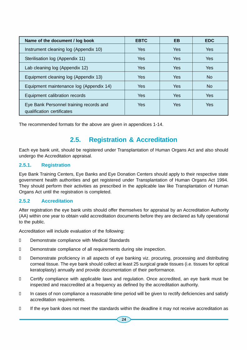
The following documents, logbooks and records are to be maintained by EBTC, EB and EDCs:
Name of the document / log book EBTC EB EDC
Donor initial information form (Appendix 1) Yes Yes Yes
Death Certificate / Eye Donor Medical Particulars(Appendix 2) Yes Yes Yes
Consent form (Appendix 3) Yes Yes Yes
Donor Information Sheet (Appendix 4) Yes Yes Yes
Serology Report (Appendix 5) Yes Yes No
Tissue Evaluation Report (Appendix 6) Yes Yes No
Haemodilution form (Appendix 7) Yes Yes Yes
Tissue distribution form (Appendix 8) Yes Yes No
Surgeons’ adverse finding report form Yes Yes Yes(Appendix 9)
24
The recommended formats for the above are given in appendices 1-14.
2.5. Registration & AccreditationEach eye bank unit, should be registered under Transplantation of Human Organs Act and also shouldundergo the Accreditation appraisal.
2.5.1. Registration
Eye Bank Training Centers, Eye Banks and Eye Donation Centers should apply to their respective stategovernment health authorities and get registered under Transplantation of Human Organs Act 1994.They should perform their activities as prescribed in the applicable law like Transplantation of HumanOrgans Act until the registration is completed.
2.5.2 Accreditation
After registration the eye bank units should offer themselves for appraisal by an Accreditation Authority(AA) within one year to obtain valid accreditation documents before they are declared as fully operationalto the public.
Accreditation will include evaluation of the following:
l Demonstrate compliance with Medical Standards
l Demonstrate compliance of all requirements during site inspection.
l Demonstrate proficiency in all aspects of eye banking viz. procuring, processing and distributingcorneal tissue. The eye bank should collect at least 25 surgical grade tissues (i.e. tissues for opticalkeratoplasty) annually and provide documentation of their performance.
l Certify compliance with applicable laws and regulation. Once accredited, an eye bank must beinspected and reaccredited at a frequency as defined by the accreditation authority.
l In cases of non compliance a reasonable time period will be given to rectify deficiencies and satisfyaccreditation requirements.
l If the eye bank does not meet the standards within the deadline it may not receive accreditation as
Name of the document / log book EBTC EB EDC
Instrument cleaning log (Appendix 10) Yes Yes Yes
Sterilisation log (Appendix 11) Yes Yes Yes
Lab cleaning log (Appendix 12) Yes Yes Yes
Equipment cleaning log (Appendix 13) Yes Yes No
Equipment maintenance log (Appendix 14) Yes Yes No
Equipment calibration records Yes Yes Yes
Eye Bank Personnel training records and Yes Yes Yes
qualification certificates

25
an eye bank and may be re-designated as an eye collection center. The State Registration Authorityshall be informed about failure to meet accreditation requirements and to cancel registration underTransplantation of Human Organs Act.
2.5.3. Accreditation Authority
Shall be a body comprising of nominees by Government of India, State Government, any other nominatedby NPCB and EBAI.
2.6. Eye Bank InspectionThe Accreditation Committee shall be responsible for inspecting each Eye Bank as outlined in the writtenprocedures of the EBAI and the Government of India.
Accreditation and reaccreditation site inspections shall be scheduled following written notification of theimpending inspection. Unannounced inspections may be conducted in case of receipt of any allegation ofviolation of “Medical Standards” by any eye bank. Failure to permit an inspection will result in suspensionor revocation of an eye bank’s accreditation and registration under Transplantation of Human Organs Act.
2.6.1. Procurement of Eye Bank Essentials
The Eye Bank should have a policy and procedure for maintaining sufficient stocks of essential eye banksupplies and also a procedure for procuring eye bank supplies. Procurement procedure has to bedocumented and produced at the time of site inspection.


STANDARD OPERATINGPROCEDURES OF EYE BANKS
Chapter-3Chapter-3Chapter-3Chapter-3Chapter-3


CHAPTER - 3
Standard Operating Procedures of Eye Banks3.1. Scope of the Manual
Personnel
3.1.1 Recruitment 33
3.1.2 Personnel & Responsibilities 33
3.1.3 Medical Director 33
3.1.4 Executive Director 34
3.1.5 Technical Staff 34
3.1.6 Eye Donation Counselors 34
3.1.7 Secretary 35
3.2. Appraisal & PromotionSkill EnhancementTraining & Certification (In case of an Eye Bank Training Center)
3.2.1 Training Centre 35
3.3. Eye Bank Facilities
3.3.1 Eye Bank Laboratories & Operations 35
3.3.2 Laboratory 1 – Handling Contaminated Materials 36
3.3.3 Laboratory 2 - Laboratory for Corneal Excision 36
3.3.4 Laboratory 3 – Cornea Evaluation 36
3.4. Cleaning, Maintenance & Calibration
3.4.1 Cleaning of Laboratories 36
3.4.2 Cleaning, Packing and Sterilization of Instruments 36
3.4.3 Cleaning, Maintenance and Calibration of Equipment 37
3.4.4 Equipment Cleaning 37
3.4.5 Equipment Maintenance & Calibration 37
3.5. Complaints 383.5. New Work 383.5. Waste Disposal 393.5. Infection Control & Safety 39
3.5.1 Infection 393.5.2 Accidents 403.5.3 Immunisation 40
3.5.4 Training 41
3.6. Donor Tissue Recovery & Screening of Donor Blood
3.6.1 Introduction 41
3.6.2 Objectives 41

3.7. Pre Recovery Procedures
3.7.1 Pre Recovery Review and Donor Preparation 423.7.2 Consent for Ocular Tissue Recovery 423.7.3 Identify the Donor 423.7.4 Worksite Preparation 433.7.5 Gross Inspection of the Donor 43
3.8. Donor Preparation
3.8.1 Donor Face Cleaning 433.8.2 5% Povidone-Iodine Solution Preparation 433.8.3 Lid Margin and Lash Prep 433.8.4 Conjunctiva Prep 443.8.5 Globe Prep 443.8.6 Lids and Face Prep 443.8.7 Sterile Field Preparation 453.8.8 Attire of Field Personnel 45
3.9. In Situ Corneal ExcisionMaterials
3.9.1 Corneo Scleral Rim Excision 463.9.2 Lab Corneal Excision 483.9.3 Donor Cornea Evaluation 493.9.4 Slit Lamp Examination 503.9.5 Specular Examination 54
3.10. Sclera Processing & PreservationSerological Testing of Donor Blood Sample
3.10.1 Blood Sample for Serological Testing 573.10.2 Drawing of Donor Blood Sample 583.10.3 Transferring Blood from Syringe to Vacutainer Tubes 593.10.4 Blood Tube Labeling and Handling 593.10.5 Screening of Human Donor Blood Sample for HIV, 59
Hepatitis B, Hepatitis C & Syphilis
Appendix - 1 60Donor Call- Initial Information
Appendix - 2 61Eye Donor Medical Particulars
Appendix - 3 62Familiy Pledge Form For Eye Donation
Appendix - 4 63Donor Information Sheet

31
Appendix - 5 65Immunology Laboratory Serology Report
Appendix - 6 66Tissue Evaluation Report
Appendix - 7 68Hemodilution Assessment
Appendix - 8 69Tissue Distribution InformationDuplicate form of Appendix - 8 71
Appendix - 9 72Donor Tissue Tentative Utility Data & Adverse Report Form
Appendix - 10 74Instrument Cleaning Log
Appendix - 11 75Instrument Sterilization Log
Appendix - 12 76Laboratory Cleaning Log
Appendix - 13 77Humanorgan Transplantation Act (HOTA)
Appendix - 14 97Laboratory Equipment Cleaning Log
Appendix - 15 98Ferquently asked questions About Eye Donation
Appendix - 16 100Facts and Myths About Donating Eyes
Appendix - 17 101Eye Donation Counsellor (EDC)Daily Report Form
Appendix - 18 102Eye Donation Counsellor (EDC) ReportCase Summary
Appendix - 19 103Eye Donation Counsellor (EDC)Monthly Report Form
Appendix - 20 106Financial Assistance available under NPCB
Appendix - 21 109Eye Banks* In India

32

33
Chapter-3
Standard Operating Procedures of Eye Banks
3.1 Scope of the Manual
The Eye Bank manual on ‘Policies and Standard Operating Procedures’ is structured to comply with thelaws, regulations and prescribed standards in eye banking so that the information contained in the manualwill contribute to the establishment, application and maintenance of effective, efficient and safe technicaland medical standards in eye banking.
The manual is subject to change, to allow acceptable variation within the scope of eye bank policies andophthalmologic practice. The Medical Director/Eye Bank Incharge and the Executive Director or Designatewill review the manual once a year.
The manual describes i). policies related to recruitment of staff, trainees (from eye donation centers forCME), HCRP, accreditation, data documentation and reporting, and ii). procedures related to maintenanceof eye bank laboratories, instruments and equipment, infection control and safety, identification andscreening of donors, donor cornea recovery, tissue evaluation, preservation & distribution and scleraltissue processing.
Personnel3.1.1. Recruitment
The staff requirement of the organization is initially discussed between the Directors (Medical Directorand the Executive Director or Designate) of the eye bank. A written request is then submitted to theBoard or competent authority after whose approval, it is forwarded to the Personnel department forreleasing an advertisement. In the Eye Banks attached to Govt. Institutions the staff available from Govt.source to be designated and trained specifically for Eye Bank purpose.
All recruits shall be under observation for the first 6 months. They are required to submit their bio dataalong with copies of educational and experience certificates to the Personnel department.
3.1.2. Personnel & Responsibilities
3.1.3. Medical Director/Officer Incharge Eye Bank
The Medical Director of the eye bank shall be an Ophthalmologist appointed by the Management and asper the Standards of Eye Banking in India.
1. The Medical Director shall provide advice for all medical aspects of the eye bank operations.
2. He/She shall be responsible for the formulation, approval, and implementation of medical policiesand procedures.
3. He/She shall ensure that the medical issues are in compliance with the existing Medical standards.
4. The final responsibility for determination of suitability of each tissue for transplantation is with thecorneal transplant surgeon. In cases of discrepancies, the Medical Director shall be consulted.
5. He/She shall contribute to the training and CME programs of the eye bank.
6. The Medical Director shall work in close association with the Executive Director or Designate of theeye bank in all administrative and scientific matters concerning eye banking.

34
3.1.4. Executive Director or Designate
The Executive Director shall be a qualified and experienced person with experience in a laboratoryatmosphere or hospital management, appointed by the Board or competent authority, and trained in alltechnical, administrative, and scientific aspects of eye banking.
1. All administrative policies and procedures of the eye bank shall be under the supervision of theExecutive Director or designate.
2. He/She shall be responsible for the day to day administrative, medical and scientific operations ofthe eye bank under the supervision of the Medical Director.
3. He/She shall devise and implement systems for quality assurance at all levels of eye bank operations.
4. He/She shall be responsible for delegating responsibilities to the technical and other staff of the eyebank.
5. He/She shall train and supervise technical staff in operations related to donor cornea procurement,tissue storage and distribution.
6. He/She shall be responsible for conducting the annual appraisals of the technical staff of the eyebank.
7. He/She shall organise training and CME programs of the eye bank..
8. He/She shall be responsible for organising the accreditation procedures as per requirements.
9. He/She shall be responsible for monitoring the Cornea Retrieval programs in various hospitals.
10. He/She shall be responsible for providing donor tissue utility reports/project reports to the eye donationcenters, funding agencies, EBAI, Government and other agencies.
3.1.5. Technical Staff
An eye bank technician shall be qualified (who has undergone certification course in eye bankingtechniques from certified training centers) and appointed by the Board as per “ Standards of Eye BankingIn India” document.
1. He/She Shall carry out work related to donor cornea procurement, donor cornea storage & distribution,screening of donor blood, data documentation, cleaning and maintenance of instruments andequipment, scleral tissue.
2. He/She shall maintain the daily log forms and laboratory log books as presciibed in the “Standardsof Eye Banking In India” .
3. He/She shall generate daily, monthly, quarterly and annual reports and submit to the ExecutiveDirector or Designate of the eye bank.
4. He/She shall report to the Medical Director / Executive Director or Designate of Eye Bank in allmatters related to eye banking.
3.1.6. Eye Donation Counselors
An eye donation counselor shall be selected by the Directors of the eye bank selection committee of GOIwho shall be familiar with all issues related to eye banking, and trained in the art of grief counseling.
1. He/She shall work in a multi specialty hospital to motivate family members of the deceased to makean eye donation.
35
2. He/She shall generate daily and monthly reports and submit it to the Executive Director or Designate.
3. He/She shall report to the Executive Director or Designate in all matters related to HCRP.
3.1.7. Secretary
The Secretary shall be appointed by the board or competent authority and provides full time secretarialservice to the eye bank as per the requirements of the organization.
1. He/She shall handle work related to donor call, pledge cards, providing certificate of appreciation todonor families, all correspondence, reports, monthly statement of expenditure, purchase,documentation of donor data entry in the system, leave status of eye bank staff
2. He/She shall report to the Executive Director or Designate of Eye Bank in all matters.
3.2. Appraisal & PromotionAnnual appraisal for all recruits of the eye bank (technical staff and the Secretary) is held by an appraisalcommittee consisting of the Nominees from the Board, Executive Director or Designate and MedicalDirector of the Eye Bank. The appraisal letters are issued to the staff through the Executive Director orDesignate of Eye Bank.
Skill Enhancement
The Eye Bank Directors (Medical Director and the Executive Director or Designate) are required toparticipate in meetings and workshops related to eye banking. The technical staff members are requiredto regularly attend CME programs.
TRAINING & CERTIFICATION (In case of an Eye Bank Training Center)
3.2.1 TRAINING CENTRE ( Applicable for Eye Bank Training Centers Only.)
The center shall strive to offer certification course to different cadres of eye bank personnel, and shall bea pioneer in setting standards of excellence in the field of eye banking. Eye bank professionals from Indiaand other countries shall be educated in the planning of eye banking services, policy making in eyebanking and eye banking techniques. The eye bank shall conduct workshops periodically to update eyebank professionals in the basics and advances in eye banking. The eye bank training PROSPECTUSshall be made available to all members of EBAI. The details of different training courses/Observershipoffered at the training center, admission and certification procedures etc shall be provided.
3.3 EYE BANK FACILITIESThe eye bank is away from areas that are open to unrestricted traffic flow and has area for carrying out itsoperations. The eye bank has a reception room and 4 eye bank laboratories, each separated from theother by a self- closing door. All laboratories are fitted with UV lamps.
3.3.1. Eye Bank Laboratories & Operations
3.3.2. Laboratory 1 – Handling Contaminated Materials
All materials for decontamination are handled in this laboratory. The laboratory has a, sink and a workbench.

36
3.3.3. Laboratory 2 - Laboratory Corneal Excision
Laboratory 2 is used for i). laboratory corneal excision from whole globes received from other eye banks/eye donation centers ii). processing of sclera . The laboratory houses a Laminar Flow Hood and a workbench.
3.3.4. Laboratory 3 – Cornea evaluation
This laboratory houses the specular microscope for the evaluation of donor corneas. The donor corneas(in storage medium) are also stored in a refrigerator kept in this laboratory.
3.4. Cleaning, Maintenance & Calibration3.4.1. Cleaning of Laboratories
The floor is first cleaned with a disinfectant (phenyl 1:100) before and after work. The floor is then moppedwith a clean dry cloth. The work- benches are wiped with Dettol or equivalent before and after work andthen mopped with a clean dry cloth. Any spill is first wiped with absorbent cotton soaked in the disinfectant(70% ethanol or 0.5 to 1.0% sodium hypochlorite). The surface is wiped twice again with fresh clothsoaked in the disinfectant.
3.4.2. Cleaning, Packing and Sterilization of Instruments
Used instruments are placed in a tray containing 1% ACEPTIK (Chlorhexidine gluconate (7.5%), cetrimide(15%), isopropyl alcohol 7%) for 15 minutes. After soaking the instruments for 15 minutes, instrumentsare scrubbed with a soft bristled brush. Instruments are brushed under tap water flow to avoid splashingof contaminated material with foam water. After scrubbing, each instrument is rinsed thoroughly. Theprocedure is repeated to ensure sterility.
The rinsed instruments are placed on a fresh washed cloth to allow the instruments dry. No instrumentshould be wiped with a cloth.
Wrapping Instruments for Sterilization
The washed instruments are placed in a clean washed cloth before placing them in a washed steel tray.The cloth is laid to protect the instruments. Then the instruments are covered with 2 cotton pads to giveextra security for the instruments. A SIGNALOC indicator is placed inside the tray before wrapping theinstrument tray. The tray is then double wrapped. A label giving the item number, contents, mode ofsterilization, date and time of packing, signature of the technician, along with another SIGNALOC indicatoris stuck on the outside of the wrapped instrument tray before sending for sterilisation to the OperationTheatre.
Corneal excision set
l Drapes, torch, loupe
l Disposable Syringes (5 ml-1, 10 ml - 1) with disposable needles (21 G & spinal needle) with 2 testtubes or plain vials to collect blood sample.
l Conjunctival Scissors
l Corneal scissors (Right & Left)

37
l Fixation forceps
l Iris forceps
l Spring or Wire Speculum
l Bard Parker handle with sterile surgical blade (no. 11 or 15)
Enucleation set
l Torch
l Disposable Syringes (5 ml-1, 10 ml - 1) with disposable needles (21 G & spinal needle) with 2 testtubes or plain vials to collect blood sample.
l Conjunctival Scissors
l Fixation forceps
l Artery forceps
l Muscle hook
l Enucleating spoon
l Strabismus Scissor
l Wire or Spring Speculum
l Bard Parker Handle with sterile surgical blade (no. 11 or 15)
3.4.3. Cleaning, maintenance and calibration of equipment
The following equipment are maintained in the eye bank
l Refrigerators
l Laminar Flow Hoods
l Specular Microscope
l Slit Lamp Microscope
3.4.4. Equipment cleaning
The outside and inside of the equipment is wiped with 70% alcohol once daily. When there is a spill ofinfected material on any of the equipment, the surfaces are wiped with absorbent cotton or gauze piecesoaked in 70% alcohol or sodium hypochlorite solution.
3.4.5. Equipment maintenance & calibration
The equipment is maintained as per the instruction manual supplied along with the equipment by themanufacturer. It is calibrated by the manufacturer or manufacturer authorized representative once a year.
An EQUIPMENT INSTRUCTION MANUAL is maintained on each equipment with the following detailspertaining to the equipment.
l Name of the equipment

38
l Date of purchase/Model Number
l Supplier/Service Agent (Name & Address)
l Calibration certificate
l Service report
l Instruction manual
l Performance record
l List of authorised users
A tag is displayed on each of the equipment, mentioning the following details
l Date of calibration
l Calibration due on
l Service due on
6. Refrigerators
In house calibration for the Refrigerator.
Laminar Flow Hood: An external agency has to be used for calibration and maintenance. The LFHcalibration using a “Laser Particle Counter (0.5 and 5.0µ size) velocity is measured by velometer and theresults and date is to be recorded.
3.5. ComplaintsA COMPLAINT/ACCIDENTS/ERRORS LOG for documenting the following information is maintained atthe Eye Bank.
l Date of complaint
l Complaint made by
l Complaint recorded by
l Corrective measure taken
l Date of corrective action
l Complaint closed on
l Signature of the Medical Director
Complaints (errors, accidents) are brought to the notice of the Executive Director or Designate whodeputes a person to take corrective action.
New Work (Applicable for Eye Bank Training Centers)
New work/test is undertaken or any modification in the methodology is introduced based on the feasibilityand the logistics of introducing new tests. Once a decision is made for introducing the new work, SOP will

39
be prepared, technician will be trained and new records will be introduced. The NEW WORK LOG ismaintained in the eye bank for the documentation of all details pertaining to the work initiated.
Waste Disposal
Sharps (needles and blades) are discarded in puncture proof containers (containing 1% sodium hypochloritesolution) through a hole in the lid of the container.
Bio Medical Waste is segregated in color coded bags as followed by local regulations:
Yellow Bag (Infectious)
Discarded eye balls
Used cotton
Massage pads
Red or Blue Bag (Infectious)
Gloves
Disposable syringes
Swabs
Saline bottle
Black Bag (Non infectious)
Paper
Stationary
Separate plastic bins containing thick yellow and red plastic covers are placed in all the Laboratories ofthe eye bank. Plastic bins with black bags are placed in the reception room.
All waste bags depending upon the classification are handled by authorised personnel and disposed inappropriate manner as prescribed in the Transplantation of Human Organs Act.
All waste bags placed at a designated place for the pick up by Biomedical waste collection agency. Therecords of disposable material are maintained by the eye bank.
WASTE DISPOSAL LOG is maintained in the eye bank for recording daily activities related to wastedisposal.
Infection Control & Safety
The laboratory personnel are required to be trained to minimise the risk of contracting infectious diseaseand accidents.
3.5.1. Infection
1. The eye bank shall be away from areas that are open to unrestricted traffic flow.
2. The eye bank laboratory doors and windows shall be kept closed all the time.
3. The laboratory furnishings shall be cleaned and disinfected immediately after use.

40
4. Universal precautions shall be followed while dealing with donor samples.
i. Attire: Confine long hair and loose clothing, wear shoes and not sandals, wear front opening
laboratory coats.
ii. Handling of clinical samples: Handle clinical samples only with gloved hands while working in
serology laboratory, avoid contaminating ungloved hands, skin or eyes while handling clinical
samples, remove gloves and wash hands with soap and water after handling clinical samples,
avoid dropping clinical samples, avoid spillage.
5. UV lights shall be provided in the eye bank.
6. Donor materials shall be handled in biosafety cabinets.
7. Surfaces of LFH shall be decontaminated after a spill.
8. Eye bank personnel shall be vaccinated against Hepatitis B.
9. Pipette aids shall be used.
10. Protective clothing such as gloves, gown and mask shall be used by the eye bank technician while
dealing with donor material.
11. Hands shall be washed before and after handling infected materials.
12. No drinking and eating shall be allowed inside the eye bank.
13. Air sampling shall be performed at regular intervals and recorded.
14. Particle count of the LFH shall be performed at regular intervals.
15. LFH filters shall be replaced if particle count is beyond the acceptable limit.
16. UV lamps shall be replaced as per the manufacturers’ specifications.
17. Proper system of waste disposal shall be followed.
3.5.2. Accidents
1. Smoke detectors shall be provided in the eye bank.
2. A safety station with showers and an eye wash facility shall be provided close to the eye bank
3. Open cuts shall be bandaged while working in the eye bank
4. First aid kit shall be placed in a reachable place in the eye bank laboratory.
3.5.3. Immunisation
Eye bank technicians who comes in contact with needles, blood or blood products are immunized for
Hepatitis B.
41
3.5.4. Training
The eye bank personnel are trained in the following
1. to follow the instructions of the manufacturer for using the equipment
2. to follow SOPs in the cleaning of laboratories, equipment and instruments.
3. to report to the Executive Director or Designate of the eye bank if equipment is found non functional.
4. to maintain the log on a daily basis.
5. to use the FIRST AID KIT which is placed in strategic position in the laboratory.
6. to check the contents of the first aid kit expiry dates and replace the items.
7. to manage cuts and bruises (Clean the foreign body, clean the area with a disinfectant, bandageadequately, and to consult a physician if required)
8. to manage burns (Plunge the affected part in cold water to soothe the pain, dry the surface, applyburn ointment, cover the part loosely with a dry gauze piece, consult a physician if required.
9. to manage eye injuries (wash out the chemical with large quantity of clean water, rush the subject tosafety station for eye wash, apply antibiotic drops, consult a physician if there is foreign matter to beremoved.
Standards in Eye Banking Medical Standards - Manual
This manual is a document based on the Medical Standards of Eye Banking, prescribed by Ministry ofHealth & Family Welfare, Government of India and EBAI. This is to serve as a guideline and each eyebank should maintain procedures and follow regulations as mentioned in the Standards document.
3.6. Donor Tissue Recovery & Screening of Donor Blood3.6.1. Introduction
This manual describes the standard operating procedures followed at the Eye Bank:
1. Pre recovery procedures
2. In situ corneal excision & preservation
3. Cornea evaluation
4. Scleral processing & preservation
5. Drawing of blood sample from the donor
6. The screening of donor blood sample for HIV, Hepatitis B surface antigen, Hepatitis C and Syphilis.
3.6.2. Objectives
To provide comprehensive uniform eye donor screening, a standard method for the aseptic removal ofhuman donor corneas for surgical use (in situ and laboratory excision) that would minimise the endothelialcell loss, to describe the donor cornea evaluation procedures (slit lamp & specular microscopy), preparationand storage of sclera, procedure for obtaining blood sample from a donor for the purpose of serologicaltesting, and serological screening methods.

42
3.7. Pre Recovery Procedures
3.7.1. Pre Recovery Review and Donor Preparation
l Check expiration dates on sterile instrument kits and other supplies before traveling to the donorsite
l Pack necessary instruments kits as well as all necessary supplies into a clean transport case (steelbin) for transport to the donor site.
l Transport supplies to the donor site
l The following documentation forms are required for all donor recoveries:
t Donor call - initial information sheet
t Consent form
t Donor information sheet
t Eye donor medical particulars
t Hemodilution Assessment sheet
3.7.2. Consent for Ocular Tissue Recovery
Ensure documentation of consent is consistent with existing policy as prescribed by EBAI.
Ensure that the consent has been obtained from the correct individual according to hierarchy of personswho may make an anatomical gift specified by state law.
Ensure that the consent clearly authorizes recovery of the specific tissues for which the recovery isplanned.
Ensure that the consent gives authorization to obtain blood specimen for infectious disease screening,including HIV, Hepatitis B, and syphilis
If legislative consent is applicable, ensure that all required conditions are met.
Review the donor’s medical history before proceeding with the recovery.
Verify the accuracy of any verbally reported information.
Complete medical history in the Eye Donor Medical Particulars sheet.
Complete Hemodilution form.
3.7.3. Identify the donor
l Match the name on the consent form to the name on the donor’s ID tag, toe tag, bracelet, etc.
l Do not presume the identity of the donor or rely on a nurse or morgue attendant to point out thedonor.
l Institutional staff shall participate in documentation of steps taken to confirm the ID of an untaggedor unidentified potential donor.

43
3.7.4. Worksite Preparation
l Identify a suitable workplace (counter space near the donor on which to set up your paperwork andsterile fields).
l Clean this area with a suitable disinfectant and wipe it dry with paper towels.
l Wear gloves and mask
3.7.5. Gross Inspection of the Donor
l Examine the donor’s entire body for evidence of high risk behaviour and infectious disease includingphysical evidence for risk of sexually transmitted disease such as herpes simplex, Syphilis, physicalevidence of non medical percutaneous drug use such as needle tracks, disseminatedlymphandenopathy, oral thrush, blue or purple spots consistent with Kaposi’s sarcoma, unexplainedjaundice, hepatomegaly or icterus.
If the body was rejected for routine autopsy due to infectious criteria or if the autopsy was done in aninfectious disease control room or under any special precautions obtain reasons for these procedures.
If evidence of high risk behaviour or infectious disease is identified DO NOT proceed with therecovery.
l Perform a penlight examination of the donor’s eyes and periorbital area.
l Obtain a suitable blood sample for serological testing (See section H8.520)
3.8. Donor Preparation3.8.1. Donor face cleaning
The following steps should be performed only when gross debris is observed on the donor’s face duringthe periocular exam.
l Don non-sterile examination gloves.
l Irrigate the donor’s eyes and face with a sufficient quantity of sterile balanced salt solution to removeall visible debris from the eyes and to moisten the face.
l Use non-sterile 4 x 4 inch cotton squares to remove residual debris from the moistened face.
l Remove the non-sterile exam gloves and place them in an appropriate biohazard waste container.
3.8.2. Application of 5% Povidone-Iodine Solution Preparation
3.8.3 Lid Margin and Lash Prep
Use a Cotton Tip Applicator (CTA) to open the upper lid and roll the lid margin and lashed away from theglobe.
Use a second CTA dipped in 5% povidone-iodine solution to scrub the upper lid margin and lashes onetime across in a circular motion for approximately 20 seconds.
Next, use a CTA to open the lower lid and roll the lid margin and lashes away from the globe.
Use another CTA dipped in 5% povidone-iodine solution to scrub the lower lid margin and lashes one timeacross in a circular motion for approximately 15 seconds.

44
Repeat the povidone-iodine solution scrubbing of the upper and lower lid margin and lashes two moretimes with fresh CTAs.
Repeat this procedure for the other eye.
3.8.4. Conjunctiva Prep
Dip a cotton tipped applicator (CTA) in the 5% povidone-iodine solution and sweep the lower conjunctivalfornix from the lateral canthus towards the nose on time.
The bulb of the CTA should be fully immersed since dry cotton will stick to the eye. Be careful not to rubthe CTA across the cornea.
Holding the CTA perpendicularly to the donor will make the sweep easier to perform correctly. If necessary,a CTA may be used to open the eye.
Dip a second CTA in 5% povidone-iodine solution and sweep the upper conjunctival fornix from thelateral canthus towards the nose one time.
Repeat this procedure for the other eye.
3.8.5. Globe Prep
Fill the sterile syringe with approximately 1.5cc of the 5% povidone-iodine solution.
Use a CTA to gently roll back the upper lid and slowly drip approximately 0.75 cc of the 5% povidone-iodine solution on the eye.
Gently roll back the lower lid with a CTA and slowly drip the rest of the povidone-iodine solution on theeye.
Repeat this procedure for the other eye.
3.8.6. Lids and Face Prep
Prep the skin and facial area extending from the scalp to baseline of the nose by swabbing with foldedsterile 4 x 4’s dipped in 5% povidone-iodine solution.
Blot off the excess 5% povidone-iodine solution on the side of the plastic tray to avoid dripping on thework area.
The prep begins at the lid margins and works in a circular pattern outward.
Repeat this procedure for each eye two more times alternating eyes.
It is very important that the povidone-iodine is left on the eyes and skin for at least three to five minutesbefore irrigating the eyes. Irrigation will be performed after opening sterile supplies for the appropriatesurgical procedure performing a 3 – 5 minute surgical scrub, and donning sterile gloves and sleeves.
Carefully place the bottle of balanced salt solution off the sterile field in an upright position.
Discard remaining prep supplies in a biohazard waste container.
Remove sterile prep gloves and discard them in an appropriate biohazard waste container.

45
3.8.7. Sterile Field Preparation
Open the outside packages of two pair of sterile gloves then open the inner envelopes using aseptictechnique and leaving gloves on their sterile fields.
Use a dry foam skin disinfectant product on hands and forearms to reduce microbial flora prior togloving and sleeving.
Open the outer wrap of the appropriate surgical instrument pack taking care to avoid contaminatingthe sterile field being created by touching anything other than the wrapper edges or reaching overthe field.
Carefully open and aseptically drop sterile 4 x 4 inch gauze squares, sterile cotton tipped applicators,and sterile rubber bands on to the sterile portion of the outer wrap of the surgical instrument pack.
Open the sterile eye drape, taking care to avoid contaminating the sterile field being created bytouching anything other than the wrapper edges or reaching over the field and leave the drape on itssterile field.
Label the eye jars or corneal storage media vials (a unique number specific to the donor, or thedonor’s name, and whether the specimen is left or right), loosen the caps to the top thread and placethe jars or vials adjacent to a top corner of the instrument sterile field.
Place labeled containers for blood and/or vitreous samples near the sterile field along with hypodermicequipment and cosmetic restoration materials.
3.8.8. Attire of Field Personnel
Don sterile gloves taking care to fully extend the cuffs and to avoid contaminating the outer surface ofthe gloves.
Open the inner wrap of the instrument kit taking care to avoid contaminating gloves by contact withunsterile surfaces while handling the edges of the instrument wrapper. If necessary, place a sterilerubber band over each cuff of the sterile sleeves to ensure a good seal.
Irrigate the povidone-iodine solution from the eyes then drape the donor.
Use one hand to pick up the balanced salt solution bottle and the other hand to pick up a sterile CTAfrom the sterile instruments.
The hand used to pick up the balanced salt solution is now contaminated. Do not use the contaminatedhand to retrieve items from the sterile field or to touch the donor.
Gently open the lids with the sterile CTA, then flush the povidone-iodine solution off the glove andconjunctiva. Be certain to irrigate off all povidone-iodine solution.
Pick up a sterile CTA with the sterile hand and repeat the irrigation procedure for the other eye.
Using the hand that was used to open the lids (clean hand) pick up the sterile 4 x 4 inch gauze and
gently blot dry the lid of the first eye to remove excess moisture from the lid skin. Take care not to
touch the cornea with the 4 x 4 inch gauze.

46
Perform the same blotting procedure on the other eye.
Remove outer pair of gloves being careful not to contaminate the inner pair of gloves and ensure that
they are discarded in an appropriate biohazard container.
Carefully position the sterile bilateral eye drape on the donor.
3.9. In Situ Corneal Excision Materials1. Corneal excision set (autoclaved)
2. Enucleation set (autoclaved)
3. Linen pack (autoclaved)
4. Other materials
1. Corneal Excision set
i. S.S. Tray (Small) 1
– Disposable Syringes (5 ml, 10 ml) 1 (each with disposablesneedles (21 G)
– Test tubes or plain vials 2
ii. Spring Scissors 1
iii. Castro-veijo Corneal scissors (L&R) 1 (each)
iv. Fixation forceps 2
v. Iris forceps 1
vi. Spring Speculum 1
vii. Wire Speculum 1
viii. B.P. Blade Handle 1
ix. Lens Spoon 1 (optional)
x. Surgical Blades (11 & 15) 1 (each)
2. Enucleation set
i. S.S. Tray 1
– Disposable Syringes (5 ml, 10 ml) 1 (each with disposablesneedles (21 G)
– Test tubes or plain vials 2
ii. Spring Scissors 1
iii. Strabismus Scissors 1
iv. Enucleating Scissors 1
v. Fixation forceps 1
vi. Artery forceps 1
vii. Enucleating spoon 1
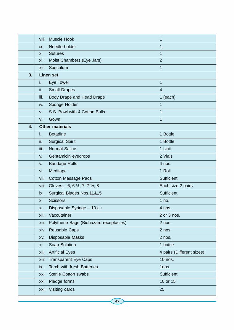
47
viii. Muscle Hook 1
ix. Needle holder 1
x Sutures 1
xi. Moist Chambers (Eye Jars) 2
xii. Speculum 1
3. Linen set
i. Eye Towel 1
ii. Small Drapes 4
iii. Body Drape and Head Drape 1 (each)
iv. Sponge Holder 1
v. S.S. Bowl with 4 Cotton Balls 1
vi. Gown 1
4. Other materials
i. Betadine 1 Bottle
ii. Surgical Spirit 1 Bottle
iii. Normal Saline 1 Unit
v. Gentamicin eyedrops 2 Vials
v. Bandage Rolls 4 nos.
vi. Meditape 1 Roll
vii. Cotton Massage Pads Sufficient
viii. Gloves - 6, 6 ½, 7, 7 ½, 8 Each size 2 pairs
ix. Surgical Blades Nos.11&15 Sufficient
x. Scissors 1 no.
xi. Disposable Syringe – 10 cc 4 nos.
xii.. Vaccutainer 2 or 3 nos.
xiii. Polythene Bags (Biohazard receptacles) 2 nos.
xiv. Reusable Caps 2 nos.
xv. Disposable Masks 2 nos.
xi. Soap Solution 1 bottle
xii. Artificial Eyes 4 pairs (Different sizes)
xiii. Transparent Eye Caps 10 nos.
ix. Torch with fresh Batteries 1nos.
xx. Sterile Cotton swabs Sufficient
xxi. Pledge forms 10 or 15
xxii Visiting cards 25

48
3.9.1. Corneo scleral rim excision
Removal of the corneal-scleral rim requires rigid sterile technique employing accepted aseptic practice.The potential for endothelial cell damage, contamination and infection are greatly increased and thisprocedure therefore shall be performed by individuals specifically trained in in-situ retrieval by RMPstrained in removal.
Open the eyelids using a sterile cotton tipped applicator or a muscle hook and insert a spring/wire speculum.Take care to avoid scratching the cornea.
Use small clawed forceps and iris or tenotomy scissors to lift and cut the conjunctiva at the limbus 360degrees. Any adhesion between the conjunctiva and the anterior globe is separated using blunt andsharp dissection technique to ensure that the conjunctiva is not in contact with the anterior globe within 5mm of the limbus. It may be necessary to make several relaxing cuts in the conjunctiva radially to accomplishthis.
The exposed sclera is carefully scraped from the limbus outward with a scalpel blade (#11 or #15) toremove all remaining conjunctival tissue. It is important to remove all conjunctiva flush to the limbusbecause the conjunctival tissue may contain microbial contaminants which should not accompany thecornea to the storage media.
Isolate the scalpel and other instruments used to scrape the conjunctiva from the other instruments onthe sterile field. These instruments should be placed mid peripherally on the sterile field and never placedcentrally on top of the sterile instruments, which have not been contaminated by conjunctiva. Use theinstruments which have touched conjunctiva only for the conjunctival removal on the other eye.
Use a second scalpel with a #15 blade and small clawed forceps to make an incision through the sclera2 mm to 4 mm from the limbus and parallel to the limbus approximately 5 mm in length. Care must betaken to cut all the way through the sclera without perforating the choroid, as, this would cause vitreousleakage which may cause collapse of the globe including the anterior chamber and compromise thecornea.
Continue the scleral incision 360 degrees using corneal section scissors (Castroviejo or Abeli) and smalltissue forceps to stabilize the posterior aspect of the scleral incision if necessary. Take great care to avoidperforating the choroid breaking into the anterior chamber or causing any deformation of the cornea’snormal curvature.
Trauma to the cornea during scissoring due to bending, loss of the anterior chamber or collapse of theglobe through vitreous loss would severely compromise its suitability for surgical use. If such trauma issuspected to have occurred, make a note on the recovery form,carefully. Evaluate the cornea with a slitlamp,and if damage cannot be ruled out the corneal shall not be offered for penetrating keratoplasty. If theincision has been made properly, the corneal-scleral button should be attached to the ciliary body-choroidonly at the scleral spur.
Complete the corneal removal by using one pair of small smooth dressing forceps to hold the scleral rimstationary and a second set of small smooth dressing forceps or an iris spatula to gently push the ciliarybody-choroid downward and away from the corneal-scleral button. Aqueous fluid should escape from theanterior chamber at this point assuring that the anterior chamber was indeed intact.

49
Remaining adhesions should be pushed gently away from the corneal-scleral button working side to sideand taking great care to avoid pulling on the cornea and creating folds. The corneal-scleral button may berolled side to side but never pulled in such a way as to cause cross cornea tension. Do not contaminatethe cornea by allowing it to touch the eyelids or other facial skin. In the event that this occurred, make anote on the tissue recovery form.
Continue to hold the cornea by the scleral rim with the small smooth dressing forceps and asepticallytransfer it to a labeled vial of storage media.
Lift the pre-loosened cap off the corneal media vial using a plastic backed sterile 4 x 4 inch gauze spongeimmediately prior to placing the cornea in the media and replace the cap immediately after placing thecornea in the media. Do not touch the threads on the media vial with the contaminated underside of theplastic backed gauze square. Remove the speculum and place it with the conjunctiva instruments on themid peripheral area of the instrument field.
After the second cornea is placed in storage media, remove contaminated surgical gloves, dispose of thegloves in a bio hazardous waste container, don non sterile exam gloves, then tighten the caps on bothmedia vials.
Gently palpate the iris and pupil of each remaining posterior segment with a blunt instrument to rule outaphakia or pseudophakia. Do not tear the iris or remove the lens.
Insert eye caps in front of the remaining posterior poles and gently close the eyelids. Do not use forcepsto close the lids, use only cotton tipped applicators or gloved fingers.
Place the corneal media vials in an ice box to which ordinary ice (NOT DRY ICE) has previously beenadded.
Discard all used disposables in a biohazard bag and all sharps in sharps container.
Rewrap instruments for transport, cleaning and sterilization.
Place a biohazard label on the contaminated instrument pack.
Replace the instrument pack in the bin until such time as it may be cleaned and re sterilized.
Clean the work area with disinfectant solution.
Discard personal protective attire in a biohazard bag.
Record information about the recovery (time of recovery, recovered by, etc.) on the donor recovery formand also on a form to leave in the donor’s medical record to document the donation.
Place a garland before the donor as a mark of respect.
Thank the donor family before leaving the donor site (hospital or residence).
3.9.2. Lab Corneal excision
Removal of the corneal-scleral rim requires rigid sterile technique employing accepted aseptic practice.The potential for endothelial cell damage contamination and infection are greatly increased and thisprocedure therefore shall be performed by individuals specifically trained in corneal excision.

50
The corneal excision from a whole eye donor shall be done in a laminar flow hood.
Don hair cover, surgical mask, and non sterile exam gloves then wipe down the work surface of thelaminar flow hood and any side tables which will be used to set up supplies with disinfectant solution.
Remove exam gloves and dispose of them in a suitable biohazard waste container.
Turn the laminar flow hood blower on and allow it to run for 15 minutes prior to beginning the procedure.
Place the sterile instrument pack, jars containing the donor eyes, labeled corneal storage medium vials,two 5 or 10cc syringes, two sterile specimen cups, 10% povidone-iodine solution, a non sterile hemostat,and balanced salt solution in the work area.
Open the outer wrap of the instrument pack on the work surface of the laminar flow hood taking care toavoid reaching over the field or touching anything except the outside edges.
Open two sterile 5 or 10 cc syringes and drop them on to the sterile field.
Place sterile specimen cups with the lids removed adjacent to the sterile field.
The eye jars are positioned so that they are adjacent to the top corner of the sterile field. Eye jars andmedia vials should be positioned to insure that left and right specimen identification remain correct. Theeye jar lids are removed and placed with the inner side up next to their respective jars.
The labeled storage media vials are positioned so that they will be next to one side of the sterile field andthe caps of the vials are removed and placed with the inner side up next to their respective vials.
Don sterile gloves.
Use the first sterile syringe to obtain 2.5 – 5.0 cc of the 5% povidone-iodine solution. The solution iscarefully dropped on to the eye making sure to coat the entire globe. In order to ensure complete coatingof the globe it may be necessary to move or tilt the eye cage using a sterile cotton tipped applicator(CTA). Repeat steps for the other eye. It is very important that the povidone-iodine solution is left on theeyes for at least five minutes before irrigation.
Discard sterile gloves in appropriate biohazard container.
Don sterile gloves taking care to fully extend the cuffs and to avoid contaminating the outer surface of thegloves.
Pick up the second sterile syringe and draw up at least 5cc of balanced salt solution.
Irrigate the eyes until all the povidone-iodine is rinsed off.
Aseptically open the inner wrap of the instrument kit.
Unfold a sterile 4 x 4 gauze sponge then refold it to form a long strip of gauze.
Aseptically lift the eye cage from the jar with a sterile hemostat, CTA, or plastic backed sterile gauze. Besure to allow excess povidone-iodine and balanced salt solution to drip off the globe and eye cage (ifapplicable) before bringing the globe on to the sterile field, to avoid wetting the sterile field. If eye jar doesnot have an eye cage, use a sterile forceps to lift the eye from the jar to the sterile field and allowingexcess fluid to drip off to avoid wetting the sterile field.

51
Remove the eye from the cage or dental roll using a small clawed forceps to grasp the rectus muscle thenwrap the eye securely several times around the equator with the long strip of gauze.
Use small clawed forceps and iris or tenotomy scissors to lift and cut any remaining conjunctiva at thelimbus and extending out 5 mm from the limbus.
Scrape the exposed sclera carefully from the limbus outward with a scalpel blade (#11 or 15) to removeall remaining conjunctival tissue.
It is important to remove all conjunctiva flush at the limbus because the conjunctival tissue may containmicrobial contaminants that should not accompany the cornea to the storage media.
The scalpel and any other instruments used to remove the conjunctiva should be isolated from the otherinstruments on the sterile field. Place these instruments mid peripherally on the sterile field (not on top ofthe centrally located instruments which have not been contaminated by conjunctiva).
These instruments should not be used again except to remove the conjunctiva of the second eye.
l Use a second scalpel with a #15 blade to make an incision through the sclera 2 mm to 4mm from thelimbus and parallel to the limbus approximately 5 mm in length.
l Place the wrapped eye down near the center of the sterile field.
l Gently separate the corneal-scleral button from the posterior segment of the globe.
l After the second cornea is placed in storage media, remove contaminated surgical gloves and nonsterile exam gloves, then replace and tighten the caps on both media vials.
l Replace and tighten the lids on both eye jars.
l Discard all used disposables in a biohazard bag and all sharps in a sharps container.
l Rewrap instruments for cleaning and sterilization.
l Place a biohazard label on the contaminated instrument pack until such time as it may be cleanedand re sterilized.
l Leave the hood blower motor on for 15 minutes following completion of the procedure.
l Clean the hood and work area with a suitable disinfectant solution.
Discard personal protective and surgical attire in a biohazard bag.
3.9.3. Donor Cornea Evaluation
3.9.4. Slit lamp examination
An Ophthalmologist is responsible for performing this procedure. Allow the cornea in storage media vialor whole globe in eye jar to reach room temperature to make the endothelium easier to visualize. Corneaor whole globe shall not be allowed to remain in room temperature for more than 1 hour. Limit the numberof warming and cooling cycles no more than 3 times.
The slit lamp examination shall be an orderly and stepwise examination of the different layers of thecornea. Each layer of the cornea shall be evaluated from front to back, and from back to front.

52
A wide slit of light is directed on the cornea with a 20 to 30 degree angle of incidence and moved to scanthe entire cornea.
Epithelium: Examine the epithelium for integrity and overall condition.
The location, extent, and depth of any epithelial defect should be fully examined and described on theCornea Evaluation Form.
Note any abrasions, lacerations, foreign bodies, microcystic edema, or severe drying.
Note any epithelial haze, exposure, or sloughing if present.
If epithelium is missing, it is necessary to carefully rule out underlying stromal injury.
Note the degree, location, type, and amount of epithelial haze, exposure and sloughing on the CorneaEvaluation Form.
Bowman’s layer
Examine the Bowman’s layer for any defect.
Examine the Bowman’s layer for corneal lacerations and scars by focusing a hairline slit on the epitheliumand carefully observing the defect margin for reflection off the Bowman’s layer.
If the hairline slit reforms at a deeper level, the depth of the defect is apparent and is usually minimal.
If the hairline slit does not reform at a deeper level, the Bowman’s layer may have been injured and thecornea would not be suitable for transplant.
Stroma
Examine the stroma to determine overall clarity and rule out marked edema.
Stromal edema may be evaluated by directing a hairline slit on the cornea at a 15 to 20 degree angle ofincidence, and observing the distance between the epithelial and endothelial reflexes.
Corneas without significant stromal edema results with the two reflexes to converge centrally and divergeperipherally.
Corneas with significant stromal edema results with the two reflexes nearly parallel or will diverge centrally.
Examine for imbedded foreign bodies, old scars, or stromal infiltrates.
At high magnification use a narrow slit beam to define the extent, depth, location and appearance ofstromal opacities.
A central stromal opacity would rule out a cornea for transplant surgery.
l Small peripheral stromal opacity (extending no more than 1-2 mm from the limbus) may or may notrule out a cornea for transplant surgery depending on whether or not the stromal opacity is determinedto be an infectious stromal infiltrate.
l Stromal scars generally have a more gray appearance without adjacent stromal edema.
l Examine for degenerative conditions such as arcus senilis.
l The presence of moderate to heavy arcus senilis that is evident within the central 8 mm of thecornea should be described and may disqualify the cornea for elective penetrating keratoplasty.

53
l Note the degree, location, type, and amount of stromal arcus, stretch lines, cloudiness, and opacitieson the Corneal Evaluation Form.
Descemet’s membrane
l Examine the Descemet’s membrane for the presence, location and severity of Descemet’s folds(striae).
l The presence of Descemet’s folds are determined by directing a narrow slit on the mid-peripheralcornea at an angle of 30 to 40 degrees, and focusing on the endothelial reflex at a medium power ofmagnification.
l The endothelial mosaic may be observed in the light colored areas, while the Descemet’s folds mayappear like dark lines, which transect the illuminated areas of the endothelium.
l The severity of the folds may generally be determined by the width of the folds and the amount ofendothelial area that they obscure from view.
The degree, location, type, and amount of folds shall be noted on the Corneal Evaluation Form.
Endothelium
Examine the overall integrity of the endothelial cell layer.
Examination of the endothelial cell layer is done by directing a narrow slit on the mid-peripheral cornea atan angle of 30 to 40 degrees, and focusing on the endothelial cell layer.
The endothelial layer is examined to rule out major defects or missing areas of the endothelium.
General appearance of the endothelium shall be compared from one area to another.
Examine the limbal area of excised corneas on the endothelial side with a broad slit at low to mediummagnification to rule out delamination of the Descemet’s membrane.
Examine the endothelial layer using specular illumination at high power magnification with a narrow slitdirected on the mid-peripheral cornea at a 30 to 40 degree angle of incidence.
Note the uniformity of cell size, estimated cell population, overall health of the cells, and uniformity of cellshape.
Look for presence of guttata, that occur with Endothelial Dystrophy. The presence of guttata shall rule outthe cornea for surgical use.
Examine for vacuolated cells that may be precursor to Decemet’s folds or striae due to trauma duringrecovery. The presence of vacuolated cells must be considered to be a degenerative condition of theendothelium and whether or not the cornea would be suitable for penetrating keratoplasty would dependon the relative number observed and their location.
A cornea with less than 15 or 20 vacuolated cells located mainly in the peripheral area may be entirelysuitable whereas a cornea with numerous bodies centrally located would not.
Examine for keratic precipitates, which appear as opaque and white piles of debris on the endothelialsurface.
The presence of significant number of keratic precipitates maybe the result of inflammatory response andmay exclude the cornea for penetrating keratoplasty procedure.

54
Determine an overall rating of the endothelium based on the estimated cell population, overall health ofthe cells, uniformity of the cell size and shape.
The rating is expressed in terms of Excellent, Very Good, Good, Fair or Poor.
The rating of less than Good renders the cornea unsuitable for elective penetrating keratoplasty.
A final endothelial rating shall be established after a specular examination is performed.
3.9.5. Specular examination
Eye bank personnel are responsible for performing this procedure.
Corneas may be examined in standard corneal storage media vials when the Konan eye bankkeratoanalyser is used.
Allow the cornea in viewing chamber or storage media vial to reach room temperature to make theendothelium easier to visualize.
Cornea shall not be allowed to remain in room temperature for more than 1 hour prior to viewing.
l Limit the number of warming and cooling cycles no more than 3 times.
If corneas are brought to the Eye Bank laboratory within 1 hour of recovery, corneas may be examinedprior to refrigeration.
Secure the viewing chamber or storage media vial on the holding device of the specular microscope.
Examine Endothelium
Methods of bringing the endothelium into focus may vary for different specular microscopes.Refer to the manufacturer’s handbook for proper focusing procedure.
View endothelial cells.
Adjust the focus knob on the specular microscope to move the objective lens until it almost contacts thebottom of the viewing chamber window or storage media vial. Avoid touching the media vial or viewingchamber with the objective lens.
Turn the knob slowly to move the objective lens away from the media vial or viewing chamber until darkcells come into focus and scan the cornea for light areas.
Once light areas are in view, slowly move the focus knob or use the fine focus to reveal the endothelialmosaic.
Examine the central endothelium in focus for the following:
Uniformity of the endothelial cell shapes and sizes.
Density of the cells.
Presence of ghost vessels in the stroma.
Vacuolated cells.
The presence of inflammatory cells, bacteria, or debris on the endothelial surface.

55
Select a representative area on the central endothelium for cell counts.
When the endothelial mosaic is in view, scan the entire central endothelium to select the most representativearea for the cell count.
Perform cell count according to equipment specific protocol found in the manufacturer’s instruction manual.
Cell counts are expressed in cells per square millimeter.
Save the image of the representative area of the central endothelium in focus.
The image of the representative area of the central endothelium shall be archived by a printed photo,video, or electronic photo image. The image may be included in the donor chart.
Photos may be provided to surgeons routinely except by special order at time of tissue request.
Criteria for Penetrating keratoplasty
The following criteria eliminates the cornea for penetrating keratoplasty procedure:
l Cell density less than 2000 cells per square millimeter. Corneas with cell density less than2000 cells / sq. mm may be suitable for lamellar procedures.
l Extreme polymegathism or pleomorphism.
l Presence of significant guttata.
l Presence of many non-hexagonal or abnormally shaped cells.
l Presence of inflammatory cells, bacteria, or debris on endothelial surface.
l Numerous vacuolated cells.
l Never to base decision for suitability for the use of cornea purely or specular microscope reportcombine with clinical examination.
3.10. Sclera processing & PreservationEye bank personnel are responsible for performing this procedure.
Preparation of Alcohol Tissue Storage Units
Aseptically set up a sterile field in the HEPA hood using a sterile moisture impermeable table drape orsimilar.
Place the vials, sterile 30 ml syringe and sterile beaker on the sterile field using aseptic technique.
Don sterile gloves and place the beaker upright near the side of the sterile field.
Remove sterile gloves and pour 95% ethyl alcohol in the beaker ensuring not to contaminate the sterilefield.
Don surgical mask, hair cover, eye protection and surgical gown or similar.
Open the outer and inner wrap of sterile gloves and sterile sleeves using aseptic technique and set aside.
Perform surgical hand scrub and don sterile gloves and sterile sleeves using aseptic technique.
Create alcohol storage units for sclera preservation.
Draw 20 ml of alcohol from the beaker using the syringe and add the 20 ml of alcohol to each vial.

56
Cap the alcohol storage vials tightly.
Label the vial “ALCOHOL PRESERVATION UNIT” along with the date of manufacture, an expiration date3 years after the date of manufacture, and initials of technician preparing units.
Store the alcohol preservation units at room temperature.
Scleral Processing
l Processing of sclera shall be performed in the HEPA hood using aseptic technique.
l Clean the HEPA hood before use. Refer to the HEPA Hood Cleaning, Operation and Maintenancestandard operating procedure in the Laboratory & Facilities Module.
l Aseptically set up a sterile field in the HEPA hood using a sterile moisture impermeable table drapeor similar.
l Aseptically open sterile 4 x 4 gauze pads, Ocular Donor Surgical Instrument Kit, cotton tippedapplicators, and a #10 scalpel blade and present them onto the sterile field.
l Remove the specimen cups or eye jars containing the scleral tissue from the “Quarantine” area ofthe tissue storage refrigerator and place in the HEPA hood near the sterile field.
Verify the tissue identification number on the specimen cups or eye jars.
Remove the specimen cup covers and place near the cup away from the sterile field.
l Label the alcohol vials with the unique tissue identification number and fraction of sclera, which willbe in the vial after processing.
l Remove the caps of the alcohol vials and place near outside the sterile field. Ensure not to contaminatethe inside of the vials.
Set up the sterile ocular irrigation solution (i.e. AK Rinse) and the antibiotic solution (i.e. Gentamycinor Neosporin).
l Pour one 10ml bottle of the sterile ocular antibiotic into each of the specimen cups containing irrigatingsolution.
l Pour one half of the sterile ocular irrigation solution into two specimen cups. Ensure to not contaminatethe inside of the specimen cup.
l Label the specimen cups with the tissue identification number and indicate right or left sclera.
Don surgical mask, hair cover, eye protection and moisture impermeable apron.
Open the outer and inner wrap of sterile gloves and sterile sleeves using aseptic technique and set aside.
Perform surgical hand scrub and don sterile gloves and sterile sleeves using aseptic technique.
Processing Procedure
Pick up one of the posterior poles with a tissue forceps and place on sterile 4 x 4 inch gauze in the sterilefield.
Remove remaining conjunctiva and cut the remaining ocular muscles attachments at their insertion pointusing iris forceps and scissors.
Remove remaining intra-ocular material using sterile gauze and forceps.
Clean the inside of the posterior pole with cotton tipped applicators and gauze to remove all choroids andtissue fragments.
Pick up the posterior pole with tissue forceps and place into the appropriate specimen cup containing the

57
sterile ocular irrigation and antibiotic solution mix. Ensure the posterior pole is completely filled with thesolution and let soak for 5 minutes.
Remove the first posterior pole from the antibiotic solution using forceps and allow solution to drain beforetransferring to a clean sterile 4 x 4 gauze in the sterile field.
Dissect the posterior pole into halves, quarters, and eights or leave whole as directed by supervisor.
Remove all pigments and blood vessels from both surfaces of the sclera by scraping the sclera with thescalpel blade. Ensure sclera surfaces are clean, smooth and free of any pigmentation and blood vessels.
Presence of pigmentation may cause inflammation when transplanted.
If the sclera is stained with pigments return into the antibiotic solution for additional soak.
If the pigmentation or blood vessels cannot be removed after second attempt of scraping with scalpelblade, discard the scleral tissue.
Scleral Preservation
Alcohol Fixation of Scleral Tissue
Transfer a piece of processed scleral tissue into appropriately labelled alcohol storage vials using aseptictechnique.
Assure each piece of scleral tissue is placed into a vial which specifies whether the sclera is a whole, half,quarter, or eighth.
Each piece of scleral tissue shall have a unique identification number.
Post-Processing and Preservation
Remove the sterile gloves and don new pair of non-sterile examination gloves.
Place the caps on the eye jars ensuring not to contaminate the inside of the eye jar.
Discard non-biohazard waste in the waste receptacle and biohazard waste in the biohazard wastereceptacle.
Document appropriate processing information on the Ocular Tissue Processing Record form.
Serological testing of donor blood sample3.10.1. Blood sample for serological testing
All eye tissue that is used for transplantation shall be screened for HIV Ab, HBs Ag, and Syphillis Ab.
Never draw blood downstream from an IV site to avoid hemodilution.
Draw blood only from intact vasculature.
Angle the needle the same direction as the vessel flows to avoid puncturing all the way through the vein.
As soon as the tip of the needle is under the skin, apply a slight back pressure on it by withdrawing theplunger about one half inch.
This creates suction to draw in blood when the vessel is found, indicated by the sudden flow of blood intothe syringe.

58
When this occurs, withdraw the plunger enough to fill the syringe completely.
It usually takes gentle probing with the needle to locate the vessel.
3.10.2. Drawing of donor blood sample
1. Subclavian Vein
Use a 10cc syringe with a 18 gauge x 1½ inch needle.
Turn the head in the opposite direction of the blood vessel being used.
Feel for the notch one-third of the way away from the midline of the body along the clavicle (collarbone).
Insert the needle at a 30 degree angle into the vein toward the sternum.
2. Cardiac Puncture
Cardiac puncture shall not be used on a coroner’s or medical examiner’s case except when specificpermission for the cardiac puncture has been given by the coroner or medical examiner.
Use a large gauge 3 ½ inch spinal needle and a 50 – 60 cc syringe.
From the left clavicle count down four ribs and over two inches from the sternum (breast-bone).
Puncture the chest wall between the ribs at the nipple line just left of the sternum. You will need to usequite a bit of force.
Angle the needle toward the midline in a slightly upward direction.
If the flow does not begin, try repositioning the needle until you feel it puncture the wall of the heart.
Aspirate the syringe as the needle is slowly withdrawn.
3. Femoral vein
This vein is found in the groin region and is usually the easiest site to obtain the blood sample.
Use a 10cc syringe with an 18 gauge x 1 1/2 inch needle.
Palpate the groin crease for a soft area-this area is the femoral ring.
Insert the needle into this area, aspirate the syringe and slowly withdraw the needle.
When blood appears in the syringe, hold the needle in place and aspirate the syringe intermittently, thisallows blood to fill the collapsed vessel and reach the needle opening.
It is understood that sometimes it is not possible to obtain a proper blood sample, however, every effortmust be made, since without a proper blood sample a donor ocular tissue is not suitable for transplant. Ifyou are unable to obtain blood after reasonable attempts (it may take several sticks), note it on the DonorWork-up Sheet and advise your supervisor right away. All donor eyes must be accompanied by a 10 cc,standard clotted blood sample or serum.
Ensure that the blood sample obtained has not been diluted by transfusion or infusion to a degree sufficientto affect test results. Check with the hospital laboratory to see if any blood samples from the donoravailable. Verify the time and site of the draw to rule out serodilution. A sample of 10 cc or more ispreferred-minimum acceptable sample size would be 2 cc of serum.
Either serum or plasma may be used for testing. Record if the sample is serum or plasma. Anticoagulantstypically used for blood collection do not interfere with the tests; however, it must be recognized that

59
serum from heparinized patients may be incompletely coagulated and could cause falsely reactive resultsfor the HBs Ag test. If it is known that the patient had heparin therapy, the specimen should be treated withthrombin or protamine sulfates to assure complete clotting. The serum or plasma should be removedfrom the clot or red cells as soon as possible to avoid hemolysis.
Specimens may be stored, prior to testing, in a refrigerator at 2oC to 8oC for up to 7 days or frozen at –15°C or colder for short term storage. For frozen specimens multiple thaws should be avoided.
3.10.3. Transferring blood from syringe to vacutainer tubes
Hold the plunger of the syringe firmly as the needle is withdrawn from the skin and also as the rubberstopper of the vacutainer is pierced to prevent blood from escaping the syringe.
When the needle point enters the tube, the vacuum within the tube draws the blood in.
Hold the plunger firmly between tubes.
Discard the syringe in a safe manner. Never attempt to replace the sheath that covered the needle in itsoriginal wrapper since many needle stick injuries have occurred while attempting to do this.
The needle and syringe must be placed in a sharps container. Never dispose of needles in an ordinarytrash container or in a biohazard bag.
3.10.4. Blood tube labeling and handling
All tubes must be labeled with the corresponding donor number. If the donor received transfusions, theblood tubes must be labeled as pre or post transfusion.
Upon return to the laboratory , blood samples are centrifuged, a serum separator is pushed into the spuntube, separated serum is placed in a new labeled tube, then the separated serum sample is refrigerated(2oC to 8oC) until tested.
Used test tubes and serum separators are disposed in a biohazard sharps container.
3.10.5. Screening of Human Donor Blood Sample for HIV, Hepatitis B, Surface Antigen,Hepatitis C & Syphilis
ELISA methodology is followed using the ELISA kits, as per the instructions of the manufacturer.
NACO approved Rapid test kits can also be used as per the instructions of the manufacturer.

60
Appendix - 1
DONOR CALL – INITIAL INFORMATION
Time Date
Name of the donor
Age of the donor Sex of the donor
Time of death Cause of death
Name of the caller
Relationship with the donor Contact Phone
Body availability
Placed at: House Hospital Mortuary
Will be taken away to the house from Hospital / mortuary in about mins./hrs.
Address (where the body is available)
Specific landmarks & detailed directions to reach the above address:
Death Certificate Available Not available
Comments & additional remarks
Receiver’s Name Designation
Case# Month Year
Signature

61
Appendix - 2
EYE DONOR MEDICAL PARTICULARS
1. Name of the Deceased :
2. Age & Sex :
3. Permanent address :
4. Date of Death :
5. Time of Death :
6. Place of Death :
7. Manner of Death : Natural Accident Suicide Homicide Pending investigation
8. Cause of Death :
9. Secondary Causes :
10.Visible Identification Marks :
11. Information given by next-of-kin : & Relationship with the donor
12. Death Certificate : Available Not Available
Reason for not obtaining death certificate copy:
13.The certificate is under process & will be submitted later
Signature
Name of Next-of-kin
I hereby certify the death of Mr. / Mrs. / Ms.
Signature
(Doctor attending Eye donation call)
Date: Name:
Place: Regn. No.

62
Appendix - 3
FAMILY PLEDGE FORM FOR EYE DONATION
In the hope that I/We may help others. I/We hereby make this anatomical gift, if medically acceptable,
to take effect upon my/our death. The statement below indicates my/our desire (s).
I and the following members of our family give my / our eyes for the purposes of transplatation,
theraphy, medical research or education.
Relationship / Name in Block Date of SignatureWitnesss Letters Birth
Self_________________________ Mr./Ms _________________________________________________________
_________________________ Mr./Ms _________________________________________________________
_________________________ Mr./Ms _________________________________________________________
_________________________ Mr./Ms _________________________________________________________
_________________________ Mr./Ms _________________________________________________________
_________________________ Mr./Ms _________________________________________________________
_________________________ Mr./Ms _________________________________________________________
_________________________ Mr./Ms _________________________________________________________
_________________________ Mr./Ms _________________________________________________________
_________________________ Mr./Ms _________________________________________________________
_________________________ Mr./Ms _________________________________________________________
Addres____________________________________________________________________________________
_____________________________________________________________________City ___________________
Pin Code ___________________ District _______________________ State ____________________________
Tel. (O/R) _________________________________________________ Date _____/____ /_________

63
Appendix - 4
DONOR INFORMATION SHEET
Eye Bank No: _______ / _______ / _______ / _______ / (Right / Left)
Name of the donor ______________________________________________________________________________________
Institution _____________________________________________________________________________________________
Autopsy / History Number ________________________________________________________________________________
Medical Examiner / Attending Physician _____________________________________________________________________
Age : _______________ Religion : ____________________________ Sex : _______________________________
Date of injury / Admittance : ______________________________________________________________________________
Death : Date________________ Hours__________________________
Enucleation / In situ Excision : Date________________ Hours__________________________
Type of Tissue : ____________________ ____________________________
Type of Preservation : ____________________ ____________________________
Lot Number : ____________________ ____________________________
Death to Preservation : ____________________ ____________________________
Immediate cause of death : ____________________ ____________________________
Medical / Case History : ____________________ ____________________________
__________________________________________________________________________________________________
Physical Appearance of Body _____________________________________________________________________________
PATHOLOGIST’S COMMENTS concerning autopsy & Date of Interview __________________________________________
If Pathologist observed signs of IV drug use or infection? Yes / No _______________________________________________
Donor visited a hospital? Yes / No if yes, length of stay ________________________________________________________
Hospital chart available to examine : Yes / No _______________________________________________________________
Donor on a respirator? Yes / No if Yes, how long _____________________________________________________________
Donor received blood products within 48 hours of death? Yes / No_______________________________________________
If yes : Number of Units : ________________ Last date / time obtained_________________________
In Situ Observation : ____________________________________________________________________________
Check / If any of the following apply : DO NOT USE FOR SURGERY
AIDS OR HIGH RISK GROUP _______________ LYMPHOMAS / LYMPHOSARCOMA_____________________________
ACTIVE HEPATITIS _______________________ INTRINSIC EYE DISEASE___________________________________
BACTEREMIA / SEPTICEMIA _______________ BLAST FORM S ____________________________________
CONGENITAL RUBELLA __________________ RABIS___________________________________________________
CNS DISEASE OF UNKNOWN ETIOLOGY ___________________ REYES SYNDROME __________________________
CREUTZFELD / JACOB DISEASE ___________________________ ACTIVE SYPHILLIS ___________________________
ENCEPHALITIS __________________________________________ CONJUNCTIVITIS ____________________________
MULTIFOCAL LEUKOENCEHALOPATHY______________________ DEMENTIA _________________________________
JAUNDICE / except when due to non-infectious causes / ______________________________________________________
SUBACUTE SCLEROSING PANENCEPHALITS ____________________________________________________________
Donor refrigerated? Yes / No, If Yes : Date & Time of refrig ____________________________________________________

64
NA : NOT APPLICABLE NRA : NO REPORT AVAILABLE
DEATH TO REFRIGERATION TIME : _________________________________________________________________
LAB TEST RESULTS : _________________________________________________________________
WBC / DATES / COUNT : _________________________________________________________________
Temp, trends : _________________________________________________________________
CULTURE TYPE : Blood________________ Date_________________Growth ______________
CULTURE TYPE : Blood________________ Date_________________Growth ______________
MEDICATIONS : _________________________________________________________________
Name & Signature of fellow
HARVESTED DONOR EYE DATE
EYE DONATION Voluntary [ ]
Motivated [ ] Motivated by :______________________________
Designation : ______________________________DONOR DATA
Consent taken at : ___________________________ a.m. / p.m.
Consent given by : _____________________________________________________________________________________
Position of body : _____________________________________________________________________________________
Phakic : ______________________________________ Aphakic : ______________________________________________
# of tissues : [ ] Blood sample obtained : Yes / No [ ]
During the procedure : Problems :____________________________________________________________
: Solutions :____________________________________________________________
Procedure done by : ___________________________________ Time____________________________a.m./ p.m.
Procedure done at : ____________________________________________________________________________
Assisted by : ____________________________________________________________________________
Team members : ____________________________________________________________________________
Mode of transport: Eye Bank to donor site_________________________________________________________________
Donor site to Eye Bank_______________________________________________________________________________
Case # _____________________________ Month __________________________ Year____________________________
Date received ____________________________ Time received________________________

65
Appendix - 5
IMMUNOLOGY LABORATORY SEROLOGY REPORT
Name : _____________________________________ Date : _____________________________________________
MR. No : _____________________________________ Serology No : _______________________________________
Age & Sex : _____________________________________ Category : Eye Bank _________________________________
Nature of specimen : Blood :
__________________________________________________________________________________________________
Referring Eye Bank :
__________________________________________________________________________________________________
Reference No. :
__________________________________________________________________________________________________
Tests Done Report
1. HIV :
2. HBs Ag :
3. VDRL :
__________________________________________________________________________________________________
Date :
Note :1. The results relate only to items tested2. This report shall not be reproduced except in full without written approval of the laboratory.
__________________________________________________________________________________________________
Technician :
__________________________________________________________________________________________________
Microbiologist :
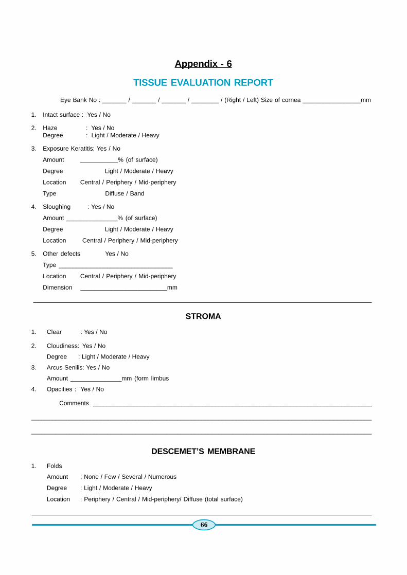
66
Appendix - 6
TISSUE EVALUATION REPORT
Eye Bank No : _______ / _______ / _______ / ________ / (Right / Left) Size of cornea _________________mm
1. Intact surface : Yes / No
2. Haze : Yes / NoDegree : Light / Moderate / Heavy
3. Exposure Keratitis: Yes / No
Amount ___________% (of surface)
Degree Light / Moderate / Heavy
Location Central / Periphery / Mid-periphery
Type Diffuse / Band
4. Sloughing : Yes / No
Amount _______________% (of surface)
Degree Light / Moderate / Heavy
Location Central / Periphery / Mid-periphery
5. Other defects Yes / No
Type _________________________________
Location Central / Periphery / Mid-periphery
Dimension _________________________mm
STROMA
1. Clear : Yes / No
2. Cloudiness: Yes / No
Degree : Light / Moderate / Heavy
3. Arcus Senilis: Yes / No
Amount _______________mm (form limbus
4. Opacities : Yes / No
Comments __________________________________________________________________________________
__________________________________________________________________________________________________
__________________________________________________________________________________________________
DESCEMET’S MEMBRANE
1. Folds
Amount : None / Few / Several / Numerous
Degree : Light / Moderate / Heavy
Location : Periphery / Central / Mid-periphery/ Diffuse (total surface)

67
ENDOTHELIUMExcellent / Very Good / Good / Fair / NSFS
Comments ___________________________________ Cells per mm____________________________________________
___________________________________________________________________________________________________
OVERALL RATING OF TISSUE
EXCELLENT / VERY GOOD / GOOD / FAIR / POOR (Reseason fro rejection-Preventable / Partly preventable / Non-preventable)
Suitability for Surgical Use < Yes
No Reason_______________________________________________
Particles in Medium Yes / No Medium (if transferred) __________________________________
Ratings changed Yes / No Media changed Yes / No
Lables Very Good / Adequate / Poor
Scleral Rim Very Good / Adequate / Poor
Checked by Technician _________________________ Date & Time___________________________________________
Fellow ____________________________ Date & Time___________________________________________
Consultant _________________________ Date & Time __________________________________________
DONOR DATA FOR CORNEAL TRANSPLANTAITON
1. Consignment No: ____________ No. of tissues received Eyes Corneas Blood Sample / Report received
2. Name of Eye Bank / Place / Code__________________________________________________________________
3. Time lapse between Enucleation & Receipt of Consignment___________Hours ____Minutes
4. Age of the Donor / Initials _______________________________________________________________________
5. Time of Death / Date __________________________________________________________________________
6. Time of Enucleation / Date ______________________________________________________________________
7. Time lapse between Death and Enucleating______________ Hours _______________Minutes
8. Cause of Death _____________________________________________________________________________
9. Method of Preservation:Moist Chamber : Medium Preserved :
Condition of bottle ______________________ Name of Medium ___________________________
Condition of Cap ________________________ Lot No. ___________________________________
Bottle had __________1/4 ½ ¾ Full______Water Condition of Vial ____________________________
Colour of the water present _______________ Colour of Medium ___________________________
Trimming of conjunctiva Yes / No Any foreign body present in medium_____________
Discolouration of sclera __________________ Position of cornea ___________________________
Presence of foreign matter _______________
10. Condition of thermocol box ______________________________________________________________________
11. Position of ice in the thermocol box ________________________________________________________________
12. General Evaluation of Eye Ball ____________________________________________________________________
13. Aphakia : Yes / No
14. Lens ….. Slit Lamp Evaluaion :
Nuclear Sclerosis _______________________________________________________________Posterior Sub Capsular_____________________________________________________________________________Cortical Cataract __________________________________________________________________________________
Total Cataract ____________________________________________________________________________________Clear ___________________________________________________________________________________________
Lens No. ________________________________________________________________________________________

68
Appendix - 7

69
Appendix - 8
Case # _______________ Month ____________ Year______________
TISSUE DISTRIBUTION INFORMATION
Eye Bank No :________/ ________ / _________ / _________ / (Right / Left)
Age :__________ Religion :______________ Sex :________________
Injury / Admittance Date _________________ Hour ________________
Death : Date _________________ Hour ________________
Enucleation / Excision : Date _________________ Hour ________________
Preservation : Date _________________ Hour ________________
Cause of Death : _________________________________________________________________________________
_________________________________________________________________________________
Medical History : _________________________________________________________________________________
_________________________________________________________________________________
Ocular History : _________________________________________________________________________________
Medications : _________________________________________________________________________________
TISSUE PRESERVATION AND CORNEAL STATUS INFORMATION
Death – Preservation : (Time) ___________ Hrs. _______________ Mins.
Tissue Type :_________________ Storage Method:_____________
Lot Number: _________________
Status: _____________________Corneal Rating : _________________________________________________________________________________
Epithelium : _________________________________________________________________________________
Stroma : _________________________________________________________________________________
Desceme’t : _________________________________________________________________________________
Endothelium : _________________________________________________________________________________
Count : ___________ Cells / mm
Serology test for : HIV Antibody(Performed –Yes / No) Results _____________________(Hepatitis B surface Antigen test(Performed – Yes / No) Results______________________
Test for : HCV(Performed – Yes / No) Results ______________________
Test for : Syphilis(Performed – Yes / No) Results _______________________
General Comments : ________________________________________________________________________________
________________________________________________________________________________
Research only –
Name & Signature of EB Tech
ORIGINALShould be filed and
kept in the eyebank

70
RECIPIENT DATA (TO BE FILLED AT THE TIME OF SURGERY)
M.R. No: __________ Name: _______________________________________Age: _______________
Eye / Diagnosis _____________________________________________________________________
Time lapse between Enucleation and Utilization ______________________________________ Hours
Date of Surgery / Time ________________________________________________________________
Operating Surgeon’s Name ____________________________________________________________
Surgical Procedure ___________________________________________________________________
Name & signature of the surgeon

71
(Duplicate form of Appendix - 8)
Case # _______________ Month ____________ Year______________
TISSUE DISTRIBUTION INFORMATION
Eye Bank No :________/ ________ / _________ / _________ / (Right / Left)Duplicate Should be kept in the patient folder
Age :__________ Religion :______________ Sex :________________
Injury / Admittance Date _________________ Hour ________________
Death : Date _________________ Hour ________________
Enucleation / Excision : Date _________________ Hour ________________
Preservation : Date _________________ Hour ________________
Cause of Death : _______________________________________________________________________________
Medical History : ____________________________________________________________________________
Ocular History : ____________________________________________________________________________
Medications : ____________________________________________________________________________
TISSUE PRESERVATION AND CORNEAL STATUS INFORMATION
Death – Preservation : (Time) ___________ Hrs. _______________ Mins.
Tissue Type :_________________ Storage Method:_____________
Lot Number: _________________
Status: _____________________Corneal Rating : _________________________________________________________________________________
Epithelium : _________________________________________________________________________________
Stroma : _________________________________________________________________________________
Descement : _________________________________________________________________________________
Endothelium : _________________________________________________________________________________
Count : ___________ Cells / mm
Name & Signature ofDoctor / Technician
Serology test for : HIV Antibody
(Performed –Yes / No) Results _____________________
(Hepatitis B surface Antigen test
(Performed – Yes / No) Results______________________
Test for : HCV
(Performed – Yes / No) Results ______________________
Test for : Syphilis
(Performed – Yes / No) Results _______________________
General Comments : ________________________________________________________________________________
________________________________________________________________________________
Research only –
Name & Signature of EB Tech
Duplicate(Should be kept
in thepatient folder)

72
Appendix - 9
DONOR TISSUE TENTATIVE UTILITY DATA & ADVERSE REPORT FORM
ID # : Date : Time :
Age : Death :
Sex : Preservation :
Received :
Tissue sent by : _______________________________________________________________________________________
Proposed date to use the tissue : _________________________________________________________________________
Name of the Surgeon :__________________________________________________________________________________
Name of the Patient : ___________________________________________________________________________________
Hospital registration /Medical records I.D. no. of Patient : _____________________________________________________
Date of Surgery : ______________________________________________________________________________________
Date :
COUNSELLOR’S SIGNATURE & NAME
ADVERSE REPORT
Details of adverse findings:
Likely Reasons for adverse findings:
Suggestions:
Signature of Operating Surgeon / Team
ORIGINAL(Should be
sent back tothe eye bank)
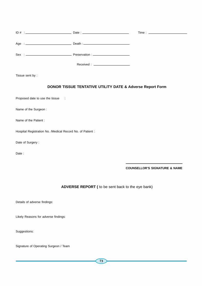
73
ID # : Date : Time :
Age : Death :
Sex : Preservation :
Received :
Tissue sent by :
DONOR TISSUE TENTATIVE UTILITY DATE & Adverse Report Form
Proposed date to use the tissue :
Name of the Surgeon :
Name of the Patient :
Hospital Registration No. /Medical Record No. of Patient :
Date of Surgery :
Date :
COUNSELLOR’S SIGNATURE & NAME
ADVERSE REPORT ( to be sent back to the eye bank)
Details of adverse findings:
Likely Reasons for adverse findings:
Suggestions:
Signature of Operating Surgeon / Team

74
Appendix - 10
INSTRUMENT CLEANING LOG
DATE INSTRUMENT # OF INSTRUMENT INSTRUMENT INITIALSSET BATCHES CLEANED BY PACKED BY

75
Appendix - 1 1
INSTRUMENT STERILIZATION LOG
STERILIZATION RUN CONTENTSBIOLOGICAL INDICATOR TESTING
PERFORMED? YES NO

76
Appendix - 12
LABORATORY CLEANING LOG
Month :_______ / Year :___________
Week 1 Week 2 Week 3 Week 4
Initials of person (s) performing cleaning
Cleaning Agent Used
Date of Cleaned
Counters
Sink (s)
Light Switches / Donor and Cabinet Handles
HEPA Hood / Biosafety Cabinet
Floors Mopped
Instrument Lubricant Solution Changed
Other____________
Other____________
Cabinets Monthlycleaning only
Centrifuge – do–
Slit Lamp Microscope – do–
Specular Microscope – do–
Centrifuge – do–
Refrigerator (Internal & External) – do–
Freezer (External) – do–
Other ___________
Other __________
Refer to the Laboratory and Facilities Module for SOPs regarding Laboratory Cleaning and Maintenance

77
Appendix - 13
Human Organs Transplantation Act (HOTA)

78

79

80

81

82

83

84

85
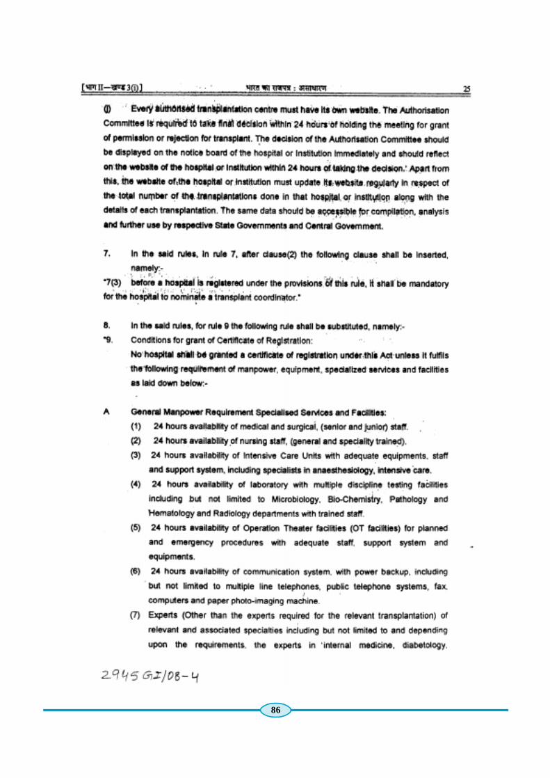
86

87

88
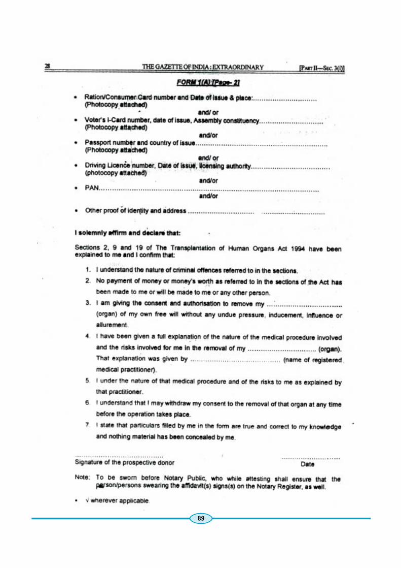
89

90

91

92

93

94

95

96
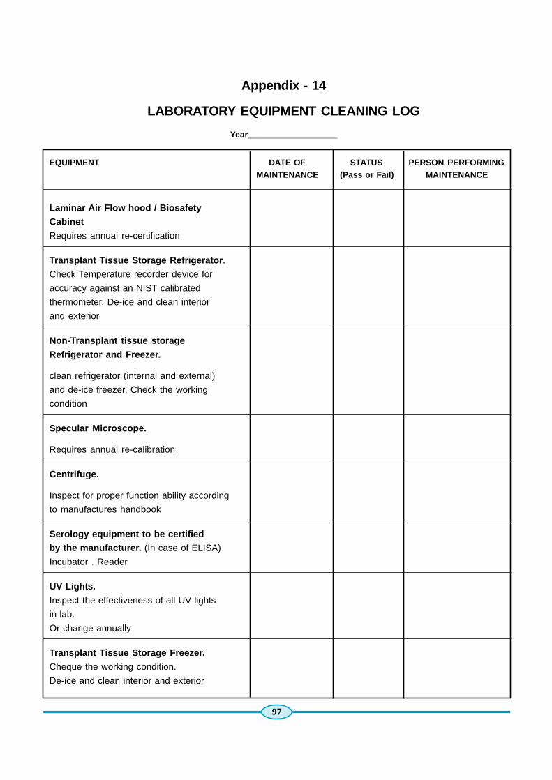
97
Appendix - 14
LABORATORY EQUIPMENT CLEANING LOG
Year___________________
EQUIPMENT DATE OF STATUS PERSON PERFORMINGMAINTENANCE (Pass or Fail) MAINTENANCE
Laminar Air Flow hood / Biosafety
Cabinet
Requires annual re-certification
Transplant Tissue Storage Refrigerator .
Check Temperature recorder device for
accuracy against an NIST calibrated
thermometer. De-ice and clean interior
and exterior
Non-Transplant tissue storage
Refrigerator and Freezer.
clean refrigerator (internal and external)
and de-ice freezer. Check the working
condition
Specular Microscope.
Requires annual re-calibration
Centrifuge.
Inspect for proper function ability according
to manufactures handbook
Serology equipment to be certified
by the manufacturer. (In case of ELISA)
Incubator . Reader
UV Lights.
Inspect the effectiveness of all UV lights
in lab.
Or change annually
Transplant Tissue Storage Freezer.
Cheque the working condition.
De-ice and clean interior and exterior
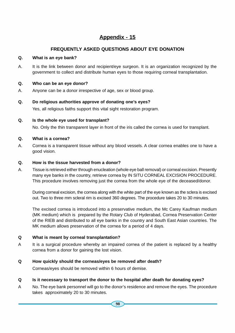
98
Appendix - 15
FREQUENTLY ASKED QUESTIONS ABOUT EYE DONATION
Q. What is an eye bank?
A. It is the link between donor and recipient/eye surgeon. It is an organization recognized by thegovernment to collect and distribute human eyes to those requiring corneal transplantation.
Q. Who can be an eye donor?
A. Anyone can be a donor irrespective of age, sex or blood group.
Q. Do religious authorities approve of donating one’s eyes?
Yes, all religious faiths support this vital sight restoration program.
Q. Is the whole eye used for transplant?
No. Only the thin transparent layer in front of the iris called the cornea is used for transplant.
Q. What is a cornea?
A. Cornea is a transparent tissue without any blood vessels. A clear cornea enables one to have agood vision.
Q. How is the tissue harvested from a donor?
A. Tissue is retrieved either through enucleation (whole eye ball removal) or corneal excision. Presentlymany eye banks in the country, retrieve cornea by IN SITU CORNEAL EXCISION PROCEDURE.This procedure involves removing just the cornea from the whole eye of the deceased/donor.
During corneal excision, the cornea along with the white part of the eye known as the sclera is excisedout. Two to three mm scleral rim is excised 360 degrees. The procedure takes 20 to 30 minutes.
The excised cornea is introduced into a preservative medium, the Mc Carey Kaufman medium(MK medium) which is prepared by the Rotary Club of Hyderabad, Cornea Preservation Centerof the RIEB and distributed to all eye banks in the country and South East Asian countries. TheMK medium allows preservation of the cornea for a period of 4 days.
Q What is meant by corneal transplantation?
A It is a surgical procedure whereby an impaired cornea of the patient is replaced by a healthycornea from a donor for gaining the lost vision.
Q How quickly should the corneas/eyes be removed after death?
Corneas/eyes should be removed within 6 hours of demise.
Q Is it necessary to transport the donor to the hospital after death for donating eyes?
A No. The eye bank personnel will go to the donor’s residence and remove the eyes. The proceduretakes approximately 20 to 30 minutes.
99
Q Do cataracts or the use of spectacles render the corneas unfit?
A. No. Both these conditions relate to the lens of the eye and not the cornea.
Q. Does eye donation disfigure the donor’s face?
A. No. The removal of corneas/eyes does not cause disfigurement.
Q. Is there any delay in funeral arrangements?
A. No. Tissue procurement is performed within 20 to 30 minutes. Therefore family members of thedeceased may proceed as planned with funeral arrangements.
Q. What conditions render corneas unfit for donations?
A. Corneas of persons suffering from AIDS, jaundice, rabies, syphilis, tetanus, septicemia and viraldiseases are considered unfit for donation.
Q. What about diabetes or hypertension?
A. Even donors with these conditions can donate their eyes.
Q. Does the human body reject the transplanted donor cornea?
A. A cornea does not have direct blood supply. Therefore the risk of rejection is very low. If rejectionoccurs, it can be suppressed by timely medication.
Q How will the donated eyes be used?
A. After the cornea is removed from the whole eye, it is evaluated and then supplied to the eyesurgeon for use in a patient.
Q. Is there any use of corneas that are for some reason not utilised for surgery?
A. Corneas that are rejected for technical reasons may be used for research or education purposes.
Q. Will the donor or recipient family be told who donated or received the cornea?
A. No. The Donor - recipient information is maintained confidential.
Q. Will the donor family be given fees?
A. No. It is illegal to buy or sell human eyes, organs or tissues. Any cost involved with cornea retrievalis borne by the eye bank.

100
Appendix - 16
FACTS AND MYTHS ABOUT DONATING EYES
Myth - Eyes can be removed out of living human beings.
Fact - Eyes are removed only after death.
Myth - Eyes can be donated even by a live person.
Fact - Eyes can only be pledged by a live person. Eyes can be donated only after death.
Myth - Removal of eyes causes disfigurement of the face.
Fact - Removal of eyes does not produce any disfigurement of the face.
Myth - Eye donation interferes with, or delays customary final rites.
Fact - Eye donation does not interfere with or delay final rites, as the corneal excision proceduretakes less than 20 minutes.
Myth - Eyes of aged donors are not acceptable.
Fact - All donor eyes are acceptable irrespective of the donor’s age, including eyes of premature/still-born babies.
Myth - Indian eyes are not good to be used for corneal transplantation.
Fact - Eyes of any deceased person anywhere in the world can be used for corneal transplantationfollowing evaluation.
Myth - An entire eye can be transplanted.
Fact - Only the cornea is used for transplantation.
Myth - Corneal transplantation is an experimental procedure.
Fact - Corneal transplantation is a proven, routinely performed surgery and is a successfulprocedure.
Myth - Corneal transplantation is not effective and successful in Indian eyes.
Fact - Corneal transplantation is effective in all eyes, if performed under optimal conditions.
Myth - Human eyes can be bought or sold.
Fact - Selling or buying of human eyes is illegal.
Myth - Only those who have pledged their eyes can donate them after death.
Fact - Pledging of eyes is not important, because even in the case of a pledgee, the consent ofthe family member is essential, without which an eye cannot be removed.

101
Appendix - 17
Eye Donation Counsellor (EDC)
DAILY REPORT FORM
1. DATE/MONTH/YEAR : _______________________________________________
2. HOSPITAL ATTENDED : _______________________________________________
3. PLACE : _______________________________________________
4. NO. OF HRS WORKED : _______________________________________________
FROM : TO: _______________
5. NO. OF DEATHS DECLARED : _______________________________________________
FROM 6.00 AM TO 6.00 PM : _______________________________________________
FROM 6.00 PM TO 6.00 AM : _______________________________________________
6. NO. OF DEATHS DECLARED : _______________________________________________WITHIN THE WORK PERIOD
7. NO. OF CASES APPROACHED : _______________________________________________
8. NO. OF CASES NOT : _______________________________________________APPROACHED(Give reasons)
9. NO. OF CASES SUCCEEDED : _______________________________________________
10. NO. OF CASES NOT : _______________________________________________SUCCEEDED
11. Donors: Male: Nos Female: Nos
NAME OF THE EDC/TRAINEE : _______________________________________________
SIGNATURE WITH DATE : _______________________________________________
SIGNATURE OF THE EYE BANK : _______________________________________________
MANAGER / DIRECTOR

102
Appendix - 18
Eye Donation Counsellor (EDC) Report
CASE SUMMARY
1. Date / Month / Year : _____________________________________________
2. Case No : _____________________________________________
3. Particulars of the Donor :
Name : _____________________________________________
Religion : _____________________________________________
Age & Sex : _____________________________________________
Cause of death : _____________________________________________
Pledge : Yes __________ No ____________
4. Particulars of the Donor Family:
No. of family members present atThe time of motivation : _____________________________________________
Family Educated : Yes __________ No. ____________
Awareness about eye donation : Yes __________ No ____________
Encountered difficulty in convincing : Yes __________ No ____________
Questions raised by the family members:
_____________________________________________________________________________________________________
_____________________________________________________________________________________________________
_____________________________________________________________________________________________________
_____________________________________________________________________________________________________
5. Time Lapse Between death and Motivation : _______________________________________
6. Time taken for motivation : _______________________________________
7. Motivation outcome : _______________________________________
8. Consent Given by : _______________________________________
9. Briefly describe how motivation was carried Out : _____________________________________
10. Reasons for getting the consent (Describe) : _______________________________________
_____________________________________________________________________________________________________
_____________________________________________________________________________________________________
11. Reasons for not getting the consent (Describe): ____________________________________
_____________________________________________________________________________________________________

103
Appendix - 19
Eye Donation Counsellor (EDC)
MONTHLY REPORT FORM
01. MONTH/YEAR : _______________________________________
02. HOSPITAL ATTENDED : _______________________________________
03. PLACE : _______________________________________
04. NO. OF DAYS WORKED : _______________________________________
05. NO. OF HOURS WORKED PER DAY : _______________________________________
06. NO. OF DEATHS DECLARED ________________________________________
IN THE MONTH (DAY & NIGHT) : _______________________________________
07. NO. OF DEATHS DECLARED
WITHIN THE WORK PERIOD : _______________________________________
08. NO. OF CASES APPROACHED : _______________________________________
09. NO. OF CASES NOT APPROACHED : _______________________________________(Give reasons)
10. PARTICULARS OF THE CASES : _______________________________________APPROACHED
Religion :Hindu: __ ; Muslim: __ ; Christian: __
Age Group :0 – 9 _________; 10 – 19 _________;
20 – 29 ________; 30 – 39 _________;
40 – 49 ________; 50 – 59 _________;
60 – 69 ________; 70 – 79 _________;
80 and above ________.
Sex : Male _________; Female __________
Cause of Death : Heart Disease __________________
Cancer ___________________
Trauma ___________________
Brain Disease ___________________
Infectious Disease ________________
Others _________________________
Pledgees : No. of Pledgees ________________
Not pledgees _________________

104
Family educated : _____________________________
Awareness about eye donation : _____________________________
Encountered difficulty in counselling : _____________________________
11. AVERAGE TIME LAPSE : _____________________________BETWEEN DEATH & MOTIVATION
12. AVERAGE TIME TAKEN : _____________________________FOR MOTIVATION
13. NO. OF CASES SUCCEEDED : _____________________________(DONORS)
14. NO. OF CASES NOT SUCCEEDED :_____________________________
15. PARTICULARS OF DONORS : _____________________________
Religion :Hindu: __ ; Muslim: __ ; Christian: __
Age Group :0 – 9 _________; 10 – 19 _________;
20 – 29 ________; 30 – 39 _________;
40 – 49 ________; 50 – 59 _________;
60 – 69 ________; 70 – 79 _________;
80 and above ________.
Sex : Male __________________; Female ___________
Cause of Death : Heart Disease _____________________________
Cancer ___________________________________
Trauma ___________________________________
Brain Disease _____________________________
Infectious Disease__________________________
Others __________________________________
Pledgees : No. of Pledgees ___________________________
: Not pledgees ____________________________
Family educated : ________________________________________
Awareness about eye donation : ________________________________________
Encountered difficulty in counselling :
Utilisation: Op.PK Th.PK L.K Others

105
Miscellaneous
TEMPERATURE RECORD REFRIGERATOR No.
Month : ______________ Year : ______________
Date 7.00 A.M. 1.00 P.M 7.00P.M
1
2
3
4
5
6
7
8
9
10
11
12
13
14
15
16
17
18
19
20
21
22
23
24
25
26
27
28
29
30
31

106
Appendix - 20
Financial Assistance available under NPCB
II Non-recurring grant-in-aid for Eye Banks in Government/Voluntary Sector (uptomaximum Rs. 15.00 lakhs);
The objective of this scheme is to promote Eye banking activity in the country through Governmentfacilities, NGOs and other stake holders to get adequate tissue for corneal transplantation for treatment ofcorneal blindness.1. Financial assistance : Under the scheme, financial assistance will be provided up to a maximum of
Rs. 15 Lakh for purchase of equipment, furniture and fixtures (list attached)
2. Eligibility criteria : The organization should:
i. Satisfy general eligibility conditions mentioned.
ii. Should fit into the definition of Eye Bank as mentioned.
iii. Organizations having experience in providing eye care services will be given preference.iv. Should have collected at-least 100 Eye Balls in any of the preceding two years of application.
In case of difficult terrain (eg. North eastern states), relaxed criteria of 25 Pair of Eyes shall beapplicable
ORv. Should have conducted at least 600 cataract operations including other eye disease operations
in the proceeding two years. In case of difficult terrain (eg. North eastern states), relaxedcriteria of 300 cataract operation including other eye disease operations shall be applicable.
3. Infrastructure Requirement:
a. Manpower Requirement:
l Ophthalmic Surgeons (Full time / on Panel) 1
l Ophthalmic Technician 1
l Eye Donation Counselor / Social Worker / Health 2Educator / Clerk
4. Expected Output NGOs receiving non-recurring grants shall:
i. Utlize the entire grant within period of 12 months from receipt of grant after following dueprocedures
ii. Provide & maintain detailed records of Eye Balls collected and utilized in the prescribed fomatand submit monthly report to the District Health Society.
iii. The NGO should be committed to collect at-least 200 eye balls in the next two years. In caseof difficult terrain (eg. North eastern states), relaxed criteria of 100 cataract operation includingother eye disease operations shall be applicable.
5. Procedure for Approval of Grants
Two copies of application in prescribed formats would be submitted by applicant NGO along with

107
necessary documents in support of qualifying criteria to the State Programme Officer (SPO), NPCB. TheSPO would examine the proposal in terms of eligibility criteria, and depute a team of expert(s) (2-3) fromthe State to visit the NGO for assessing present facilities and requirements. This entire work should becompleted within maximum of three months from the date of receipt of applications complete in all respects.The SPO may thereafter, forward his recommendation to the competent authority for final disposal.
S.No Equipment/Furnishing
1. Slit Lamp Microscope
2. Specular Microscope
3. Laminar Flow
4. Serology Equipment
5. Instruments for corneal excision and enucleation including containers
6. Autoclave
7. Keratoplasty instruments
8. Transport Facility ( One 2 Wheeler)
9. Refrigerator
10. Computer & Accessories
11. Telephone Line
12. Air-Conditioner
13. Renovation, Repair, Furniture & Fixtures
Maximum Assistance = Rs. 15 Lakh
III Non-recurring grant-in-aid for Eye Donation Centres (EDC) in Government/ VoluntarySector (upto maximum Rs. 1.00 lakh)
1. Eye Donation Centre: For the purpose of the above scheme, an Eye Donation Centre will mean anorganization that is:
i) Is affiliated to a registered Eye Bank
ii) Harvest corneal tissue and collect blood for serology;
iii) Ensure safe transportation of tissue to the parent eye bank
iv) Provide a round the clock public response system for eye donation;
v) Coordinate with donor families and hospitals to motivate eye donation;
vi) Conduct Public and professional awareness on eye donation;
2 Financial Assistance:
Under the scheme, financial assistance will be provided up to a maximum of Rs. 1 Lakh(Rupees One Lakh Only) for the purchase of equipment.

108
3 Eligibility criteria:
(a) Should satisfy general eligibility conditions mentioned. (except the 2 year clause, i.e. neworganization can also apply)
(b) The organization should have the following staff as a minimum requirement:
Sl. No Personnel Number
1 Ophthalmic Technician 1
2 Eye Donation Counselor/ Social Worker / Health 1Educator
4 Expected Output: NGOs receiving non-recurring grants shall:
i. Utilise the entire grant within period of 12 months from the receipt of grant after following due
procedures
ii. Provide & maintain detailed records of Eye Balls collected and deposited in linked Eye Bank
in the prescribed format and submit monthly report to the District Health Society.
iii. The NGO should be committed to collect at-least 20 eye balls in the next two years.

109
Appendix - 21
Eye Banks* in IndiaANDHRA PRADESH
• Netra Eye Bank Rotary Netra Eye Hospital, Saripalli, Pendurthi, Visakhapatnam - 531173 , Ph.:2763574 • Ongole Eye Bank, Kurnool Road, Near RTC Bus Stand, Ongole -523002 , Ph.: 233767 •Aravind Eye Bank, Aravind Eye Hospital, Venkataratnam Street, Suryaraopet Vijayawada -520002, Ph.:866433018 • L.V. Prasad Eye Institute, L.V. Prasad Marg, Road No-2, Banjara Hills, Hyderabad -500034,Ph.: 30612345 • Swetcha Gora Eye Bank, C/o Vasaya Mahila Mandali, Benz Circle, Vijayawada -520010,Ph.: 2472370 • S.L. Mishra Memorial Eye Bank Lions Club of Hyderbad, Sadhuram Eye HospitalCharitable Trust & P.G. Inst. of Ophthalmology, 1-2-8 Gagan Mahal Road, Domalguda, Hyderabad -500029,Ph.: 23221094 • Chiranjeevi Eye & Blood Bank Research and Development Centre, D.No. 8-2-293/82/A/CEBB, Road No-1, Jubilee Hills Check Post Hyderabad -500033, Ph.: 23554849 • Sarojini DeviEye Hospital, T.L. Kapadia Eye Bank, Hymayun Nagar, Hyderabad -500028, Ph.: 23317274 • TheKhammam Eye Bank, Near Munneru Bridge, Ranganayakulagutta Road, Khammam , Ph.: 223756 •V.T. Thakur Memorial Lions Hospital & Eye Bank , Godown Road, Kamareddy - 503111 , Ph .: 225888• Mohsin Eye Bank, C/o. L.V. Prasad Eye Institute, GMR Varalakshmi Campus, D.No. 11-113/1,Hanumanthuwaka Junction, Visakhapatnam -530040, Ph.: 2714000 • Lions Citizens Eye Bank-ONGOLE,C/o. Urban Health Centre, Papa Colony, ONGOLE, Ph.: 82835 • Khammam District Police Eye Bank,Distt. Police Office, Khammam . Ph.: 25333 • Eye Bank, Govt. General Hospital, Guntur , Ph.: 2221818• Eye Collection Centre, District Hospital, Cuddapah , Ph.: 242152 • Eye Collection Centre, DistrictHospital, Mahaboob Nagar , Ph.: 243032 • Eye Collection Centre, District Head Quarters Hospital, KarimNagar , Ph.: 2262478 • Dr. Smita Sriman Memorial Eye Bank, Sudarsani Eye Hospital, Kothapet MainRoad, Guntur -522001, Ph.: 2220028 • Indian Red Cross Society Eye Bank, Near Govt. General Hospial,Opp. Ravi Talkies Kurnool -518002, Ph.: 255347 • N.J.R. Eye Bank, Rose Trust, QTR. No-98-B, Sector-I, Ukkunagaram, Visakhapatnam -530032 • Badam Balakrishna Eye Bank, 34-1-8, Temple Street,Kakinada -533001, Ph.: 2379184 • Lions Manibai Bhagathram Guptha Eye Bank, Lions Dundoo EyeInstitute, 10-1-254, West Marredpalli, Secunderabad - 500026, Ph . : 27802124 • Narayana MedicalCollege Hospital & Eye Bank, Narayana Medical Institutions, Chintareddypalem, Nellore -524002, Ph.:2317966 • Jnanananda Eye Bank Association, Jnanananda Ophthalmic Institute, Mayur Town, RailwayStation Road, P.O. Siva Rao Pet, Bhimavaram -534202, Ph.: 223321 • Swargdham Cornea CollectionCentre, A unit of Lions Club of Medchal Charitable Trust, B/H-Job bank, Madhavi Nagar Colony , Alwal,Secunderabad • Red Cross Society Eye Donation Center, IRCS & INTAC BUILDING, BESIDE-I-TownP.S. Potti Sriramulu Jn. Old Bus Stand Srikakulam , Ph.: 226555
ASSAM
• Jeuti Eye Bank, Chandra Prabha Eye Hospital, K.K. Handigue Road, Jorhat-785001, Ph.: 2304500 •Lions Drishti Eye Bank, Amolapatty Railway Gate, Dibrugarh-786001, Ph.: 2320507
ANDAMAN & NICOBAR
Department of Ophthalmology, DBCS DHS G.B. Pant Hospital, Port Blair -744104
BIHAR
• Piyush Eye Bank, Pujya Tapaswi Sri Jagjeevanjee Maharaj Chakchu Chikitsalaya, Petarbar-Post -829121, Ph.: 265609 • Dr. Kashyap Memorial Eye Bank, C/o Kashyap Eye Hospital, Purulia Road,Ranchi -834001, Ph.: 651301198

110
CHANDIGARH
• Department of Ophthalmology P.G. Institute of Medical Education & Research Centre, Chandigarh -160012, Union Territory of Chandigarh , Ph.: 2747837 • Command Hospital, Western Command,Chandigarh -160022, Ph.: 867549 • Department of Ophthalmology, Govt. Medical College & Hospital,Sector-32, Level-IV, B-Block, Chandigarh -160022, Ph.: 2607707 • Eye Bank Society, Eye Department,P.G. Institute of Medical Education & Research Centre, Ph.: 172544589 • Chandigarh Eye Bank, C/oGrewal Eye Institute, SCO 166-169, Madhya Marg, Sector 9C, Ph.: 2747118 • City Eye Bank Chandigarh,SCO-833-834, Sector-22A, Ph.: 3099731
DELHI
• Delhi Central Rotary Eye Bank, Sir Ganga Ram Hospital, Rajinder Nagar, New Delhi -110060, Ph.:42251356, 25861463, Extn. 1356 (M) 9811771213 • Rotary Regency Gift of Sight Co-ordination &Processing Centre, Venu Eye Institute & Research Centre, 1/31, Sheikh Sarai Phase-II InstitutionalArea, New Delhi -110017, Ph.: 29250952/1155/1156 Extn. 135 (M) 9899396838 • National Eye Bank,Dr. Rajendra Prasad Centre for Ophthalmic Sciences AIIMS, Ansari Nagar, New Delhi -110029, Ph.:26589461, 26588500, 26588700, 26593444 • Guru Nanak Eye Centre Eye Bank, Maharaja Ranjit SinghMarg, New Delhi-110002, Ph.: 23234612, 23234622, 23235145, 23232400 Extn. 4376 • Dr. Shroff’sCharity Eye Bank, Dr. Shroff’s Charity Eye Hospital, 5027, Kedar Nath Road, Daryaganj, New Delhi -110002, Ph.: 43524444 Extn. 268 (M) 9873208336/26 • Army Hospital (R & R Centre) Eye Bank, Deptt.of Ophthalmology, Delhi Cantt., New Delhi -110010, Ph.: 23338181/23338183, 23338185 • Guru GovindSingh International Eye Bank, 31, Defence Enclave, Vikas Marg, New Delhi -110092, Ph.: 22542325• Indraprashtha Apollo Hospitals, Apollo Eye Bank, Sarita Vihar, Delhi-Mathura Road, New Delhi -110044, Ph.: 26925858, 26925801 • Safdarjang Hospital & Vardhman Mahavir Medical College,Department of Ophthalmology, New OPD Block, New Delhi -110029, Ph.: 26198126, 26707217 • SewaEye Bank, A-Unit of Sewa Sansthan Charitable Society, M.M. Eye Tech., 29, Link Road, Lajpat Nagar-III,New Delhi -110024, Ph.: 41551811, 29841919, 9212035119. Dial 1919 Toll Free MTNL Number.
GOA
Rotary Club of Panaji Eye Bank Trust, C/o Metal Fabrik India, 16, Lydia Garden, St. INEZ, Panaji -403001, Ph.: 2425692
GUJARAT
• Rotary Eye Institute, Sant Punit Chakshu Bank, Smt. Lilavatiben Mohan Lal Shah (Bilimora) EyeHospital, Dudhia Talav, Navsari -396445, Ph.: 258920 • Indian Red Cross Society, Dholka Branch, RedCross International Eye Bank, Atulya Bhawan, Sarkhej, Gandhi Nagar Highway, Ahmedabad -380054• Lokdrishti Eye Bank, Gayatri Hospital, A/26, Rangavdhut Society Matawadi, L.H.Road, Surat -395001,Ph.: 2553653 • Mehsana Jaycees Charitable Trust, Sanchalit The Mehsana Nagarik Sahkar Bank Ltd.Eye Bank, Super Market, Dairy Road, Mehsana -384002, Ph.:. 276252814 • C.J. Patel Eye Bank, SanjivaniHospital, AT-Chalthan, Chalthan-Post - 394305, Ph.: 262771184 • Samsan Vyausta Samiti Eye Bank,Vahivati Committee, C/O. Yashuant C. Khakhar, Grain Market, Jamnagar -361001, Ph.: 558854 • R.N.C.Free Eye Hospital & Eye Bank, Dr. Paragji Desai Road, Opp-Mamlatdar Office, Valsad -396001, Ph.:244263 • Maharaja Agarsen Chakshu Bank (Eye Bank), C/o Agarwal Vikash Trust City Light Area,Surat -395007, Ph.: 2214713 • ASO PALOV Eye Bank, Retina Foundation Near Shahibaug Under Bridge,Rajbhavan Road Shahibaug, Ahmedabad -380004, Ph.: 22865537 • S.S.G. Hospital Eye Bank, Ward-E-1/2, Deptt. of ophthalmology, Surgical Building, 4th Floor, SSG Hospital, Baroda -390001, Ph.: 3133393• Sanjivani The Hanuman Welfare Society, Dr. Ashok Agarwal Eye Donation Centre, Government Hospital

111
Campus, Marvad -396210, Ph.: 3133393 • Nagri Eye Bank, C.H.Nagri Eye Hospital, Ellisbridge,Ahmedabad -380006, Ph.: 26466724 • Eye Bank, S.S.G.Hospital, Baroda -390001, Ph.: 2424748 • ShreeSwaminarayan Gurukul Hospital, Shree Swaminarayan Gurukul Eye Institute Bank, Gondal Road,Near Water Tank, Rajkot -360004, Ph.: 2377708 • Sewa Rural Jhagadia, Society For Education Welfare& Action Rural, Jhagadia -393110, Ph.: 220021 • Eye Bank-Department of Ophthalmology, GovernmentMedical College, Majuragate, Surat -395001, Ph.: 2244147 • Raghudeep Eye Clinic-Eye Bank, GurukulRoad, Memnagar, Ahmedabad -380052, Ph.: 27490909 • Dr. Dhanijisha E Anklesaria Eye Bank, M & JInstitute of Ophthalmology, Civil Hospital Campus, Asarwa , Ahmedabad - 380016, Ph.: 22681010 •Lions Karnavati Eye Bank, Lions Karnavati Shantaben Vishnubhai Patel Eye Hospital, At-OGNAJ Village,Post-New High Court, Daskroi -380060, Ph.: 244052
HARYANA
• Mahavir International, Faridabad, Kewal Prem, 614, Sector 15 A, Faridabad -121007, Ph.: 282595 •Shakti Eye Bank, Sri Shakti Sewa Dal Chimnibai Dharmsala, NH-3, NIT Faridabad, Ph.: 5421047 •International Eye Bank, Shah Satnam Ji Speciality Hospitals,(DSS), Near Shah Satnam Ji Dham, NejiaKhera -125055, Ph.: 45588 • Jeevan Eye Bank, C/o. Gayatri Eye Care Institute, Jindal Chowk, Hisar -125005, Ph.: 220052 • Y.P. Mahendru Niramaya Eye Bank, 560/1, Dayanand Colony, New RailwayRoad, Gurgaon -122001, Ph.: 2321234 • Eye Bank-Deptt. of Ophthalmology, Pt. B.D.Sharma, P.G.I.M.S.Rohtak -124001, Ph.: 244536 • Shanti Smriti Shri Madhav Eye Bank, C/o. Shanti Smriti Eye & GeneralHospital, Canal Road, Narwana -126116, Ph.: 243322 • Hisar Fort Lions Eye Bank, #119-120, UrbanEstate - II , Hisar – 125005 • Lions Eye Bank, Dr. Praveen Eye Hospital, Hissar Road, Sirsa -125055,Ph.: 221211 • Madhav Netra Bank, Ambala City, C/o. Kapil Eye Hospital, 240, Vivek Vihar, Ambala City- 134002, Ph.: 2553533 • Madhav Eye Bank, Run by Akhil Bhartiya Drishtiheen Kalyan Sangh- Nagpur,Thakur Eye & Maternity Hospital, Near Post office, Karnal -132001, Ph.: 2202880
HIMACHAL PRADESH
Kangra Districted Cross Society, Eye Collection Centre, Red Cross Bhawan, Dharmasala -176215,Ph.: 189224888
JHARKHAND
• Bihar Eye Bank Trust, Bariatu Road, Ranchi -834009, Ph.: 2545333 • Drishti Eye Bank, Departmentof Ophthalmology, Patliputra Medical College Hospital, Dhanbad -826005, Ph.: 2204730 • M.G.M.MedicalCollege & Hospital, Jamshedpur, Ph.: 2432138
KARNATAKA
• Narayana Netralaya, Dr. Raj Kumar Eye Bank, 121/C, Chord Road, Rajaji Nagar, R-Block, Bangalore -560010, Ph.: 23325311 • Netra Enterprises Prabha Eye Clinic Eye Bank, 504, 40th, Cross, VIII-Block,Jayanagar, Bangalore -560070, Ph.: 2644131 • S.G.M. Eye Bank Trust & Research Foundation C/oJoshi Eye Institute, Padma Nayanalaya, Gokul Road, Husur Cross, Hubli -580021, Ph.: 2228431 • KichanChand Chellaram Eye Bank & Cornea Grafting Centre, Regional Institute of Ophthalmology, Bangalore -560002, Ph.: 26707176 • Lions International Eye Bank, B.W.L. Superspeciality Eye Hospital, #5, LionsEye Hospital Road, (Off. J.C.Road) Bangalore - 560002, Ph.: 22235005 • Kollegal Eye Bank, ShantiNursing Home, Kollegal -571440, Ph.: 222230 • The Mysore Eye Bank & Research Centre, 1205,Kantharaja URS Road, Chamarajapuram, Mysore -570005, Ph.: 2331216 • Satyavathi Eye Bank, A Div.of Netralakshmi Vaidyalay, near B.D.O. Officer, Hospet -583201, Ph.: 839457997 • Sri AkkamahadeviNetradan Prerna Samithi, Sri Akkamahadevi Samaja , P. J. Extension, Davangare - 577002, Ph.: 255112

112
• Lions Eye Bank-Gokak, Lions Eye Trust, Dr. Kulkarani Nursing home, Lions Club Gokak -591307,Ph.: 226414 • Command Hospital, Indian Air Force- Eye Deptt. Agram-Post Banglore -560007, Ph.:25369030 • Bhagwan Mahaveer Jain Rotary Eye Bank, No-3, Platform Road, Seshadripuram,Bangalore -560020, Ph.: 23462559 • The Mandya Eye Donation Centre, B.G.Nethralaya, V.V. Road,Mandya -571401, Ph.: 220841 • Vasudevamurthy Memorial Eye Bank, Shekar Nethralaya, No-633,100 Ft.Ring Road, J.P.Nagar, 3-Phase, Bangalore -560078, Ph.: 26593210 • Shraddha Eye Bank, #256/14, Kanakapura Main Road, 7-Block, Jayanagar, Bangalore -560082, Ph.: 26634200 • Friends CentreEye & Blood Bank, C/o. Nagarajy.V. Ing Vysya Bank, B.H.Road, Barandur -577201, Ph.: 273573 • Dr.A.K.Nadagouda Patil Eye Bank, Shree Padma Nursing Home, Krishna River Road, Rabkavi, Jamkhandi- 587314 , P h . : 230004 • KLES Hospital & Eye Bank, Nehru Nagar, Belgaum, Belgaum -590010, Ph.:2473777 • Eye Bank, District Hospital Near Channamma Circle, Belgaum -590001, Ph.: 2430728 • EyeBank Basaveswar Hospital, H.K.E. Society’s Mahadevappa Rampure Medical College, MahadevappaRampure Marg, Gulbarga -585105, Ph.: 247988 • Basaveshwara Punarjyothi Eye Bank (R.),Basaveshwara Medical College Hospital & Research Centre, NH-4, Bypass, Near Housing Board Colony,Chitradurga - 577501, Ph.: 222045 • Ramaiah Memorial Eye Bank, M.S. Ramaiah Medical TeachingHospital Eye, M.S.R. Nagar, M.S.R.I.T.-Post, Bangalore -560054, Ph.: 23608888 • Padamabhushan Dr.M.C.Modi Charitable Eye Hospital, Dr. M.C.Modi Road, Mahalakshmipuram, Bangalore -560086, Ph.:23492233
KERALA
• Ahalya Eye Bank, P.B. # 120, Karnal Pirivu, Near Walayar, Palakkad (Dist.), Kerala -678557, Ph.:04923-235100, 235900, 235101• Medical College Hospital, Trichur -680001, Ph.: 24231050 • ChaithanyaEye Bank, Chaithanya Eye Hospital & Research Centre, Institute Complex, Kesavadasapuram,Thiruvananthapuram -6950044, Ph.: 2447183 • Eye Bank Association Kerala, Little Flower Hospital,Angamally -683572, Ph.: 2454779 • Regional Institute of Ophthalmology, Government OphthalmicHospital, Thiruvananthapuram, Ph.: 2307749 • Help Rotary Eye Donation Centre, XVI/1667, M.P.Commercial Centre, Nayak’s Road, Kasaragod -671121, Ph.: 420578 • Department of Ophthalmology,Medical College, Kozhikode -673008, Ph.: 9846071637/ 2354906 • Department of Ophthalmology,Medical College, Kottayam, Ph.: 2597311
MADHYA PRADESH
• Dr. Narula Eye Hospital & Eye Bank, 77, Vikas Nagar, Neemuch -458441, Ph.: 220050 • Eye BankRegional Hospital (Govt.), Dhanpuri -484114, Ph.: 76526330 • Lions District Eye Bank, Indore EyeHospital Society, M O G Lines, Dhar Road, Indore - 452002 , Ph . : 731380554 • Chhavi Eye Bank, GitaBhawan Eye Hospital, Manoramaganj, Indore -450001, Ph.: 535536 • Lions Club Neemuch CentralEye Bank, Mahavir Netra Chikitsalaya, 3, Bihar baghm Neemuch -458441, Ph.: 742321515 • Departmentof Ophthalmology, M.G.M. Medical College, M.Y.Hospital, Indore -452001, Ph.: 427301 • Khushi EyeBank, C/o. Subhishi Hospital Pvt. Ltd., Diversion Road, Gouridham, Khargone - 451001, Ph.: 234786 •Kasyap Rotary Eye Bank & Corneal Clinic, 101-B, Nahar Pura, Gali No-1, Ratlam -457001, Ph.: 232556• Department of Ophthalmology, Bhopal Memorial Hospital & Research Centre, Raisen Bypass Road,Karond, Bhopal -462038, Ph.: 2742212 • Smt. Anasuyadevi Eye Bank, Rotary Club of Indore Magdhoot,4/7, Lasudia Mori, Indore -452010, Ph.: 5033019 • Netra Jyoti Microsurgery Eye Hospital, Birla Road,Satna , Ph.: 251077 • Arihant Eye Bank, Arihant Hospital & Research Centre, 283-A, Gumasta Nagar,Indore -452009, Ph.: 2785173 • Murlidhar Kripa Eye Bank, Murlidhar Kripa Hospital & Research Centre,Ujjain Road, Maski -465106, Ph.: 233426 • Dada Virendra Puri Ji Maharaj Eye Bank, C/o. Jan Jyoti EyeHospital, 1051, Golbazaar, Jabalpur , Ph.: 2404608
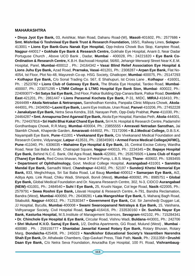
113
MAHARASHTRA
• Divya Jyot Eye Bank, 03, Avishkar, Main Road, Dahanu Road-(W), Masoli -401602, Ph.: 2577686 •Smt. Mishribai G Toshniwal Eye Bank Trust & Research Foundation, 165/1, Railway Lines, Solapur -413001 • Lions Eye Bank-Guru Nanak Eye Hospital, Opp-Indora Chowk Bus Stop, Kamptee Road,Nagpur -440017 • Gokhale Eye Bank & Research Centre, Gokhale Eye Hospital, Anant-3, Near DadarPortuguse Church , Above UCO Bank , Dadar, Mumbai - 400028, Ph.: 24221820 • Eye Bank Co-Ordination & Research Centre, K.B.H. Bachooali Hospital, 56/60, Jehangir Merwanji Street Near K.E.M.Hospital, Parel, Mumbai -400012 , Ph.: 24164342 • Vasai Blind Relief Association Eye Hospital &Lions Juhu Eye Bank, Azad Road, Prarnaka, Vasai -401201, Ph.: 2308287 • Arpan Eye Bank, Flat No-4054, Ist Floor, Plot No-48, Mayuresh Co-op. HSG. Society, Ghatkoper, Mumbai -400075, Ph.: 25147293• Kolhapur Eye Bank, C/o Sonal Trading Co. 567, E Shahupuri, Ist Cross Lane , Kolhapur - 416001,Ph. : 2523782 • Lions Club of Gateway Eye Bank, The Bhatia Eye Hospital, Tardeo Road, Mumbai -400007, Ph.: 223071295 • LTMM College & LTMG Hospital Eye Bank Sion, Mumbai - 400022, Ph.:224093077 • Sri Satya Sai Eye Bank, 2nd Floor, Patkar Building Opp-Canara Bank, Patkar Road, DombivliEast -421201, Ph.: 2861447 • Lions Parasmal Kocheta Eye Bank, P-31, MIDC, MIRAJ -416410, Ph.:2644499 • Akola Netradan & Netraropan, Sanshodhan Kendra, Panpalia Clinic Milipura Chowk, Akola -444001, Ph.: 2434050 • Laxmi Eye Bank, Laxmi Eye Institute, Uran Road, Panvel -410206, Ph.: 27452228• Janakalyan Eye Bank, Netra Seva Kendra, 318, Narayan Peth, Opp-Daily Prabhat, Pune -411030, Ph.:24464287 • Smt. Annapurna Devi Agarwal Eye Bank, Akola Eye Hospital, Ramdas Peth, Akola -444001,Ph.: 724437816 • Sri Hathi Bhai Kakal Chand Eye Bank, Sri H.N. Hospital & Research Centre, PadamshriGordhanbapa Chowk, R.R.Marg, Mumbai -400004, Ph.: 23855555 • Amravati Netradan Sansthan, JaiStambh Chowk, Khaperde Garden, Amaravati -444602, Ph.: 72172096 • B.J.Medical College, D.B.S.K.Nayampalli Eye Bank, Pune -411001 • Vivekanand Eye Bank, C/o Vivekanand Medical Foundation andResearch Centre, Vidyanagar, Latpur -413531, Ph.: 238345901 • Armed Force Medical College(AFMC),Pune -411040, Ph.: 6306035 • Mahatme Eye Hospital & Eye Bank, 16, Central Excise Colony, WardhaRoad, Near Sai Baba Mandir, Chatrapati Square, Nagpur -440015, Ph.: 2234345 • Dr. Bapaye HospitalEye Bank, Behind N.C.C. Bank, Old Agra Road, Nasik -422001, Ph.: 253576505 • Lions Club of Kopri(Thane) Eye Bank, Red Cross bhavan, Near 3-Petrol Pump, L.B.S. Marg, Thane - 400602, Ph.: 5393455• Department of Ophthalmology, Govt. Medical College Hospital, Aurangabad -431001 • SanmitraMandal Eye Bank, Samadhi Ward, Chandrapur -442402, Ph.: 52197 • Sunderji Khetsi Memorial EyeBank, 833, Meghchhaya, Sri Sai Baba Road, Lal Baug Mumbai -400012 • Samarpan Eye Bank, A/2,Aditya Apts. Link Road, Chiku Wadi, Shimpoli, Borvli (West), Mumbai -400092, Ph.: 8985751 • GlobalEye Bank, Global Medical Foundation and Dr. Nayana Research Centre, 302, N-3, CIDCO Aurangabad(NEW)-431001, Ph.: 2484540 • Suhi l Eye Bank, 25, Krushi Nagar, Col lege Road, Nasik -422005, Ph.:2579791 • Sewa Roshni Eye Bank, Lilavati Hospital & Research Centre, A-791, Bandra Reclamation,Bandra (West), Mumbai -400050, Ph.: 26438281 • Lata Mangeshkar Eye Bank, 5, Maharaj Bagh Road,Sitabuildi, Nagpur -440012, Ph.: 712530347 • Government Eye Bank, Col. Sir Jamshedji Duggan Lal,JJ Hosptial, Byculla, Mumbai -400008 • Swami Swaroopanand Netralaya & Eye Bank, 15, Vaishana,Vidhyanagar Society, Civil Hospital Road, Sangli -416416, Ph.: 233530160 • Dr. Sushila Nayar EyeBank, Kasturba Hospital, M.S.Institute of Management Sciences, Sevagram -442102, Ph.: 715284341• Dr. Chinchole Eye Hospital & Eye Bank, Circular Road, Vishnu Wadi, Buldana -443001, Ph.: 242706• Shri Mulund K.V.O. Samaj Eye Bank, 1/2, Dedhia Apartments, GG Road, Mulund (West) , Mumbai -400080 , Ph. : 259155777 • Shantabai Jawarilal Kawad Rotary Eye Bank, Rotary Bhuvan, RotaryMarg, Dondaicha -425408, Ph.: 245023 • Nandkishor Educational Society’s Vasantiben NarendraShah Eye Bank, Dr. Athalwale Chambers, Opp-Gavkari Press, Tilak Path, Nasik , Ph.: 2311358 • DrushtiDaan Eye Bank, C/o Netra Seva Foundation, Anuradha Eye Hospital, 100 Ft. Road, Vishrambaug -
114
416415, Ph.: 2301939 • S.S.L. Hospital Eye Bank- With Madhav Bank, Shankar Rao Shewak NearBank of Baroda, Kalyan , Ph.: 2205148 • Bhaiyasaheb Jawade Eye Bank, Jawade Compound Mall-HillRoad, Amaravati -444602, Ph.: 2663334 • Late Smt. S.M. Mehta Eye Bank, C/o. Jain Social Group(Dailana), D/17, Ground Floor, Ishwarnagar, LBS Marg, Bandup(W), Mumbai -400078, Ph.: 25953785 •Rotary Eye Bank, E-3, Dwarkesh, L.T.Road, Near Diomond Cinema, Borivali (West), Mumbai -400092,Ph.: 28980707 • Madhav Eye Bank, Vardhaman Eye Hospital, Kamal Apts. Ground Floor, Opp- TelephoneExchange, Saharanpur Road, Canada Corner, Nasik -422002, Ph.: 2571800
MANIPUR
Department of Ophthalmology, Regional Institute of Medical Sciences, (RIMS), IMPHAL -795004, Ph.:2414750
NAGALAND
Mokokchung Wood Land Multi Purpose, Co-operative Society Ltd.
ORISSA
• Eye Bank-Rotary Club of Bhubaneswar Metro, C/o. Brig. N.Dhal, B-21, H.I.G. H.B. Colony, Baramunda,Bhubaneswar -751003, Ph.: 556184 • Drushti Daan, L.V. Prasad Eye Institute, Bhubaneswar -751002,Ph.: 3987999 • Maa Tarini Netradham (A Mission Drishti Project), Mining Road, Near Gayatri Mandir,Keonjhar , Ph.: 253016 • Daitari Satyabhama Eye Bank, Institute of Medical Sciences & Sum HospitalKalinga Nagar, Bhubaneswar
PONDICHERRY
• Department of Ophthalmology, Jawaharlal Nehru Institute of Post Graduate Medical Education &Research, Dhanvantari Nagar , Ph.: 41372380 • Gothi Eye Bank, 17/5B, Vazhudavur Road, Opp. KamarajKrishi Vigyan Kendra, Kurumbapet , Ph.: 41371115
PUNJAB
• Sri Guru Harkrishan Sahib Charitable Eye Hospital Trust, Akal Ashram near Chandigarh, Sohana -140308, Ph.: 2258262 • Dr. Dajlit Singh Eye Hospital, Sheranwalagate, Amritsar , Ph.: 183551600 •Mata Kulwant Kaur Bhai Sunder Singh Baweja Eye Bank, Milap Chowk, Jalandhar -144001, Ph.:2455213 • Bharat Vikas Parishad, Eye Bank Faridkot, C/o Dr. S.P.S. Sodhi Sidhi Cottage, HarindraNagar, Faridkot -151203, Ph.: 51131 • Eye Bank Dayanand Medical College & Hospital, OphthalmologyDeptt, DMCRH , LUDHIANA - 141001 , Ph. : 2471500 • Department of Ophthalmology, Christian Medicalcollege, Ludhiana -141008, Ph.: 161643171 • Muktisar Eye Bank Society, C/o Sohan Singh BhagwanKaur Charitable Eye & General Hospital, Tibbi Sahib Road, Muktsar -152026, Ph.: 163360465 • Dr. BansalEye Institute & Research Centre, 105, New Lal Bagh, Patiala , Ph.: 175223345 • Punarjyot Eye Bankof Punjab, 65-A, BRS Nagar, Firozpur Road, Ludhiana , Ph.: 2464999 • Eye Donation AssociationHoshiarpur, 10-R, Model Town, Hoshiarpur -146001, Ph.: 222622 • Eye Bank Society-Mansa, TailorsStreet, Mansa -151505, Ph.: 236390 • Sunam Nettar Bank Samiti, Opp- Satnarain Mandir Alibazar,Sunam -148028, Ph.: 222827 • Netradaan Samiti, Satsang Road Near Punjab National Bank, Bareta -151501 • Eye Bank Welfare Society (Regd), Government Medical College, Rajindra Hospital, Patiala ,Ph.: 2371477
RAJASTHAN
• Jain Eye Charitable Trust C/o Dr. Jain Eye Hospital, Dhohad Road, Banswara -327001, Ph.: 240425
115
• Sri Jagdamba Charitable Eye Hospital, Hanuman Garh Road, Sri Ganganagar -335001, Ph.: 2464358• Lions Eye Bank, Lions Club of Udaipur, C/O Alok Eye Hospital, 27, Ashwini Bazar, Udaipur -313001,Ph.: 2524255 • Rotary Eye Bank, Ch. Uda Ram Sihag Memorial, Sukharia Circle, Sri Ganganagar -335001, Ph.: 2433933 • Rotary City Eye Bank, Dawar Eye Hospital, Sukhakida Road, Sri Ganga Nagar -335001, Ph.: 434020 • Rajasthan Eye Bank, Eye Research Centre Society, Bhabha Marg, Moti Dungri,Jaipur -302004, Ph.: 621888 • J.W. Global Hospital & Research Centre Eye Bank, C/o Global HospitalInstitute of Ophthalmology, Taleti, Post-Shantivan, Mount Abu-307501, Ph.: 238347 • Eye Bank Societyof Rajasthan, 429, Near Geeta Bhawan, Adarsh Nagar, Jaipur -302004, Ph.: 2604117 • Than ChandMehta Eye Bank, Department of Ophthalmology, M.D.M. Hospital, Jodhpur • Eye Bank, Department ofOphthalmology, S.M.S. Hospital, Jaipur
TAMILNADU
• Vazum Kangal Eye Bank, M.N. Eye Hospital (F.S.), 783, T.H. Road Opp-Todiarpet Police StationChennai -600021, Ph.: 25956403 • C.U.Shah Eye Bank Sankara Netralaya, 18, College Road, Chennai -600006, Ph.: 28261268 • Regional Institute of Ophthalmology, Govt. Ophthalmic Hospital EGMOREChennai -600008, Ph.: 28267709 • Sankara Eye Bank, Sri Kanchi Kama Koti Medical Trust, 77, WestPonnurangam Road, R.S.Puram Coimbatore -641002, Ph. : 2544680 • Sri Ramachandre Eye Bank, SriRamachandra Medical College & Research Institute (D.U.) Porur, Chennai -600116, Ph.: 24765515 •Lions Eye Bank Trust, District 324- A2, 72, Mutt Street, Kumbakonam -612001, Ph.: 23523520 • NatrajHospital Eye Bank, Sri Kanchi Kama Koti Medical Trust, Sivanandapuram, Sathy Road, Coimbatore -641035, Ph.: 2866450 • Rotary Rajan Eye Bank, Rajan Eye Care Hospital No-3, Vidyodaya East, 2ndStreet, T- Nagar, Chennai -600017, Ph.: 28259635 • Nagercoil Eye Bank , C/ o Bejan Singh Eye Hospital, 82 , College Road, Nagercoil-629001, Ph.: 237491 • Aravind Eye Hospital, Aurolab, 1, Anna Nagar,Madurai - 625020, Ph.: 2535573 • Sankar Eye Bank, Sankar Charitable Trust,32-B, Nellukara Street,Kanchipuram -631502, Ph.: 2223452 • Aravind Eye Bank, Aravind Eye Hospital & Post Graduate Instituteof Ophthalmology, Avinashi Road, Peelamedu, Coimbatore -641014, Ph.: 2578901 • Lions Eye BankTrust Research Foundation, Govt. Ophthalmic Hospital New Building, District 324-A1, Chennai -600008,Ph.: 28553840 • J.P. Gandhi Eye Bank, A Unit of P.D.Gandhi & M.P. Gandhi Charitable Trust, 23, BungalowRoad, Tirupur -641602, Ph.: 2205890 • Lotus Eye Care Hospital & Eye Bank, Civil Aerodrome-Post,Avinashi Road, Coimbatore -14, Ph.: 2626464 • Vision Mission Eye Bank, 34, Gandhi Road, WestTambaram, Chennai -600045, Ph.: 22381130 • Salem Lions Eye Bank, Salem Eye Hospital & Institute ofOphthalmology, 11, A/1, Gandhi Road, Salem -636007, Ph.: 2319993 • Uma Eye Clinic-Rotary Club-Vision Foundation Eye Bank, 1957, 10th Main Road, Anna Nagar, Chennai -600040, Ph.: 26213670
TRIPURA
Society For Promotion of Eye Donation, C/o Banakutir, 2, Nutanpalli, Krishnanagar, Agartala -799001,Ph.: 2302072
UTTAR PRADESH
• Department of Ophthalmology, LLRM Medical College, Meerut , Ph.: 2763133 • Eye Department,OIC, Army Medical Corps, Meerut Cantt -250001, Ph.: 696535 • Varanasi Eye Bank Society Sah ModernClinic, K-16/102, Bulanala(East), Varanasi - 221001, Ph.: 2333272 • Eye Bank-Gandhi Eye Hospital,Dr. Mohan Lal Memorial Gandhi Eye Hospital, Ram Ghat Road, Aligarh -202001, Ph.: 2505968 • EyeBank Dhampur, Nagina Road, Dhampur -246761, Ph.: 20222 • A-1040, Indira Nagar, Lucknow -226016,Ph.: 2347930 • Sarfrazganj, Moosabagh, Picnic Spot, Hardoi Road, Lucknow -226003, Ph.: 2248122 •Lucknow Eye Bank, Precision Eye Care, B-145, Sector-A, Mahanagar, Lucknow , Ph.: 2381919 • Denaji

116
Eye Bank, Rajani Eye Care Centre, Parkar College Road, Moradabad -244001, Ph.: 2317975 • IndianMedical Association, Banaras Branch, C7/31, Chetganj, I.M.A. House, Varanasi -221001, Ph.: 2354197• ICARE Charitable Eye Hospital & P.G. Inst. Glucoma Research Centre, E-3A, Sector-26, NOIDA-201301, Ph.: 4068800 • Sahyog Viklang Seva Sansthan, 426-9, Lalkothi Baruzai, Shahjahanpur -242001,Ph.: 222472 • B.H.U. Eye Bank, Deptt. of Ophthalmology, Institute of Medical Sciences, Banaras HinduUniversity, Varanasi -221005, Ph.: 2307590 • C.L. Gupta Eye Bank Welfare Society, C.L. Gupta EyeHospital, Swami Vivekananda Road, Opp-Govt. Polytechnic, Moradabad , Ph.: 2450681 • Ganga MataCharitable Eye Hospital & Research Institute, Sapt Rishi Link Road, Hardwar -249410, Ph.: 260190 •SRMS Eye Bank, Shri Ram Murthi Smarak Trust, N-3, Rampur Garden, Bareilly , Ph.: 2582014 •Department of Ophthalmology, King George’s Medical University Chowk, Lucknow -226003, Ph.: 225840• Eye Bank & Keratoplasty Centre, C/o. Commandant, Command Hospital (CC), Lucknow , Ph.: 2296548• Roshni Eye Bank, Flat No-7, Guru Nanak Marke, Ambala Road, Saharanpur -247001, Ph.: 2663779 •Institute of Ophthalmology, J.N.Medical College, Aligarh Muslim University, Aligarh -202002, Ph.:2504576 • Raj Eye Bank Association, C-168/18, Near Jhankar Cinema, Naseerabad, Saraidhola,Gorakhpur , Ph.: 2292340 • Post-Pharsar, Barhalganj, Gorakhpur , Ph.: 2325055 • Apex Eye Hospital, Polytechnic Chouraha , Jaunpur - 222002, Ph.: 69532 • House No. 491, Near Water Tank, Brahm Puri,Fafran Road, Modi Nagar - 201204, Ph.: 245542 • 94, Civil Lines, Sitapur , Ph.: 242769 • RajbahadurSingh Smarak Jan Kalyan Seva Sansthan, 187, Raghav Nagar, Deoria, Ph.: 222029 • Jankikund,Chitrakoot - 210320, Ph.: 265330 • Agrasen Chowk , Masani Chouraha, Mathura, Ph.: 2401665 • 165,Nehru Nagar, Near Shree Talkies, Agra -282002, Ph.: 2156515 • S.N. Medical College, Agra , Ph . :2530619 • Lohandi Kalan, Rewa Road, Mirzapur -231001, Ph.: 245200 • Swaroop Nagar, Kanpur -208002, Ph.: 294134
UTTARAKHAND
• Jolly Grant, Doiwala, Dehradun -248140, Ph.: 2412081 • Shree Swami Bhumanand Charitable Eye& Dental Hospital & Research Institute, Bhuma Niketan, Nyassansthnam, Sapt Sarowar Marg, Haridwar -249410, Ph.: 260722 • Gandhi Satabdi Eye Hospital, 2-C, Preetam Road, Dalanwala, Dehradun -248001,Ph.: 2654279 • Ganga Mata Charitable Eyes Hospital & Research Institute, Sapt Rishi Link Road,Hardwar -249410, Ph.: 260190
WEST BENGAL
• Gujarati Relief Socity Mukta Eye Bank, 20, Pollock Street, Kolkata -700001, Ph.: 33255188 •International Eye Bank, Calcutta, AD-202, Salt Lake, Kolkata -700064, Ph.: 23585758 • Prova EyeBank, B.C.Memorial Eye Foundation, Disha Eye Hospital & Research Centre, 88 (63A), Ghosh ParaRoad, Barrackpore -700120, Ph.: 25931729 • Howrah Lions Hospital (Unit of Corneal Grafting), 9,Nityadhan Mukherjee Road, Howrah -711101, Ph.: 26605470 • Regional Institute of Ophthalmology,Medical College & Hospital Campus, Kolkata -700073, Ph.: 2413853 • Midnapore Eye Bank & Eye CareUnit, Adibasipare, Prembazar , P.O. Hijli Co-Operative Kharagpur -721306, Ph.: 2277331 • Eye Bank-Command Hospital, Eastern Command, Alipore Road, Kolkata -700027, Ph.: 22226391 • SeramporeSeva Kendra & Chakshu Bank, 51, Thakurdas Babu Lane, Serampore -712201, Ph.: 26524612 •Vanmukta Eye Bank, Susrut Eye Foundation & Research Centre, HB-36/A/1, Sector-III. Salt Lake,Kolkata -700091, Ph.: 23580201 • Ramraja Nabin Sangla Eye Bank & Seba Kendra, 31, Ram CharanSeth Road, Santragachi, Howrah -711104, Ph.: 26272995 • Lions Eye Bank & Visual Care Centre, 118,Raja Ram Mohan Roy Sarani, Kolkata -700009, Ph.: 22414168 • Siliguri Lions Eye Bank, Siliguri LionsNetralaya Hill Cart Road, Near Mahananda Bridge, Siliguri -734403, Ph.: 2511004 • Prayash AtreyeeEye Bank, Raghunathpur, Post-Beltalapark, Balurghat -733103, Ph.: 255989 • Siliguri Greater Lions

117
Eye Bank, 2-Mile Sevoke Road, B/HV Vaishali Cinema Hall , Siliguri - 734402, Ph.: 2543301 • M.P. BirlaEye Donation Centre, C/o M.P. Birla Eye Clinic, 8-Floor, Maruthi Building, 12, DR. U.N.BrahmachariStreet, Kolkata -700017, Ph.: 22817780 • Basirhat Chowmatha Sebayan Eye Collection Centre, SirR.N.Mukherjee Foad, Chowmatha, Basirhat -743411 • Netraloke Eye Collection Centre, P.O.Taki, Taki -743429 , Ph. : 233003 • Red Cross Eye Collection Centre, Indian Red Cross Society, Madhaitala More,Katwa -713130, Ph.: 257308 • Alokdisha Eye Collection Centre, Than Road, Tarakeswar, Hooghly -712410, Ph.: 276080 • Atul Ballav Eye Bank, Department of Ophthalmology, N.R.S. Medical College &Hospital, Kolkata -700014.
* The list of Eye banks is an extract from the list maintained by the Eye Bank Association of India.

Directorate General of Health ServicesMinistry of Health & Family Welfare
Government of IndiaNew Delhi - 110108
www.mohfw.nic.in / www.npcb.nic.in