Standardization – CBER update June 12, 2007 XX SoGAT Indira Hewlett, Ph.D. Chief, Lab. of...
35
Standardization – CBER update June 12, 2007 XX SoGAT Indira Hewlett, Ph.D. Chief, Lab. of Molecular Virology DETTD/CBER/FDA
-
Upload
antony-goodwin -
Category
Documents
-
view
220 -
download
2
Transcript of Standardization – CBER update June 12, 2007 XX SoGAT Indira Hewlett, Ph.D. Chief, Lab. of...

Standardization – CBER update
June 12, 2007XX SoGAT
Indira Hewlett, Ph.D.Chief, Lab. of Molecular Virology
DETTD/CBER/FDA
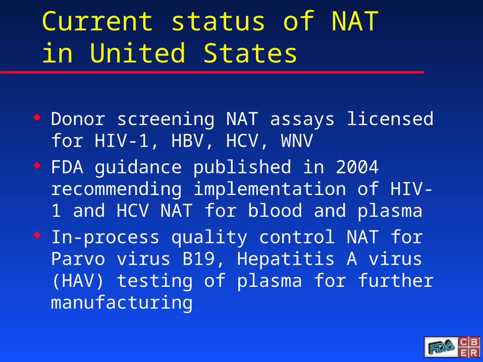
Current status of NAT in United States
Donor screening NAT assays licensed for HIV-1, HBV, HCV, WNV
FDA guidance published in 2004 recommending implementation of HIV-1 and HCV NAT for blood and plasma
In-process quality control NAT for Parvo virus B19, Hepatitis A virus (HAV) testing of plasma for further manufacturing
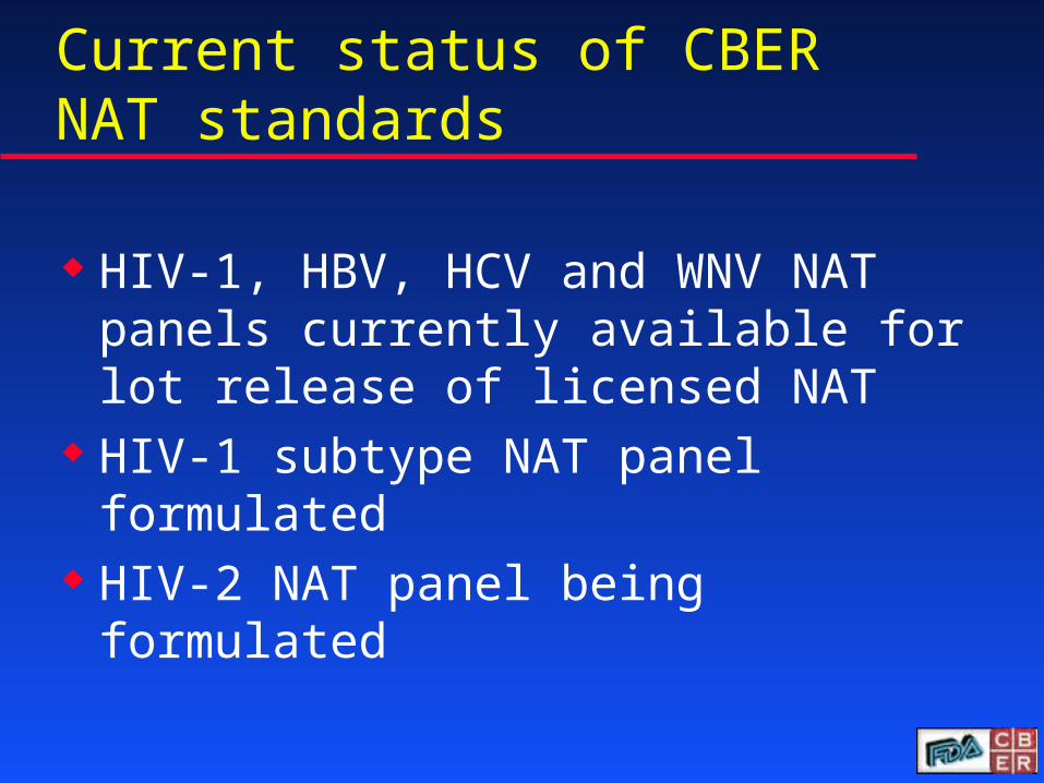
Current status of CBER NAT standards
HIV-1, HBV, HCV and WNV NAT panels currently available for lot release of licensed NAT
HIV-1 subtype NAT panel formulated HIV-2 NAT panel being formulated
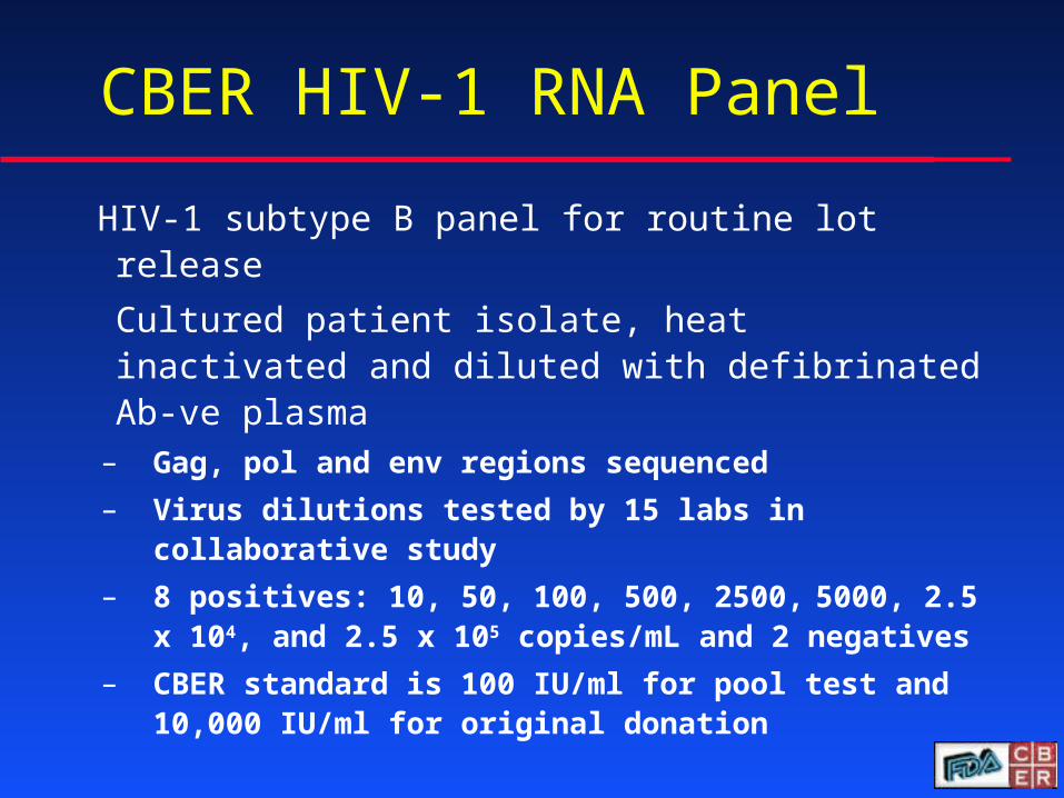
CBER HIV-1 RNA Panel
HIV-1 subtype B panel for routine lot release
Cultured patient isolate, heat inactivated and diluted with defibrinated Ab-ve plasma
– Gag, pol and env regions sequenced
– Virus dilutions tested by 15 labs in collaborative study
– 8 positives: 10, 50, 100, 500, 2500, 5000, 2.5 x 10⁴, and 2.5 x 10⁵ copies/mL and 2 negatives
– CBER standard is 100 IU/ml for pool test and 10,000 IU/ml for original donation
5
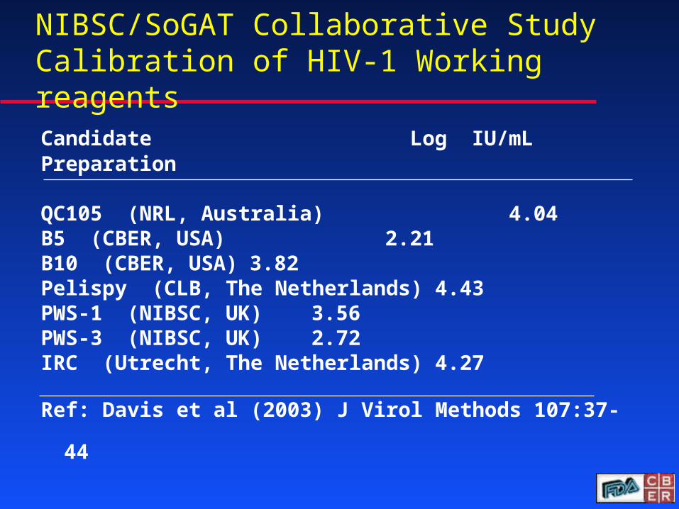
NIBSC/SoGAT Collaborative Study Calibration of HIV-1 Working reagents
Candidate Log IU/mLPreparation QC105 (NRL, Australia) 4.04B5 (CBER, USA) 2.21B10 (CBER, USA) 3.82Pelispy (CLB, The Netherlands) 4.43PWS-1 (NIBSC, UK) 3.56PWS-3 (NIBSC, UK) 2.72IRC (Utrecht, The Netherlands) 4.27
Ref: Davis et al (2003) J Virol Methods 107:37-44
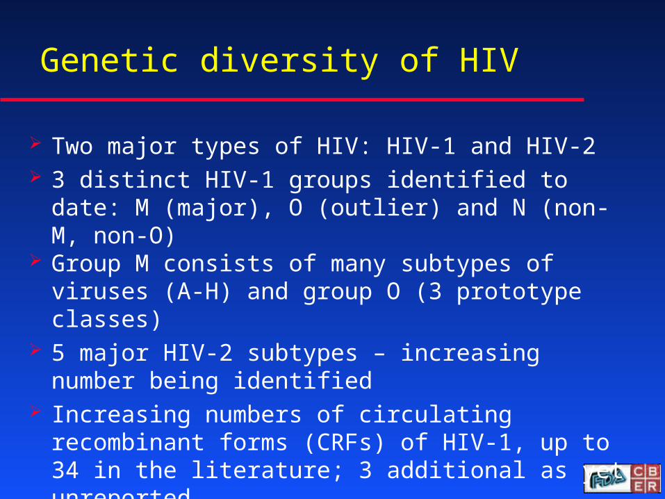
Genetic diversity of HIV
Two major types of HIV: HIV-1 and HIV-2 3 distinct HIV-1 groups identified to date: M
(major), O (outlier) and N (non-M, non-O) Group M consists of many subtypes of viruses
(A-H) and group O (3 prototype classes) 5 major HIV-2 subtypes – increasing number
being identified Increasing numbers of circulating recombinant
forms (CRFs) of HIV-1, up to 34 in the literature; 3 additional as yet unreported
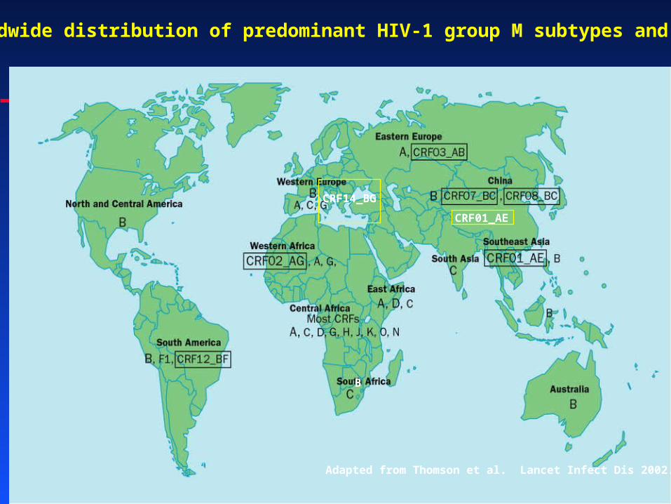
Adapted from Thomson et al. Lancet Infect Dis 2002.
Worldwide distribution of predominant HIV-1 group M subtypes and CRFs
CRF14_BG
CRF01_AE
B
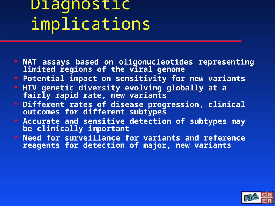
Diagnostic implications
NAT assays based on oligonucleotides representing limited regions of the viral genome
Potential impact on sensitivity for new variants HIV genetic diversity evolving globally at a fairly
rapid rate, new variants Different rates of disease progression, clinical
outcomes for different subtypes Accurate and sensitive detection of subtypes may
be clinically important Need for surveillance for variants and reference
reagents for detection of major, new variants
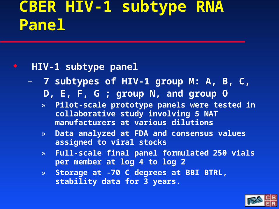
CBER HIV-1 subtype RNA Panel
HIV-1 subtype panel
– 7 subtypes of HIV-1 group M: A, B, C, D, E, F, G ; group N, and group O
» Pilot-scale prototype panels were tested in collaborative study involving 5 NAT manufacturers at various dilutions
» Data analyzed at FDA and consensus values assigned to viral stocks
» Full-scale final panel formulated 250 vials per member at log 4 to log 2
» Storage at -70 C degrees at BBI BTRL, stability data for 3 years.

HIV-1 Subtype Isolates used in current CBER Panel
Clade Origin ID # Viral copies/ml
A Djbouti DJ/258/91 6.6 x 10⁶
B United States 9697 1.9 x 10⁹
C Senegal SE/364/90 1.9 x 10⁷
D Uganda UG/021/92 4.2 x 10⁷
E Thailand TH/022/92 1.9 x 10⁷
F Brazil BZ/162/90 1.6 x 10⁷
G Nigeria G3/Nigeria 1.2 x 10⁷
N Cameroon Sinnousi 2.0 x 10⁷
O Spain 1422/German 8.1 x 10⁶

Current status of CBER HIV-2 panel development
Seven isolates of HIV-2 belonging to subtype A from Spain
Isolates were tested by three manufacturers at different serial dilutions
Statistical analysis of data for value assignment
Panel being formulated with 2 isolates to include log ranges of 2 – 4
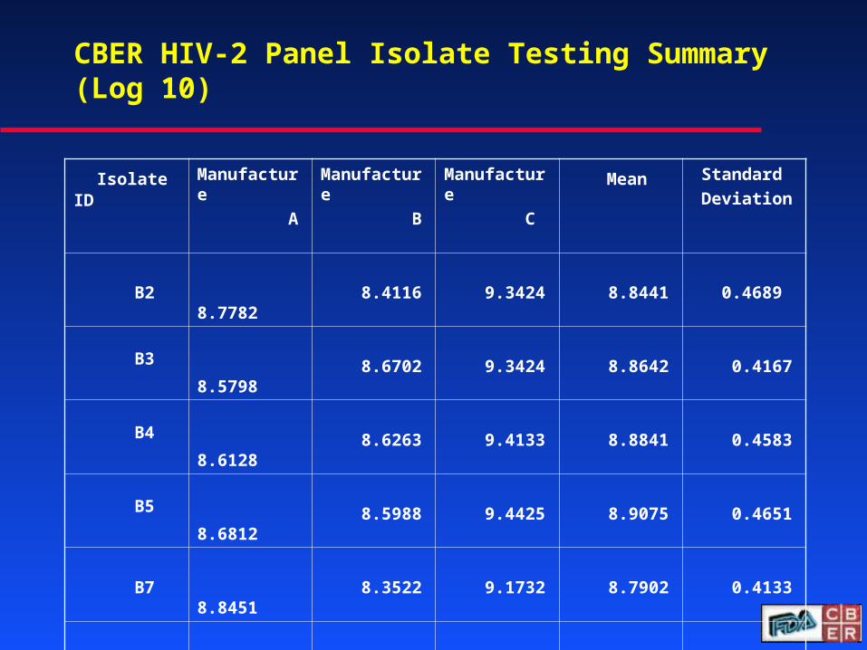
CBER HIV-2 Panel Isolate Testing Summary (Log 10)
Isolate ID Manufacture
A
Manufacture
B
Manufacture
C Mean Standard
Deviation
B2
8.7782 8.4116 9.3424 8.8441 0.4689
B3 8.5798 8.6702 9.3424 8.8642 0.4167
B4 8.6128 8.6263 9.4133 8.8841 0.4583
B5 8.6812 8.5988 9.4425 8.9075 0.4651
B7 8.8451 8.3522 9.1732 8.7902 0.4133
B8 8.6721 8.1847 9.1732 8.6767 0.4943
B9 8.2788 7.7924 9.0969 8.3894 0.6593
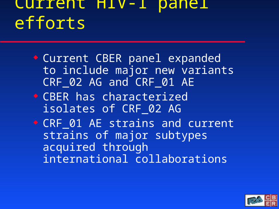
Current HIV-1 panel efforts
Current CBER panel expanded to include major new variants CRF_02 AG and CRF_01 AE
CBER has characterized isolates of CRF_02 AG
CRF_01 AE strains and current strains of major subtypes acquired through international collaborations
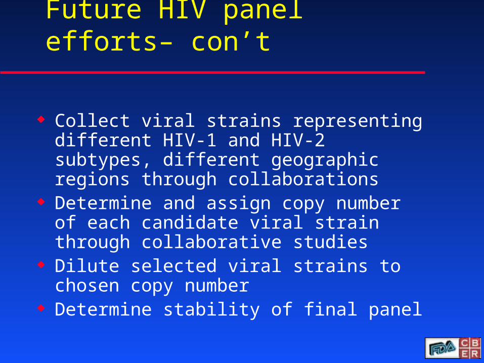
Future HIV panel efforts– con’t
Collect viral strains representing different HIV-1 and HIV-2 subtypes, different geographic regions through collaborations
Determine and assign copy number of each candidate viral strain through collaborative studies
Dilute selected viral strains to chosen copy number
Determine stability of final panel
15
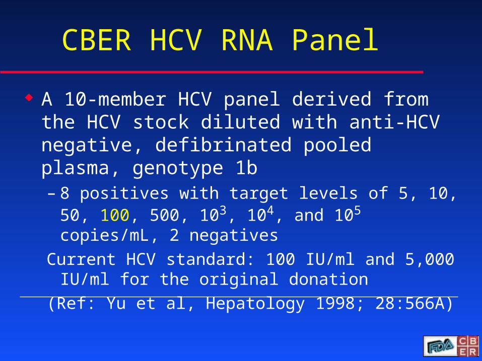
CBER HCV RNA Panel
A 10-member HCV panel derived from the HCV stock diluted with anti-HCV negative, defibrinated pooled plasma, genotype 1b – 8 positives with target levels of 5, 10, 50, 100,
500, 103, 104, and 105 copies/mL, 2 negatives
Current HCV standard: 100 IU/ml and 5,000 IU/ml for the original donation
(Ref: Yu et al, Hepatology 1998; 28:566A)
16
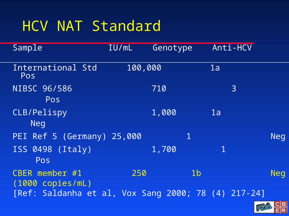
HCV NAT Standard
Sample IU/mL Genotype Anti-HCV International Std 100,000 1a Pos
NIBSC 96/586 710 3 Pos
CLB/Pelispy 1,000 1a Neg
PEI Ref 5 (Germany) 25,000 1 Neg
ISS 0498 (Italy) 1,700 1 Pos
CBER member #1 250 1b Neg(1000 copies/mL)[Ref: Saldanha et al, Vox Sang 2000; 78 (4) 217-24]
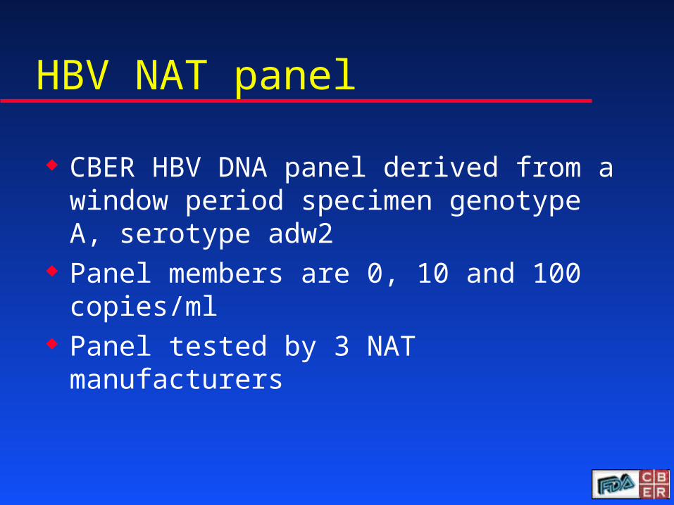
HBV NAT panel
CBER HBV DNA panel derived from a window period specimen genotype A, serotype adw2
Panel members are 0, 10 and 100 copies/ml
Panel tested by 3 NAT manufacturers
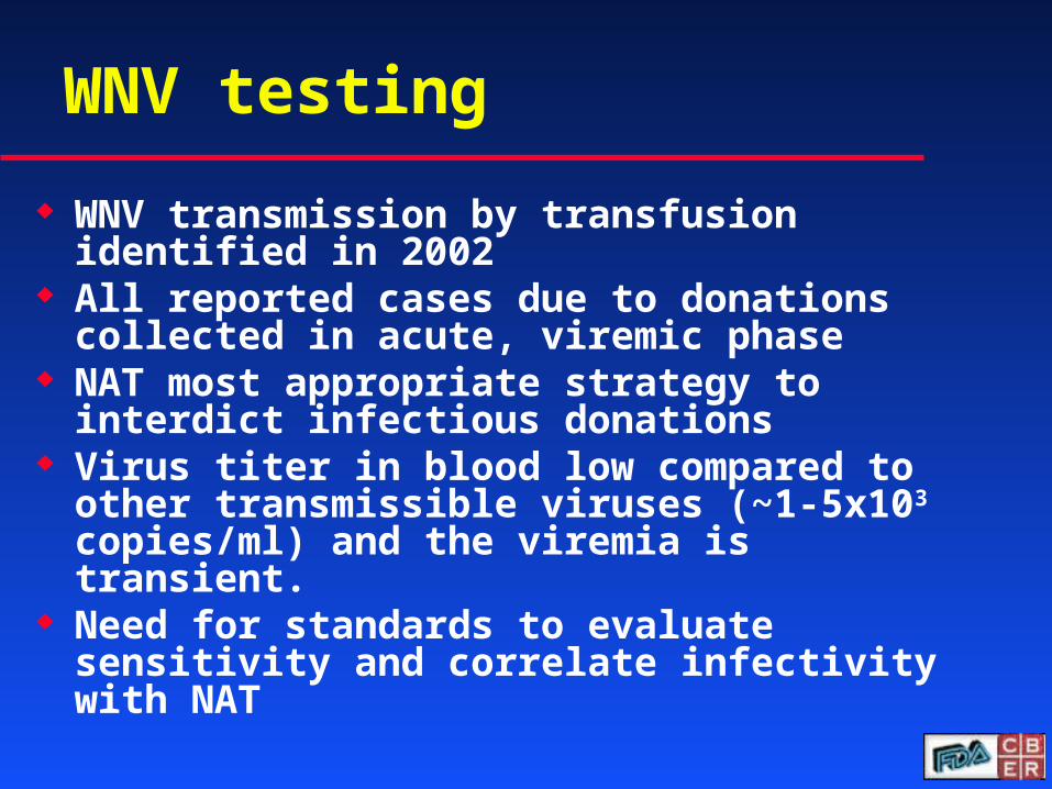
WNV testing
WNV transmission by transfusion identified in 2002
All reported cases due to donations collected in acute, viremic phase
NAT most appropriate strategy to interdict infectious donations
Virus titer in blood low compared to other transmissible viruses (~1-5x10³ copies/ml) and the viremia is transient.
Need for standards to evaluate sensitivity and correlate infectivity with NAT
WNV NAT Panel
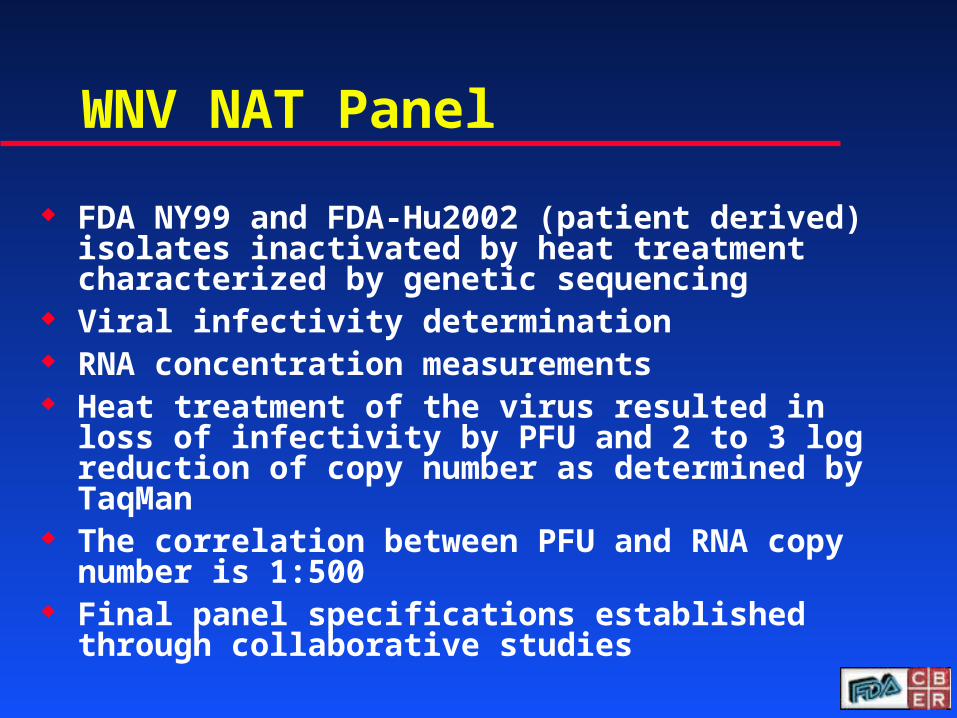
FDA NY99 and FDA-Hu2002 (patient derived) isolates inactivated by heat treatment characterized by genetic sequencing
Viral infectivity determination RNA concentration measurements Heat treatment of the virus resulted in loss of
infectivity by PFU and 2 to 3 log reduction of copy number as determined by TaqMan
The correlation between PFU and RNA copy number is 1:500
Final panel specifications established through collaborative studies
WNV Panel Formulation and Evaluation in Collaborative Studies
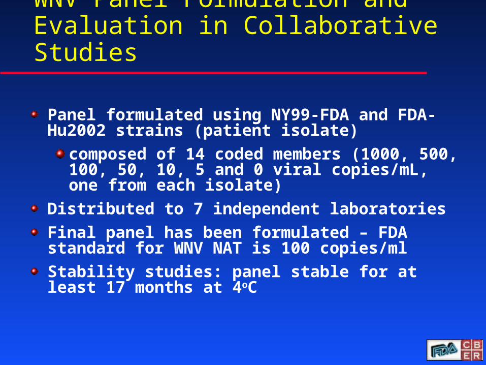
Panel formulated using NY99-FDA and FDA-Hu2002 strains (patient isolate)
composed of 14 coded members (1000, 500, 100, 50, 10, 5 and 0 viral copies/mL, one from each isolate)
Distributed to 7 independent laboratories
Final panel has been formulated – FDA standard for WNV NAT is 100 copies/ml
Stability studies: panel stable for at least 17 months at 4oC
Dengue
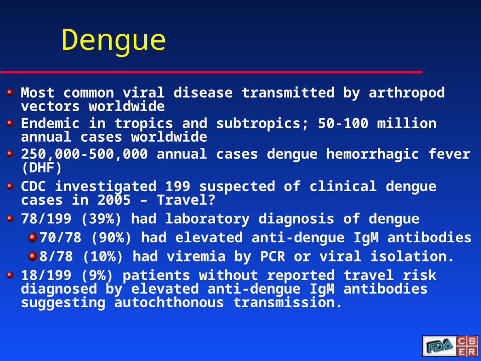
Most common viral disease transmitted by arthropod vectors worldwideEndemic in tropics and subtropics; 50-100 million annual cases worldwide 250,000-500,000 annual cases dengue hemorrhagic fever (DHF)CDC investigated 199 suspected of clinical dengue cases in 2005 – Travel?78/199 (39%) had laboratory diagnosis of dengue
70/78 (90%) had elevated anti-dengue IgM antibodies8/78 (10%) had viremia by PCR or viral isolation.
18/199 (9%) patients without reported travel risk diagnosed by elevated anti-dengue IgM antibodies suggesting autochthonous transmission.
Future efforts - collaborative study
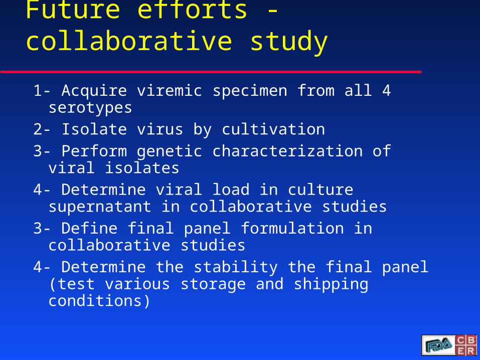
1- Acquire viremic specimen from all 4 serotypes2- Isolate virus by cultivation 3- Perform genetic characterization of viral
isolates 4- Determine viral load in culture supernatant in
collaborative studies3- Define final panel formulation in collaborative
studies 4- Determine the stability the final panel (test
various storage and shipping conditions)

Parvovirus B19 NAT as an In-Process Control
Require validation as an analytical test and approve it under relevant product’s license
Proposed limit: <104 IU of B19 DNA per mL in all manufacturing pools
– B19 transmissions associated with S/D Treated Pooled Plasma in a phase 4 study in healthy donors
» <104 GE/mL in non-transmitting lots
– Viral neutralization by anti-B19 in pools
– Viral clearance by manufacturing procedures
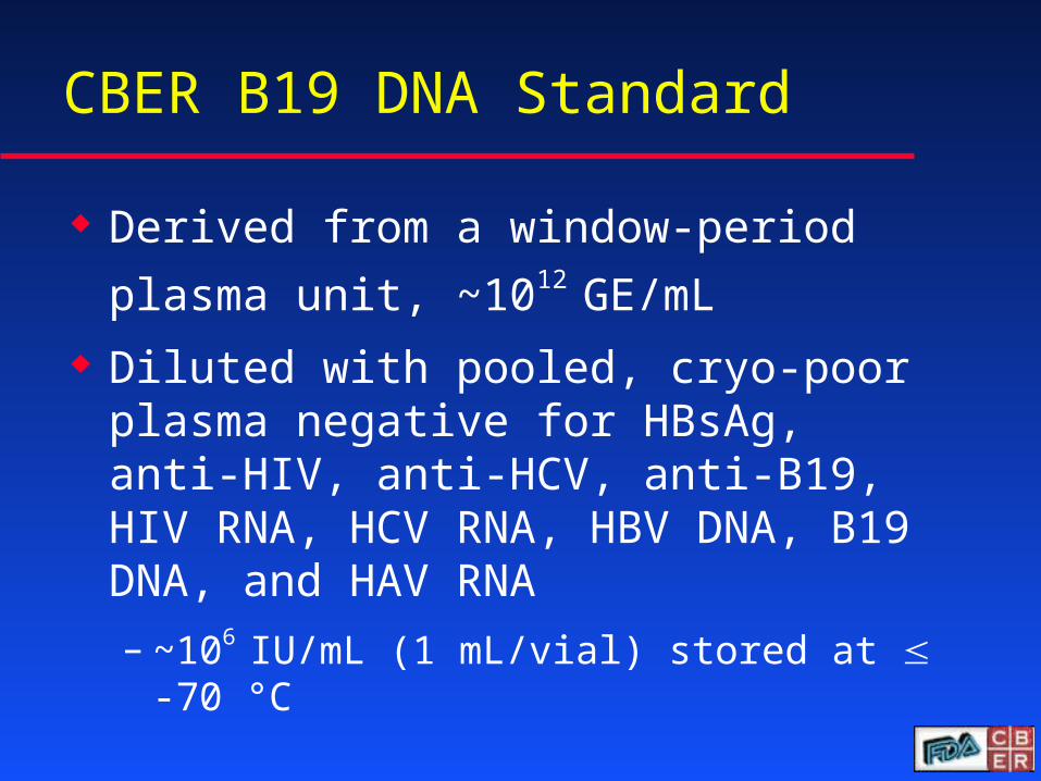
CBER B19 DNA Standard
Derived from a window-period plasma
unit, ~1012 GE/mL
Diluted with pooled, cryo-poor plasma negative for HBsAg, anti-HIV, anti-HCV, anti-B19, HIV RNA, HCV RNA, HBV DNA, B19 DNA, and HAV RNA
– ~106 IU/mL (1 mL/vial) stored at -70 °C
25
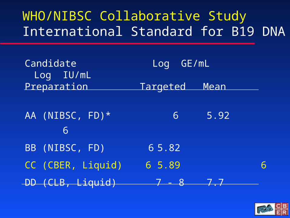
WHO/NIBSC Collaborative Study International Standard for B19 DNA
Candidate Log GE/mL Log IU/mL
Preparation Targeted Mean
AA (NIBSC, FD)* 6 5.92 6
BB (NIBSC, FD) 6 5.82
CC (CBER, Liquid) 6 5.89 6
DD (CLB, Liquid) 7 - 8 7.7
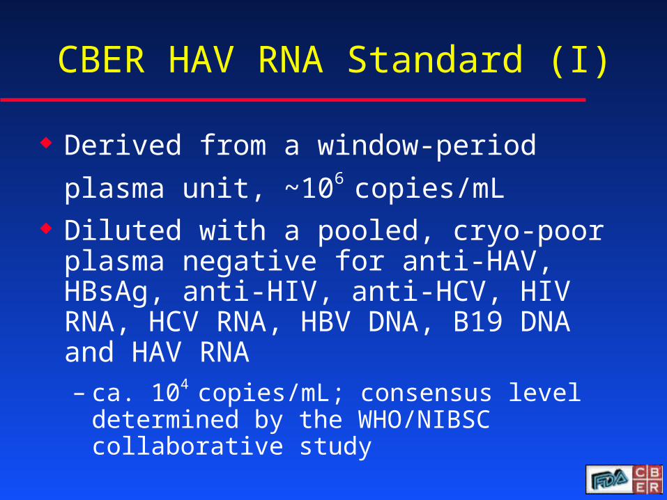
CBER HAV RNA Standard (I)
Derived from a window-period plasma
unit, ~106 copies/mL Diluted with a pooled, cryo-poor plasma
negative for anti-HAV, HBsAg, anti-HIV, anti-HCV, HIV RNA, HCV RNA, HBV DNA, B19 DNA and HAV RNA– ca. 104 copies/mL; consensus level
determined by the WHO/NIBSC collaborative study
Summary
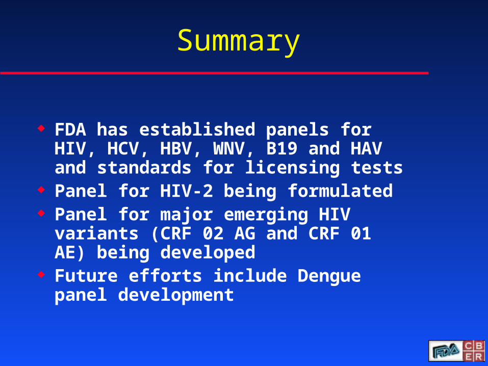
FDA has established panels for HIV, HCV, HBV, WNV, B19 and HAV and standards for licensing tests
Panel for HIV-2 being formulated Panel for major emerging HIV variants
(CRF 02 AG and CRF 01 AE) being developed
Future efforts include Dengue panel development
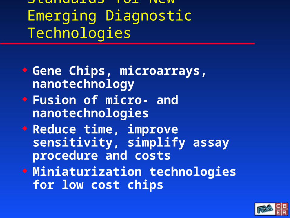
Standards for New Emerging Diagnostic Technologies
Gene Chips, microarrays, nanotechnology
Fusion of micro- and nanotechnologies
Reduce time, improve sensitivity, simplify assay procedure and costs
Miniaturization technologies for low cost chips
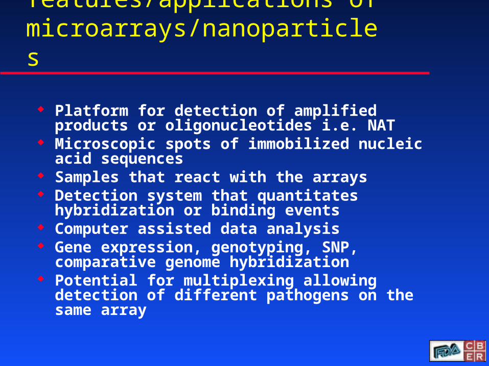
General features/applications of microarrays/nanoparticles
Platform for detection of amplified products or oligonucleotides i.e. NAT
Microscopic spots of immobilized nucleic acid sequences
Samples that react with the arrays Detection system that quantitates hybridization
or binding events Computer assisted data analysis Gene expression, genotyping, SNP, comparative
genome hybridization Potential for multiplexing allowing detection of
different pathogens on the same array
McNeil, (2005), J. Leuk. Biol., 78:585-594
Common Nanoscale Particles in Biological Use
Nanoparticle/microarray detection of avian Nanoparticle/microarray detection of avian influenza virus subtypesinfluenza virus subtypes
RNA targetHybridization
viral RNA
Au-probeHybridization
H1 H3M H5 N1 N2 control
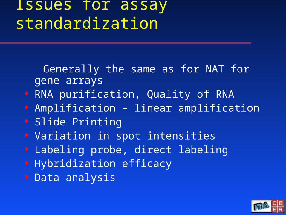
Issues for assay standardization
Generally the same as for NAT for gene arrays RNA purification, Quality of RNA Amplification – linear amplification Slide Printing Variation in spot intensities Labeling probe, direct labeling Hybridization efficacy Data analysis
Reference reagents
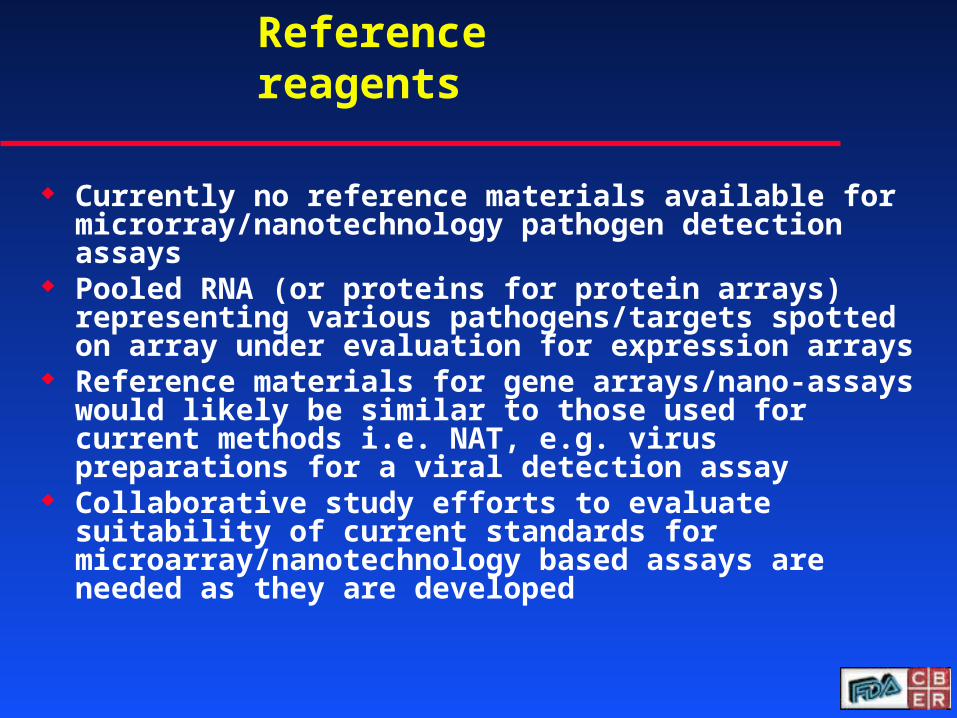
Currently no reference materials available for microrray/nanotechnology pathogen detection assays
Pooled RNA (or proteins for protein arrays) representing various pathogens/targets spotted on array under evaluation for expression arrays
Reference materials for gene arrays/nano-assays would likely be similar to those used for current methods i.e. NAT, e.g. virus preparations for a viral detection assay
Collaborative study efforts to evaluate suitability of current standards for microarray/nanotechnology based assays are needed as they are developed
Summary
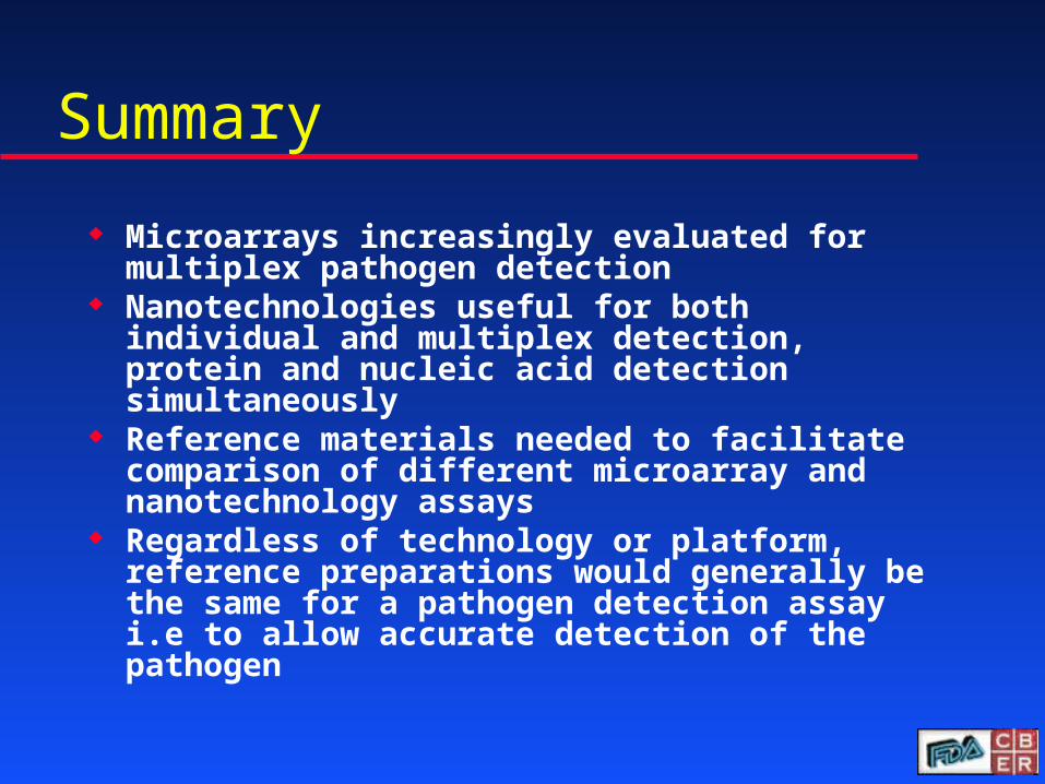
Microarrays increasingly evaluated for multiplex pathogen detection
Nanotechnologies useful for both individual and multiplex detection, protein and nucleic acid detection simultaneously
Reference materials needed to facilitate comparison of different microarray and nanotechnology assays
Regardless of technology or platform, reference preparations would generally be the same for a pathogen detection assay i.e to allow accurate detection of the pathogen
Acknowledgements
CBER/FDA WRAIRS. Lee N. MichaelM. Yu ARCO. Wood S. StramerM. Rios Carlos SaludS. Kerby V. SorianoR. Biswas NYDOHR. Duncan L. KramerC. Hsia NYUJ. Zhao P. Nyambi