STAINING OF THE CRAB LEG JOINT RECEPTORS...
16
1 STAINING OF THE CRAB LEG JOINT RECEPTORS By Zana R. Majeed 1,2 , Josh Titlow 1 , H. Bernard Hartman 3 , Ellen Burns 1 and Robin L. Cooper 1 1 Department of Biology, University of Kentucky, Lexington, KY 40506, USA; 2 Department of Biology, College of Sci, Univ. of Salahaddin, Erbil, Iraq; 3 Oregon Institute of Marine Biology, University of Oregon, Charleston, OR 97420, USA 1. Purpose To learn histological techniques for staining nerve cells to aid in describing the general organization of a chordotonal organ. 2. Preparation The chordotonal organs in the joints of walking legs of the blue crab Callinectes sapidus or the Dungeness crab Cancer magister will be employed. Other large Cancer species are also suitable. 3. Introduction The walking leg of a crab has six joints, each having a means of detecting joint proprioception (Figure 1). The proprioceptive organs that monitor joint position and movement in crustaceans are called chordotonal organs. Using Alexandrowiz's (1967) designation, these receptors are named according to which joint they are monitoring (i.e., "PD-organ" is the organ between the propodite (P) and the dactylopodite (D)). Joint receptors consist of an elastic strand into which are inserted the dendrites of neurons. These neurons signal joint movement, direction of movement, and static position (Wiersma, 1959). Analysis of individual cells in the chordotonal organ spanning the propodite-dactylopodite joint (PD) revealed an orderly arrangement of these neurons along the elastic strands according to function
Transcript of STAINING OF THE CRAB LEG JOINT RECEPTORS...

1
STAINING OF THE CRAB LEG JOINT RECEPTORS
By
Zana R. Majeed1,2, Josh Titlow1, H. Bernard Hartman3, Ellen Burns1 and Robin L.
Cooper1 1Department of Biology, University of Kentucky, Lexington, KY 40506, USA; 2Department of Biology, College of Sci, Univ. of Salahaddin, Erbil, Iraq; 3Oregon Institute of Marine Biology, University of Oregon, Charleston, OR 97420, USA
1. Purpose
To learn histological techniques for staining nerve cells to aid in
describing the general organization of a chordotonal organ.
2. Preparation
The chordotonal organs in the joints of walking legs of the blue crab Callinectes
sapidus or the Dungeness crab Cancer magister will be employed. Other large Cancer
species are also suitable.
3. Introduction
The walking leg of a crab has six joints, each having a means of detecting joint
proprioception (Figure 1). The proprioceptive organs that monitor joint position and
movement in crustaceans are called chordotonal organs. Using Alexandrowiz's
(1967) designation, these receptors are named according to which joint they are
monitoring (i.e., "PD-organ" is the organ between the propodite (P) and the
dactylopodite (D)). Joint receptors consist of an elastic strand into which are inserted
the dendrites of neurons. These neurons signal joint movement, direction of
movement, and static position (Wiersma, 1959). Analysis of individual cells in the
chordotonal organ spanning the propodite-dactylopodite joint (PD) revealed an
orderly arrangement of these neurons along the elastic strands according to function

2
(Hartman and Boettiger, 1967) as well as in other chordotonal organs (Cooper and
Hartman, 1993; Cooper, 2008).
Figure 1: First walking leg of a crab with the tendon shown as an X-ray. The
chordotonal organs are the hatched regions.
The anatomical arrangement of chordotonal organs in crabs allows one to
analyze each individual neuron according to function (Figure 2). In addition,
developmental questions can be addressed as the animal grows or when the animal
regenerates a limb (Cooper, 2008; Cooper and Govind, 1991; Hartman and Cooper,
1994).

3
Figure 2: The functional arrangement of the sensory neurons on the PD organ and the
tension receptors associated with the closer muscle.
4. Methods 4.1 Materials 1. Crab Saline (see Tables 1 & 2) 2. Methylene blue: This is made of crayfish saline at a concentration of 0.25% 3. Cobalt chloride (300 mM)
4. 4-di-2-ASP or Lucifer yellow stain (10 µM). 5. Sylgard coated dishes (Dow Corning, SYLGARD® 184 silicone elastomer kit; Dow Corning Corporation, Midland, MI. USA) 6. Dissecting tools 7. Insect pins 8. Glass rod/tools for dissecting and manipulating nerves 9. Pipettes and beakers 10. Petroleum jelly (clear) 4.2 Setup
1. The microscope, high intensity illuminator, and the saline bath is needed 2. Setup the microscope in a position where it is overlooking the microscope stage. 3. Position the high intensity illuminator in a convenient position. 4. Place the previously prepared crab saline in the Sylgard dish and position the dish on the stage of the microscope. Staple the dissected leg in the dish. 4.3 Dissection
Walking legs of the swimming crab Callinectes sapidus caught in the Gulf of
Mexico (http://www.crabplace.com/crabs.asp) or from Cancer magister, found off the
northwest coast of the USA, will be used. If the crab is dropped, its carapace will crack
and the animal will bleed to death. Therefore, handle them carefully.
Using a net obtain a crab from the aquarium and place into an empty bucket.
While holding the crab with the net or large tongs across the carapace from behind,
and avoiding the claws, cut across the merus of the second-right walking leg with a
stout pair of scissors. The animal will/should autotomize the remaining basal portion of
that limb thus sealing the wound and preventing blood loss. If the crab does not break
it off, help it by inserting the scissor tips into the wound and twisting the stump. Return

4
the animal to its aquarium. Put the leg in the Sylgard-lined dissecting dish and cover it
with the species correct cooled (12-19oC) crab saline.
With scissors, make a cut between the propus and carpus. Discard the carpus
and the attached merus. Use Figure 3 to guide you through the rest of this dissection.
Cut a large window in the cuticle on the pigmented (anterior) side of the propus with a
scalpel with a #11 blade. (Note: Do not cut deeply). Remove the cuticle layer by sliding
the scalpel blade beneath and parallel to the cuticle. This severs the opener and
closer muscle fibers attached to the cuticle. Using the same technique cut a smaller
window on the pigmentless (posterior) side of the propus, but leaving the condyle
attachment intact.
Figure 3: Cut along the dotted line on the propodite
You must exchange the saline in the bath with fresh cold saline throughout the
dissection so that the neurons stay alive. Pin the preparation to the recording dish in
the locations illustrated using stainless-steel staples with the pigmented side facing
upwards. For further dissection, place the preparation dish under a dissecting
microscope and use fiber-optic illumination and transmitted light through the base of
the dissection platform.
Using sharp-pointed intermediate-size scissors carefully cut the opener tendon
from its attachment to the dactyl. Be very careful not to disturb the main leg nerve
which should be clearly visible, remove and discard the opener muscle and tendon.
This will require minimal cutting and trimming.
Locate the PD organ by careful probing with the glass needles. The elastic
strand spanning the joint has a silver appearance. Ask for help if you cannot find it.
Now pin the main leg nerve so that the PD organ nerve is exposed (see Figure 4). The
fine dissection that follows requires iris scissors. They are only used to cut small soft
tissue. You will receive guidance with this part of the dissection because it is not

5
initially easy. Remove muscle fibers that obscure your view of the organ and its nerve
from both sides of the tendon being very careful not to injure the PD organ or its nerve.
Once this has been accomplished, firmly re-attach the preparation to the dish with the
pigment side facing you.
Figure 4: Exposed PD organ and nerve.
Using a fragment of a plastic millimeter ruler, measure and record in your
notebook the length of the elastic strand when the dactyl is in the opened and closed
positions. Use the attachment points on the protuberance and the tendon as reference
markers.
Trace the PD organ nerve in the propus as far proximally as possible in order to
free-up a long length of nerve (1.5 cm) for recording purposes. This is best done while
the PD nerve is still attached to the main leg nerve for a cm or so since leaving the
nerve attached to the main leg nerve adds support. After separating the PD nerve from
the main leg nerve with the aid of glass needles, sever the PD nerve proximally with
the iris scissors. (Note: Do not stretch or pull on the nerve during the dissection).
After finding the PD nerve the rest of the preparation not required can be cut
way. The opener muscle and the carpopodite can be removed. The distal half of the
dactylopodite can also be removed. Carefully remove the proximal half of the closer
muscle and apodeme as well as the ventral part of the propodite cuticle. One should

6
have gone through a series of steps as shown in Figure 5.
Figure 5: Trimming the closer apodeme to the expose the PD organ and nerve.
4.4 Staining
4.5.1 Methylene Blue:
This technique was explained in the physiology protocol for the PD organ.
Refer to this earlier text for details.
4.5.2 Cobalt back-fill of the PD nerve:
Using the in situ preparation, back-fill the entire PD organ nerve according to
the following procedure. A one-day back-fill in the incubator at 13 (oC) is suggested.

7
Expose the nerve. Keep it immersed in cold saline that is refreshed every 10-15
minutes during the operation and in all the steps to follow except where noted. A
petroleum jelly well is required to be made to hold the CoCl2. If any CoCl2 spills into
the saline bath the entire preparation will stain black, and the preparation should be
discarded. One needs to slip a small cut of a polystyrene sheet, to make a plastic
platform, and pin it in such a manner that it will not float away or become immersed in
the saline bath (Figure 6).
Figure 6: Polystyrene sheet with pins to hold in place in dish.
Ejecting petroleum jelly or silicone stopcock grease from a fine hypodermic
needle fastened to a disposable syringe, make a barrier (a circle might work well) on
top of the slip of polystyrene. The barrier should be about 1-1.5 mm high except for a
shallow "V" at midpoint where the nerve will be draped across. Make a small puddle of
saline on either side of the barrier near the "V". Taking care not to stretch or pinch the
nerve. Lift the nerve carefully from the dish and place it in the saline puddle in the

8
petroleum jelly well. Working quickly so that the section of nerve does not dry out,
carefully eject petroleum jelly or stopcock grease to cover the exposed nerve. Now
test the barrier with saline and make sure it is not leaking. To assure yourself that the
two sides are isolated from one another, blot away saline on the inside of the barrier
and see if it fills up when the bath saline is high around the wall of petroleum jelly. If
saline does not leak across in a few seconds, the barrier is probably sealed (Figure
7).
Figure 7: Petroleum jelly well with saline and PD nerve spanning the well
Using the rolled up point of a piece of tissue paper, blot up the saline in the well
without the paper wrapping up the nerve. Make a new puddle using a few small drops
of distilled water, and then cut the nerve end. Be very careful not to pull the nerve cord
through the barrier when making the cut. The osmotic shock of the distilled water will
"balloon" the axons of the connectives. Within 30 seconds add a small drop of CoCl2
to the water, blot up this solution, and then add enough CoCl2 to form a puddle over
this shortened section of nerve (Figure 8). Preparations are best kept refrigerated at
4oC for 12-24 hrs

9
Figure 8: The cut PD nerve being exposed to CoCl2.
The rest of the procedure involves precipitation of CoCl2 which has been taken
up by the axons and transported to neural processes and cell bodies. Remove the
humidifying puddles. Blot away the cobalt solution using a tissue and wash away the
remnants of cobalt with several changes of saline. Transfer the isolated nerve cord to
a small glass Petri dish containing about 10 ml of saline. The neurons are washed and
the following steps are done in situ. Good metal tools are not to be used to handle the
preparation after this step (you should use specific tools for this). Add 1-2 drops of
ammonium sulfide (NH4)2S to the saline. Cap the bottle tightly and place back into the
hood. Observe the reaction in the preparation under a dissecting microscope (Figure
9).
Within a minute or two, cobalt filled neurons and their processes should begin
to appear. After 5-10 min, replace the development solution with fresh saline. Make
certain that the development solution you have poured into the sink drain is followed
by running tap water for a few minutes.

10
Figure 9: Neurons that were filled with CoCl2 and processed (A). Traced outline of the
stained preparation shown (B).
Pour out the saline and fix the nerve preparation for about 15 minutes with two
changes of Bouin’s solution fixative (Sigma). For larger tissues, increase the duration
of fixation. Dehydrate in an ascending order of ethanol concentration beginning at
70% (i.e. 70%, 80%, 90%, 100%). About 10 minutes at each concentration is sufficient
for small tissues. After about 10-15 minutes in two changes in 100 % alcohol clear the
tissue by replacing alcohol with methyl salicylate. The preparation will stay in this
solution permanently for repeated viewing. With time the filled cells will be come more
apparent because the surrounding tissue will become clearer.
4.5.3. 4-di-2-ASP or Lucifer yellow stain:
Fluorescent dyes can also be used to back-fill the PD neuron; however a
microscope with abilities to view the fluorescent stain is required.
Use the same approach as described above for cobalt filling in making a petroleum
jelly well to contain the dye and the nerve of interest. In the well place a 0.01 to 0.04
mM 4-Di-2-ASP solution or a 0.02 mM Lucifer yellow solution and leave the
preparation in the refrigerator for 12 to 24 hours. These fluorescent dyes have the

11
advantage over the cobalt dye, in that the amount of filling can be assessed at various
stages of filling, although, these fluorescent dyes do fade relatively quickly. So
photograph the preparations quickly and avoid over exposure to the mercury light
(Figure 10). If one has a high enough magnification the sensory endings can be seen
inside of the supportive scolopales (Whitear, 1962,1965; Hartman and Cooper, 1994;
Figure 11-13).
Figure 10: A back-fill of the PD nerve in Cancer magister with 4-Di-2-ASP.
Figure 11: Higher magnification of neurons shown in Figure 14. The PD nerve in
Cancer magister was back filled with 4-Di-2-ASP.
12
Figure 12: Movement sensitive sensory endings in the PD organ of Cancer magister.
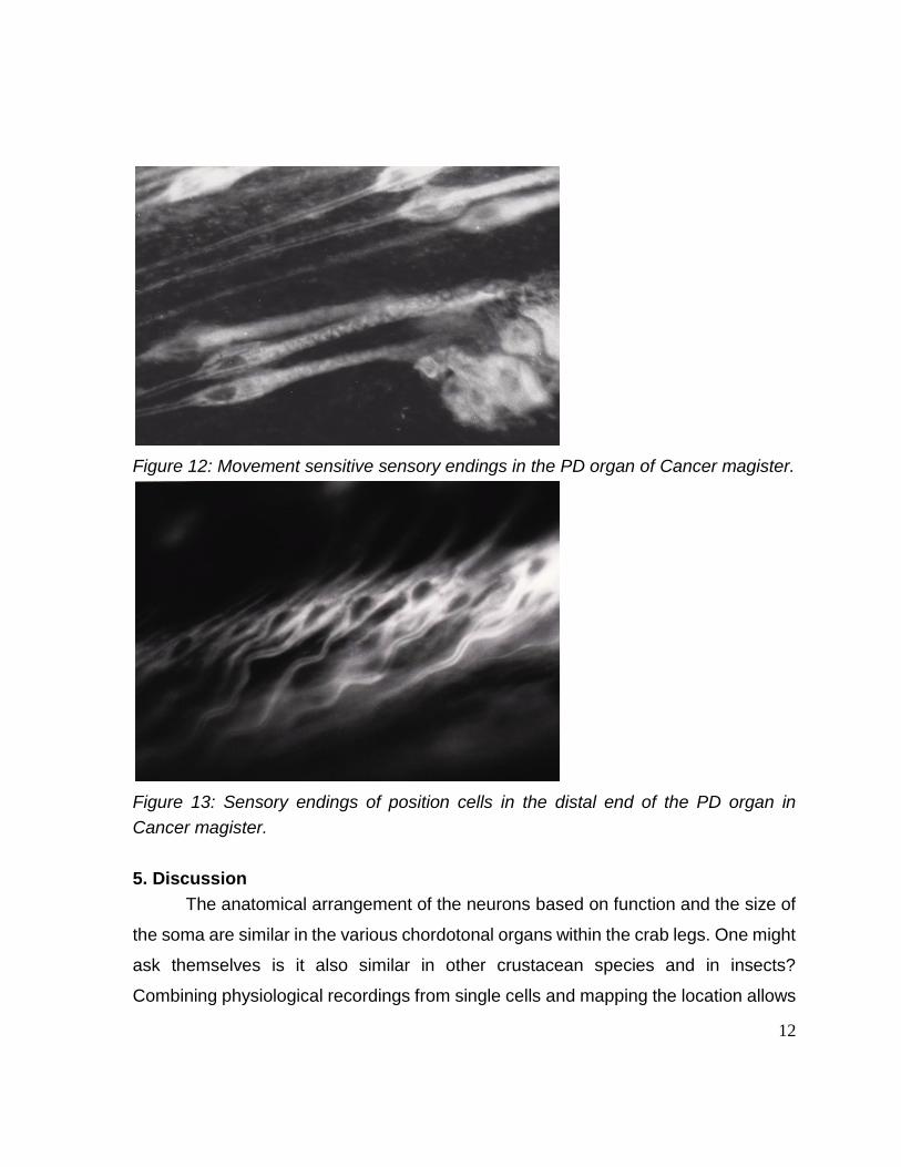
Figure 13: Sensory endings of position cells in the distal end of the PD organ in
Cancer magister.
5. Discussion
The anatomical arrangement of the neurons based on function and the size of
the soma are similar in the various chordotonal organs within the crab legs. One might
ask themselves is it also similar in other crustacean species and in insects?
Combining physiological recordings from single cells and mapping the location allows

13
direct structure function relationships. The large somata located proximally on the
strand tend to belong to the dynamic movement sensitive neurons; the neurons
signaling static positions by firing tonically have small somata and are located distally
(Figure 9).
REFERENCES: Alexandrowicz, J.S. (1972) The comparative anatomy of leg propriocetors in some
decapod Crustacea. J. Mar. Biol. Ass. UK 52:605-634
Burgess, P.R., Wei, J.Y., Clark, F.J., and Simon, J. (1982) Signaling of kinesthetic
information by peripheral sensory receptors. Ann. Rev. Neurosci. 5:171-187
Burke, W. (1953) An organ for proprioception and vibration sense in Carcinus
maenas. J. Exp. Biol. 31:127-138
Bush, B.M.H. (1962) Proprioceptive reflexes in the legs of Carcinus maenas. J. Exp.
Biol. 39:89-105
Bush, B.M.H. (1965a) Proprioception by chordotonal organs in the mero-carpopodite
and carpo-propodite joints of Carcinus meanas legs. Comp. Biochem. Physiol.
14:185-199
Bush, B.M.H. (1965b) Proprioception by the coxo-basal chordotonal organ, CB, in
legsä of the crab, Carcinus maenas. J. Exp. Biol. 42: 285-297.
Chung,Y-S. Cooper, R.M., Graff, J. and Cooper, R.L. (2012) The acute and chronic
effect of low temperature on survival, heart rate and neural function in crayfish
(Procambarus clarkii) and prawn (Macrobrachium rosenbergii) species. Open
Journal of Molecular and Integrative Physiology 2:75-86.
Cooper, R.L. (2008) Mapping proprioceptive neurons on chordotonal organs in the
crab, Cancer magister. Crustaceana 81(4):447-475.
Cooper, R.L. (1998) Development of sensory processes during limb regeneration in
adult crayfish. Journal of Experimental Biology 201:1745-1752
Cooper, R.L. and Govind, C.K. (1991) Axon composition of the proprioceptive PD nerve during growth and regeneration of lobster claws. Journal of Experimental Zoology 260:181-193.
Cooper, R.L. and Hartman, H.B. (1994) Responses of the bender apodeme tension receptors in the Dungeness crab, Cancer magister. Comparative Biochemistry and Physiology 109A:479-486.
Cooper, R.L. and Hartman, H.B. (1989) Effects of neuromodulators on proprioceptive
neurons in the limbs of the crab, Cancer magister. Soc. Neurosci. 15:565.

14
Cooper, R.M., Schapker, H. Adami, H. and Cooper, R.L. (2011) Heart and ventilatory
measures in crayfish during copulation. Open Journal of Molecular and
Integrative Physiology 1(3):36-42.
Dasari, S. and Cooper, R.L. (2004) Modulation of sensory–CNS–motor circuits by
serotonin, octopamine, and dopamine in semi-intact Drosophila larva.
Neuroscience Research 48:221-227.
Hartman, H.B. (1985) Tension receptors on the closer muscle apodeme in the walking
legs of the blue crab Callinectes sapidus. J Comp Physiol 157:355-362
Hartman, H.B. and Austin, W.D. (1972) Proprioceptor organs in the antennae of Decapoda Crustacea. J. Comp. Physiol. 81: 187-202.
Hartman, H.B., and Boettiger, E.G. (1967) The functional organization of the
propys-dactylus organ in Cancer irroratus Say. Comp. Biochem. Physiol.
22:651-663
Hartman, H.B. and Cooper, R.L. (1994) Regeneration and molting effects on a
proprioceptor organ in the Dungeness crab, Cancer magister. J. Neurobiology
25:461-471.
Hartman HB, Johnson EE, and Storer PD (1997) Manipulation by the crab claw is dependent upon chordotonal organ afference. J.Exp. Zool. 280:215-221
Macmillan, D. L. and Dando, M.R. (1972). Tension receptors on the apodemes of
muscles in the walking legs of the crab, Cancer magister. Mar. Behav. Physiol.,
1: 185-208.
Marder, E., Thirumalai, V., 2002. Cellular, synaptic and network effects of neuromodulation. Neural Networks. 15, 479–493. Mill, P.J., and Lowe, D.A. (1973) The fine structure of the PD proprioceptor of Cancer
pagurus. I. The receptor strand and the movement sensitive cells. Proc. R. Soc.
Lond. B 184:179-197
Pitman RM, Tweedle CD, Cohen MJ. (1972). Branching of central neurons:
intracellular cobalt injection for light and electron microscopy. Science.
176(4033):412-4.
Rossignol, S., Giroux, N., Chau, C., Marcoux, J., Brustein, J., Reader, T.A., 2001.
Pharmacological aids to locomotor training after spinal injury in the cat. J.
Physiol. 533, 65–74.
Rossignol, S., Bouyer, L., Barthelemy, D., Langlet, C., Leblond, H., 2002. Recovery of
locomotion in the cat following spinal cord lesions. Brain Res.—Brain Res. Rev.
40, 257–266.
Strawn, J.R., Neckameyer, W.S., and Cooper, R.L. (2000) The effects of 5-HT on
sensory neurons, central, and motor neurons driving the abdominal superficial

15
flexor muscles in the crayfish. Comparative Biochemistry and Physiology B
127:533-550.
Vedel, J. P., Clarac, F., Combined reflex actions by several proprioceptive inputs in
the rock lobster walking legs. J. Comp. Physiol. 130, 251-258 (1979).
Whitear, M. (1962) The fine structure of crustacean proprioceptors. I. The chordotonal
organs in the legs of the shore crab, Carcinus meanas. Phil. Trans. R. Soc.
Lond. B 245:291-325
Whitear, M. (1965) The fine structure of crustacean proprioceptors. II. The
thoracico-coxal organs in Carcinus, Pagurus and Astacus. Phil. Trans. R. Soc.
Lond. B 248:437-462
Wiersma, C.A.G. (1959a) Movement receptors in decapod crustacea. J. Mar. Biol.
Ass. UK 38:143-152
Wiersma, C.A.G., and Boettiger, E.G. (1959b) Unidirectional movement fibres from a
proprioceptive organ of the crab, Carcinus maenas. J. Exp. Biol. 36:102-112.

16
Table 1: Solution for C. sapidus saline (Blundon 1989, J. Comp. Physiol B 158:
689-696)
SALT g/l g/2l g/3l
NaCl 27.47 54.94 82.41
KCl 0.59 1.18 1.77
MgCl2 6H20 1.421 2.842 4.269
CaCl2 2H20 2.205 4.41 6.615
Dextrose 1.982 3.964 5.945
HEPES acid (5mM) 1.19 2.38 3.57
HEPES salt (5mM) 1.30 2.60 3.90
Adjust to pH 7.5 with NaOH or HCl.
With an osmotic pressure of 1000 mm/l.
Table 2: Solution for C. magister saline (Macmillan & Dando, 1972; Hartman and
Cooper, 1994)
SALT g/l
NaCl 27.29
KCl 0.81
MgSO4 7H20 4.81
CaCl2 2H20 1.85
Na2SO4 10H20 0.97
Dextrose 1.982
HEPES acid 0.476
HEPES salt 2.08
Adjust to pH 8.1 with NaOH or HCl.