STA - NZIMLS
44
Transcript of STA - NZIMLS


Diagnostica Stago S.A.SRCS Nanterre B305 151 4093 allée Thérésa92600 Asnières sur Seine FrancePh. +33 (0)1 46 88 20 20Fax +33 (0)1 47 91 08 [email protected]
STA®-Liatest® D-Di PlusMake the sa fe cho ice
DiET study: A 5-year multi-national prospective management study;
fully compliant with CLSI H59-A guidelines
ww
w.e
pice
a.co
m -
©20
15 D
iagn
ostic
a S
tago
- A
ll rig
hts
rese
rved
- 0
3/20
17
Suspicion of VenousThromboembolism?
Make the best choice
5 countries, 16 sites
980 patients suspected of DVT
with low or moderate pre-test probability
5 countries, 9 sites
1130 patients suspected of PE
with low or moderate pre-test probability
DVTDeep Venous Thrombosis
NPV*
99,7 %Sensitivity
97 %Specificity
75 %
Ex c l u s i o n V T
E
Cl in
ical Val idation
PEPulmonary Embolism
NPV*
100 %Sensitivity
100 %Specificity
55,2 %
STA®-Liatest® D-Di shows outstanding clinical performance for VTE exclusion with a clinical threshold at 0,5 µg/mL FEU
*NPV: Negative Predictive Value

Editor Rob Siebers, PGCertPH FNZIC FNZIMLS FRSB, University of Otago, Wellington
Deputy Editor Mike Legge, PhD MRSB FIBMS FNZIMLS FFSc (RCPA)
Editorial Board Julie Creighton, DipMLS, Canterbury Health Laboratories, Christchurch Chris Kendrick, GradDipSci MSc MNZIMLS, Massey University, Palmerston North Mike Legge, PhD MRSB FIBMS FNZIMLS FFSc(RCPA), University of Otago, Dunedin Holly Perry, MAppSc(Hons) DipMLT MNZIMLS, Auckland University of Technology Cat Ronayne, BMLSc DipGrad MNZIMLS, University of Otago, Dunedin Mohd. Shahid, MBBS MD PhD FNZIMLS, Arabian Gulf University, Bahrain John Sterling, BSc(Hons) MLett AFRCPA MAIMS FRS-SA, Flinders University, Australia Terry Taylor, BSc DipMLS MNZIMLS, Southern Community Laboratories, Dunedin Tony Woods, BA BSc(Hons) PhD MAIMS FFSc(RCPA), University of South Australia
Statistical Editor Nevil Pierse, PhD, University of Otago, Wellington
Formatting Sharon Tozer, AT DipBusStud, Executive Office NZIMLS, Rangiora
About the Journal The New Zealand Journal of Medical Laboratory Science (the Journal) is the official publication of the New Zealand Institute of Medical Laboratory Science (NZIMLS). The Journal is peer reviewed and publishes original and review articles, case studies, technical communications, and letters to the Editor on all subjects pertaining to the practice of medical laboratory science. The Journal is open access (www.nzimls.org.nz/nzimls-journal.html) and is published three times per year in April, August, and November. Hard copies are circulated to all NZIMLS members and universities and research units in New Zealand and overseas. Current circulation is about 2,200 copies per issue. Printing is by Griffin Press, Christchurch on environmentally responsible paper using elemental chlorine free third party certified pulp sourced from well managed and legally harvested forests and manufactured under the strict ISO14001 Environmental Management System. The Journal is indexed by CINAHL, EMBASE, SCOPUS, Informit, Thomson Gale, EBSCO and Biosis Citation Index, and the Journal Editors are members of the World Association of Medical Editors (www.wame.org).
Brief instructions to authors The Journal accepts original submissions from anyone and anywhere. Comprehensive instructions can be found on the NZIMLS website (www.nzimls.org.nz/instructions-to-authors.html). All submissions will undergo single-blind peer review and possibly plagiarism checking with iThenticate™ software. If accepted for publication, copyright is vested in the author(s) under terms of the Creative Commons Attribution License (www.creativecommons.org/licenses/by/2.5/legalcode). The authors are responsible for the scientific content and views. Opinions expressed in the Journal are not necessarily those of the Editors, Editorial Board, or Council of the NZIMLS.
Advertising and subscription Advertisement bookings and enquiries should be addressed to the NZIMLS Executive Officer at [email protected]. Enquiries regarding subscription and address changes should be addressed to [email protected]. Address for the NZIMLS is PO Box 505, Rangiora 7440, New Zealand. Phone +64 3 313 4761
Original articles Co-occurrence of extended-spectrum and AmpC-β-lactamase-encoding genes in Enterobacteriaceae isolated from sewage and drinking water from North-India: analysing the genetic environment with special reference to insertion sequences, integrase, and integrons Anuradha Singh, Haris M Khan and Mohammad Shahid............................................................ 44-52
Prevalence of thrombocytopenia among apparently healthy newborns in Delta State, Nigeria Collins O Adjekuko, Matthias A Emokpae, Humphrey B Osadolor and Zaccheaus A Jeremiah........... 54-56
Document review checklists for ISO 22870:2016 internal auditing: an applied tool for medical laboratories Dennis Mok, Rana Nabulsi and Sharfuddin Chowdhury.....59-64
Case studies Methylene blue stained reticulocytes in a Giemsa blood film – a case study Leah Pringle................................................ 66-68
Neonatal giant cell hepatitis: a case study Vanita Patil and Samarina Musaad.................................... 70-72
Letter to the Editor Shiga-toxigenic Escherichia coli Nandina Ghosh .......................................................................43
Erratum Thomas RR, Gaastra MLH, Brooks HJL. Shiga (Vero) -toxigenic Escherichia coli: epidemiology, virulence and disease. N Z J Med Lab Sci 2018; 72: 3–10 .........................................43
New Zealand Journal of
Medical Laboratory Science
Volume 72 Number 2 August 2018 ISSN 1171-0195
Regular features
Advertisers...............................................................................43
In this issue..............................................................................43
Journal questionnaire ..............................................................56
Journal questionnaire - April answers......................................78
Journal reviewers August 2017-April 2018 ..............................64
Media release: Claro................................................................52
NZIMLS Calendar ....................................................................72
Pacific Way.........................................................................74-75
Science Digest.........................................................................77
Fellowship of NZIMLS..............................................................69
Barrie Edwards and Rod Kennedy Scholarship.......................69
New Zealand Journal of Medical Laboratory Science 201842

In this issue Rob Siebers, Editor
The appearance of antibiotic-resistant bacteria in the aquatic ecosystem is alarming. In this issue, Anuradha Singhan and colleagues from India showed the occurrence of blaampC and blaESBLs in a significant number of Enterobacteriaceae isolates isolated from sewage, indicating gut colonisation of the human population with resistant bacteria. Presence of these resistant bacteria in drinking water indicates possible fecal contamination in some areas and warrants implementation of stringent policies relating to safe drinking water.
Thrombocytopenia is recognised as an important cause of mortality and morbidity in otherwise healthy neonates. In developed countries a prevalence of 0.9% to 5% has been reported. In this issue, Collins Adjekuko and colleagues from Nigeria showed an overall prevalence of 9.4% of thrombocytopenia among apparently healthy newborns in the Delta State of Nigeria. Caesarian delivery, but not gestational age or gender, was significantly associated with thrombocytopenia in the newborns.
Dennis Mok and colleagues from Australia and Saudi Arabia have developed an applied tool based on the administrative
requirements of ISO 22870:16. The specified check lists can be used by medical laboratories to perform routine document reviews to support the verification of conformity status.
Leah Pringle from Southern Community Laboratories in Dunedin presents a case study of a female breast cancer patient with an increase in mean cell volume and a raised reticulocyte count. Her blood film showed basophilic stippling that was morphologically distinct from Pappenheimer bodies or Howell Jolly bodies. The patient had undergone a sentinel node biopsy where ethylene blue was injected into the tumour. This resulted in time dependant blue inclusions inside the polychromatic cells apparently caused by the circulating methylene blue.
Vanita Patil and Samarin Musaad from Labtests Auckland present a case study of elevated conjugated bilirubin arising from giant cell neonatal hepatitis. Awareness of elevated conjugated bilirubin in the neonate, together with radiological investigations and liver biopsy, is needed to differentiate biliary atresia from giant cell neonatal hepatitis.
Advertisers in this issue Abacus .......................................................................................................................................57
Bio-Strategy ...............................................................................................................................65
Biomerieux .................................................................................................................................58
Diagnostica Stago Pty Ltd .................................................................................. Inside front cover Mediscope................................................................................................................................. 68
Roche Diagnostics .................................................................................................................... 53
Sysmex ........................................................................................................... Outside back cover
LETTER TO THE EDITOR
Shiga-toxigenic Escherichia coli On page 7 of their review article on Shiga-(Vero) toxigenic Escherichia coli, Thomas et al. state that Eculizumab is an anti-Shiga toxin antibody (1). Whilst I am aware its use is indicated in atypical haemolytic uraemic syndrome, I understood it is a monoclonal antibody targeted against the C5 component of the complement system, and its action on complement is the mechanism of action in this and other disorders, such as paroxysmal nocturnal haemoglobinuria (2).
Nandini Ghosh, PGDip Haematol, Medical Laboratory Scientist Surface Markers Laboratory, Canterbury Health Laboratories, Christchurch
REFERENCES 1. Thomas RR, Gaastra MLH, Brooks HJL. Shiga (Vero) –
toxigenic Escherichia coli: epidemiology, virulence anddisease. N Z J Med Lab Sci 2018; 72: 3-10.
2. Kelly RJ, Hill A, Arnold LM, Brooksbank GL, Richards SJ,Cullen M, et al. Long-term treatment with eculizumab inparoxysmal nocturnal haemoglobinuria: sustained efficacyand improved survival. Blood 2011; 117: 6786-6792.
ERRATUM
Thomas RR, Gaastra MLH, Brooks HJL. Shiga (Vero) -toxigenic Escherichia coli: epidemiology, virulence and disease. N Z J Med Lab Sci 2018; 72: 3–10.
Correction from the authors Eculizumab is erroneously described as an anti-Shiga toxin antibody. It is a terminal complement inhibitor consisting of monoclonal antibody against C5. The early use of Eculizumab has been shown to be beneficial in the treatment of typical haemolytic uraemic syndrome where there is central nervous system involvement (1).
REFERENCE 1. Pape L, Hartmann H, Bange FC, Suerbaum S, Bueltmann
E, Ahlenstiel-Grunow T. Eculizumab in typical hemolyticuremic syndrome (HUS) with neurological involvement.Medicine (Baltimore) 2015; 94: e1000.
New Zealand Journal of Medical Laboratory Science 201843

ORIGINAL ARTICLE
Co-occurrence of extended-spectrum- and AmpC-β-lactamase-encoding genes in
Enterobacteriaceae isolated from sewage and drinking water from North-India: analyzing
the genetic environment with special reference to insertion sequences, integrase,
and integrons Anuradha Singh, Haris M. Khan, Mohammad Shahid
Aligarh Muslim University, Aligarh, India
INTRODUCTION Waterborne infections are common in developing countries because of limited access to clean and safe water, and improper sanitation (1). An Indian study suggested mortality due to water-associated diseases exceeded 5 million people per year (2). Of these, more than 50% are due to intestinal infections, cholera being the most dominant (3). Microbial infections are increasing due to ingestion of water contaminated with human and animal feces, and occurrence and spread of
antibiotic resistant bacteria in aquatic ecosystems is considered as a serious problem worldwide (3). Irrational use of antibiotics in the treatment of human and animal infections, and in animal food may contribute to the development of antibiotic resistance through selective pressure (4). A common antibiotic resistance mechanism is the production of antibiotic-degrading enzymes (4-6). Resistance due to production of β-lactamases in bacteria is now considered a global health problem (7).
ABSTRACT Objective: The appearance of antibiotic-resistant bacteria in aquatic ecosystem is alarming. Here we detected co-occurrence of ESBLs (blaCTX-M, blaTEM a n d blaSHV) and AmpC genes with mobile genetic elements (MGEs) in Enterobacteriaceae isolates from sewage and drinking water samples collected from various places in Aligarh, a North Indian City. Methods: Paired samples comprising sewage and drinking water were processed for bacterial growth on blood and MacConkey agar plates. Growths of Gram-negative bacteria were identified by biochemical testing and subjected to antibiotic susceptibility testing. Third generation cephalosporins-resistant isolates were characterized for extended-spectrum beta-lactamase genes by PCR methods. Results: A total of 48 Enterobacteriaceae isolates were isolated from 70 water samples (including sewage and drinking water). 66.6% (32/48) of isolates were resistant, showing significant resistance to cefotaxime (71.8%), cefpirome and aztreonam (53.1% each). The majority of isolates were from sewage samples. On phenotypic ESBL testing 56.2% (n=18) were found to be ESBL producers. However, on molecular testing bla genes were observed as: blaCTX-M ( 1 5 . 6 % , n = 5 ) ; blaTEM (6.2%, n=2); blaSHV (3.1%, n=1); and blaampC (28.1%, n=9). On detection of CTX-M genogroups, genogroup-1 of CTX-M was found to be the dominant group. Co-occurrence for class A and C types beta-lactamases were observed in 12.5% (n=4) of environmental isolates. MGEs were observed in 34.3% of isolates; Sul-1 type integrons in 21.8% (n=7) and Int-1 integrase gene were observed in 40.6% (n=13) of isolates. Of the seven resistant isolates from drinking water, only one isolate was found to harbour blaCTX-M, blaampC and mobile genetic elements (ISEcp1, Sul-1, and Int-1 type integrase genes). Conclusion: This study showed occurrence of blaampC (28.1%) and blaESBLs (21.8%) in a significant number of bacterial population isolated from sewage indicating gut colonization of the human population with resistant bacteria. Presence of these resistant bacteria in drinking water indicates possible fecal contamination in some areas and warrants implementation of stringent policies relating to safe drinking water. Gut colonization with resistant bacteria has clinical implications and warrants screening of water sources where people bathe and swim, such as swimming pools, ponds, and rivers. Key words: Extended-spectrum beta-lactamases; environmental samples; AmpC beta-lactamases; integrons; Enterobacteriaceae; mobile genetic elements.
N Z J Med Lab Sci 2018; 72: 44-52
New Zealand Journal of Medical Laboratory Science 201844

Extended-spectrum β-lactamases (ESBLs), which mediate resistance to oxyimino-cephalosporins but not to cephamycins, are observed in Enterobacteriaceae worldwide (8). AmpC type β-lactamases mediate resistance to both oxyimino- and 7-α-methoxy-cephalosporins and monobactams (9). Rapid and widespread emergence of multidrug-resistance patterns is due to the presence of mobile genetic elements (plasmids, insertion-sequences and integrons). Class 1 integrons are prevalent in clinical isolates which comprise two conserved segments, 5’CS and 3’CS, separated by a variable region that contains one or more gene cassettes. The 5’CS includes the integrase gene (intI1), the recombination site (att1) and promoter Pc that directs transcription of gene cassettes. The 3’CS region usually consists of qacE∆1 (encoding resistance to quaternary ammonium compounds), Sul1 (encoding resistance to sulphonamide), and orf5 of unknown function (10). Many resistance genes, like dfr, aad, and β-lactamase genes are reported to be present as gene cassettes in an integron (11).
These resistance genes can move to other genetic sites or transfer horizontally to other bacteria (11). Rapid dissemination of β-lactamases also involves mobile elements like insertion sequences (12). These insertion sequences may include ISEcp1 (13), ISCR (previously known as orf513) (14), and IS26 (15). Several studies examining the presence of these MGEs have been conducted worldwide (16,17). The molecular mechanisms of the acquisition and dissemination of resistance genes among bacterial isolates have been studied thoroughly (11,18). The most common mechanism by which Gram-negative bacteria capture gene cassettes is the incorporation of resistance genes into resistance integrons.
There are studies from other countries that have studied ESBLs in Enterobacteriaceae isolated from water samples (19,20). However, such studies are scarce in India (21). Therefore the present study was conducted to characterise the co-occurrence of ESBLs and AmpC genes with associated mobile genetic elements in Enterobacteriaceae isolates from water samples (drinking and sewage water) collected from a north-Indian city. Randomly amplified polymorphic DNA (RAPD) genotyping in all resistant isolates was performed to find any clonal relationship among the isolates collected from the various sites.
MATERIALS AND METHODS Sample collection A total of 70 water samples (35 sewage water, 33 drinking-water, and 2 river stream-water) were collected from 35 collection sites from various rural and urban areas in the Aligarh district, a north-Indian city (Figure 1). From these respective sites, a paired sample comprising sewage and drinking water was collected in 50 ml sterile sample vials. Most of the sites of water collection were from areas where people used untreated water for drinking. Sewage water samples were collected in sterilized sample vials from surface water of sanitary drainage systems. Drinking water samples included tap water (pipeline source) and hand pump water (ground penetration deeper than 6 meters). No repeat sampling was done from any site.
All samples were processed for bacterial growth on blood and MacConkey agar plates for culture growth. Growths of Gram-negative bacteria were processed by biochemical testing for identification of bacterial species. Bacterial isolates were identified according to standard procedures as previously described (22).
Antibiotic susceptibility testing Antibiotic susceptibility testing was performed on Mueller-Hinton agar (HiMedia Lab. Ltd., India) against first and second line antibiotics prescribed in our institution and interpreted according
to CLSI (23). The antibiotics disks used and their concentrations were: cefoperazone (75µg), cefixime (5µg), cefpirome (30µg), cefotaxime (30µg), cefoxitin (30µg) ceftriaxone (30), ceftazidime (30µg), cefpodoxime (10µg), cefepime (30µg), ofloxacin (5µg), gentamicin (10µg), amikacin (30µg), gatifloxacin (5µg), aztreonam (30µg), imipenem (10µg), piperacillin/tazobactam (100/10µg), and ceftriaxone/sulbactam (30/15µg). The antibiotics disks used were purchased from HiMedia Lab. Ltd., India.
Double disk synergy test using piperacillin/tazobactam disks The double disk synergy test was carried out as previously described (24). For ESBL detection the test inoculum (0.5 McFarland turbidity) was streaked on Mueller-Hinton agar. Disks of piperacillin-tazobatam (100/10µg) were placed 20 mm, centre to centre, from disks containing cefotaxime (30µg) and ceftazidime (30µg). Plates were incubated at 37˚C overnight to analyse the sensitivity of the respective cephalosporins for ESBL detection. Enhancement of zones of inhibition of cephalosporins towards piperacillin-tazobatam indicated ESBL-producing isolates.
ESBL detection by combination disk method The combination disk test (CDT) using ceftriaxone versus ceftriaxone/sulbactam was used for detection of ESBL producers. An increase in zone diameters in combination disks (containing inhibitor) of ≥8 as compared to their respective antibiotic was taken as indication for isolates to be presumptive ESBL producers as per the protocol described by Shahid et al. (24).
Molecular studies for detection of beta-lactamases and mobile genetic elementsAll isolates found resistant to third generation cephalosporins were characterized for detection of ESBL genes (blaCTX-M, blaTEM and blaSHV) by monoplex PCR (25) and further processed for the presence of various genogroups of CTX-M by multiplex PCR (26). Detection of mobile genetic elements (ISEcp1, I S 26, ORF513) was performed as described previously (25). Sul-1-type integrons were detected as described previously (27). Int-1 and Int-2 type integrase genes were detected as described previously by Koeleman et al. (28). Co-occurrence of blaampC was also detected by PCR using methodology described by Feria et al. (29), with some modification as described previously (25). Details of the primers used in our study are shown in Table 1.
RAPD genotyping RAPD typing of resistant isolates (n=32) was done by PCR. The ERIC primer (5’-AAGTAAGTGACTGGGGTGAGCG-3’) was used to determine any diversity or clonal relationships as described by Shahid 2010 (25). Results were analysed using a Gel DocTM XR Documentation System (Bio-Rad, Hercules, CA). A neighbor joining dendrogram was prepared using the online software PyElph 1.4 (https://sourceforge.net/projects/pyelph/). Cluster profiling of the RAPD pattern was given on the basis of dendrogram.
RESULTS Bacterial identification Of the 70 water samples, 52 showed bacterial growth in culture. Sixty-eight Gram-negative bacteria were isolated from cultures from these 52 water samples, of which 48 were identified as Enterobacteriaceae; 16 Escherichia coli, 16 Citrobacter spp., 8 Shigella spp., 5 Klebsiella spp., 2 Edwardsiella, 1 Enterobacter, while the remaining 20 isolates were identified as other Gram-negative bacteria (13 Acinetobacter, 7 Pseudomonas) and thus excluded from the study. Antibiotic susceptibility testing was done on all 48 isolates.
New Zealand Journal of Medical Laboratory Science 201845

Antibiotic resistance rates and patterns Of the 48 Enterobacteriaceae isolates, 66.6% (n=32) were found to be resistant to various antibiotics. Of these 32 resistant isolates, higher resistance was observed to cefotaxime (71.8%), followed by cefpirome (53.1%), aztreonam (53.1%), ceftazidime (50%), ceftriaxone (46.8%), and imipenem (34.3%) [Table 2]. Phenotypic and genotypic tests for ESBLs, AmpC and mobile genetic elements were done on these resistant isolates only (n=32).
Table 1: List of primers used in the study.
Phenotypic detection of ESBL producers by DDST and CDT On phenotypic characterisation of the antibiotic resistant isolates (n=32), 56.2% (n=18) were found as ESBL producers by any of the phenotypic method used; 28.1% (n=9) and 56.2% (n=18) isolates were found as ESBL producers by DDST and CDT, respectively (Table 3).
Target gene Primer used Primer sequence Amplicon size
blaCTX-M CTX-MU1 5’-ATG TGC AGY ACC AGT AAR GT-3’ 593 bp
CTX-MU2 5’-TGG GTR AAR TAR GTS ACC AGA-3’
blaCTX-M group-1 CTX-M gp1F 5’-AAA AAT CAC TGC GCC AGT TC-3’ 415 bp
CTX-M gp1R 5’-AGC TTA TTC ATC GCC ACG TT-3’
blaCTX-M group-2 CTX-M gp2F 5’-CGA CGC TAC CCC TGC TAT T-3’ 552 bp
CTX-M gp2R 5’-CCA GCG TCA GAT TTT TCA GG-3’
blaCTX-M group-9 CTX-M gp9F 5’-CAA AGA GAG TGC AAC GGA TG-3’ 205 bp
CTX-M gp9R 5’-ATT GGA AAG CGT TCA TCA CC-3’
blaCTX-M group-8 CTX-M gp8F 5’-TCG CGT TAA GCG GAT GAT GC-3’ 666 bp
blaCTX-M group-25 CTX-M gp25F 5’-GCA CGA TGA CAT TCG GG-3’ 327 bp
CTX-M gp8/25R 5’-AAC CCA CGA TGT GGG TAG C-3’
blaTEM TEM-F 5’-KAC AAT AAC CCT GRT AAA TGC-3’ 936 bp
TEM-R 5’-AGT ATA TAT GAG TAA ACT TGG-3’
blaSHV SHV-F 5’-TTT ATC GGC CYT CAC TCA AGG-3’ 930 bp
SHV-R 5’-GCT GCG GGC CGG ATA ACG-3’
blaampC AmpC-F 5’-CCC CGC TTA TAG AGC AAC AA-3’ 634 bp
AmpC-R 5’-TCA ATG GTC GAC TTC ACA CC-3’
ISEcp1 ISEcp1U1 5’-AAA AAT GAT TGA AAG GTG GT-3’ ~1100 bp
P2D 5’-CAG CGC TTT TGC CGT CTA AG-3’
IS26 IS26 5’-GCG GTA AAT CGT GGA GTG AT-3’ Variable
SHA 5’-ATT CGG CAA GTT TTT GCT GT-3’
ORF513 (ISCR1) ORF513D3 5’-CTC ACG CCC TGG CAA GGT TT-3’ 600 bp
ORF513D5 5’-CTT TTG CCC TAG CTG CGG T-3’
Sul-1 Sul1A 5’-CTT CGA TGA GAG CCG GCG GC-3’ ~420 bp
Sul1B 5’-GCA AGG CGG AAA CCC GCG CC-3’
Int-1 type integrase Int-1 F 5’-CAG TGG ACA TAA GCC TGT TC-3’ 160 bp
Int-1 R 5’-CCC GAG GCA TAG ACT GTA-3’
Int-2 type integrase Int-2 F 5’-TTG CGA GTA TCC ATA ACC TG-3’ 288 bp
Int-2 R 5’-TTA CCT GCA CTG GAT TAA GC-3’
Y, Wobble (C+T); R, Wobble (A+G); S, Wobble C+G; K, Wobble (G+T)
New Zealand Journal of Medical Laboratory Science 201846

Table 2: Antibiotic resistance rates and patterns of Enterobacteriaceae isolates from water samples.
Cefotaxime (Ctx); Ceftazidime (Caz); Cefepime (Cpm); Cx (Cefoxitin); Ofloxacin (Of); Gentamycin (G); Cefpirome (Cfp); Ceftriaxone (Ci); Aztreonam (Ao); Imipenem (I)
Table 3. Phenotypic and genotypic detection of resistance in various Enterobacteriaceae isolates.
Antibiotics used
% resistance (n=48)
Escherichia coli (16)
Klebsiella spp. (5)
Citrobacter spp. (16)
Shigella spp. (8)
Edwardsiella (2)
Enterobacter (1)
66.6(32) 13 2 11 5 1 -
Ctx 71.8(23) 11 2 6 3 1 -
Caz 50(16) 5 1 5 4 1 -
Cpm 28.1(7) 2 - 2 3 - -
Cx 28.1(9) 8 - - 1 - -
Of 15.6(5) 1 - 3 1 - -
G 6.2(2) - - 2 - - -
Cfp 53.1(17) 4 1 7 4 1 -
Ci 46.8(15) 5 1 6 2 1 -
Ao 53.1(17) 4 1 7 4 1 -
I 34.3(11) 3 1 7 - - -
Detection % occurrence (n=32)
Escherichia coli (10)
Klebsiella spp. (2)
Citrobacter spp. (9)
Shigella spp. (5)
Edwardsiella (1)
DDST 28.1(9) 3 - 4 1 1
CDT 56.2(18) 8 - 6 3 1
CTX-M 15.6(5) 3 - 1 - 1
CTX-M-gp1 15.6(5) 3 - 1 - 1
TEM 6.2(2) 2 - - - -
SHV 3.1(1) - 1 - - -
AmpC 28.1(9) 8 - - 1 -
ISEcp1 15.6(5) 3 - 1 - 1
IS26 - - - - - -
ORF513 3.1(1) 1 - - - -
Sul-1 21.8(7) 4 - 3 - -
Int-1 40.6(13) 6 - 5 1 1
Int-2 - - - - - -
New Zealand Journal of Medical Laboratory Science 201847

Co-occurrence of class A (blaCTX-M, blaTEM and blaSHV) and class C type beta-lactamases Of all 32 resistant isolates, 21.8% (n=7) isolates were found to harbor ESBL genes including: 15.6% (n=5) blaCTX-M; 6.2% (n=2) blaTEM; and 3.1% (n=1) blaSHV; one isolate showed genes of both CTX-M and TEM type. On detection of CTX-M genogroups, all CTX-M positive isolates were found to harbor genogroup-1. Comparatively higher occurrence of blaampC (28.1%; n=9) was observed than class A type beta-lactamases. Co-occurrence for class A and C type beta-lactamases was observed in 12.5% (n=4) environmental Enterobacteriaceae isolates. The detailed results of presence of resistance genes in various Enterobacteriaceae species are shown in Table 3. Figure 1 represents different locations from where various beta-lactamases were detected.
Molecular characterisation of mobile genetic elements (MGEs) Of all the 32 resistant isolates, 50% (n=16) were found to harbor MGEs including: 15.6% (n=5) ISEcp1; 3.2% (n=1)
ORF513; 21.8% (n=7) Sul-1 type integrons; and 40.6% (n=13) Int-1 integrase gene for class-1 integron. Various combinations of these genetic elements were noticed: ISEcp1+Sul-1+Int-1 (n=1); ORF513+Sul-1+Int-1 (n=1); Sul-1+Int-1 (n=5). The details are shown in Table 3. Figure 1 represents different locations from where various MGEs were detected.
RAPD analysis Out of the 32 resistant isolates, RAPD profiling could be obtained in only 19 isolates (the remaining 13 isolates did not show any visual band) (Figure 2A, Table 4). When the gel was run through PyElph 1.4 software, it could automatically detect only 16 lanes which had prominent bands. The remaining three lanes (lanes 3,16,18) were not detected due to faint bands. Cluster profiling of the RAPD pattern by PyElph software elicited three broad clusters. The Neighbor joining tree dendrogram is shown in Figure 2B.
Figure 1. Water collection sites, occurrence of various beta-lactamases, and MGEs.
Water samples collected from different locations
Beta-lactamases along with MGEs
MGEs only
Beta-lactamases only
New Zealand Journal of Medical Laboratory Science 201848

Figure 2A. RAPD typing of Enterobacteriaceae isolates of water samples. Lanes 1 and 21: Genei High range DNA Ruler, lanes 2-20: Isolates showing RAPD banding pattern.
Figure 2B. Neighbor joining tree showing RAPD profiling of 16 isolates in three broad clusters.
New Zealand Journal of Medical Laboratory Science 201849

DISCUSSION It is alarming to see that apart from sewage samples, drinking water also showed growth of Enterobacteriaceae; with a significant number of these bacteria (66.6%; n=32) showing resistance to antibiotics. Phenotypic detection of these 32 antibiotic resistant isolates showed ESBL production in 56.2% (n=18) isolates. However, on genotypic detection, beta-lactamases were observed in 15.6% (n=5), 6.2% (n=2), 3.1% (n=1) and 28.1% (n=9) isolates showing presence of blaCTX-M, blaTEM, blaSHV and blaampC genes, respectively. In contrast, a study from northeast India showed higher genotypic occurrence of ESBL genes (78.52% E. coli and 79.45% K. pneumoniae) in comparison to phenotypic ESBL detection (57.78% E. coli and 53.42% K. pneumoniae) (30). This could be due to either unexpressed genes in their collection or subjectivity of phenotypic tests. In our study the majority of the isolates harboring bla genes were from sewage samples, only one isolate from drinking water had bla genes. Our study also
showed a 12.5% (n=4) co-occurrence of class A and C type beta-lactamases in environmental isolates. On characterization of the genetic environment of Enterobacteriaceae, from water samples isolates we found 50% (n=16) isolates harboring MGEs i.e. ISEcp1, ORF513, Sul-1 and Int-1 in 15.6% (n=5), 3.2% (n=1), 21.8% (n=7), and 40.6% (n=13), respectively. None of the isolates showed presence of IS26.
On analysis of various combinations of beta-lactamases with MGEs we observed the following combinations: blaCTX-
M+blaampC+ISEcp1+Sul-1+Int-1 (n=1); blaCTX-M+blaampC+ISEcp1 (n=1); blaCTX-M+ISEcp1 (n=1), blaCTX-M+ISEcp1+Int-1 (n=1); blaCTX-M+blaTEM+blaampC+ISEcp1 (n=1); blaTEM+blaampC+Int-1 (n=1), blaSHV (n=1); blaampC (n=5), Sul-1+Int-1 (n=5); ORF513+Sul-1+Int-1 (n=1); and Int-1 (n=4). CTX-M genogroup-1 was found to be the dominant group of CTX-M ESBLs in this study, which has also been reported as a common genogroup in clinical isolates from other parts of India (26,28).
Table 4. RAPD Analysis of various Enterobacteriaceae isolates of water samples collected from rural and urban areas of Aligarh district.
Gel lane No.
Isolate No. Organism Sample
type Sample collection site Clusters
Antibiotic resistance pattern and phenotypic detection
Beta-lactamases Mobile genetic elements
19 3Y Citrobacter freundii Sewage Harduaganj Cluster 1 Ao - -
9 21ST Escherichia coli Sewage Dodhpur Cluster 1 Ao - -
20 23YL+ Shigella flexneri Sewage Nagla Dileep Cluster 1 Ctx, Caz, Cpm, Ao,
Ci, Cfp, Cx AmpC -
8 23wbm Escherichia coli Sewage Nagla Dileep Cluster 1
Ctx, Caz, Cpm, Ao, Cfp, Cx DDST+, CDT+
AmpC -
13 35ym Citrobacter koseri Sewage Bhopatpur village Cluster 2 I, Ao - -
11 10Ew Escherichia coli Sewage Devi Nagla Cluster 2 I, Ao - -
15 68wb Edwardsiella Sewage Nagla Mai village Cluster 2 Ctx, Caz, Ao, Ci, Cfp DDST+, CDT+ CTX-M ISEcp1+Int-1
14 40y Citrobacter freundii Sewage Atrauli Cluster 2 Ctx, Caz, I, Ao, Ci,
Cfp CDT+ - Sul-1+Int-1
17 57 Enterobacter Drinking Water Barola Jafrabad Cluster 2 Ao - -
12 24wbm Shigella flexneri
Drinking Water Ikeri village Custer 2 I - -
10 32y Escherichia coli Sewage Barotha Village Cluster 3 Ctx, Caz, Ao, Ci,
Cfp - Sul-1+Int-1
5 45w Escherichia coli Sewage Peepli, Atrauli Cluster 3
Ctx, Caz, Cpm, I, Ao, Ci, Cfp, Cx DDST+, CDT+
CTX-M + AmpC ISEcp1
7 62ws Escherichia coli Sewage Gadiya Village Cluster 3 Ctx, Cx TEM + AmpC Int-1
2 17YS Escherichia coli Sewage Rawantila Cluster 3 Ctx, I, Ao, Cx AmpC -
6 56 Escherichia coli Sewage Durgabadi Cluster 3 Ctx, Cx AmpC -
4 4B Escherichia coli
Drinking Water Keshav Nagar Cluster 3
Ctx, Caz, Cpm, Ao, Of, Ci, Cfp, Cx DDST+, CDT+
CTX-M + AmpC ISEcp1+Sul-1 +Int-1
New Zealand Journal of Medical Laboratory Science 201850

This study also showed a higher occurrence for blaampC gene as opposed to that of ESBLs. A higher association for class 1 integron specific genes Sul-1 (21.8%) and Int-1 (40.6%) was also observed. Integrons are mobile elements that participate in a powerful site-specific recombination system to capture, accumulate, excise, and organize gene cassettes (11,31-33). Integrons play a major role in spreading antibiotic resistance genes which are present in Gram-negative bacteria (34,35). We speculate that a higher occurrence of Sul-1 and Int-1 genes (specific for class 1 integrons) can disseminate resistance genes in environmental bacterial isolates and can migrate through water to other reservoirs also. Clustering of the tested environmental isolates could elicit at least three broad clusters, suggesting that few clones are circulating in the environment.
CONCLUSION A higher occurrence of blaESBLs and blaampC was noticed in sewage samples, indicating gut colonization with resistant bacteria in our population. Only a few isolates harbored AmpC and SHV genes without any association with MGEs, demonstrating plasmid-mediated dissemination of these genes in environmental isolates. Higher occurrence of MGEs, especially integrons, show the potential of environmental bacteria to capture and migrate resistance genes. Occurrence of these resistance genes and MGEs in the environment could lead to potential health issues. The similar threat of dissemination of resistance genes through MGEs in other geographic regions, both developed and developing, cannot be ignored. Drinking water contamination with resistant bacteria is also of concern. There is a need to make strict policies on safe drinking water and proper household disposal.
ACKNOWLEDGMENTS Anuradha Singh is grateful to the Department of Science & Technology, Science and Engineering Research Board, Government of India, for the award of a Young Scientist Project (SB/YS/LS-93/2013). This study was partially funded by the Department of Science & Technology, Science and Engineering Research Board, Government of India (SB/YS/LS-93/2013).
AUTHOR INFORMATION Anuradha Singh, MSc PhD, SERB Young Scientist Haris M Khan, MBBS MD, Professor Mohammad Shahid, MBBS MD PhD FNZIMLS PGDipHSM, Professor
Section of Antimicrobial Resistance Research and Molecular Biology, Department of Microbiology, Jawaharlal Nehru Medical College & Hospital, Aligarh Muslim University, Aligarh, India
Corresponding author: Dr. Anuradha Singh , Section of Antimicrobial Resistance Research and Molecular Biology, Department of Microbiology, Jawaharlal Nehru Medical College & Hospital, Aligarh Muslim University, Aligarh, India. E-mail: [email protected]
REFERENCES 1. WHO. Guidelines for Drinking-water Quality, Incorporating
1st and 2nd Addenda, Volume 1, Recommendations, 3rd
ed.; World Health Organization: Geneva, Switzerland;2008.
2. Kanungo S, Sah BK, Lopez AL, Sung JS, Paisley AM, SurD, et al. Cholera in India: an analysis of reports, 1997–2006. Bull World Health Organ 2010; 88: 185-191.
3. Cabral JP. Water microbiology. bacterial pathogens andwater. Int J Environ Res Public Health 2010; 7: 3657-3703.
4. Kümmerer K. Resistance in the environment. J AntimicrobChemother 2004; 54: 311-320.
5. Murray BE. Antibiotic resistance. Adv Intern Med 1997; 42:339-367.
6. Smets BF, Barkay T. Horizontal gene transfer:perspectives at a crossroads of scientific disciplines. NatRev Microbiol 2005; 3: 675-678.
7. Shahid M, Singh A, Sobia F, Patel M, Khan HM, Malik A,et al. An overview of CTX-M β-lactamases. Rev MedMicrobiol 2011; 22: 28-40.
8. Bradford PA. Extended-spectrum β-lactamases in the 21stcentury: characterization, epidemiology, and detection ofthis important resistance threat. Clin Microbiol Rev 2001;14: 933-951.
9. Shahid M, Sobia F, Singh A, Khan HM, Hawkey PM, HuqA, et al. AmpC β-lactamases and bacterial resistance: anupdated mini review. Rev Med Microbiol 2009; 20: 41-55.
10. Fluit AC, Schmitz FJ. Resistance integrons and super-integrons. Clin Microbiol Infect 2004; 10: 272-288.
11. Hall RM, Collis CM. Mobile gene cassettes and integrons:capture and spread of genes by site-specificrecombination. Mol Microbiol 1995; 15: 593-600.
12. Mahillon J, Chandler M. Insertion sequences. MicrobiolMol Biol Rev 1998; 62: 725-774.
13. Rossolini GM, D’Andrea MM, Mugnaioli C. The spread ofCTX-M-type extended-spectrum β-lactamases. ClinMicrobiol Infect 2008; 14 Suppl. 1: 33-41.
14. Toleman MA, Bennett PM, Walsh TR. ISCR elements:novel gene-capturing systems of the 21st century?Microbiol Mol Biol Rev 2006; 70: 296-316.
15. Mollet B, Iida S, Shepherd J, Arber W. Nucleotidesequence of IS26, a new prokaryotic mobile geneticelement. Nucleic Acids Res 1983; 11: 6319-6330.
16. Frank T, Gautier V, Talarmin A, Bercion R, Arlet G.Characterization of sulphonamide resistance genes andclass 1 integron gene cassettes in Enterobacteriaceaei,Central African Republic (CAR). J Antimicrob Chemother2007; 59: 742-745.
17. Shaheen BW, Oyarzabal OA, Boothe DM. The role ofclass 1 and 2 integrons in mediating antimicrobialresistance among canine and feline clinical E. coli isolatesfrom the US. Vet Microbiol 2010; 144: 363-370.
18. Yum JH, Yong D, Lee K, Kim HS, Chong Y. A newintegron carrying VIM-2 metallo-beta-lactamase genecassette in a Serratia marcescens isolate. Diagn MicrobiolInfect Dis 2002; 42: 217-219.
19. De Boeck HD, Miwanda B, Lunguya-Metila O, Muyembe-Tamfum JJ, Stobberingh E, Glupczynski Y, et al. ESBL-positive Enterobacteria Isolates in drinking water. EmergInfect Dis 2012; 18: 1019-1020.
20. Zurfluh K, Hächler H, Nüesch-Inderbinen M, Stephan R.Characteristics of extended-spectrum β-lactamase- andcarbapenemase-producing Enterobacteriaceae isolatesfrom rivers and lakes in Switzerland. Appl EnvironMicrobiol 2013; 79: 3021-3026.
21. Shahid M, Khan HM, Sobia F, Singh A, Khan F, Shah SM,et al. Molecular epidemiology of various antibioticsresistance genes, including blaNDM-1, in Indianenvironmental and clinical bacterial isolates: acomparative study. N Z J Med Lab Sci 2014; 67: 9-14.
22. Collee JG, Miles RS. Watt B. Tests for the identification ofbacteria. In: JG Collee, BP Marmion, AG Fraser, ASimmons, Editorss.,Mackie & McCartney Practical MedicalMicrobiology, 14th Edition, Churchill Livingstone, NewYork, 1996; 131-151.
23. Clinical and Laboratory Standards Institute. PerformanceStandards for Antimicrobial Susceptibility Testing; Twenty-Second Informational Supplement. CLSI document M100-S22, Vol. 32, No. 3. Wayne PA: Clinical and LaboratoryStandards Institute; 2012.
New Zealand Journal of Medical Laboratory Science 201851

24. Shahid M, Singhai M, Malik A, Shukla I, Khan HM,Shujatullah F, et al. In vitro efficacy of ceftriaxone/sulbactam against Eschericia coli isolates producing CTX-M-15 extended-spectrum β-lactamase. J AntimicrobChemother 2007; 60: 187-188.
25. Shahid M. Citrobacter spp. simultaneously harboringblaCTX-M, blaTEM, blaSHV, blaampC, and insertion sequencesIS26 and orf513: an evolutionary phenomenon of recentconcern for antibiotics resistance. J Clin Microbiol 2010;48: 1833-1838.
26. Woodford N, Fagan EJ, Ellington MJ. Multiplex PCR forrapid detection of genes encoding CTX-M extended-spectrum β-lactamases. J Antimicrob Chemother 2006;57: 154-155.
27. Singh A, Shahid M, Sobia F, Umesh, Khan HM.Occurrence and molecular epidemiology of blaCTX-M,including co-occurrence of blaTEM and blaSHV genes, andSul-1 association in Indian Enterobacteriaceae. Int JAntimicrob Agent 2012; 39: 184-185.
28. Koeleman JG, Stoof J, Van Der Bijl MW, Vandenbroucke-Grauls CM, Savelkoul PH. Identification of epidemicstrains of Acinetobacter baumannii by integrase genePCR. J Clin Microbiol 2001; 39: 8-13.
29. Féria C, Ferreira E, Correia JD, Gonçalves J, Caniça M.Patterns and mechanisms of resistance to beta-lactamsand beta-lactamase inhibitors in uropathogenicEscherichia coli isolated from dogs in Portugal. JAntimicrob Chemother 2002; 49: 77-85.
30. Bora A, Hazarika NK, Shukla SK, Prasad KN, Sarma JB,Ahmed G. Prevalence of blaTEM, blaSHV and blaCTX-M genesin clinical isolates of Escherichia coli and Klebsiellapneumoniae from Northeast India. Indian J PatholMicrobiol 2014; 57: 249-254.
31. Bennet PM. Integrons and gene cassettes: a geneticconstruction kit for bacteria. J Antimicrob Chemother1999; 43: 1-4.
32. Carattoli A. Importance of integrons in the diffusion ofresistance. Vet Res 2001; 32: 243-259.
33. Mazel D. Integrons: agents of bacterial evolution. Nat RevMicrobiol 2006; 4: 608-620.
34. Fluit AC, Schmitz FJ. Class 1 integrons, gene cassettes,mobility and epidemiology. Eur J Clin Microbiol Infect Dis1999; 18: 761-770.
35. Yu HS, Lee JC, Kang HY, Jeong YS, Lee EY, Choi CH, etal. Prevalence of dfr genes associated with integrons anddissemination of dfrA17 among urinary isolates ofEscherichia coli in Korea. J Antimicrob Chemother 2004;53: 445-450.
Copyright: © 2018 The authors. This is an open-access article distributed under the terms of the Creative Commons Attribution License, which permits unrestricted use, distribution, and reproduction in any medium, provided the original author(s) and source are credited.
Media Release Claro launches online legal training for health professionals
Claro, New Zealand’s specialist health sector law firm, has launched New Zealand’s first online training service to help health professionals minimise avoidable mishaps through a better understanding of New Zealand health law.
Traditional on-the-job training in the health sector can require considerable commitment of time and money in the face of existing workload pressures. This, in turn, can create unnecessary risk for the approx. 150,000 regulated and non-regulated health professionals working in the sector, their employers, and, ultimately, patients.
www.clarify.co.nz aims to be an inexpensive and convenient way for health professionals to keep abreast of their legal obligations. Featuring senior members of Claro’s team presenting on different aspects of health law, the six online tutorials each include an online test and certification which can be used for CPD and other purposes.
Claro’s Managing Partner, Dr Jonathan Coates, says that the development of Clarify was a natural extension of Claro’s prevention-ahead-of-cure philosophy and commitment to training and education. “Health sector professionals are not lawyers, yet they work at the crossroads of health and the law. The law can have a major effect on the quality of patient outcomes and many negative outcomes can be prevented with greater awareness of regulatory requirements,” Dr Coates says.
The NZ Artificial Limb Service and the Midwifery Council of New Zealand are early adopters of Clarify. The Midwifery Council has already endorsed Clarify as a provider of continuing midwifery education, and the NZALS says of it:“The New Zealand Artificial Limb Service has been an early adopter of Clarify’s e- learning modules. We are committed to ensuring our expert workforce are appropriatelytrained and supported to get the best outcomes for the people we care for.
This includes understanding their legal and ethical obligations when dealing with patients. As an employer of health professionals, we like Clarify’s certification process – which provides senior management with a way to assure the organisation that our team understand the key issues they need to be across and a formal record of their compliance.“ Sean Gray, CEO, NZALS
The first six Clarify modules cover: • An overview of New Zealand health law• Obligations of health practitioners under the Health
Practitioners Competence Assurance Act• Health information privacy• The law and informed consent• Maintaining professional boundaries• Treating incompetent patients
From launch in late April there will be an introductory price of $55 per module – the full price of $85 begins on 1 November 2018. More modules will be added as demand requires – and Claro welcomes suggestions on issues that users would like to be covered in future modules.
Claro was established six years ago with the goal of reducing the health sector’s exposure to legal risk, with a prevention-ahead-of-cure philosophy and commitment to training and education.
Contact: Jonathan Coates, Managing Partner [email protected] 021 444 356
Rachael Heslop, General Manager [email protected] 21 232 7421
New Zealand Journal of Medical Laboratory Science 201852

The cobas® Liat® SystemRedefi ning Point of Care with PCR Diagnostics
Fast, Easy, and Reliably AccurateThe cobas® Liat® System is an innovative real-time polymerase chain reaction (PCR) solution that delivers laboratory-quality results at the point of care (POC). Expertly designed for size, speed, and ease of use, the cobas® Liat® System is ideal for on-demand STAT testing to support time-sensitive decisions.
Start your journey with the next generation in PCR point-of-care diagnostics
• Small, compact design to fi t any clinic, laboratory, or satellite location• Turnaround time of 20 minutes* or less• Fully automated workfl ow, intuitive user interface, and easy-to-interpret results• Robust design to ensure quality data output and reduced risk of user error• POC environment connectivity for streamlined data and workfl ow management
ƗTurnaround times vary by assay.
cobas® Strep A
cobas® Infl uenza A/B
cobas® Infl uenza A/B & RSV
Expanding assay portfolio
Virus Bacteria
cobas® Cdiff
Others in development
Check the product specifi cations and instructions for use. Roche Diagnostics NZ Ltd, AucklandTAPS No: NA 10234

ORIGINAL ARTICLE
Prevalence of Thrombocytopenia among apparently healthy newborns in Delta State,
Nigeria
INTRODUCTION There is growing concern about the development of thrombocytopenia among apparently healthy newborns at birth. Thrombocytopenia in newborns is often silent, and can result in disastrous complications if not detected early (1). Thrombocytopenia is recognised as an important cause of mortality and morbidity in otherwise healthy neonates (2). In developed countries, prevalences of 0.9% - 5% have been reported (1, 3). Most studies on neonatal thrombocytopenia from developing countries like Nigeria have only centered on neonates admitted in intensive care units (4,5). Platelet counts are not routinely performed on newborns except in cases of neonatal bleeding and purpura (1). Therefore, the actual prevalence of newborns with thrombocytopenia is unknown. Against this background, this study aimed to determine the prevalence of thrombocytopenia at birth as well as the effects of location, gender, mode of delivery, and gestational age of newborns on the prevalence of thrombocytopenia in Delta State, Nigeria.
MATERIALS AND METHODS Study area Delta State with Asaba as the State capital is located in the Niger-Delta region of the South-South geopolitical zone of Nigeria. The State has a population of about 4,112,445, with Asaba having a population of 150,032 while that of Warri is 564,657 (6). Asaba is a commercial city with domestic activities, while Warri is an industrial city with petrochemical and oil refining activities.
Study population The study was carried out in the Central Hospital, Warri and the
Federal Medical Centre, Asaba, from April to November 2016. A
total of 374 apparently healthy newborns were recruited for this
study. The physicians who delivered the newborns adjudged
those to be healthy to be eligible for the study. Gender of the
newborns, mode of delivery and gestational age (pre-term are
newborn delivered<37 weeks while full term are those delivered
at ≥37 weeks gestation) were recorded. Informed consent was
obtained from the mothers of the newborn before delivery. The
Ethical Committees of Central Hospital, Warri, and Federal
Medical Centre, Asaba, approved the protocol for this study.
Specimen collection and processing Cord blood (5ml) was collected from the umbilical cord of all
neonates immediately after delivery into an EDTA container,
mixed and labelled. Platelet counts were estimated using an
Sysmex KX-21N analyser (Sysmex Corporation, Kobe, Japan)
following the manufacturer’s instructions. Thrombocytopenia
was defined as a platelet count of <150 x109/L (7).
Statistical analysis The data obtained was analysed with the chi square (X2) test and odds ratio (OR) with 95% confidence interval (95% CI) analysis using the statistical software INSTAT® (Graph Pad Inc., La Lolla, CA, USA). Statistical significance was set at the p 0.05 level.
ABSTRACT Objective: Against the background of no available report on the prevalence of thrombocytopenia among newborns in Delta State, Nigeria, this study aimed to determine the prevalence of thrombocytopenia among apparently healthy newborns. Methods: Cord blood was collected from 374 apparently healthy newborns immediately after delivery. Platelet count was estimated using a haematology auto-analyser. Results. The prevalence of thrombocytopenia among the newborns was significantly (p=0.0170) higher among newborns in Warri (13.2%) compared to those in Asaba (5.4%). Similarly, caesarean delivery was significantly associated with thrombocytopenia in the newborns (OR=5.646, 95%CI=2.702, 11.796; p<0.0001). Gender and gestational age of the newborns did not significantly affect the prevalence of thrombocytopenia (p>0.05). Conclusion: An overall prevalence of 9.4% of thrombocytopenia was observed among apparently healthy newborns. Routine screening of newborns for thrombocytopenia is advocated. Keywords: thrombocytopenia; prevalence; newborn, platelet count; Nigeria.
N Z J Med Lab Sci 2018; 72: 54-56
Collins O. Adjekuko1,2, Mathias A. Emokpae1, Humphrey B. Osadolor1 and Zaccheaus A. Jeremiah3
1College of Medical Sciences, University of Benin, Benin City, Nigeria, 2Department of Medical Laboratory Services, General Hospital, Okwe-Asaba, Nigeria and 3College of Health
Sciences, Niger Delta University, Wilberforce Island, Bayelsa State, Nigeria
New Zealand Journal of Medical Laboratory Science 201854

RESULTS A total of 35 (9.4%) out of the 374 newborns had thrombocytopenia. The prevalence of thrombocytopenia was significantly (p=0.0170) higher in newborns from Warri (13.2%)
DISCUSSION A total of 35 (9.4%) out of the 374 newborns had thrombocytopenia in this study. This is the first report of the prevalence of thrombocytopenia in apparently healthy newborns in Delta State, Nigeria. This is higher than the 0.9% reported by Dreyfus et al. (1) and 1-5% reported by Roberts and Murray (3). The prevalence of 9.4%in this study is lower than those reported by other authors (25-55%) (4,5,8). The difference in the prevalence observed in this study and those of other authors could be due to the geographical location and type of subjects studied. The studies of Dreyfus et al. (1) and Roberts and Murray (3) were carried out in Paris, France and London, United Kingdom respectively while this study was conducted in Delta State, Nigeria. The studies of Jeremiah and Oburu (4), Fernandez and de Alarcon, (8) and Sharma and Thapar (5), were on neonates who had other clinical conditions and were on admission in the intensive care units, in contrast to this study on apparently healthy newborns at birth.
The prevalence of neonatal thrombocytopenia was significantly (p=0.0170) higher among new-borns delivered in Warri (13.2%) compared with those in Asaba (5.4%).The reason for this is unclear; however, pollutions from oil and gas exploration has been reported to affect humans especially their haematological parameters (9). Benzene and other toxic metals are environmental pollutants found during oil and gas exploration activities such as oil refining industries, and these have been reported to cross the placental barrier (10). Some studies have demonstrated an association between benzene inhalation during pregnancy and low birth weight, delayed bone formation and causing bone marrow damage (10). The Warri metropolis has been reported to be more prone to environmental pollutants such as toxic metals compared to Asaba (11-13). This may explain the finding of this study.
The prevalence of thrombocytopenia did not differ significantly (p=0.7975) between male and female gender in this study and this agrees with previous studies (5,14).
compared with those from Asaba (5.4%). Caesarean delivery was significantly associated with thrombocytopenia in newborns (OR=5.65, 95% CI: 2.70- 11.80; p<0.0001). Gender and gestational age of newborns did not significantly (p>0.05) affect the prevalence of thrombocytopenia in newborns (Table 1).
Caesarean delivery has often been indicated in mothers with immune thrombocytopenic purpura to prevent the risk of intracranial bleeding associated with vaginal delivery. In this study, mode of deliveries were significantly associated with neonatal thrombocytopenia (p<0.0001). This is in contrast with the findings of Gupta et al. (15) and Saini et al. (16) who reported that, although the prevalence of neonatal thrombocytopenia was higher in neonates delivered by vaginal delivery compared with those delivered by caesarean section, there was no significant association between thrombocytopenia and type of delivery. The difference with this study could be due to geographical locations and type of subjects studied. This study was conducted on newborns at birth in Nigeria while the studies of Gupta et al. (15) and Saini et al. (16) were on neonates admitted in the intensive care units in India.
Preterm newborns are often associated with thrombocytopenia following chronic hypoxia-induced suppression of megakaryopoiesis (17). In this study, 9.6% of preterm babies developed thrombocytopenia compared with 9.3% full term babies, but the difference was not statistically significant (p=0.9337). This was in agreement (p=0.480) with the study of Sharma and Thapar (5), but in contrast with the study of Saini et al.(16) who observed a significant association between preterm and thrombocytopenia. Thrombocytopenia in newborns is caused by both immune and non-immune mechanisms. The mechanisms of thrombocytopenia among the newborns in this study were not investigated. This is a limitation of this study.
In conclusion, an overall prevalence of 9.4% thrombocytopenia
among apparently healthy newborns was observed. Location
and caesarean delivery were risk factors for thrombocytopenia
in newborns.
Table 1: Effect of location, gender, mode of delivery and gestational age on the prevalence of newborns with thrombocytopenia.
Characteristics No. tested No. with thrombocytopenia
(%) OR 95%CI P value
Location
Warri 190 25 (13.2) 2.636 1.228, 5.659 0.0170
Asaba 184 10 (5.4)
Gender
Male 216 19 (8.8) 0.856 0.425, 1.723 0.7975
Female 158 16 (10.1)
Mode of delivery
Caesarean 60 16 (26.7) 5.646 2.702, 11.796 <0.0001
Vaginal 314 19 (6.0)
Gestational age
Preterm 94 9 (9.6) 1.242 0.550, 2.801 0.7593
Full term 280 22 (9.3)
New Zealand Journal of Medical Laboratory Science 201855
Journal QuestionnaireBelow are ten questions based on articles from the August 2018 issue. Read the articles carefully as most questions require more than one answer.
Answers are to be submitted through the NZIMLS website. Make sure you supply your correct email address and membership number. It is recommended that you write your answers in a word document and then cut and paste your answers on the web site.
The site has been developed for use with Microsoft's Internet Explorer web browser. If you are having problems submitting your questionnaire and you are using the Firefox web browser, try re-submitting using Microsoft's Internet Explorer.
You are reminded that to claim valid CPD points for successfully completing the journal questionnaire you must submit an individual entry. It must not be part of a consultative or group process. In addition, members who have successfully completed the journal questionnaire cannot then claim additional CPD points for reading the articles from which the questions were derived. The site will remain open until Friday 19th October 2018. You must get a minimum of eight questions right to obtain five CPD points.
The Editor sets the questions but the CPD Co-ordinator, Jillian Broadbent, marks the answers. Direct any queries to her at [email protected]
AUGUST 2018 JOURNAL QUESTIONNAIRE 1. Name possible aetiologies for biliary atresia.2. What are the usual causes of giant cell hepatitis?3. What are the two main functions of bile?4. Basophilic stippling caused by denatured RNA fragments is
associated with what?5. Caesarean delivery is often indicated in mothers with
immune thrombocytopenic purpura to prevent what?6. Preterm newborns are often associated with
thrombocytopenia following what?7. The emergence of multi-drug resistance patterns is due to
the presence of which mobile genetic elements.8. What is the most common mechanism by which Gram-
negative bacteria capture gene cassettes?9. What may contribute to the development of antibiotic
resistance through selective pressure?10. Integrons play a major role in spreading what.
Answers to the April Journal Questionnaire can be found on page 78.
Copyright: ©NZIMLS
AUTHOR INFORMATION Collins O Adjekuko, BMLS MSc AMLSCN, PhD Student1,2 Mathias A Emokpae, PhD FMLSCN, Associate Professor1 Humphrey B Osadolor, PhD FMLSCN, Professor1 Zaccheaus A Jeremiah, PhD FRCPath, Professor3 1Department of Medical Laboratory Science, School of Basic
Medical Sciences, College of Medical Sciences, University of Benin, Benin City, Nigeria 2Department of Medical Laboratory Services, General Hospital,Okwe-Asaba, Nigeria 3Haematology and Blood Transfusion Science Unit,Department of Medical Laboratory Sciences, College of Health Sciences, Niger Delta University, Wilberforce Island, Bayelsa State, Nigeria
Correspondence: Collins Adjekuko Email: [email protected]
REFERENCES 1. Dreyfus M, Kaplan C, Verdy F, Schlegel N, Durand-Zaleski I,
Tchernia G. Frequency of immune thrombocytopenia innewborns: a prospective study. Immune ThrombocytopeniaWorking Group. Blood 1997; 89: 4402 – 4406.
2. Raju U, Arora P. Neonatal immune thrombocytopenia. Med JArmed Forces India 2004; 60: 333-336.
3. Roberts I, Murray N. A. Neonatal thrombocytopenia. SeminFetal and Neonatal Med 2008; 13: 256-264.
4. Jeremiah ZA, Oburu JE. Pattern and prevalence of neonatalthrombocytopenia in Port Harcourt, Nigeria. Pathol Lab Med Int2010; 2: 27-31.
5. Sharma A, Thapar K. A prospective observational study ofthrombocytopenia in high risk neonates in a tertiary careteaching hospital. Sri Lanka J Child Health 2015; 44: 213-219.
6. National Population Commission. Population and HousingCensus of the Federal Republic of Nigeria. 2006. Available at:http://www.population.gov.ng.
7. Ruggeri M, Fortuna S, Rodeghiero F. Heterogeneity ofterminology and clinical definitions in adult idiopathicthrombocytopenia purpura: a clinical appraisal from a systematicreview of the literature. Haematologic. 2008; 93: 98-103.
8. Fernández KS, de Alarcón P. Neonatal thrombocytopenia.NeoReviews 2013; 14: e74-e82.
9. Adienbo OM, Nwafor A. Effect of prolonged exposure to gasflaring on some haematological parameters of humans in theNiger Delta region of Nigeria. J Appl Sci Environ Manage2010; 14: 13-15.
10. Tilley SK, Fry RC. Priority environmental contaminants:understanding their sources of exposure, biological mechanisms,and impacts on health. In: Systems Biology in Toxicology andEnvironmental Health (Editor: R Fry). Elsevier Inc. 2015:pp.117-169. Available from DOI: 10.1016/B978-0-12-801564-3.00006-7.
11. Etchie TO, Etchie AT, Adewuyi GO. Source identification ofchemical contaminants in environmental media of a ruralsettlement. Res J Environ Sci 2011; 5: 730-740.
12. Osakwe SA, Akpoveta OV, Okoh BE, Ize-Iyamu OK. Chemicalforms of heavy metals in soils around municipal waste dumpsitesin Asaba Metropolis, Delta State. Chem Spec Bioavail 2012; 24:23-30.
13. Edosomwan EU, Ainerua MO, Izevbizua DO. Heavy metallevels in dried fish consumed in Benin and Warri Metropolis;levels and health risk assessment. Afr Sci 2016; 17: 93-96.
14. Tirupathi K, Swarnkar K, Vagha, J. Study of risk factors ofneonatal thrombocytopenia. Int J Contemp Pediatr 2017; 1:191-196.
15. Gupta A, Mathai SS, Kanitkar M. Incidence of thrombocytopeniain the neonatal intensive care unit. Med J Armed Forces India2011; 67: 234-236.
16. Saini R., Saini P, Sehra RN., Saini L, Gehlot, Y.Thrombocytopenia burden and its associating risk factors: Across-sectional study at a tertiary care set up. Int Multispec JHealth 2017; 3: 237-243.
17. Saxonhouse MA, Rimsza LM, Stevens G, Jouei, N. ChristensenRO, Sola MC.. Effects of hypoxia on megakaryocyte progenitorsobtained from umbilical cord blood of term and preterm neonates.Biol Neonates 2006; 89: 104-108.
Copyright: © 2018 The authors. This is an open-access article distributed under the terms of the Creative Commons Attribution License, which permits unrestricted use, distribution, and reproduction in any medium, provided the original author(s) and source are credited.
New Zealand Journal of Medical Laboratory Science 201856

New Zealand Journal of Medical Laboratory Science 201857

WORKFLOW
OPTIMISATION
VITEK® MS
VITEK® 2
RAPIDEC® CARBA NP
BIOFIRE
FILMARRAY® BCIDETEST®
SEPSIS
PREVI®
COLOR GRAMVIDAS®
B•R•A•H•M•S PCT™
MYLA®
ANTIBIOTIC
SUSCEPTIBILITY
TESTING
RESULTS& METRICS
VITEK® 2
IDENTIFICATION
DIRECT
EXAMINATION
BACT/ALERT® VIRTUO®
BACT/ALERT® 3D
SAMPLE COLLECTION
& TRANSPORTATION DIAGNOSIS
EMERGENCY / ICU
SEPSIS DIAGNOSTIC MANAGEMENT
Time is of the essence
Sepsis is now defined as a life-threatening organ dysfunction caused by a dysregulated host response to infection1. This serious
condition incurs long hospital stays, and high morbidity and mortality. The WHO has recognised sepsis as a Global Health
Priority2, and the Surviving Sepsis Campaign’s latest guidelines3 (2016) offer very clear strategies for better patient outcomes.
When time is critical, you need accurate information - and you need it fast!
bioMérieux is your partner along the sepsis management pathway, bringing you rapid and
reliable results to support clinical decisions for better patient care. A partner you can count on
for sepsis management.
bioMérieux is acutely aware of the challenges the microbiology lab faces daily in the fight
against sepsis and the need for leading-edge diagnostics. Along with our continual innovation
in rapid diagnostic solutions and tests, we are also a sponsor of World Sepsis Day.
Like you, providing the best possible patient care is at the heart of our concerns. So you
can count on us to be by your side, facing the sepsis management challenge.www.world-sepsis-day.org
Worl
d Sepsis Day, September 13, 2018
Freephone: 0800 633 472Order online: www.mediray.co.nz
1. Singer et al. The Third International Consensus Definitions for Sepsis and Septic Shock (Sepsis-3). JAMA. 2016;315(8):801-8102. Reinhart et. Al, Recognizing Sepsis as a Global Health Priority- A WHO resolution. N Engl J Med 2017; 377:414-417August 3, 20173. Rhodes A., et al. “Surviving Sepsis Campaign: International Guidelines for Management of Sepsis and Septic Shock: 2016”. Intensive Care Med (2017) 43:304–377

ORIGINAL ARTICLE
Document review checklists for ISO 22870:2016 internal auditing: an applied
tool for medical laboratories Dennis Mok1, Rana Nabulsi2 and Sharfuddin Chowdhury3
1Medical Management Consulting, Birkdale, Australia, 2Dubai Health Authority, Dubai, United Arab Emirates and 3King Saud Medical City, Riyadh,
Saudi Arabia
INTRODUCTION Medical laboratory professionals add value to the delivery of health care by providing pathology information required for the diagnosis, monitoring and treatment of conditions. More specifically, medical laboratory professionals follow authoritative and contemporary standards produced by reputable non-governmental organisations, such as the International Electrotechnical Commission and the International Organization for Standardization, to produce appropriate medical laboratory services based on best management and technical practices (1- 3). In addition to the pathology services industry-specific standard, such as ISO 15189:2012 entitled ‘Medical laboratories — Requirements for quality and competence’ (4), the International Organization for Standardization has recently updated ISO 22870:2006 entitled ‘Point-of-care testing (POCT) — Requirements for quality and competence’ (5) toISO 22870:2016 entitled ‘Point-of-care testing (POCT) — Requirements for quality and competence’ (6) to support the specific field of point-of-care testing (POCT) for medical laboratories. The implementation of ISO 22870:2016 reflects current concepts and opinions relating to POCT areas of operations.
The ISO 22870:2016 implementation process has the potential to pose administrative challenges even to the most seasoned
medical laboratory professionals. The administrative challenges have the potential to range from minor administrative errors (7) to failure to implement certain mandatory processes (8-10). The implementation process is further complicated by the rapid advancements in biotechnology (11-16), in addition to the enhanced best practices and regulatory requirements that medical laboratories are expected to follow (17-19). One way for laboratory management to ensure the quality management-related processes fulfil the relevant expectations and compliance requirements is to conduct a comprehensive document review of all relevant documents on a regular basis (9). The recommended guidance on how to conduct a document review relating to ISO 22870:2016 remains to be explored. However, there are common administrative requirements that must be fulfilled regardless of the form of POCT. A requirement has been defined by the International Electrotechnical Commission and the International Organization for Standardization as an ‘expression in the content of a document conveying objectively verifiable criteria to be fulfilled and from which no deviation is permitted if compliance with the document is to be claimed’ (20). More specifically, Subclause 4.14 (Internal audits) of ISO 22870:2016 (6,p.6) requires that all conformance requirements must be internally audited at planned intervals (8,21). This provides an assurance that the
ABSTRACT Objectives: The purpose of the current study was to develop an applied tool based on administrative requirements identified in ISO 22870:2016 which can be used by internal auditors when conducting the document review. The objectives include the identification and quantitation of administrative requirements as well as the design of clause-specific checklists that can provide sound assessment of the administrative aspects of implementation. Methods: The administrative requirements were identified in Clauses 4 and 5 of ISO 22870:2016 by conducting content analysis. The administrative requirements were defined and quantified for the development of checklists. Results: A total of 103 administrative requirements were identified in Clauses 4 and 5 of ISO 22870:2016. Clauses 4 and 5 contained 42/103 (41 %) administrative requirements and 61/103 (59 %) administrative requirements respectively. Clause-specific checklists alongside the selected administrative requirements and an interpretation table were developed as an applied tool to support internal auditors when conducting document reviews. There are considerable advantages for medical laboratories that use such a tool, especially the internal auditors, including enhancement of situational awareness in quality, knowledge management of critical information and enabling support of risk management. Conclusions: The present study makes noteworthy contributions with respect to the practical application of the document review for the implementation of ISO 22870:2016. The clause-specific checklists enable internal auditors to conduct comprehensive and effective assessments of administrative requirements implementation by medical laboratories. Key words: clinical competence, continuous quality management, point-of-care testing, quality control, quality improvement, total quality management.
N Z J Med Lab Sci 2018; 72: 59-64
New Zealand Journal of Medical Laboratory Science 201859

implementation-specific requirements for achieving conformity to ISO 22870:2016 are fulfilled for the medical laboratory as specified in Subclause 4.1.3 of ISO 22870:2016 (6,p.2) which requires that the medical laboratory ‘shall meet the requirements of this International Standard when carrying out work at its permanent facilities, or in associated or mobile facilities’.
The aim of this paper is to develop an applied tool based on the identified administrative requirements in Clause 4 (Management requirements) of ISO 22870:2016 (6,pp.1-6) and Clause 5 (Technical requirements) of ISO 22870:2016 (6,pp.6-10). This paper begins by defining the terms used in the administrative requirements. The terms customised for this analysis were based on previously introduced definitions (8). The administrative requirements were then identified and quantified with the support of a computer-aided qualitative data analysis package. Clause-specific checklists for conformity verification purposes were developed based on the distribution of the administrative requirements. The final scoring of overall results was then summarised on a final checklist for interpretation. The design of this particular checklist format has the specific advantage of complete coverage of all intended administrative requirements (9,22). The specified checklists can be used by medical laboratories to perform routine document reviews to support the verification of conformity status.
MATERIALS AND METHODS Content analysis The content analysis was performed using ISO 22870:2016 (6) published by the International Organization for Standardization. Content analysis offers one of the more practical ways to assess the content of ISO 22870:2016 by analysing clauses and subclauses (23-27). The specific areas of interest were Clauses 4 (Management requirements) and 5 (Technical requirements) of ISO 22870:2016 (6,pp.1-10). The headings used to describe the administrative requirements are based on previous research (8). A total of 23 headings were used in this study. Textual analysis was used to identify the occurrences, as previously described (9).
Computer-aided qualitative data analysis A computer-aided qualitative data analysis package, NVivo™ 10 for Windows® (version 10.0.638.0) (QSR International, Doncaster, Victoria, Australia), was used for the quantitation of administrative requirements during the content analysis (28-30), as previously described (9).
Terminological clarification The administrative requirements were defined using international standards, such as the Institute of Electrical and Electronics Engineers (31), the International Electrotechnical Commission (32) and the International Organization for Standardization (33), depending on the availability of definitions.
RESULTS Quantitation of ISO 22870:2016 administrative requirementsContent analysis was used to identify and locate the administrative requirements in Clauses 4 (Management requirements) and 5 (Technical requirements) ofISO 22870:2016 (6,pp.1-10). A total of 103 administrative requirements were identified based on the headings (n = 23) (Table 1), with Clause 4 containing 42/103 (41 %) administrative requirements and Clause 5 containing 61/103 (59 %) administrative requirements (published as supplementary information in Table S1). The overall percentage
ranged from 1 % to 23 % (Table S1). The term ‘documented procedure(s)’ appears in 24/103 (23 %) administrative requirements, representing the most frequently presented term.
Table 1. The definition of administrative requirements in ISO 22870:2016.
* Definition is for the term ‘interval’.
Terms and definitions (n = 23)
Agreement Mutual acknowledgement of terms and conditions under which a work-ing relationship is conducted (34). Alert interval Interval of examination results for an alert (critical) test that indicates an immediate risk to the patient of injury or death (4). Arrangement Something that has been planned, agreed or put into order (35). Biological reference interval Specified interval of the distribution of values taken from a biological reference population (4). Contingency plan A plan for dealing with a risk factor, should it become a problem (36). Criteria Work-related measures or outcomes that are used to judge the mean-ingfulness, predictive value or utility of the assessment results (37). Documented procedure Specified way to carry out an activity or a process that is documented, implemented and maintained (4). Instruction Provision that conveys an action to be performed (38). Inventory control system The set of policies and controls that monitor levels of inventory and determine what levels should be maintained, when stock should be replenished, and how large orders should be (39). List Finite, ordered set of related items (40). Measurement uncertainty Non-negative parameter characterizing the dispersion of the quantity values being attributed to a measurand, based on the information used (41). Plan Information item that presents a systematic course of action for achiev-ing a declared purpose, including when, how, and by whom specific activities are to be performed (42). Planned interval A short pause between two actions ⃰ (43). Policy Intentions and direction of an organization as formally expressed by its top management (44). Procedure Specified way to carry out an activity or a process (45). Process Set of interrelated or interacting activities which transform inputs into outputs (4). Programme Group of projects managed in a coordinated way to obtain benefits not available from managing them individually (46). Quality indicator Measure of the degree to which a set of inherent characteristics fulfils requirements (4). Quality manual Specification for the quality management system of an organization (45). Quality objective Something sought, or aimed for, related to quality (4). Quality policy Overall intentions and direction of a laboratory related to quality as formally expressed by laboratory management (4). Reportable interval The range of analyte values that a method can measure, allowing for specimen dilution, concentration or other pretreatment used to extend the direct analytical measurement range (47). Turnaround time Elapsed time between two specified points through pre-examination, examination and post-examination processes (4).
New Zealand Journal of Medical Laboratory Science 201860

Administrative requirements checklist for Clause 4 (Management requirements) of ISO 22870:2016 The administrative requirements checklist for Clause 4 of ISO 22870:2016 was developed based on the relationship between the headings (n = 16) and distribution in Subclauses 4.1.1 to 4.15.1 (n = 22) (published as supplementary information in Figure S1). Clause 4 of ISO 22870:2016 contains 42/103 (41 %) administrative requirements under 16 headings (Figure S1). More specifically, administrative requirements are distributed in Subclauses 4.1.1 to 5.15.1. The term ‘documented procedure (s)’ appears in 13/42 (31 %) administrative requirements, representing the most frequently presented term in Clause 4.
Administrative requirements checklist for Clause 5 (Technical requirements) of ISO 22870:2016 The administrative requirements checklist for Clause 5 of ISO 22870:2016 was developed based on the relationship between the headings (n = 12) and distribution in Subclauses 5.1 to 5.8.1 (n = 11) (published as supplementary information in Figure S2). Clause 5of ISO 22870:2016 contains 61/103 (59 %) administrative requirements under 12 headings (Figure S2). Administrative requirements are distributed in Subclauses 5.1 to 5.8.1. The terms ‘documented procedure(s)’, ‘procedure(s)’ and ‘process(es)’ each appear in 11/61 (18 %) administrative requirements, representing the most frequently presented terms in Clause 5.
Verification of conformity status by using the document review results The overall frequency distribution of administrative requirements was developed based on the headings (n = 23) and distribution in Subclauses 4.1.1 to 5.8.1 (published as supplementary information in Figure S3). The conformity status can be performed with the support of the interpretation table (Figure S3).
DISCUSSION The present study was designed to develop an applied tool for internal auditors by using the identified administrative requirements in Clauses 4 (Management requirements) and 5 (Technical requirements) of ISO 22870:2016 (6,pp.1-10). The results of this study by content analysis show that there is a total of 103 administrative requirements, with Clause 4 containing 42/103 (41 %) administrative requirements and Clause 5 containing 61/103 (59 %) administrative requirements. The findings were used to construct checklists that can be used for reviewing of relevant documents during internal auditing. The checklists can provide direct support for fulfilment of Subclause 4.14 (Internal audits) of ISO 22870:2016 (6,p.6) by providing an estimation of one aspect of conformity to the quality management system. The analysis of administrative requirements in ISO 22870:2016 has the potential to enhance the reliability and validity of internal auditor’s ability to collect objective evidence during reviewing of documents relating to the medical laboratory quality management system.
The proliferation of medical laboratories deciding to use international standards to support core operations has led to the development of various forms of assessment using checklist-based formats to determine the extent of conformity (48). Similar approaches have been proposed for such purposes (49-52).
The proposed checklists for administrative requirements offer a simple and standardised approach towards the conduct of either initial or planned document review using a nominal scale of measurement (53). The design of this particular checklist format has the competitive advantage of being technically practical for medical laboratory professionals who are not well trained in internal auditing. The final results can be used as objective evidence for the medical laboratories to determine whether relevant conformance requirements are addressed during the implementation process.
The quantitative analysis of the administrative requirements for conformity has three potential areas that are likely to add value to the medical laboratories at the completion of a document review by using the proposed checklists. The first area is the enhancement of situational awareness in relation to quality for medical laboratory professionals who are involved in the document review process. Although personnel are likely to have received training in the quality management system as specified in Subclause 5.1 (Personnel) of ISO 22870:2016 (6,p.6), it is highly likely that the internal auditors will gain further insights into the quality capabilities and intentions for compliance with policies and procedures by reviewing the related documents (54) with the effect of countering potential deskilling (55,56). The proposed checklists are able to cover almost the entire spectrum of contexts in which quality management operations are conducted, therefore by reviewing the relevant documents the internal auditors can gain a comprehension of the current situation, particularly in the source of strength and status of quality elements (57,58). Another area that is closely associated with the situational awareness is the support of management of corporate knowledge. If the laboratory management intends to incorporate document reviews into the internal auditing programme as specified in Subclause 4.14 (Internal audits) of ISO 22870:2016 (6,p.6), then it is highly likely that the selected internal auditors are different personnel at the planned intervals. It is also probable that the internal auditors are selected from personnel who have primary responsibilities in medical testing, therefore there is a potential for a lack of positional clarity and specific training (59). The proposed checklists are clearly structured for medical laboratory professionals to use, enabling the production of results with consistent in the quality of information (60-62). In summary, the document review process using the proposed checklists has the capacity to retain organisational knowledge (63).
Finally, the incorporation of document reviews into the internal auditing programme can support routine reporting. The identification of any shortfalls which may impede implementation can add value to both project and risk management (64-66). The early confirmation and detection of issues in documentations can give the earliest possible warning that laboratory management may be required to plan time to address relevant countermeasures and risks. It is important to note that some administrative requirements can take considerable time to implement and can have tremendous impact on the success of audits by the relevant regulators. The document review process has the potential to maintain the momentum of implementation by allocating available time more effectively.
First, the medical laboratory should note that this study is not an attempt to provide complete coverage of all administrative requirements. Each medical laboratory has its own role and undertakes different tasks from its parent organisation, therefore further decision whether to include more terms may be required. Further terms, such as ‘job descriptions’ in Subclause 5.1 (Personnel) of ISO 22870:2016 (6,p.6), ‘communication systems’ in Subclause 5.2.1 of ISO 22870:2016
Terminological clarification of ISO 22870:2016 administrative requirement termsThe basic definition of each term is provided by using international standards if available. A total of 19/23 (83%) definitions were offered by international standards and 4/23 (17%) definitions were from other published resources (Table 1).
New Zealand Journal of Medical Laboratory Science 201861

(6,p.8) and ‘information systems’ in Subclause 5.3.1 of ISO 22870:2016 (6,p.8), can be included to strengthen the administrative requirements checklists. The appointed internal auditors need to exercise an appropriate level of flexibility to balance the need for compliance with the requirement to adapt and respond.
Second, the medical laboratory should determine whether the term of measurement uncertainty is an auditable item. The internal auditors must ensure the determination of measurement uncertainty is required for quantitative measurements as specified in Subclause 5.5.1 of ISO 22870:2016 (6,p.8). It is not a requirement for qualitative testing in any circumstance, although it has been suggested that it is possible to determine measurement uncertainty for qualitative measurements (67). The appointed internal auditors need to exercise skilled judgement to ensure the administrative requirements checklist (Figure S2) is applicable to only quantitative testings of measurement uncertainty.
Third, the medical laboratory should understand that final results derived from the total score does not correlate either to operational effectiveness or efficiency. When internal auditors present the overall findings to the laboratory management using the proposed administrative requirements checklists (Figures S1, S2 and S3), it is important to note that it is an indication on the documents relating to quality management system contain mandatory administrative elements from a proposed list only (Table 1). The appointed internal auditors need to be aware of the limitation of the administrative requirements checklists because the results are merely indicators of whether suggested administrative tasks are carried out in a relatively standardised manner.
CONCLUSIONS The present study was undertaken to develop an applied tool to support the document review process. The tool consists of checklists for Clauses 4 and 5 of ISO 22870:2016 against selected administrative requirements and an interpretation table. The use of such checklists has the potential to add value to both medical laboratories and personnel associated with the document review process. These include the enhancement of situational awareness in relation to quality, maintenance of organisational knowledge and promotion of coordination with laboratory management relating to risk assessment. The present study contributes further information on how the document review process can be performed in a comprehensive and structured manner. Taken together, the results derived from the tool can alert the relevant areas of operations to the presence of administrative gaps with description and magnitude.
Finally, the interpretation of results is subject to certain limitations. The internal auditors need to decide whether the level of coverage is appropriate to the medical laboratory’s particular areas of operations. Additional administrative requirements can be inserted to balance the level of complexity of quality management system if required. Furthermore, the rate of conformance of administrative requirements does not have any correlation with either the operational effectiveness or efficiency of the medical laboratory. More research will be needed to determine the level of coverage appropriate for medical laboratories as well as to estimate the experience necessary to enable internal auditors to reveal further expectations and design direction of checklists. Overall, the proposed applied tool offers a practical and reasonable approach to supplement the document review process by enabling internal auditors to have an adequate scan of the medical laboratory quality management system.
ACKNOWLEDGMENTS The authors would like to thank Jacqueline Larkin, BA MAppPsych PhD CAHRI MAPS, Lecturer in Management, School of Business and Law, Central Queensland University, Sydney, New South Wales, Australia, for reading the manuscript and suggesting substantial improvements.
This article includes studies and findings of research supported by King Saud Medical City, under support order no.H1RI-08-Jan-18-03.
AUTHOR INFORMATION Dennis Mok, CSci MIBMS MIHRM(HK) CAHRI CMgr FCMI FIML FNZIMLS, Medical Laboratory Scientist1
Rana Nabulsi, BSc MSc PhD CPHQ CSSGB, Senior Clinical Quality Specialist2
Sharfuddin Chowdhury, MBBS MMed FCS(SA), Consultant Trauma Surgeon3
1Medical Management Consulting, Birkdale, Queensland, Australia 2Pathology and Genetics Department, Dubai Health Authority, Dubai, Dubai, United Arab Emirates 3King Saud Medical City, Riyadh, Riyadh, Saudi Arabia
Author for correspondence: Rana Nabulsi. Email: [email protected]
REFERENCES 1. Yang C-C. The evolution of quality concepts and the related
quality management. In: Kounis LD, ed. Quality control andassurance - An ancient Greek term re-mastered. In Tech,Rijeka, 2017: 3-21.
2. Bogusz MJ, Hassan H. Role of accreditation procedures inmaintaining quality. In: Bogusz MJ, ed. Quality assurance inthe pathology laboratory: forensic, technical, and ethicalaspects. Taylor & Francis Group, Boca Raton, Florida,2011: 139-204.
3. Burnett D. ISO standards for pathology - a step too far? AnnClin Biochem 2015; 52: 712-714.
4. International Organization for Standardization. Medicallaboratories — Requirements for quality and competence.3rd edn. ISO 15189:2012. International Organization forStandardization, Geneva, 2014.
5. International Organization for Standardization. Point-of-caretesting (POCT) — Requirements for quality andcompetence. ISO 22870:2006. International Organization forStandardization, Geneva, 2006.
6. International Organization for Standardization. Point-of-caretesting (POCT) — Requirements for quality andcompetence. 2nd edn. ISO 22870:2016. InternationalOrganization for Standardization, Geneva, 2016.
7. Wilson IG, Smye M, Wallace IJ. Meta-audit of laboratoryISO accreditation inspections: measuring the old emperor'sclothes. MicrobiologyOpen 2016; 5: 95-105.
8. Mok D, Lim E, Eckersley K, Hristov L, Kirsch C.ISO 15189:2012 implementation: an applied guide formedical laboratories. Aust J Med Sci 2013; 34: 134-173.
9. Mok D, Chowdhury S. Document review checklists forISO 15189:2012 internal auditing: an applied tool formedical laboratories. Aust J Med Sci 2016; 37: 48-55.
10. Mok D, Ang E. ISO 15189:2012 implementation: an updateof related international standards and guidance documentsfor medical laboratory quality management. N Z J Med LabSci 2016; 70: 42-66.
11. Iannone E. Labs on chip: principles, design, and technology.Devices, circuits, and systems. Taylor & Francis Group,Boca Raton, 2014.
New Zealand Journal of Medical Laboratory Science 201862

12. Carrara S. Lab-on-a-chip. In: Carrara S, Iniewski K, eds.Handbook of bioelectronics: directly interfacing electronicsand biological systems. Cambridge University Press,Cambridge, 2015: 425-429.
13. Cheng CM, Kuan CM, Chen CF. In-vitro diagnostic devices:introduction to current point-of-care diagnostic devices.Springer International Publishing, Cham, 2016.
14. Jung W, Han J, Choi J-W, Ahn CH. Point-of-care testing(POCT) diagnostic systems using microfluidic lab-on-a-chiptechnologies. Microelectron Eng 2015; 132: 46-57.
15. Sydemoradi L, Daneshpour M, Alvandipour M, Gomez FA,Hajghassem H, Omidfar K. Point of care testing: the impactof nanotechnology. Biosens Bioelectron 2017; 87: 373-387.
16. Esfandyarpour R, DiDonato MJ, Yang Y, Durmus NG, HarrisJS, Davis RW. Multifunctional, inexpensive, and reusablenanoparticle-printed biochip for cell manipulation anddiagnosis. Proc Natl Acad Sci Unit States Am 2017; 114:E1306-E1315.
17. Robinson F, Thompson M. Accreditation, licensure,certification, and surveying bodies. In: Troyer GT, ed. Riskmanagement handbook for health care organizations. 6thedn. Vol. 3. Business risk: legal, regulatory and technologyissues. John Wiley & Sons, San Francsico, 2011: 53-82.
18. Roseff SD, Russell DE, Anderson CE, Mercer R. The impactof regulatory requirements. In: Garcia LS, Bachner P,Baselski VS, Lewis MR, Linscott AJ, Schwab DA, Steele JCJr, Weissfeld AS, Wilkinson DS, Wolk DM, eds. Clinicallaboratory management. 2nd edn. ASM Press, Washington,2014: 84-138.
19. ISO 9001 Auditing Practices Group. Guidance on statutoryand regulatory requirements. International Organization forStandardization, Geneva, 2016.
20. International Organization for Standardization, InternationalElectrotechnical Commission. Principles and rules for thestructure and drafting of ISO and IEC documents. 7th edn.ISO/IEC DIR 2:2016. International ElectrotechnicalCommission, Geneva, 2016.
21. Russell J. Is your lab ready? Biomed Sci 2017; February:32-33.
22. Bowie P, Atkinson S. Participatory design of a preliminarysafety checklist for the general practice work system. In:Sharples S, Shorrock S, Waterson P, eds. Contemporaryergonomics and human factors 2015. Taylor & FrancisGroup, Boca Raton, 2015: 197-200.
23. Coffey A. Analysing documents. In: Flick U, ed. The SAGEhandbook of qualitative data analysis. SAGE Publications,London, 2014: 367-379.
24. Crane K. Analyzing qualitative data. In: Phillips PP, ed.ASTD handbook of measuring and evaluating training.ASTD Press, Alexandria, 2010: 165-171.
25. Hignett S, McDermott H. Qualitative methodology. In:Wilson JR, Sharpes S, eds. Evaluation of human work.4th edn. Taylor & Francis Group, Boca Raton, 2015: 119-138.
26. Seale C, Charteris-Black J. Keyword analysis: a new tool forqualitative research. In: Bourgeault I, Dingwall R, de VriesR, eds. The SAGE handbook of qualitative methods inhealth research. SAGE Publications, London, 2010: 536-556.
27. Hartley J. Is this chapter any use? Methods for evaluatingtext. In: Wilson JR, Corlett N, eds. Evaluation of humanwork. 3rd edn. Taylor & Francis Group, Boca Raton, 2005:335-356.
28. Cogin J, Ng JL. Computer-supported qualitative research.In: Townsend K, Loudoun R, Lewin D, eds. Handbook ofqualitative research methods on human resourcemanagement: innovative techniques. Handbooks ofresearch methods in management. Edward ElgarPublishing, Cheltenham, 2016: 221-234.
29. Hall M. CAQDAS: computer-aided qualitative data analysis.In: Holt R, Thorpe R, eds. The SAGE dictionary ofqualitative management research. SAGE Publications,London, 2008: 36-38.
30. Gibbs GR. Using software in qualitative analysis. In: Flick U,ed. The SAGE handbook of qualitative data analysis. SAGEPublications, London, 2014: 277-294.
31. McCall M. IEEE computer society. In: Laplante PA, ed.Encyclopedia of software engineering. Vol. I. Taylor &Francis Group, Boca Raton, 2010: 404-413.
32. Moore JW. International standards organizations. In:Marciniak JJ, ed. Encyclopedia of software engineering. 2ndedn. Vol. 1. John Wiley & Sons, New York, 2001: 643-648.
33. Marsden A, Shahtout A. International Organization forStandardization. In: Garcia LS, Bachner P, Baselski VS,Lewis MR, Linscott AJ, Schwab DA, Steele JC Jr, WeissfeldAS, Wilkinson DS, Wolk DM, eds. Clinical laboratorymanagement. 2nd edn. ASM Press, Washington, 2014: 447-450.
34. International Organization for Standardization, InternationalElectrotechnical Commission. Systems and softwareengineering — Software life cycle processes. 2nd edn.ISO/IEC 12207:2008. International Organization forStandardization, Geneva, 2008.
35. A & C Black Publishers. Dictionary of information and librarymanagement. 2nd edn. A & C Black Publishers, London,2006.
36. International Organization for Standardization, InternationalElectrotechnical Commission, Institute of Electrical andElectronics Engineers. Systems and software engineering— Vocabulary. ISO/IEC/IEEE 24765:2010. InternationalOrganization for Standardization, Geneva, 2010.
37. International Organization for Standardization. Assessmentservice delivery — Procedures and methods to assesspeople in work and organizational settings — Part 1:requirements for the client. ISO 10667-1:2011. InternationalOrganization for Standardization, Geneva, 2011.
38. International Organization for Standardization, InternationalElectrotechnical Commission. Standardization and relatedactivities — General vocabulary. 8th edn.ISO/IEC Guide 2:2004. International Organization forStandardization, Geneva, 2004.
39. Jacobs FR, Chase RB. Operations and supply chainmanagement: the core. 4th edn. The McGraw-Hill educationseries operations and decision sciences. McGraw-HillEducation, New York, 2016.
40. International Organization for Standardization, InternationalElectrotechnical Commission. Information technology —Vocabulary. ISO/IEC 2382:2015. International Organizationfor Standardization, Geneva, 2015.
41. International Organization for Standardization. Optics andoptical instruments — Field procedures for testing geodeticand surveying instruments — Part 1: theory. 3rd edn.ISO 17123-1:2014. International Organization forStandardization, Geneva, 2014.
42. International Organization for Standardization, InternationalElectrotechnical Commission, Institute of Electrical andElectronics Engineers. Systems and software engineering— Content of life-cycle information products(documentation). ISO/IEC/IEEE 15289:2011. InternationalOrganization for Standardization, Geneva, 2011.
43. Collin SM. Dictionary of ICT. 4th edn. BloomsburyPublishing, London, 2004.
44. International Organization for Standardization. Humanresource management — Vocabulary. ISO 30400:2016.International Organization for Standardization, Geneva,2016.
45. International Organization for Standardization. Qualitymanagement systems — Fundamentals and vocabulary. 4thedn. ISO 9000:2015. International Organization forStandardization, Geneva, 2015.
New Zealand Journal of Medical Laboratory Science 201863
46. International Organization for Standardization. Spacesystems — Programme management — Part 1: structuringof a project. 2nd edn. ISO 14300-1:2011. InternationalOrganization for Standardization, Geneva, 2011.
47. Briggs C, Culp N, Davis B, D’Onofrio G, Zini G, Machin SJ.ICSH guidelines for the evaluation of blood cell analysersincluding those used for differential leucocyte andreticulocyte counting. Int J Lab Hem 2014; 36: 613-627.
48. De Feo JA. Quality assurance and audits. In: De Feo JA,ed. Juran’s quality handbook: the complete guide toperformance excellence. 7th edn. McGraw-Hill Education,New York, 2016: 289-309.
49. World Health Organization Regional Office for South-EastAsia, World Health Organization Regional Office for theWestern Pacific. Laboratory quality standards and theirimplementation. World Health Organization Regional Officefor South-East Asia, New Delhi, 2011.
50. Datema TA, Oskam L, van Beers SM, Klatser PR. Criticalreview of the Stepwise Laboratory Improvement ProcessTowards Accreditation (SLIPTA): suggestions forharmonization, implementation and improvement. Trop MedInt Health 2012; 17: 361-367.
51. Kaganov M. Checklists―A perfect tool to tune up yourquality manual. Qual Progr 2000; 33: 37-41.
52. Nasser M, Tijane M, Bouazzi O. Self-diagnosis of qualitybased on ISO 15189. Accred Qual Assur 2017; 22: 45-49.
53. Meyer W. Measuring: indicators ― Scales ― Indices ―Interpretations. In: Stockmann R, ed. A practitionerhandbook on evaluation. Trans. Bartley G, Edward ElgarPublishing, Cheltenham, 2011: 189-219.
54. Vidulich MA. Situational awareness issues at work. In:Karwowski W, ed. International encyclopedia of ergonomicsand human factors. 2nd edn. Taylor & Francis Group, BocaRaton, 2006: 2340-2342.
55. Atzeni M. Deskilling. In: Wilkinson A, Johnstone S, eds.Encyclopedia of human resource management. EdwardElgar Publishing, Cheltenham, 2016: 87.
56. Rocha R, Figueiredo V, Baptista AK. Knowledgemanagement for counterbalancing the process of loss ofskills at work: a practical study. In: Arezes PM, Rodriguesde Carvalho PV, eds. Ergonomics and human factors insafety management. Industrial and systems engineering.Taylor & Francis Group, Boca Raton, 2016: 207-230.
57. Golightly D. Situation awareness. In: Wilson JR, Sharples S,eds. Evaluation of human work. 4th edn. Taylor & FrancisGroup, Boca Raton, 2015: 549-563.
58. Endsley MR. Situation awareness. In: Salvendy G, ed.Handbook of human factors and ergonomics. 4th edn. JohnWiley & Sons, Hoboken, 2012: 553-568.
59. Brandis S, Fitzgerald A, Avery M, McPhail R, Fisher R,Booth J. The emergence of new kinds of professional workwithin the health sector. In: Wilkinson A, Hislop D, CouplandC, eds. Perspectives on contemporary professional work:challenges and experiences. New horizons in management.Edward Elgar Publishing, Cheltenham, 2016: 232-258.
60. Radziwill NM, DuPlain RF. Quality and continuousimprovement in knowledge management. In: InformationResources Management Association, ed. Organizationallearning and knowledge: concepts, methodologies, toolsand applications. Vol. I. Contemporary research ininformation science and technology. IGI Global, Hershey,2012: 431-441.
61. Abril RM. The quality attribution in data, information andknowledge. In: Schwartz DG, Te’eni D, eds. Encyclopedia ofknowledge management. 2nd edn. IGI Global, Hershey,2011: 1343-1354.
62. Maravilhas S. Information quality and value. In: Khosrow-Pour M, ed. Encyclopedia of information science andtechnology. 3rd edn. Vol. VI. IGI Global, Hershey, 2015:3981-3989.
63. Chauvel D, Poulingue G. Organisational knowledge andknowledge management. In: Wilkinson A, Townsend K,Suder G, eds. Handbook of research on managingmanagers. Edward Elgar Publishing, Cheltenham, 2015:227-244.
64. Furneaux B, Janasz T, Schild T, Klimmek R. Riskmanagement. In: Uhl A, Gollenia LA, eds. A handbook ofbusiness transformation management methodology. Taylor& Francis Group, Farnham, 2012: 85-108.
65. Clark C, Sherman L. Risk management in the laboratory. In:Brown SM, ed. Risk management handbook for health careorganizations. 6th edn. Vol. 2. Clinical risk management.John Wiley & Sons, San Francisco, 2011: 261-291.
66. Rosemann M, Recker J, Safrudin N, Marketsmueller R.Program and project management. In: Uhl A, Gollenia LA,eds. A handbook of business transformation managementmethodology. Taylor & Francis Group, Farnham, 2012: 217-244.
67. McFadden M. Measurement uncertainty in qualitativelaboratory testing. Biomed Sci 2016; 60: 370-375.
Copyright: © 2018 The authors. This is an open-access article distributed under the terms of the Creative Commons Attribution License, which permits unrestricted use, distribution, and reproduction in any medium, provided the original author(s) and source are credited.
Journal Reviewers August 2017 to July 2018The listed persons below reviewed submitted articles to the Journal between August 2017 and July 2018, some more than once. All submitted articles undergo peer review in order that the Journal maintains its high standard. Additionally, thoughtful comments and suggestions made by reviewers help authors in ensuring that their articles, if accepted, are put in front of the reader in the best possible light. The Editor thanks the reviewers for their valuable time and effort, and A/Prof Nevil Pierse, Wellington for statistical review of submitted articles.
REVIEWERS Tony Barnett, Nelson Dr Heather Brooks, Dunedin Sarah Burge, Wellington
Julie Creighton, Christchurch Navin Karan, Wellington Chris Kendrick, Palmerston North A/Prof Michael Legge, Dunedin Rhonda Lucas, Dunedin Kate McLaughlin, Hamilton Sandie Newton, Auckland Dr Martha Nicholson, Dunedin Holly Perry, Auckland Cat Ronayne, Dunedin Prof Mohammad Shahid, India A/Prof Rob Siebers, Wellington
Rob Siebers, Editor Michael Legge, Deputy-Editor
This article has supplementary data (Figures S1, S2 and S3 as noted in the text. This is available online at http://www.nzimls.org.nz under the Journal April 2018 issue.
New Zealand Journal of Medical Laboratory Science 201864


CASE STUDY
Methylene blue stained reticulocytes in a Giemsa film – a case study
Leah Pringle Southern Community Laboratories, Dunedin
INTRODUCTION Sentinel node biopsy is a commonly performed as a minimally invasive means to stage cancer. The procedure involves injecting the tumour with a blue dye, such as isosulphan blue or methylene blue to mimic the spread of metastatic cancer cells (1,2). This case study illustrates an example of a female breast cancer patient who was injected with methylene blue during a sentinel node biopsy. The dye entered the circulation and stained her reticulocytes, which were then visible in a Giemsa stained blood film. The differential diagnosis included other red cell inclusions such as Pappenheimer bodies and Howell Jolly bodies.
CASE REPORT A 69-year-old female breast cancer patient was deteriorating on the ward post-biopsy. A complete blood count (CBC) and various chemical pathology tests were sent to the laboratory to investigate the cause and guide treatment. The CBC was analysed on our automated haematology analyser (Sysmex XE5000) and a rise in mean cell volume (MCV) to 104 fL from 94 fL previously was found (reference interval: 80-99fL). To investigate the increased MCV a reticulocyte count was performed, which was raised at 140x109/L (reference interval: 20-100x109/L). The patient was anaemic, with a haemoglobin of 86g/L (reference interval: 115-155g/L).
On these indications, a blood film was made. At the time of film review, no chemistry results were available, due to the longer processing time required. The blood film was leucoerythroblastic with occasional circulating nucleated red blood cells and myelocytes. The polychromatic red cells showed fine basophilic stippling, randomly distributed throughout the cell, with variable numbers of blue inclusions within each cell (Figures 1-4). The inclusions were morphologically distinct from both Pappenheimer bodies (Figure 5) and Howell Jolly bodies (Figure 6), being finer in appearance, spread throughout the cell, and exclusively found within the polychromatic cells.
In order to determine the cause of the blue inclusions, the film was sent to be reviewed by the haematologists and was considered by the wider haematology team.
Chemical pathology results were fairly normal and did not add much to the interpretation of the film. Further investigation of the clinical situation revealed that the patient was post-sentinel node biopsy, and that methylene blue had been used to visualise the lymph node. The haematologists concluded that the methylene blue had entered the circulation and behaved in a similar fashion as a new methylene blue supravital stain, staining the reticulocytes in vivo. These inclusions were then visible in the Giemsa stained film that was prepared in the laboratory.
Figure 1. Blood film at 40x magnification.
Figure 2. Blood film at 100x magnification. The stippled polychromatic cell is clearly seen in the middle.
ABSTRACT This case study details a female breast cancer patient with an increase in mean cell volume and a raised reticulocyte count. Her blood film showed basophilic stippling within the polychromatic cells that did not morphologically resemble traditional red cell inclusions, such as Pappenheimer or Howell Jolly bodies. The patient had undergone a sentinel node biopsy, and had been injected with methylene blue. The blue dye had entered the circulation and stained the reticulocytes in vivo, which was then evident in a Giemsa stained film. The hypothesis is that the methylene blue acted in a similar fashion to new methylene blue, and worked as a supravital stain within the body, before the laboratory film was made. A film made a few hours later failed to show as many inclusions, suggesting that this rarely observed phenomenon is time-dependant.Keywords: Methylene blue; polychromatic cells; reticulocytes; basophilic stipling.
N Z J Med Lab Sci 2018; 72: 66-68
New Zealand Journal of Medical Laboratory Science 201866

Figure 3. Blood film at 100x magnification
Figure 4. Blood film at 100x magnification.
Figure 5. Pappenheimer bodies at 100x magnification.
Figure 6. A Howell Jolly body at 100x magnification.
DISCUSSION Methylene blue is used for sentinel node biopsies as it carries a lower risk of allergic reaction than the alternative blue dyes (1,2). The procedure itself is a minimally invasive way of staging the axilla in breast cancer, as metastases occur in an orderly pattern. The sentinel node is defined as any lymph node(s) receiving direct lymphatic drainage from the primary tumour. [1] Methylene blue is injected into the tumour, then follows lymphatic tracts into the lymph node(s). The sentinel node will then be stained blue and can be visualised and removed for staging. The vital dyes enter the circulation and can interfere with pulse oximetry, as well as making patients appear cyanotic. Patients undergoing the procedure need to be warned that the dye is excreted in the urine post operatively (1).
Methylene blue is a compound of dark green powder, which yields a deep blue colour in solutions of water or alcohol. It stains negatively charged components, such as DNA, and can be used to treat both cyanide poisoning and low levels of methaemoglobin (3). Methylene blue has been used to stain reticulocytes in the past, but was quickly replaced by more intensely staining compounds such as the chemically distinct new methylene blue (4). The procedure for reticulocyte staining in the laboratory requires a mixture of blood and stain that is incubated at 37◦C (4,5). Post sentinel node biopsy, the methylene blue stays in the circulation until it is removed by the kidneys (1). Theoretically this would be sufficient time for methylene blue injected into the patient to stain reticulocytes in the bloodstream.
The differentiation between the inclusions found in this film and other more well documented red cell inclusions was solely based on the morphological characteristics. The inference that it was the internal contents of the reticulocyte staining and not anything more sinister was supported by both the high reticulocyte count and the fact that the inclusions were only found in the polychromatic cells. Performing an iron stain would have excluded Pappenheimer bodies from the differential diagnosis, as they contain iron (4,5). Due to the age of the sample, this was not done. Differentiation between reticulofilmaentous material and other red cell inclusions is a difficult task. Dacie and Lewis note that Pappenheimer bodies usually present as single small dots, less commonly as multiple dots, and tend to stain a darker colour (5). However, these observations are based on a new methylene blue stained film and so may not be applicable to this case.
Howell Jolly bodies should also be considered in a differential diagnosis. These are DNA remnants seen in asplenic or hyposplenic patients (6). However, they are morphologically dissimilar from the inclusions in this case in that they are round, larger, and stain intensely in a giemsa film (5). The inclusions in this case were very fine granules and there were many in each cell, making the differential diagnosis of Howell Jolly bodies very unlikely.
In the haematologist report for the film, the inclusions were reported as basophilic stippling. Traditionally basophilic stippling is caused by denatured RNA fragments and associated with heavy metal poisoning, haemoglobinopathies, severe infections, sideroblastic anaemia and megaloblastic anaemia (6). These conditions did not fit in with the clinical picture of our case but without extensive testing it is hard to distinguish between stippling of the cell and staining of the internal contents of the reticulocyte. A point in the favour of reticulocyte staining is that the inclusions were only present in the polychromatic cells of the film, whereas basophilic stippling would be visible in polychromatic and normochromic red cells.
New Zealand Journal of Medical Laboratory Science 201867

A follow up slide made on day-old blood showed a large reduction in the numbers of inclusions present and it was considered insufficient for an iron stain to be helpful. This raises the possibility that the red cells were actively clearing the dye as they aged in the collection tube, and were only visible in the film for a short amount of time. In this instance, it was the last sample received in the laboratory before the patient passed away, negating any possible follow-up.
This case study is interesting because it is unsupported in the literature. A few papers document the use of methylene blue in sentinel node biopsy, but none were found describing the appearance of blue inclusions post-procedure. This may be because it is fairly rare to look at blood films on post-biopsy patients, as most of the changes to the complete blood count can be explained by the post-operative inflammatory status. It could also be attributed to the short time frame of when these inclusions are visible in the film. In this case study, films made the next day on the same blood sample had far fewer inclusions present. This could mean that many films made on post biopsy patients may be too late to visualise the inclusions. A further point to note is that the appearance of these inclusions could be particular to individuals and simply not there in most patient films. Our patient also had a high reticulocyte count, which may have made the inclusions more abundant and therefore more obvious in the film.
CONCLUSIONS Red cell inclusions have a diverse range of causes. Each situation needs to be evaluated independently to determine the reasons for the inclusions and any associated pathology. This case highlights the importance of relevant clinical information for the diagnostic laboratory, as full details are essential for interpreting the blood film. In this example, blue inclusions inside the polychromatic cells appear to be caused by circulating methylene blue after a sentinel node biopsy. The inclusions were visible in a Giemsa-stained film and were time-dependent, only lasting for a few hours.
AUTHOR INFORMATION Leah Pringle, BMLSc, Medical Laboratory Scientist Southern Community Laboratories, Dunedin
Correspondence: Leah Pringle. Email: [email protected]
REFERENCES 1. Somasundaram SK, Chicken DW, Keshtgar MR. Detection
of the sentinel lymph node in breast cancer. Br Med Bull2007; 84: 117-131.
2. East JM, Valentine CS, Kanchev E, Blake GO. Sentinellymph node biopsy for breast cancer using methylene bluemanifests a short learning curve among experiencedsurgeons: a prospective tabular cumulative (CUSUM)analysis. BMJ Surg 2009; 9: 2.
3. National Centre for Biotechnology Information. PubChemCompound Database; CID=6099, https://pubchem.ncib.nlm.nih.gov/compound/6099.
4. Dacie J, Lewis S. Practical Haematology – Eighth Edition.Longman Group Limited, New York, 1995.
5. Rodak B, Fritsma G, Doig K. Haematology – ClinicalPrinciples and Applications. Saunders Elsevier, Missouri2002.
6. Adewoyn AS, Nwogoh B. Peripheral blood film – a review.Ann Ib Postgrad Med 2014; 12:71-79.
Copyright: © 2018 The authors. This is an open-access article distributed under the terms of the Creative Commons Attribution License, which permits unrestricted use, distribution, and reproduction in any medium, provided the original author(s) and source are credited.
New Zealand Journal of Medical Laboratory Science 201868

Fellowship of the NZIMLS
The NZIMLS encourages members to consider Fellowship as an option for advancing their knowledge and career prospects. Fellowship provides an attractive option to academic postgraduate qualifications at a fraction of the cost.
Recently, changes to the regulations have been made, themain one doing away with the examination route to Fellowship.
Fellowship of the NZIMLS may be gained by thesis, by peer reviewed publications; or by treatise in case of a member holding an appropriate postgraduate or professional qualification.
Fellows may use the nominals FNZIMLS if a current financial member of the Institute.
Thesis The thesis must be based on the style of Master of Science by thesis requirement of New Zealand universities.
Publications A minimum of ten peer reviewed publications in international or discipline acknowledged biomedical journals. The candidate must be the 1st or senior author of at least six of these publications. A comprehensive review of the submitted publications is also required.
Treatise By submission of a treatise in the form of a dissertation of 3000 - 5000 words on a medical laboratory science subject. The dissertation may take the form of a review, a scientific study, development of a hypothesis, or any other presentation that meets with the approval of the Fellowship Committee.
Candidates considering Fellowship by this route must be holders of at least a Master’s degree in medical laboratory science or a closely related subject; or have a professional qualification such as Fellowship of the following professional bodies: the Australian Institute of Medical Science (FAIMS); the Institute of Biomedical Science (FIBMS); the Faculty of Science of the RCPA, the Australian Association of Clinical Biochemists (FAACB), or the Royal Institute of Biology, London (FRSB).
For full Fellowship regulations and the application process visit the NZIMLS website at www.nzimls.org.nz
Barrie Edwards & Rod Kennedy Scholarship
The Barrie Edwards & Rod Kennedy scholarship is one of the most significant awards offered by the NZIMLS. The scholarship provides the winner with support to attend an international or national scientific meeting up to a maximum value of $7,500.
Application for this prestigious scholarship is invited from Fellows, Members and Associate Members of the NZIMLS. Applicants must be a current financial member of the NZIMLS and have been a financial member for at least two concurrent years prior to application. To be eligible applicants must make an oral presentation or present a poster as 1st author at their nominated scientific meeting.
All applications will be considered by a panel consisting of the President and Vice-President of the NZIMLS and the Editor of the New Zealand Journal of Medical Laboratory Science
Application is by letter. Please address all correspondence to: NZIMLS Executive Officer PO Box 505 Rangiora 7440
There is one scholarship awarded in each calendar year. Closing date is December 20th in any given year.
Application details are available from our website: https://www.nzimls.org.nz/scholarships-and-grants.
Successful applicants will be required to provide a full written report on return which will be published in the Journal. If not intended to publish elsewhere, successful applicants will be required to submit their study results for consideration by the New Zealand Journal of Medical Laboratory Science.
Barrie Edwards
(who are ineligible to apply for the scholarships). The applications will be judged on your professional and academic abilities together with your participation in the profession. The panel’s decision is final and no correspondence will be entered into.
Rod Kennedy
"Icons, Geniuses and Mavericks" A Christchurch Scientific Experience
The New Zealand Institute of Medical Science Annual Scientific Meeting for 2018 will be held at the Wigram Air Force Museum, Christchurch from 21 - 24 August 2018. This is a relatively new conference facility, created post 2011 earthquake, in response to when Christchurch's existing facilities crumbled.
It is based in the Wigram Air Force museum building and incorporates a very unique display of aircraft and associated Air Force memorabilia as well as a practical layout for conference needs. The theme, Icons, Geniuses and Mavericks - a Christchurch Scientific experience has been deliberately chosen in its generalization to capture all aspects of medical science - Icons - the stalwart of pathology, Geniuses - new advances at the forefront of thinking and Mavericks - those new ideas that perhaps are unconventional but excite us for future things. The organising committee hopes to provide a conference that explores all of the above in the context of Pathology and as part of this, include our industry partners. Hopefully the worst of winter will be over - be ready to enjoy the delights of the rebuild, explore our up and coming restaurant and bar scene, hire a bike and take a slight challenge to cycle along the newly laid cycle way, through to the red zone and pushing further to our eastern beaches, walk around our extensive inner park and gardens and perhaps even bring your skis for some Spring skiing. Be ready to express your creativity for the conference dinner. We look forward to welcoming you to Christchurch to be part of a stimulating and thought-provoking conference. Jacquie Leaman - Conference Convenor
New Zealand Journal of Medical Laboratory Science 201869

CASE STUDY
Neonatal giant cell hepatitis: a case study Vanita Patil1 and Samarina Musaad1,2
1Labtests and 2LabPlus, Auckland
INTRODUCTION Bilirubin is a tetrapyrrole produced primarily by the normal breakdown of heme from haemoglobin following the removal of red cells by the spleen. It is transported to the liver bound to plasma albumin where it undergoes conjugation by the hepatocytes forming the more water soluble form, conjugated bilirubin, and is then released into the bile. Bile is a fluid made by the liver that serves two main functions: carrying toxins and waste products out of the body, including bilirubin, and facilitating the emulsification and digestion of fats. It is also important for absorption of the fat-soluble vitamins A, D, E, and K (1). Bile flow is the key mechanism for bilirubin excretion.
Accumulation of bilirubin in the body produces jaundice (icterus), which is characterized by high plasma bilirubin levels and deposition of yellow bilirubin pigments in the skin, sclera, mucous membranes, and other less visible tissues (1,2). Jaundice due to conjugated hyperbilirubinemia is less common in neonates than that due to unconjugated (albumin bound) hyperbilirubinemia. In neonatal hepatitis and biliary atresia inflammation of bile ducts soon after birth results in obstructive jaundice, and presents with elevated concentrations of conjugated bilirubin. Bile is retained in the liver where it starts damaging and scarring liver cells and if not treated in early stages can lead to end stage liver failure (3).
Possible etiologies for biliary atresia include infections such as cytomegalovirus, Reo virus III, Epstein-Barr virus, rubella virus, as well as alpha-1-antitrypsin deficiency, Down syndrome, and congenital atresia which has been associated with certain chromosome disorders. Acquired biliary atresia may be due to autoimmune inflammation (1). Giant cell hepatitis, however, is usually caused by various degrees of insults to the immature liver, or can be idiopathic. In some cases it may be caused by maternal viral hepatitis (4).
As high levels of unconjugated bilirubin can cause neurological damage (kernicterus) due to its high fat solubility, it is very important to identify the cause for jaundice in neonates (1). High conjugated bilirubin however, can damage liver cells eventually leading to cirrhosis and end stage liver failure if bilirubin is not drained from the liver (5). Both scenarios need timely management.
CASE REPORT A twenty-five day old male baby presented to the community laboratory for a bilirubin test. Neonatal bilirubin at LabTests was analysed by a Roche COBAS-501 colorimetric assay. Results are summarised in Table 1. In an attempt to identify babies with biliary atresia early, the laboratory reflex test-adds direct bilirubin to all total bilirubin requests for babies ≤ 1 year old.
The baby was born at 37 weeks weighing 2.3 Kg to an 18-year old mother. He had meconium exposure at birth and several episodes of neonatal hypoglycemia requiring intra-venous dextrose until day 6 of life. The baby was being investigated for prolonged jaundice, had light pale green stools, and a rash. The mother had reported that the jaundice worsened after feeding six-month-old cow’s milk formula. No vomiting or fever was reported.
The chest X-ray was unremarkable. Abdominal ultrasound showed a normal size gallbladder with soft markers for increased vascularity and peri-portal echogenicity. Results did not confirm nor exclude biliary atresia. Follow up ultrasound after 3-4 months was advised.
A subsequent liver biopsy showed no cirrhosis, two bile ducts were observed, bile plugging was observed in ductules. Parenchyma showed marked intracellular and extracellular cholestasis with lobular inflammation. Iron deposits were identified in the hepatocytes, peri-portal associated proteins, hepatocyte giant cell transformation, and ballooning degeneration with scattered necrotic individual hepatocytes were observed. These findings were consistent with giant cell neonatal hepatitis.
Laboratory tests for CMV, Enterovirus, Epstein Barr virus, and Parvovirus were negative (not detected). Hepatitis and HIV screen tests were negative and an alpha-1- antitrypsin test was normal.
The baby was prescribed a special milk formula and closely monitored. Bilirubin levels eventually normalized and he eventually gained weight. He is currently well and thriving.
ABSTRACT Neonatal hepatitis and biliary atresia are causes of neonatal hyperbilirubinaemia. They share a common symptom, jaundice, which is mainly due to the ineffective drainage of bile pigment thereby increasing the plasma bilirubin concentration. Here, we report a case study of a 25 day old baby with an elevated conjugated bilirubin due to giant cell neonatal hepatitis. The baby was referred to pediatric specialists at Auckland Hospital, where a liver scan and biopsy were done, and was prescribed a special milk formula following advice from the Starship hospital team. This case illustrates a rare presentation of high concentration of conjugated bilirubin, awareness of which would facilitate early diagnosis and treatment. Key words: giant cell hepatitis; biliary atresia; conjugated bilirubin; unconjugated bilirubin.
N Z J Med Lab Sci 2018; 72: 70-72
New Zealand Journal of Medical Laboratory Science 201870

Table 1. Laboratory results.
DISCUSSION This case shows the importance of testing of conjugated bilirubin in neonates with persistent jaundice. Symptoms of pathological and physiological jaundice can be quite similar but
the pathophysiology is different. It is very important to distinguish neonatal hepatitis from biliary atresia as biliary atresia requires surgical therapy but neonatal hepatitis does not. Table 2 summarises causes of neonatal jaundice.
Analyte 31/7/2017 Labtests
1/8/2017 LabPlus
17/8/2017 LabPlus Reference range
Na+ mmol/L 140 141 135-145
K+ mmol/L 4.6 4.7 3.3-5.4
Cl- mmol/L 105 103 95-110
Creatinine μmol/L <20 <20 <20
Total bilirubin μmol/L 176 167 156 up to 24 hrs <150; 24 to 48 hrs <200; 48 to 72
hrs <250; 3 to 7 days <300; 7 to 3 weeks <100 ; 3 to 4 weeks <50; 4 weeks to adult <25
Direct bilirubin μmol/L 151 145 161 one month <25; adult <5
Total protein g/L 49 50 < one month: 45-65
Albumin g/L 27 31 0 to 3 month: 25-40
ALT IU/L 289 215 <45
Alkaline phosphatase IU/L 697 1133 male 0-10 years: 80-350
Gamma glutamyl transferase IU/L 57 94 <150
AST IU/L 654 586 <80
Table 2. Physiological classification of jaundice.
New Zealand Journal of Medical Laboratory Science 201871

In this case the direct bilirubin concentration was high which indicated pathological jaundice. This could have indicated hepatitis, cholestasis, or some another critical condition such as biliary atresia, giant cell neonatal hepatitis, toxoplasmosis, and intra or extra hepatic obstruction.
X-ray and scan results on day 28 suspected hepatitis but biliary atresia was not excluded. Conjugated hyperbilirubinaemia may be due to decreased secretion of conjugated bilirubin into canaliculi or due to a blockage compromising the drainage of conjugated bilirubin. The baby was prescribed a special milk formula but the mother fed the baby with six-week old cow’s milk formula as she ran out of the prescribed formula (Pepti Junior Formula, normally given to babies with cow’s milk and soya milk allergies). This could have insulted the immature liver and led to further complications and liver inflammation, necrosis and possibly plugged bile ducts.
Biliary atresia is a life-threatening condition in infants in which the bile ducts inside or outside the liver do not have normal openings. Bile ducts in the liver, also called hepatic ducts, carry bile from the liver to the gallbladder for storage and then to the small intestine for use in digestion (9). Biliary atresia is the most common cause of pediatric end-stage liver failure. Kasai procedure, a surgical which by-passes the bile ducts in neonates, aims to restore biliary flow to the intestine and with this treatment survival rates are 80 to 90% at 4 years. However, if untreated, this disease lead to end stage liver failure and death by age of 3 years (5).
Nutrition and special diet therapy is very important for hepatitis as in the absence of bile acids protein and fats absorption is impaired (6). After three weeks the baby was doing well and had a weight gain of 4.0 Kg (birth weight: 2.3 Kg) and the total bilirubin was significantly lower (40 μmol/L).
In conclusion, neonatal giant cell hepatitis is common in neonates but rare in the adult population. Radiological investigations, liver biopsy, and series of biochemical tests are needed to differentiate biliary atresia from this disorder.
AUTHOR INFORMATION Vanita Patil, BMLSc MSc, Senior Scientist1
Samarina Musaad, PGDipPH FAACB FRCPA, Pathologist1,2
1Department of Biochemistry, Labtests, Auckland 2Department of Chemical Pathology, LabPlus, Auckland
Correspondence: Vanita Patil. Email: [email protected]
REFERENCES 1. Hartley JL, Davenport M, Kelly DA . Biliary atresia. Lancet
2009: 374: 1704-1713.2. Fieldman AG, Sokol RJ. Neonatal cholestasis. Neoreviews
2013:14: e63-e73.3. Tietz Textbook of Clinical Chemistry and Molecular
Diagnosis. (Fifth edition). 2012. Elsevier, St Louis, USA.4. Collins DL. Neonatal hepatitis; including case associated
with maternal hepatitis during pregnancy. Can Med Assoc J1956:15: 828-832.
5. Lam L, Mussad S, Kyle C, Mouat S. Utilization of reflextesting for direct bilirubin in the early recognition of biliaryatresia. Clin Chem 2017: 63: 973-979.
Copyright: © 2018 The authors. This is an open-access article distributed under the terms of the Creative Commons Attribution License, which permits unrestricted use, distribution, and reproduction in any medium, provided the original author(s) and source are credited.
2018 NZIMLS CALENDAR Dates may be subject to change
Date Council Contact
19/20 August Council Meeting [email protected]
Date Seminars Contact
29 September Biochemistry SIG, Otago Museum, Dunedin [email protected]
13 October Haematology SIG, Coachman Hotel, Palmerston North [email protected]
13 October Anatomical Pathology SIG, Airport Hotel, Hamilton
26 October Molecular Diagnostics SIG, Wellington Hospital [email protected] [email protected]
26 October Microbiology SIG, Wellington Hospital [email protected]
10 November Mortuary SIG, Wellington Hospital
17 November Immunology SIG, Wellington Hospital [email protected]
Date Conference Contact
21-24 August Annual Scientific Meeting, Air Force Museum, Christchurch [email protected] [email protected]
Date NZIMLS Examinations Contact
03 November QMLT Examinations [email protected]
New Zealand Journal of Medical Laboratory Science 201872

South Island Seminar 2018 This popular event was held at the Hutton Theatre in the Otago Museum in Dunedin on Saturday 17th of March 2018. There were over 100 delegates and a total of 15 presentations over the day. The day started with a morning coffee followed by the introduction by the conference convener, Terry Taylor.
The first presentation was from Roger Barton, Quality manager at SCL Dunedin. Roger gave a review of the Dunedin Multidisciplinary Study that has been monitoring the progress of 1,000 babies born in 1972 at Dunedin Hospital. All aspects of their lives are investigated including a wide range of laboratory tests. The participants are tested every five years and have a long list of tests performed and a total of 17 blood tubes taken. Serum is stored so there can be retrospective testing if required. Roger outlined a problem that caused a few issues between the five year testing gaps. Assays are constantly being improved, and in those five year gaps there can be a slight variability in results between samples that have the potential to sway comparison data between time periods. Roger showed that if an assay had changed, then retesting of the stored samples from the previous cohorts would show any true differences with the updated assays.
Holly Pawson from the Biochemistry laboratory at SCL Dunedin, gave a personal recount of her battle with a severe concussion and the effects it has had on her life since. She is fighting a long and slow battle to get well enough to have a normal existence - never mind going back to work in a busy Biochemistry laboratory. This was a very courageous presentation and we all hope Holly can be back close to her old self in the near future.
An insight into laboratory life in the heart of Central Otago and the Southern Lakes regions was given by Raewyn Morait and Victoria Hanrahan. The unique issues of having transient and tourist-based populations as well as extreme weather events make these very challenging places to work. Queenstown in particular has a real accommodation issue with inflated rents and house prices which makes recruiting and retaining staff very difficult.
Suzi Rishworth from the New Zealand Blood Service (NZBS) presented an overview of the use of blood products with a particular emphasis on the massive transfusion protocols (MTP) in operation around New Zealand. Suzi explained about the whole process and the importance of good communication between the laboratory and the instigator of the MTP process.
One of our Dunedin Clinical Haematologists, Shingi Chiruka, outlined his experience of being on the other side of the stethoscope. Shingi was diagnosed with a malignant brain tumour and he described the experience of this epic journey. He talked about the effects on his family and strength he gained from his spiritual beliefs. The importance of empathy and support was a reinforcing theme he has taken from this experience. Shingi had nothing but praise for the laboratory in all aspects of his care and felt very humbled by the care he has received and continues to get throughout his ordeal.
Don Mikklesen, Chair of the Medical Sciences Council (MSC), provided an excellent review of the role of the MSC and the current projects they are working on. The MSC are working on extensions to the current scopes of practice for some more specialized tasks that scientists and technicians may be performing. He also gave a brief overview of the proposal to provide an examination for assessing suitability for provisional registration with the MSC. The take-away message is make
sure everyone performing laboratory tasks in a diagnostic laboratory has registration with the MSC and an Annual Practicing Certificate (APC). If not, then that person needs to be training toward gaining provisional registration.
Christian from biochemistry at SCL Dunedin, delivered a case presentation of primary biliary cirrhosis. This was a thorough breakdown of the chemical basis for the disease and the laboratory tests required for diagnosis. The treatment regime was explained in detail along with prognosis. Christian also had to deal with a PowerPoint projector failure right in the middle of his presentation!
After a hearty southern lunch, we had Wendy Raumati and Ann Gutsell from Te Ara Hauroa (Maori health liaison service) from the Southern DHB come and entertain us. This was an interactive presentation with lots of questions and explanations of different cultural scenarios. The take home message from this presentation was good communication, and if you are not sure about a particular situation, just ask. We were shown how to get the many resources available to get the information we all need as health professionals.
Angela Dewhirst is an anaesthetic technician with the Southern DHB. She is also the immediate past present of the New Zealand Anaesthetic Technicians Society (NZATS). Her presentation was an introduction to the world of the anaesthetic technician from their role as AT’s to the training and qualifications required. Angela outlined how the processes and regulations that they are required to follow are very similar to that of scientists and technicians within our own profession. The NZIMLS has been working with NZATS on implementation and administration of a CPD programme which Angela has also been involved with.
Cat Ronayne is a senior teaching fellow within the University of Otago BMLSc course. Cat outlined the requirements, expectations and training involved with producing graduate medical laboratory scientists. Each of the three academic years were outlined and also the requirements for the fourth year placements. Cat also talked about and showed some of the fun and games that help make the Otago experience an unforgettable one.
Antonia Ahn, an Otago BMLSc graduate, who has also completed his Masters of Medical Laboratory Science and is now working towards his PhD, presented his project on ‘Epigenetic regulation of PD-L1 in melanoma’. Antonio discussed the background on the aetiology of malignant melanoma and outlined the treatment regimes and drugs used since the 1970’s. He explained how his group are looking into the genetic profile of the melanoma cell lines to see which genes are responsible for certain regulatory functions. The ultimate aim is to provide a target for future treatment therapy that will both suppress the melanoma cell and promote the T-cell response against the malignant cells.
Jessica Bungard from the Haematology laboratory at SCL Dunedin presented a case study about a coagulopathy identified in her laboratory. This patient presented with a sky-high INR which usually is the result of a warfarin overdose or a complication from other treatments, or very occasionally acute liver dysfunction. The patient was not on any warfarin or any other anticoagulants and correction studies and coagulation factor tests showed that the factors affected were all vitamin K dependant. After vitamin K treatment the INR partially corrected
New Zealand Journal of Medical Laboratory Science 201873

but then increased quickly within days of the treatment. It was surmised that the patient had ingested super warfarin which is a fortified potent rat poison. This type of warfarin has a half-life lasting up to 70 days so is incredibly difficult to neutralise. The patient fervently denied being exposed to this. It is well known among recreation drug users that lacing marijuana with super warfarin may give a bigger ‘high’.
Kayleigh Hancock from SCL Invercargill gave a fascinating insight into a local patient with a rare metabolic disorder. The now six year-old boy has Very Long-Chain Acyl-Coenzyme A Dehydrogenase Deficiency (VLCADD). This is a rare fatty acid metabolic disorder characterised by constant muscle breakdown due to a lack of ATP control within the cell. The treatment is a regime of constant supplementation to bypass the deficiency. The other issue is that any exercise causes a rapid drop in muscular ATP and accelerated breakdown. As anyone can imagine this is a harrowing situation for a six year-old and their parents to be in. Increases in serum myoglobin give the early indication of a crisis situation developing.
Jillian Broadbent, the NZIMLS CPD coordinator delivered an excellent overview of what we cannot claim as CPD points on the NZIMLS CPD programmes. She explained there have been several instances found during the audits, of points being
incorrectly claimed for some activities. The most common issue was claiming CPD points for activities that are actually part of the 60 points that are earned for the competency sign-off. Basically, if it is something that is required to be performed for your job, such as fire training, health and safety and LIS training, then that is part of your competency. Jillian went through all of the categories and explained just what can be claimed including maximum CPD points allocations for each category.
The last act of the seminar was to announce the awards. These are for the two best presentations by full delegates and members of the NZIMLS. The best presentation was awarded to Christian (one name) from SCL Dunedin and the runner up was Jessica Bungard from SCL Dunedin. The day finished with relaxing nibbles and drinks before people moved off to enjoy the last throws of St Patrick's Day and the impending Highlanders versus Crusaders game at the covered stadium.
A huge thank you to all the speakers and delegates who attended and those who chaired the sessions. And another big thank you to our sponsors for the day: Roche, Abbott Diagnostics, Beckman Coulter and Bio-Rad.
Terry Taylor South Island Seminar Convenor
NEW ZEALAND BASED TRAINING COURSES 2018 PACIFIC PARAMEDICAL TRAINING CENTRE
Laboratory Health & Safety and Quality Management Systems: 9 April – 4 May 2018 A Laboratory Health and Safety and Quality Management Systems course was provided by the PPTC in April/May 2018 at its centre in Wellington, and the following two students attended: Vaimaila Teitala from Tuvalu and Eunice Soares from East Timor.
Laboratory Health & Safety and Quality Management Systems Course: Students and Staff
Biochemistry 21 May – 15 June 2018: A Biochemistry course was provided by the PPTC in May / June 2018 at its centre in Wellington, and the following four students attended: Roddy Narayan and Ravineel Prasad both from Fiji, Augusto De Assis from Eas t T imor and Samual Bakon from Vanuatu.
Biochemistry 2018: Students and staff
New Zealand Journal of Medical Laboratory Science 201874
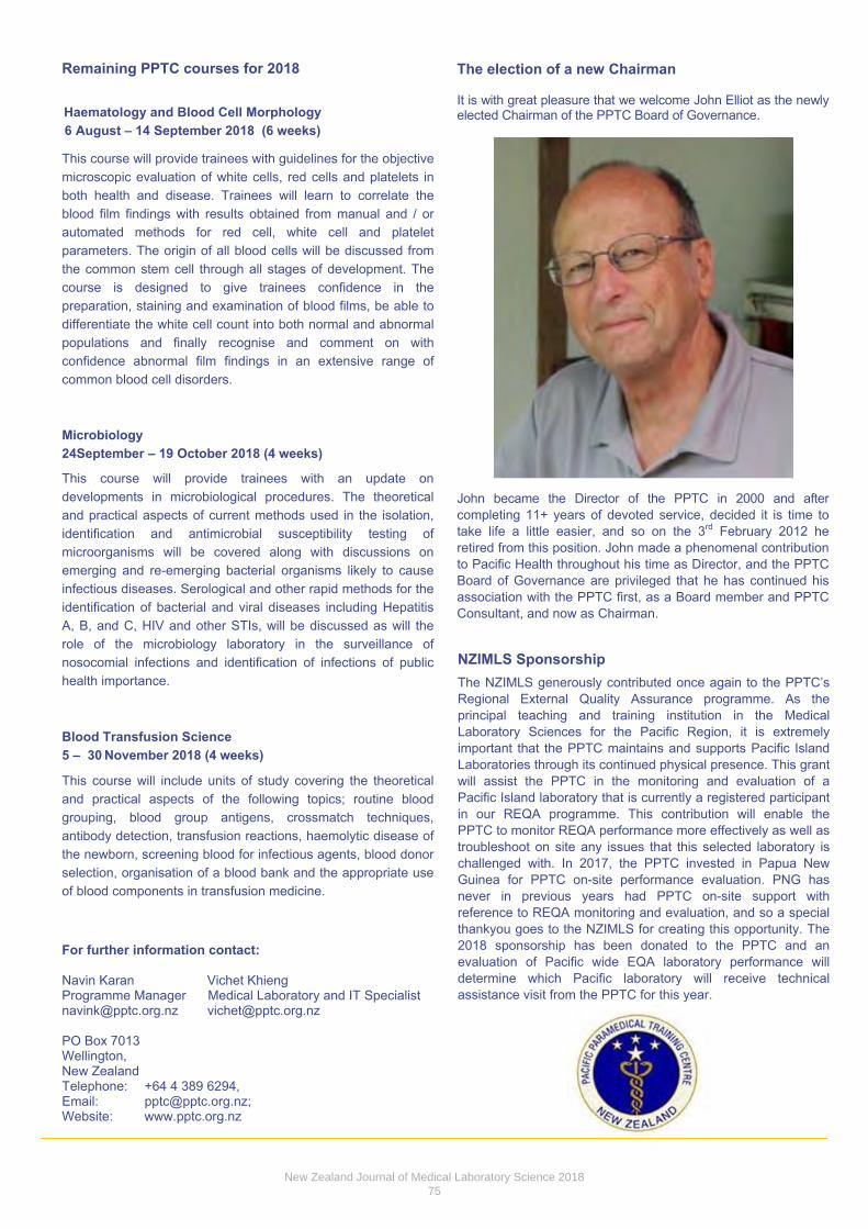
Remaining PPTC courses for 2018
Haematology and Blood Cell Morphology 6 August – 14 September 2018 (6 weeks)
This course will provide trainees with guidelines for the objective microscopic evaluation of white cells, red cells and platelets in both health and disease. Trainees will learn to correlate the blood film findings with results obtained from manual and / or automated methods for red cell, white cell and platelet parameters. The origin of all blood cells will be discussed from the common stem cell through all stages of development. The course is designed to give trainees confidence in the preparation, staining and examination of blood films, be able to differentiate the white cell count into both normal and abnormal populations and finally recognise and comment on with confidence abnormal film findings in an extensive range of common blood cell disorders.
Microbiology 24September – 19 October 2018 (4 weeks)
This course will provide trainees with an update on developments in microbiological procedures. The theoretical and practical aspects of current methods used in the isolation, identification and antimicrobial susceptibility testing of microorganisms will be covered along with discussions on emerging and re-emerging bacterial organisms likely to cause infectious diseases. Serological and other rapid methods for the identification of bacterial and viral diseases including Hepatitis A, B, and C, HIV and other STIs, will be discussed as will the role of the microbiology laboratory in the surveillance of nosocomial infections and identification of infections of public health importance.
Blood Transfusion Science 5 – 30 November 2018 (4 weeks)
This course will include units of study covering the theoretical and practical aspects of the following topics; routine blood grouping, blood group antigens, crossmatch techniques, antibody detection, transfusion reactions, haemolytic disease of the newborn, screening blood for infectious agents, blood donor selection, organisation of a blood bank and the appropriate use of blood components in transfusion medicine.
For further information contact:
Navin Karan Vichet Khieng Programme Manager Medical Laboratory and IT Specialist [email protected] [email protected]
PO Box 7013 Wellington, New Zealand Telephone: +64 4 389 6294, Email: [email protected]; Website: www.pptc.org.nz
The election of a new Chairman
It is with great pleasure that we welcome John Elliot as the newly elected Chairman of the PPTC Board of Governance.
John became the Director of the PPTC in 2000 and after completing 11+ years of devoted service, decided it is time to take life a little easier, and so on the 3rd February 2012 he retired from this position. John made a phenomenal contribution to Pacific Health throughout his time as Director, and the PPTC Board of Governance are privileged that he has continued his association with the PPTC first, as a Board member and PPTC Consultant, and now as Chairman.
NZIMLS Sponsorship The NZIMLS generously contributed once again to the PPTC’s Regional External Quality Assurance programme. As the principal teaching and training institution in the Medical Laboratory Sciences for the Pacific Region, it is extremely important that the PPTC maintains and supports Pacific Island Laboratories through its continued physical presence. This grant will assist the PPTC in the monitoring and evaluation of a Pacific Island laboratory that is currently a registered participant in our REQA programme. This contribution will enable the PPTC to monitor REQA performance more effectively as well as troubleshoot on site any issues that this selected laboratory is challenged with. In 2017, the PPTC invested in Papua New Guinea for PPTC on-site performance evaluation. PNG has never in previous years had PPTC on-site support with reference to REQA monitoring and evaluation, and so a special thankyou goes to the NZIMLS for creating this opportunity. The 2018 sponsorship has been donated to the PPTC and an evaluation of Pacific wide EQA laboratory performance will determine which Pacific laboratory will receive technical assistance visit from the PPTC for this year.
New Zealand Journal of Medical Laboratory Science 201875

Conference Report Julie Creighton
Canterbury Health Laboratories
The Australian Society of Antimicrobials Annual Scientific Meeting was held in Adelaide, Australia, from 22 to 24 February 2018. This meeting is widely attended in microbiology circles and delegates include microbiology clinicians, Infectious Diseases specialists, laboratory scientists, managers, and antimicrobial stewardship specialists. The meeting attracts delegates from around the world and the calibre of presentations and speakers is excellent. The focus is on antimicrobial resistance – mechanisms, detection, trends, new rapid methods, prevention and stewardship. The meeting runs from 7am to late, for 3 days. It is very full-on and really challenging to take it all in! However the annual ASA meeting is without doubt one of the most informative and directly relevant meetings for microbiology. It is not possible to condense all of the sessions into one review, but here are some of the highlights for me.
A wonderful presentation was given by Prof Lance Price, George Washington University, USA, titled “Food producing animals as a source of Antimicrobial Resistance (AMR) in humans”. Dr Price is an engaging speaker and very passionate about his field. In the USA, more than 8.4 million kg of antibiotics are used in animal production, including use for growth promotion. The majority is tetracycline, but also some penicillin and macrolides. Animals are housed together, sharing food, shelter and faecal flora. Antibiotic resistant organisms colonising meat-producing animals leads to contamination of their meat, which is then consumed by humans. However the problem is not just with the classic food borne pathogens such as campylobacter and salmonella, but also with the classic colonising pathogens (COPs), such as E. coli, Klebsiella pneumoniae, Staphylococcus aureus and Clostridium difficile. The problem with COPs is that they are involved in silent person-to-person transmission; colonisation, usually asymptomatic and often of lengthy duration; and are found in human, animal and environmental sources. E. coli is the major ‘bad boy’ COP due to its common cause of UTI, kidney infections and urosepsis (which can lead to death). Understanding gut colonisation is a key factor is tackling AMR.
One study that Dr Price’s group undertook was to look at E. coli collected from animals (chickens, turkeys, pigs) and compare strains collected from humans, using sequencing technology. The study looked at sequence type (ST) and host adaptive genes. Although they found over 400 different sequence types,
they found several STs that had originated in humans, but migrated to pigs and some that were originally an animal COP, but migrated to cause human infections, with clonal spread in both hospital and community settings. Globally ST131 is one of the most widely distributed E. coli STs, being responsible for many UTIs and often harbouring ESBL CTX-M as well as ciprofloxacin resistance. In this study, ST131 was found predominantly in humans but also discovered in animals and meat products.
In another presentation by Lance Price, he further discussed the rise and rise of E. coli ST131, subtype H30 (fimbrial adhesion gene H subclone), with ciprofloxacin qnr (quinolone) resistance and ESBL CTX-M-15 production: otherwise known as the Red Queen! ST131 was first described in 2008 – but by then it was already found in 9 countries and in 3 continents. It is a very successful coloniser, is often associated with UTIs, sepsis and other extraintestinal infections. Acquisition of qnr appears to be due to direct antibiotic pressure and the use of ciprofloxacin to treat other infections, leading to qnr resistant E. coli in the gut! The rapid global expansion of the highly virulent ‘Red Queen’ is most likely due to clonal spread, rather than horizontal plasmid transfer. Its ecological success appears to be a combination of efficient gut colonisation, transmission between hosts, antibiotic selection pressure, virulence and antibiotic resistance.
Prof Greg Cook, University of Otago, spoke about a Generation of New Antimicrobials for Bad Bugs. Greg’s work involves the search for new types of antimicrobials, which are fast acting, with a new mode of application, for use in the vet industry. He called them ‘green’ antimicrobials as they are exclusively for use in animals, helping to break the link between human and vet medicine and safeguard against the exchange of AMR between animals and humans. Greg’s lab is focusing on metabolism and energy generation of organisms, in order to understand how pathogens metabolise and persist in host tissues. His lab has developed a next-generation sanitiser for the control of bovine mastitis in the dairy industry. Currently the most commonly used product is TeatX (which is Chlorhexidine). Chlorhexidine is also used in human health. It must not be present in any milk sold to China. The new product is currently names ZDR22 – it has low cytotoxicity, is cheap to make, has low MICs, and can kill Strep uberis, Staph.aureus and E. coli, in both aerobic and anaerobic states. ZDR22 can also work together with Chlorhexidine, meaning Chlorhexidine can be used at much lower concentrations.
The worrying issue of increasing rates of isolation of N. gonorrhoeae, coupled with increasing multi-drug resistance, was discussed by Prof Monica Lahra, WHO Collaborating Laboratory, NSW. The highest rates off STI are in the Asia/Pacific region. Increasing rates of gonorrhoea worldwide are due to a combination of better testing (NAATS), behavioural issues, social services such as Tinder and Grindr, more frequent testing and travel. The down side of NAAT introduction is that fewer cultures are performed; on a global basis less than
ASA 2018 Brisbane
New Zealand Journal of Medical Laboratory Science 201876
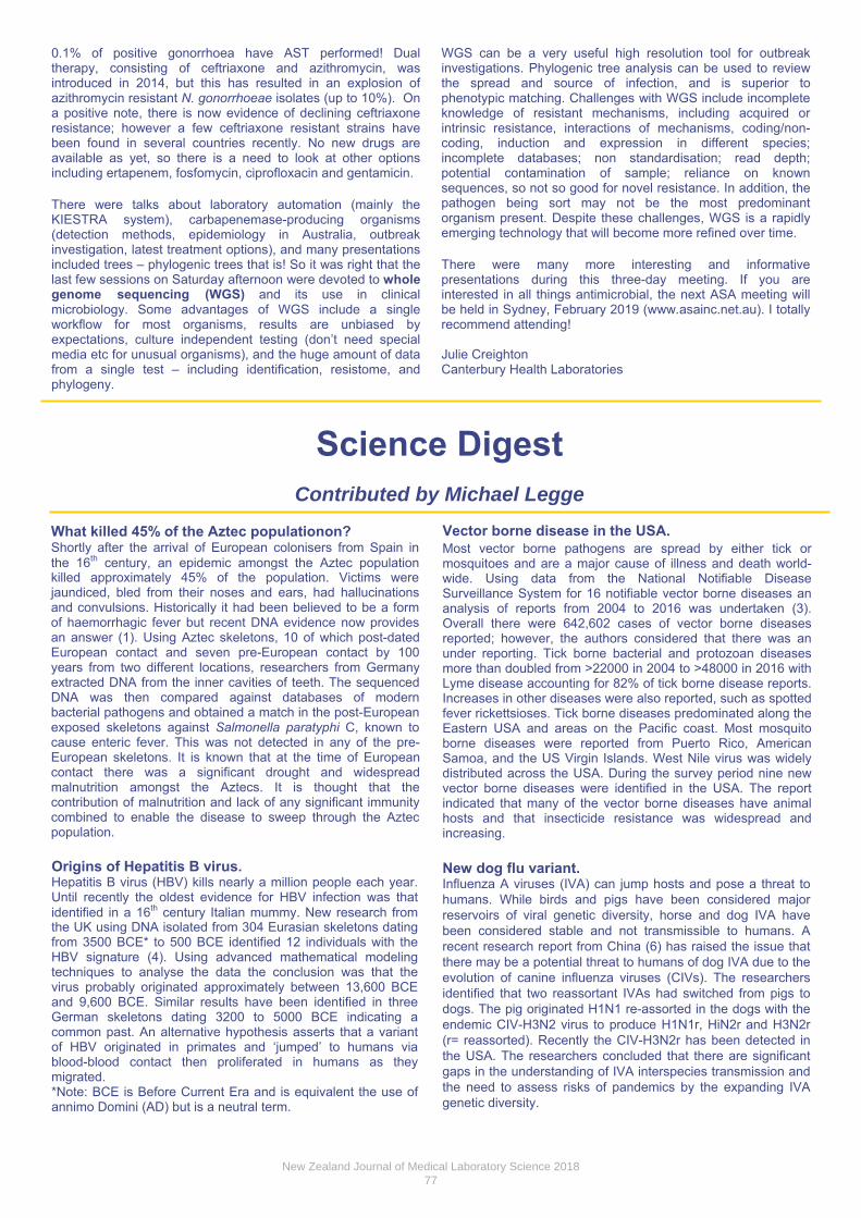
0.1% of positive gonorrhoea have AST performed! Dual therapy, consisting of ceftriaxone and azithromycin, was introduced in 2014, but this has resulted in an explosion of azithromycin resistant N. gonorrhoeae isolates (up to 10%). On a positive note, there is now evidence of declining ceftriaxone resistance; however a few ceftriaxone resistant strains have been found in several countries recently. No new drugs are available as yet, so there is a need to look at other options including ertapenem, fosfomycin, ciprofloxacin and gentamicin.
There were talks about laboratory automation (mainly the KIESTRA system), carbapenemase-producing organisms (detection methods, epidemiology in Australia, outbreak investigation, latest treatment options), and many presentations included trees – phylogenic trees that is! So it was right that the last few sessions on Saturday afternoon were devoted to whole genome sequencing (WGS) and its use in clinical microbiology. Some advantages of WGS include a single workflow for most organisms, results are unbiased by expectations, culture independent testing (don’t need special media etc for unusual organisms), and the huge amount of data from a single test – including identification, resistome, and phylogeny.
WGS can be a very useful high resolution tool for outbreak investigations. Phylogenic tree analysis can be used to review the spread and source of infection, and is superior to phenotypic matching. Challenges with WGS include incomplete knowledge of resistant mechanisms, including acquired or intrinsic resistance, interactions of mechanisms, coding/non-coding, induction and expression in different species; incomplete databases; non standardisation; read depth; potential contamination of sample; reliance on known sequences, so not so good for novel resistance. In addition, the pathogen being sort may not be the most predominant organism present. Despite these challenges, WGS is a rapidly emerging technology that will become more refined over time.
There were many more interesting and informative presentations during this three-day meeting. If you are interested in all things antimicrobial, the next ASA meeting will be held in Sydney, February 2019 (www.asainc.net.au). I totally recommend attending!
Julie Creighton Canterbury Health Laboratories
Science Digest Contributed by Michael Legge
What killed 45% of the Aztec populationon?Shortly after the arrival of European colonisers from Spain in the 16th century, an epidemic amongst the Aztec population killed approximately 45% of the population. Victims were jaundiced, bled from their noses and ears, had hallucinations and convulsions. Historically it had been believed to be a form of haemorrhagic fever but recent DNA evidence now provides an answer (1). Using Aztec skeletons, 10 of which post-dated European contact and seven pre-European contact by 100 years from two different locations, researchers from Germany extracted DNA from the inner cavities of teeth. The sequenced DNA was then compared against databases of modern bacterial pathogens and obtained a match in the post-European exposed skeletons against Salmonella paratyphi C, known to cause enteric fever. This was not detected in any of the pre-European skeletons. It is known that at the time of European contact there was a significant drought and widespread malnutrition amongst the Aztecs. It is thought that the contribution of malnutrition and lack of any significant immunity combined to enable the disease to sweep through the Aztec population.
Vector borne disease in the USA. Most vector borne pathogens are spread by either tick or mosquitoes and are a major cause of illness and death world-wide. Using data from the National Notifiable Disease Surveillance System for 16 notifiable vector borne diseases an analysis of reports from 2004 to 2016 was undertaken (3). Overall there were 642,602 cases of vector borne diseases reported; however, the authors considered that there was an under reporting. Tick borne bacterial and protozoan diseases more than doubled from >22000 in 2004 to >48000 in 2016 with Lyme disease accounting for 82% of tick borne disease reports. Increases in other diseases were also reported, such as spotted fever rickettsioses. Tick borne diseases predominated along the Eastern USA and areas on the Pacific coast. Most mosquito borne diseases were reported from Puerto Rico, American Samoa, and the US Virgin Islands. West Nile virus was widely distributed across the USA. During the survey period nine new vector borne diseases were identified in the USA. The report indicated that many of the vector borne diseases have animal hosts and that insecticide resistance was widespread and increasing.
New dog flu variant. Influenza A viruses (IVA) can jump hosts and pose a threat to humans. While birds and pigs have been considered major reservoirs of viral genetic diversity, horse and dog IVA have been considered stable and not transmissible to humans. A recent research report from China (6) has raised the issue that there may be a potential threat to humans of dog IVA due to the evolution of canine influenza viruses (CIVs). The researchers identified that two reassortant IVAs had switched from pigs to dogs. The pig originated H1N1 re-assorted in the dogs with the endemic CIV-H3N2 virus to produce H1N1r, HiN2r and H3N2r (r= reassorted). Recently the CIV-H3N2r has been detected in the USA. The researchers concluded that there are significant gaps in the understanding of IVA interspecies transmission and the need to assess risks of pandemics by the expanding IVA genetic diversity.
Origins of Hepatitis B virus. Hepatitis B virus (HBV) kills nearly a million people each year. Until recently the oldest evidence for HBV infection was that identified in a 16th century Italian mummy. New research from the UK using DNA isolated from 304 Eurasian skeletons dating from 3500 BCE* to 500 BCE identified 12 individuals with the HBV signature (4). Using advanced mathematical modeling techniques to analyse the data the conclusion was that the virus probably originated approximately between 13,600 BCE and 9,600 BCE. Similar results have been identified in three German skeletons dating 3200 to 5000 BCE indicating a common past. An alternative hypothesis asserts that a variant of HBV originated in primates and ‘jumped’ to humans via blood-blood contact then proliferated in humans as they migrated. *Note: BCE is Before Current Era and is equivalent the use ofannimo Domini (AD) but is a neutral term.
New Zealand Journal of Medical Laboratory Science 201877

Elevated troponin without myocardial infarction. Troponin elevation is widely accepted as a reliable marker for myocardial infarction. In a recent case report the authors identify an independent cause for serum troponin elevation (5). Myocardial infarction is not uncommon in cases of uncontrolled diabetes and may often be the cause of death in the first 24 hours of admission. However, elevated serum cardiac biomarkers in uncontrolled diabetes have been reported in the absence of a myocardial infarction and this has been linked to ketoacidosis. In the case report, the authors report on a male patient admitted with elevated blood glucose (31.4mmolL-1), ketoacidosis (pH 6.99, anion gap 36mmolL-1) and a normal troponin (<0.04 ng/ml). The patient was alert and an ECG was
A mechanism for morning sickness. The pathogenesis of nausea and vomiting (morning sickness) during pregnancy is unknown. This condition can affect between 70 to 90% of pregnant women. While morning sickness may be transient in its most severe form, Hyperemisis gravidarum, the resulting dehydration and electrolyte imbalance is the most common cause for hospital admission during early pregnancy. This has been associated with low birth weight and pre-term delivery of infants. Historically reproductive hormones have been considered to be causative agents; however, in non-pregnancy these hormones do not cause morning sickness. Research from the UK has identified that a growth factor, growth and differentiation factor (GDF15), response may provide a clue to morning sickness. GDF15 is expressed in the liver but during pregnancy the placenta also expresses it, with concentrations rising rapidly in maternal blood during early pregnancy. The research investigated blood concentrations of GDF15 in 791 pregnant women. Circulating GDF15 concentrations were identified in women reporting vomiting in the second trimester of pregnancy and were higher in women taking anti-emetics but lower in those women reporting no nausea or vomiting (2). As it is known that elev ated GDF15 concentrations in mice with cancer induces cancer cachexia by suppressing food intake, the authors concluded that the GDF15-GDF15 receptor signaling system evolved to provide a food aversion signaling system to help protect the fetus.
REFE RENCES
1. Vågene AJ, Herbiq A, Campana MG, Robes García NM,Warriner C, Sabins S, et al. Salmonella enterica genomesfrom victims of a major sixteenth-century epidemic in Mexico.Nat Ecol Evol 2018; 2: 520-528.
2. Petry CJ, Ong KK, Burling KA, Barker P, Perry JRB, AceriniCL, et al. GDF15 concentrations in maternal serumassociated with vomiting in pregnancy: the Cambridge BabyGrowth Study. 2018. bioRxiv 221267; doi: https://doi.org/10.1101/221267 .
3. Rosenberg R, Lindsey NP, Fischer M, Gregory CJ, HinckleyAF, Mead PS, et al. Vital signs: Trends in reported vectorborne disease cases - United States and Territories, 2004-2016MMWR Morb Mortal Wkly Rep 2018; 67: 496-501.
4. Műhlemann B, Jones TC, Damgaard PB, Allentoft ME,Shevnina I, Loqvin A, et al. Ancient hepatitis B viruses fromthe Bronze Age to the Medieval period. Nature 2018; 557:418-423.
5. Odubanjo Aa, Kalisetti R, Adrah R, Ajenifuja A, Joseph B,Zaman M. Severe myocarditis in diabetic ketoacidosis – alltroponins are not myocardial infarctions. Clin Med InsightsCase Rep 2018; 11: 1179547618763356.
6. Chen Y, Trovão NS, Wang G, Zhao W, He P, Zhou H, et al.Emergence and evolution of novel reassortment influenza Aviruses in canines in Southern China. MBio 2018; 9:e00909-18.
normal. Two days after admission he complained of a chest pain and had a rapidly rising troponin peaking at 7.3ng/ml with changes in the ECG. All other cardiac investigations were unremarkable. A diagnosis of myocarditis was made and treatment resulted in a rapid decrease in serum troponin. The authors concluded that the elevation of the serum troponin was the result of metabolic disturbances in the myocardial muscle.
APRIL 2018 JOURNAL QUESTIONS AND ANSWERS 1. The three-fold conversion of the haematocrit to derive a
haemoglobin value is not affected by which factors?Patient’s age, gender, hydration status (as measured byBUN/Cr ratio) and kidney function.
2. What were the limitations of the haematocrit to haemoglobinconversion factor study?Inability to assess the hydration status of the patientsclinically (only the BUN/Cr status was used to reflectthe hydration status). Also, as the study wasretrospective, the confounding effect of some otherfactors, such as polycythaemia, haemoglobinopathies,or blood sample haemolysis, could not be assessed.
3. What pre-analytical factors associated with muscle masscan influence blood creatinine levels?Age, sex, race, body size and haemodynamics.
4. The Jaffe assay used for measuring creatinine is prone towhich interfering substances?Glucose, ketoacids, albumin, antibiotics such ascephalosporin and streptomycin, and to ascorbic acid.
5. How do clinical cases of Shiga-toxigenic Escherichia coliinfection typically present clinically?Self-limiting, with painful abdominal cramps and non-bloody diarrhoea that occur 1-8 days post ingestion.
6. Which therapeutic medications are contra-indicated inShiga-toxigenic Escherichia coli infections, and why?Treatments involving quinolones, ciprofloxacin, inaddition to anti-motility agents. Because they havebeen associated with increased incidences ofprogression to haemolytic uraemic syndrome.
7. Shiga-toxigenic Escherichia coli toxins cause bloody andnon-bloody diahrroea through which mechanisms?Through the killing of intestinal endothelial cells eitherdirectly, or indirectly through inducing mesentericischaemia in the regional vasculature. The toxins arealso able to translocate across the gastrointestinalendothelia and enter the systemic circulation, causing ahost of pathological thrombotic and immunomodulatoryeffects on both the renal glomerulus and systemicmicrovasculature.
8. Extended spectrum ß-lactamase enzymes are capable ofhydrolysing what, but are inhibited by which antibiotics?Clavulanic acid, tazobactam or sulbactam. Inhibited byclavulanic acid, tazobactam and salbactam.
9. P. aeruginosa has been demonstrated to have intrinsicresistance to different classes of anti-bacterial drugs throughwhich mechanisms?Multidrug resistance efflux, pumps, decreasedpermeability and the loss of the OprD2 (outermembrane porin) protein.
10. The cytopathologic differential diagnosis of mammaryanalogue secretory adenoma includes which low-gradeepithelial neoplasms?Pleomorphic adenoma, low-grade mucoepidermoidcarcinoma, acinic cell carcinoma, myoepithelialcarcinoma low-grade cribriform cystadenocarcinoma,oncocytic carcinoma and salivary duct carcinoma.
Copyright: ©NZIMLS
New Zealand Journal of Medical Laboratory Science 201878

BIOCHEMISTRY SPECIAL INTEREST GROUP
SAVE THE DATE Saturday September 29th 2018
Otago Museum, Dunedin
If you are interested in presenting, please contact convenor
Sian Horan (Dunedin SCL) [email protected]
Register at www.nzimls.org.nz

Haematology Special Interest Group Seminar
Coachman Hotel, Palmerston North
Saturday 13 October 2018
Presentations welcome!
Come and spend a day in balmy Palmy
Re‐invigorate your Haematology passion
Catch up with old friends
Enthuse with new ones
For further information or to send presentations
e‐mail Steve [email protected]
Register at www.nzimls.org.nz
New Zealand Journal of Medical Laboratory Science 201880

Wellington Hospital, Newtown, Wellington
Held for the first time on the Friday before the Micro SIG with a cross-discipline function on Friday evening
Presentations (oral and poster) are invited for the following disciplines: Molecular Genetics, Biochemical Genetics, Molecular Virology,
Microbiology, Molecular Haematology and Cytogenetics.
Closing date for abstracts: Friday 17 August 2018
For further information, please contact either:
Angela Brounts (Microbiology) email: [email protected] Clive Felix (Genetics) email: [email protected]
Online Registration is available at www.nzimls.org.nz
New Zealand Journal of Medical Laboratory Science 201881

Immunology Special Interest Group 2018
Saturday 17 November 2018 Wellington Hospital, Education Lecture Theatre
Confirmed speakers:
Dr Richard Steele, Immunopathologist Dr Tim Blackmore, Microbiologist
James Rice-Davies, HIV/ID Clinical Nurse Specialist Louise Wienholt, Manager Business Development and
Blood Disciplines, RCPAQAP Dr Russell Barker, Immunopathologist
Presentations are encouraged from all specialities Auto Immune
Infectious Diseases Allergy
Contact Sarah Burge: [email protected]
REGISTER AT WWW.NZIMLS.ORG.NZ