![SSMJ South Sudan › ... › SSMJ_Vol_6_3.pdf · relatively early in the course of HIV infection tends to have the typical radiographic findings [15, 16]. With more advanced HIV disease](https://static.fdocuments.us/doc/165x107/5ed5985035c3a005a51b0a36/ssmj-south-a-a-ssmjvol63pdf-relatively-early-in-the-course-of-hiv.jpg)
SSMJ - docs.southsudanngoforum.org
24
SSMJ South Sudan Medical Journal ISSN 2309 - 4605 Volume 8. Number 1. February 2015 www.southsudanmedicaljournal.com Importance of cough • Making cassava flour safe • MUAC in paediatric malnutrition • Case report: Progressive dysarthria and ataxia • Reducing maternal deaths: the role of medical doctors in task-shifting
Transcript of SSMJ - docs.southsudanngoforum.org

Vol 8. No 1. February 2015 South Sudan Medical Journal
SSMJSouth Sudan Medical Journal
ISSN 2309 - 4605
Volume 8. Number 1. February 2015 www.southsudanmedicaljournal.com
Importance of cough•
Makingcassavafloursafe•
MUAC in paediatric malnutrition •
Case report: Progressive dysarthria and ataxia•
Reducing maternal deaths: the role of medical doctors in task-shifting

South Sudan Medical Journal Vol 8. No 1. February 2015
EdItoRIAL
Maternal mortality can be reduced through task-shifting in South Sudan Staffan Bergström ..................................................... 3
MAIN ARtICLES
Making cassava flour safe using the wetting method J. Howard Bradbury, Julie Cliff and Jean P. Banea ........................................................... 4
Task-shifting: The crucial role of medical doctors in reducing maternal deaths in South Sudan Staffan Bergström ............................... 8
CASE REPoRt
Progressive dysarthria and ataxia Lynsey McAlpine, Fiona Cran, Eluzai Hakim ................. 12
ShoRt ItEMS
The importance of coughs David Tibbutt ...16
A longitudinal study of MUAC as a measure of paediatric malnutrition in Yei, South Sudan: Lessons from a hospital link Abigail Sharpe and Simon Struthers ........................... 18
Letter to the editor ...................................21
RESoURCES ....................................... 22
BACK CoVER PoStER
Poster of the simple wetting method for removal of cyanogens from cassava flour. ........................................................ 24
CoNtENtS
EdItoR-IN-ChIEF
dr Edward Eremugo Luka
South Sudan Doctors’ Association
Ministerial Complex
Juba, South Sudan
[email protected] Twitter: @eremugo
ASSoCIAtE EdItoRS
dr Wani Mena
Department of Ophthalmology
Juba Teaching Hospital,
PO Box 88, Juba
South Sudan
dr Eluzai Abe hakim
Department of Adult Medicine & Rehabilitation
St Mary’s Hospital, Newport,
Isle of Wight PO30 5TG, UK
EdItoRIAL BoARd
Dr James Ayrton
Dr Charles Bakhiet
Professor James Gita Hakim
Dr Ayat C. Jervase
Dr David Tibbutt
Prof. John Adwok
EdItoRIAL AdVISoR
Ann Burgess
dESIgN ANd LAyoUt
Dr Edward Eremugo Luka
The South Sudan Medical Journal is a quarterly publication intended for Healthcare Professionals, both those working in the South Sudan and those in other parts of the world seeking information on health in South Sudan. The Journal is published in mid-February, May, August and November.
Reviewers are listed on the website
2
SSMJ Volume 8. No. 1. www.southsudanmedicaljournal.com
A Publication of the South Sudan Doctors’ Association
South Sudan Medical JournalISSN 2309 - 4605
Cover photo:
Trained nurse doing intubation for general anaesthesia (credit: Dr Angelo Nyamtema)
THIS ISSUE OF THE SSMJ IS PRINTED WITH A GENEROUS
SUPPORT FROM CORDAID

Vol 8. No 1. February 2015 South Sudan Medical Journal
EdItoRIAL
Maternal mortality can be reduced through task-shifting in South Sudan
3
In this issue of SSMJ there is an article entitled “The crucial role of medical doctors in reducing maternal deaths in South Sudan”. It is based on two circumstances. Firstly, South Sudan has the highest national maternal mortality ratio in the world, above 2000, which is roughly 1000 times higher than in Sweden. Secondly, leading cadres in the Ministry of Health in Juba have an open, evidence-based and scientifically updated attitude to the most fundamental underlying problem: the scarcity of human resources for health (HRH) to save maternal lives.Can “task-shifting” be part of the solution to the problem of the devastating maternal mortality in South Sudan? Massive scientific evidence from a number of other African countries tells us that - in the absence of alternative solutions in the foreseeable future - South Sudan has to seriously consider inviting medical doctors to be employed as trainers and supervisors of clinical officers in the same way as has happened in Tanzania, Malawi, Mozambique, Zambia and Ethiopia. There is no collision of interests in this approach, since medical doctors are prerequisites for any success of “task-shifting”.It is argued that any initiative to train “mid-level providers of health care” is doomed to fail if there are no competent trainers in the leadership of this training. And there are no other competent trainers than medical doctors, quoting scientific evidence from other countries, where life-saving comprehensive emergency obstetric care, including caesarean sections, has been delegated successfully to such “mid-level providers of care”. It is also underlined that there should be precautions observed not to create a sub-standard cadre of “surgeons”. Medical doctors’ active collaboration and formal employment as supervisors are crucial prerequisites for success. This is a powerful message to the Ministry of Health in South Sudan.
Staffan Bergström, MD, PhDProfessor Emeritus of International HealthSenior Consultant (Obstetrics and Gynaecology)Karolinska InstituteSE-171 77 [email protected]
Medical doctors’ active
collaboration and formal employment as supervisors are
crucial prerequisites for success.

South Sudan Medical Journal Vol 8. No 1. February 2015
MAIN ARtICLES
4
Abstract
Many people, particularly in Africa, suffer various conditions from eating bitter cassava which contains poisonous cyanogens. As well as poisoning, which sometimes causes death, these conditions include konzo, an irreversible paralysis of the legs, which affects mainly children and young women, impaired neurocognition in children, tropical ataxic neuropathy in older people, and aggravation of iodine deficiency disorders (such as goitre and cretinism) in iodine deficient areas. The wetting method removes residual cyanogens, and is an additional method of processing cassava flour after its preparation by one of the traditional methods. The wetting method is simple and easy to use, the traditional thick porridge (fufu or ugali) made from the treated flour tastes very good, and the method is readily accepted by rural women in East and Central Africa. Regular use of the wetting method by rural women in 13 villages in the Democratic Republic of Congo has prevented konzo amongst more than 9,000 people. We recommend that the wetting method be used as an additional method to traditional methods to remove cyanogens from cassava flour in tropical Africa.
Making cassava flour safe using the wetting methodJ. Howard Bradburya, Julie Cliffb and Jean P. Baneac
a EEG, Research School of Biology, Australian National University, Canberra, ACT 0200, Australia.b Department of Community Health, Faculty of Medicine, Universidade Eduardo Mondlane, Maputo, Mozambique. c Programme National de Nutrition (PRONANUT), Kinshasa, Democratic Republic of Congo)
Correspondence: J. Howard Bradbury [email protected]
Introduction
Cassava is the third most important food source in the tropics and the staple food of tropical Africa. Cassava:
is easy to grow, •
produces a good yield of starchy roots in 6-9 months • even in poor soils without added fertilizer,
is drought resistant; the roots are a reserve source of • food in drought and famine conditions [1].
However the cassava plant makes cyanogenic glucosides, (linamarin and a small amount of lotaustralin), and the enzyme linamarase, that catalyses their hydrolysis to liberate poisonous hydrogen cyanide (HCN), when the plant is attacked by predators. This defense mechanism is used by more than 2.000 plants to deter predators [2]. If the cassava plant is stressed during drought it makes 2-4 times more cyanogens than normal [3,4]. The cyanogens are present in all parts of the plant, however with ‘sweet’ cassava the inside of the starchy root has only small amounts of cyanogens. Those cassava cultivars with high cyanide content, called ‘bitter’ cassava, are dangerous to eat and must be processed before consumption.
Cyanide is extremely toxic to humans, animals, insects and plants. It causes many health problems in Africa, especially where cassava is being introduced into new areas to help feed a rapidly increasing population [1]. There are many traditional methods for processing bitter cassava roots, but these often leave large amounts of cyanogens in the resultant cassava flour [5]. The wetting method [6-8] removes residual cyanogens from cassava
flour and should be used as an additional method before the flour is cooked in the traditional way to give a thick porridge called ugali or fufu. This is eaten with a sauce to give it flavor and more nutrients. In this article we propose that the wetting method should be widely used to ensure the safety of cassava flour amongst the cassava eating populations of Africa.
Cassava cyanide diseases
Acute cyanide poisoning • from bitter cassava causes dizziness, nausea, headache, abdominal pain, vomiting, diarrhoea, weakness and sometimes death. The lethal dose of cyanide is proportional to body weight, so children tend to be more susceptible to poisoning than adults. There are widespread accounts of acute cyanide poisoning from Asia, the South Pacific and tropical Africa [9].
Konzo• is an upper motor neuron disease that occurs mainly in children and young women and is associated with high cyanogen intake amongst very poor people living on a monotonous diet of bitter cassava – which is also deficient in the sulfur amino acids methionine and cysteine/cystine [10,11]. These amino acids are needed to detoxify cyanide to thiocyanate in the body; analysis of urinary thiocyanate gives the best measure of recent cyanide intake. We have found a significant correlation between the percentage monthly incidence of konzo and the percentage of children with high urinary thiocyanate content. Therefore konzo is very likely due to high cyanide/low sulfur amino acid intake from bitter cassava [12].

Vol 8. No 1. February 2015 South Sudan Medical Journal 5
MAIN ARtICLES
Konzo occurs suddenly, is non-progressive and produces a visible spastic walk with exaggerated knee and ankle jerks and often impaired vision and speech (Figure 1). Konzo occurs in the Democratic Republic of Congo (DRC), Mozambique, Tanzania, Cameroon, Central African Republic and Angola. Reported cases to 2009 were 6,788 [13], but in DRC alone an estimate made by the Ministry of Health in 2002 was 100,000 [14]. Recently konzo has spread geographically into many new areas [13]. Konzo epidemics occur due to:
o war, where people are forced to eat poorly processed bitter cassava,
o drought, when the cassava plant produces increased amounts of linamarin [4], and
o short cut processing [15].
Neurodegeneration of children.• There are reported neuropsychological effects in children exposed to high cyanide intake from bitter cassava associated with konzo. Motor proficiency skills are affected and neurocognition is reportedly impaired compared with children from a non-konzo area [16].
Tropical ataxic neuropathy (TAN),• also called “ataxic polyneuropathy,” is a neurological disease which generally occurs in older people in Nigeria, Tanzania, Uganda, Kenya, West Indies and South India. Symptoms are burning sensations in the soles of the feet, numbness in hands, unsteady walking, blurred speech, blindness and deafness. The condition is endemic in south west Nigeria and is probably due to long term consumption of cyanogens from gari [17].
Aggravationof iodinedeficiencydisorders• which are caused by reduced production of thyroxin by the thyroid due to deficiency of dietary iodine. Where there is already dietary iodine deficiency, intake of cyanide from cassava exacerbates the disorders [18].
Processing of cassava
Low cyanide sweet cassava can be just boiled and eaten, but higher cyanide bitter cassava roots must be processed to remove cyanogens. Traditional processing methods developed over many years are very diverse, but the major ones used in tropical Africa are:
Sun drying.• The peeled root is dried in the sun, then pounded into flour, using a wooden pestle and mortar, and sieved. This method is used widely in East and Central Africa, but leaves about one third of the linamarin in the flour [5].
Heap fermentation.• The peeled roots are placed in a small heap on the ground for about 3 days after which the roots are sun dried, pounded and sieved. This leaves about one sixth of the linamarin in the flour [5]. Village women in Mozambique change to this method when cyanide intoxication occurs during drought [4], but this change is insufficient to prevent konzo.
Soaking.• The peeled cassava roots are soaked in water in a large vessel or a running stream for 3-4 days. This causes the roots to soften and the enzyme breaks down linamarin and HCN gas bubbles off. The roots are dried in the sun, pounded and sieved to produce flour [19]. This method, used in the wet tropics, removes cyanogens satisfactorily, but if the soaking time is reduced to 1-2 days (called short soaking) cyanogens are only partially removed and cassava flour has high cyanide content [15]. Short soaking can happen if:
o there is an urgent need for food for the family,
o there is the likelihood that the roots may be stolen from the river, or
Figure 1. Boy with konzo supported by his father (credit J. Howard Bradbury).
South Sudan Medical Journal Vol 8. No 1. February 2015
MAIN ARtICLES
6
o flour is being produced to be sold to traders [20].
Gari • is made throughout West Africa, particularly in Nigeria. The peeled cassava root is ground up mechanically and left in a cloth bag for 3 days. The bag is dewatered in a press and the damp product roasted in a metal dish while stirred to prevent burning. The gari contains about 10-20 ppm cyanide, compared with the World Health Organization (WHO) maximum value for cassava flour of 10 ppm. Lactic acid fermentation also occurs during preparation that reduces the pH to about 4.
Wetting method [6-8]
Cassava flour produced by sun drying, heap fermentation or short soaking nearly always contains more cyanide than the WHO safe level. The wetting method is an additional method to remove residual cyanogens after one of these traditional methods.
The poster on page 24 illustrates the wetting method. Cassava flour is placed in a bowl and the level of the flour marked on the inside of the bowl. Water is added with thorough mixing. The level of the flour initially drops and then rises again to the mark. The wet flour is spread on a mat in a layer not thicker than a finger nail for 2 hours in the sun or 5 hours in the shade to allow the produced HCN gas to escape. This removes nearly all residual cyanogens. The damp flour is then cooked in the usual way.
The wetting method is accepted by rural women because
it requires little extra work or equipment and produces fufu that is no longer bitter [21]. The free poster on page 24 (back cover) is available in 13 languages - see http://biology.anu.edu.au/hosted_sites/CCDN/.
Use of the wetting method in DRC
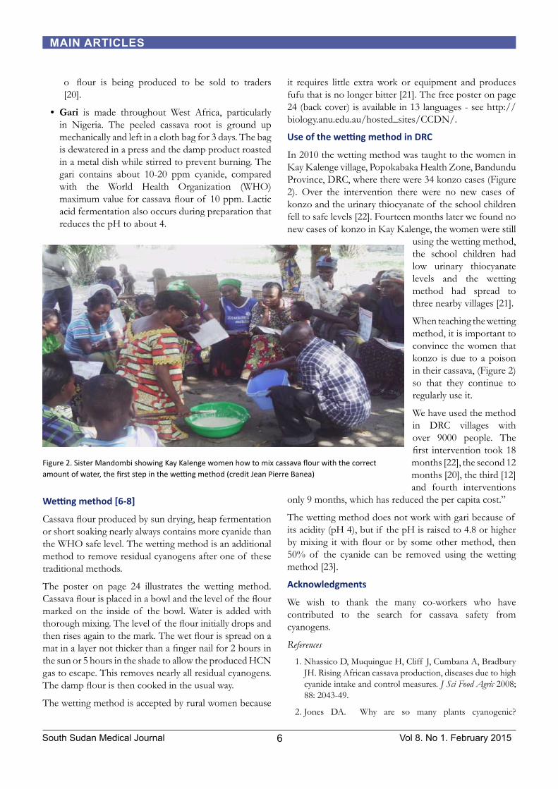
In 2010 the wetting method was taught to the women in Kay Kalenge village, Popokabaka Health Zone, Bandundu Province, DRC, where there were 34 konzo cases (Figure 2). Over the intervention there were no new cases of konzo and the urinary thiocyanate of the school children fell to safe levels [22]. Fourteen months later we found no new cases of konzo in Kay Kalenge, the women were still
using the wetting method, the school children had low urinary thiocyanate levels and the wetting method had spread to three nearby villages [21].
When teaching the wetting method, it is important to convince the women that konzo is due to a poison in their cassava, (Figure 2) so that they continue to regularly use it.
We have used the method in DRC villages with over 9000 people. The first intervention took 18 months [22], the second 12 months [20], the third [12] and fourth interventions
only 9 months, which has reduced the per capita cost.”
The wetting method does not work with gari because of its acidity (pH 4), but if the pH is raised to 4.8 or higher by mixing it with flour or by some other method, then 50% of the cyanide can be removed using the wetting method [23].
Acknowledgments
We wish to thank the many co-workers who have contributed to the search for cassava safety from cyanogens.
References
Nhassico D, Muquingue H, Cliff J, Cumbana A, Bradbury 1. JH. Rising African cassava production, diseases due to high cyanide intake and control measures. J Sci Food Agric 2008; 88: 2043-49.
Jones DA. Why are so many plants cyanogenic? 2.
Figure 2. Sister Mandombi showing Kay Kalenge women how to mix cassava flour with the correct amount of water, the first step in the wetting method (credit Jean Pierre Banea)

Vol 8. No 1. February 2015 South Sudan Medical Journal 7
MAIN ARtICLES
Phytochemistry 1998; 47: 155-162.
Bokanga M, Ekanayake IJ, Dixon AGO, Porto MCM. 3. Genotype-environment interactions for cyanogenic potential in cassava. Acta Hort 1994; 375: 131–139.
Ernesto M, Cardoso AP, Nicala D, Mirione E, Massaza 4. F, Cliff J, Haque MR, Bradbury JH. Persistent konzo and cyanogen toxicity from cassava in northern Mozambique. Acta Tropica 2002; 82: 357-362.
Cardoso AP, Mirione E, Ernesto M, Massaza F, Cliff J, 5. Haque MR, Bradbury JH. Processing of cassava roots to remove cyanogens. J Food Comp. Anal, 2005; 18: 451-460.
Bradbury JH. Simple wetting method to reduce cyanogen 6. content of cassava flour. J Food Comp Anal 2006;19: 388-393.
Cumbana A, Mirione E, Cliff J, Bradbury JH. Reduction 7. of cyanide content of cassava flour in Mozambique by the wetting method. Food Chem. 2007;101: 894-897.
Bradbury JH, Denton IC. Rapid wetting method to reduce 8. cyanogen content of cassava flour. Food Chem. 2010; 121: 591-594.
Cliff J. Acute cyanide poisoning from cassava: is it still 9. common? CCDN News 2012; No 20: 4-5.
Cliff J, Martensson J, Lundquist P, Rosling H, Sorbo B. 10. Association of high cyanide and low sulphur intake in cassava induced spastic paraparesis. Lancet 1985; 11: 1211-13.
Howlett WP, Brubaker GR, Mlingi N, Rosling H Konzo, an 11. epidemic upper motor neuron disease studied in Tanzania. Brain. 1990; 113: 223–35.
Banea JP, Bradbury JH, Mandombi C, Nahimana D, Denton 12. IC, Foster MP, Kuwa N, Tshala Katumbay D. Prevention of konzo in the Democratic Republic of Congo (DRC) using the wetting method and correlation between konzo incidence and percentage of children with high urinary thiocyanate level. African J Food Sci 2014; 8: 297-304.
Nzwalo H, Cliff J. Konzo: from poverty, cassava, and 13. cyanogen intake to toxico-nutritional neurological disease.
PloS Neglected Tropical Diseases 2011; 5: Issue 6 e1051.
Diasolua Ngudi D. Konzo and cassava toxicity: a study of 14. associated nutritional factors in the Popokabaka District, DRC. PhD Thesis, 2005. Universiteit Gent, Belgium.
Banea M, Poulter NH, Rosling H. Shortcuts in cassava 15. processing and risk of dietary cyanide exposure in Zaire. Food Nutrition Bulletin 1992; 14: 137-143.
Boivin MJ, Okitunda D, Bumoko GM, Sombo MT, Mumba 16. D, Tylleskar T, Page CF, Muyembe JJT, Tshala Katumbay D. Neuropsychological effects of konzo: a neuromotor disease associated with poorly processed cassava. Pediatrics 2013; 131: e1231-e1239.
Osuntokun BO. Chronic cyanide intoxication of dietary 17. origin and a degenerative neuropathy in Nigerians. Acta Hort 1994; 375: 311-321.
Delange F, Ekpechi LO, Rosling H. Cassava cyanogenesis 18. and iodine deficiency disorders. Acta Hort. 1994;375: 289-293.
Banea Mayambu, J.P, Cassava processing, dietary cyanide 19. exposure and konzo in Zaire. Masters Thesis, Uppsala Univesity, Uppsala, Sweden, 1993; p 55.
Banea JP, Bradbury JH, Mandombi C, Nahimana D, 20. Denton IC, Kuwa N, Tshala Katumbay D. Control of konzo by detoxification of cassava flour in three villages in the Democratic Republic of Congo. Food Chem. Toxicol., 2013; 60: 506-513.
Banea JP, Bradbury JH, Mandombi C, Nahimana D, 21. Denton IC, Kuwa N, Tshala Katumbay D. Effectiveness of wetting method for control of konzo and reduction of cyanide poisoning by removal of cyanogens from cassava flour. Food Nutrition Bull 2014; 35: 28-32.
Banea M, Nahimana G, Mandombi C, Bradbury JH, 22. Denton IC, Kuwa N. Control of konzo in DRC using the wetting method on cassava flour. Food Chem Toxicol 2012; 50: 1517-1523.
Bradbury JH, Denton IC. Simple method to reduce the 23. cyanogen content of gari made from cassava. Food Chem 2010; 123: 840-845.

South Sudan Medical Journal Vol 8. No 1. February 2015 8
MAIN ARtICLES
Task-shifting: The crucial role of medical doctorsin reducing maternal deaths in South SudanStaffan BergströmProfessor, Senior Consultant (Obstetrics & Gynaecology), Karolinska Institute, Stockholm, Sweden
Correspondence: [email protected]
Introduction
Millennium Development Goal (MDG) 5 is related to reducing maternal mortality. Against the background of the failure of this MDG, South Sudan has two features that make it interesting for the international donor community:
Firstly, South Sudan has the highest national maternal mortality ratio in the world, above 2,000 (i.e. 2,000 maternal deaths/100,000 live births), which is roughly 1,000 times higher than in Sweden.
Secondly, leading cadres in the Ministry of Health in Juba have an open and scientifically updated attitude to the most fundamental underlying problem: the scarcity of human resources for health (HRH) to save maternal lives.
Human resource management
In the “National Reproductive Health Strategic Plan 2013-2016” (November 2012) for South Sudan there is a human resources for health focus in the chapter entitled “Human resource management”. Much attention is given to two categories of mid-level personnel, namely professional midwives and clinical officers. In addressing “staff redeployment, rationalization and retention” it is stated that the government has to “give priority to the deployment of newly trained staff to under-served states and counties and classify hardship areas with the view to developing incentives, benefits and packages for staff serving in those areas”.
What is not presented convincingly in this document is the fact that any initiative to train ‘mid-level providers of health care’ is doomed to fail if there are no competent trainers to lead this training. And there are no other more competent trainers than medical doctors. This has been demonstrated in other African countries (for example in Tanzania, Mozambique, Malawi, Zambia and Ethiopia) where there have been similar initiatives and where life-saving comprehensive emergency obstetric care (CEmOC), including Caesarean sections, has been successfully delegated to ‘mid-level providers of care’. Such delegation of major surgery to thoroughly trained and supervised mid-level providers is called ‘task-shifting
of CEmOC’. Extraordinary precautions must be observed in order not to create a sub-standard cadre of ‘surgeons’. Mozambique has now almost 30 years of experience, and scientific research has clearly demonstrated that it is a success. However this success is not free of significant costs, and medical doctors’ active collaboration and formal employment as supervisors are crucial prerequisites for success.
“Who should do the Caesareans where there is no doctor?”
The article “Who should do the Caesareans where there is no doctor?” in the British Journal of Obstetrics and Gynaecology addresses the issue of task-shifting [1]. Shifting doctors’ normal duties in surgery to ‘non-doctors’ or ‘non-physician clinicians’ - or ‘associate clinicians’ (ACs) using the current terminology - has been controversial but is now recognized as the only solution in most low-income countries, if we are serious about MDG 5 (and beyond 2015 in particular) to reduce maternal mortality.
Task-shifting in surgery raises important ethical issues, as patient risk could be increased when the operating clinician lacks the expertise of a fully trained surgeon. However, comparative studies in Mozambique [1] and Tanzania [2] indicate that postoperative outcomes do not
Figure 1. Trained nurse conducts spinal anaesthesia as part of task-shifting (credit: Dr Angelo Nyamtema)
Vol 8. No 1. February 2015 South Sudan Medical Journal 9
MAIN ARtICLES
differ between physicians and ACs. However many aspects of quality of care remain unaddressed in the surgical task-shifting literature [3]. These include low referral rates to tertiary care centres due to the presence of ACs at district hospitals, the unknown quality of surgical education and training beyond the relatively small number of educational interventions described in the literature, and the degree of supportive supervision and training that providers who engage in surgical task-shifting may require but not always receive.
Surgical task-shifting remains a compelling model for surgical care delivery, and perhaps even an ethical imperative. It has been shown to be cost-effective [4, 5, 6], and may be associated with lower rates of ‘brain drain’ to higher-income countries [7] and with greater provider retention in the most underserved regions [8].
What can South Sudan learn from other African countries?
A few pertinent country examples, relevant for South Sudan, are briefly described here:
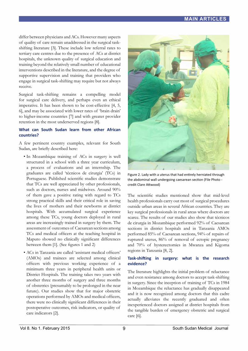
In Mozambique training of ACs in surgery is well • structured in a school with a three year curriculum, a process of evaluations and an internship. The graduates are called ‘técnicos de cirurgia’ (TCs) in Portuguese. Published scientific studies demonstrate that TCs are well appreciated by other professionals, such as doctors, nurses and midwives. Around 90% of them gave a positive rating with regard to TCs strong practical skills and their critical role in saving the lives of mothers and their newborns at district hospitals. With accumulated surgical experience among these TCs, young doctors deployed in rural areas are increasingly trained in surgery by them. The assessment of outcomes of Caesarean sections among TCs and medical officers at the teaching hospital in Maputo showed no clinically significant differences between them [1]. (See figures 1 and 2)
ACs in Tanzania are called ‘assistant medical officers’ • (AMOs) and trainees are selected among clinical officers with previous working experience of a minimum three years in peripheral health units or District Hospitals. The training takes two years with another three months of surgery and three months of obstetrics (presumably to be prolonged in the near future). Our studies show that for major obstetric operations performed by AMOs and medical officers, there were no clinically significant differences in their postoperative outcomes, risk indicators, or quality of care indicators [2].
The scientific studies mentioned show that mid-level health professionals carry out most of surgical procedures outside urban areas in several African countries. They are key surgical professionals in rural areas where doctors are scarce. The results of our studies also show that técnicos de cirurgia in Mozambique performed 92% of Caesarean sections in district hospitals and in Tanzania AMOs performed 85% of Caesarean sections, 94% of repairs of ruptured uterus, 86% of removal of ectopic pregnancy and 70% of hysterectomies in Mwanza and Kigoma regions in Tanzania [8, 2].
Task-shifting in surgery: what is the research evidence?
The literature highlights the initial problem of reluctance and even resistance among doctors to accept task-shifting in surgery. Since the inception of training of TCs in 1984 in Mozambique the reluctance has gradually disappeared and it is now recognized among doctors that this cadre actually alleviates the recently graduated and often inexperienced doctors assigned at district hospitals from the tangible burden of emergency obstetric and surgical care [6].
Figure 2. Lady with a uterus that had entirely herniated through the abdominal wall undergoing caesarean section (File Photo - credit Clare Attwood)

South Sudan Medical Journal Vol 8. No 1. February 2015 10
MAIN ARtICLES
According to the literature, health worker motivation and retention in rural assignments is a crucial response to the HRH crisis in African countries. Poor remuneration, bad working conditions, suboptimal management of human resources, limited opportunities for career progression, oppressive political climate, including insecurity and threat of violence, and a wish to provide a good education for their children influence the motivation of the health workforce, including ACs, to continue.
In Mozambique, like in South Sudan, the health workforce has generally low work motivation due to inadequate salaries and incentives, poor working conditions, absence of job description, unsatisfactory organization and management of services, heavy workload, degraded physical infrastructure preventing application of biosafety norms, and lack of supplies. In Mozambique’s task-shifting initiative the main problem of TCs is dissatisfaction due to workload, as they can rarely leave the workplace to attend training in referral hospitals or attend specific seminars to ameliorate their knowledge. In addition, there is irregular supervision by specialists as the specialist surgeons are few at provincial level.
In Tanzania’s task-shifting policy the situation is similar and the motivation is reportedly weak among health workers. AMOs face overwork, poor working conditions and lack of supportive supervision. They are rarely invited to attend meetings at the Ministry of Health in the same way as their colleagues, such as nurses and midwives, despite sharing activities in the same areas. They are seldom moved to referral hospitals for job training to ameliorate their performance, which make them feel abandoned and disoriented. Lack of career perspectives make them dissatisfied.
Attention paid to adequate supportive supervision and good management can reportedly improve work satisfaction, performance and quality of work in remote settings. In our studies, supportive supervision was not specifically addressed, but the literature reviewed indicates that, in both Mozambique and Tanzania, it is irregular or non-existent in most districts. This is an important lesson for South Sudan: medical doctors should be employed for, and closely involved in, supportive supervision of clinical officers ‘shoulder-to-shoulder’.
Supportive supervision of clinical officers: a key role for medical doctors in South Sudan
For South Sudan the experiences gained in Mozambique are extremely relevant and important. There is at present no clear retention plan of health workers in remote settings in South Sudan. Insufficient human resource
management implies a lack of job descriptions, often irregularly paid (and low) wages and lack of supportive supervision at most levels. Failure in retention policy as well as substandard human resource management have led to high turnover of HRH in all government-managed health facilities.
Retention of health staff is a crucial issue in both Mozambique and South Sudan. Our research in post-war Mozambique [8] shows that 88% of técnicos de cirurgia remained in rural areas seven years after graduation while none of the doctors assigned there stayed on in such areas after that period. Another study indicates that retention may be related to the recruitment system. If candidates are selected from each region of the country, mainly from rural areas and are integrated in scholarship schemes at provincial level with commitment to return after having finished the training, distribution of cadres and their retention are improved.
For South Sudan the issue of enabling environment in task-shifting will doubtlessly be of utmost importance. The enabling environment goes beyond the issue of numbers of health workers. Solving the problem of numbers of health professionals is not a panacea for improving access to health care. Other problems have to be addressed simultaneously in order to improve the function of the health system. For well-trained health workers in sufficient numbers to perform optimally, an enabling environment is required. Supportive supervision is presumably the most crucial ingredient of the enabling environment in South Sudan.
Addressing the issue of task-shifting in South Sudan without paying attention to the need of an enabling environment - centrally and locally - would be detrimental. Also here lessons from other countries are pertinent. In Mozambique an ‘Instituto Superior de Ciências de Saúde’ (Higher Institute of Health Sciences) was created in order to, among other things, clarify the career path for TCs. Further, the initiation of the national programme of human resources was a positive step in counteracting the human resource crisis.
The way forward for South Sudan to have fewer maternal deaths
For the foreseeable future medical doctors will be crucial for the reduction of maternal mortality in South Sudan. In the near future the country might come to stand out as a exemplary model on how to reduce the world’s highest maternal mortality ratio to well below the African average. Medical doctors cannot manage this enormous burden themselves. Clinical officers - with improved background

Vol 8. No 1. February 2015 South Sudan Medical Journal 11
MAIN ARtICLES
schooling - and trained (by medical doctors!) to perform life-saving surgery will be the backbone of emergency surgery and obstetrics at county levels. Decentralization of such emergency care to health centres (well beyond district hospitals) closer to the origin of the majority of serious obstetric complications is a success story in Tanzania [9].
An important source of information about what is going on in the field of task-shifting is the ‘African Network of Associate Clinicians (ANAC), based at the Chainama College of Health Sciences, Lusaka. Medical doctors are involved in this network in order to learn and to share knowledge of the critical issue of maternal mortality reduction, among many other issues.
The challenge to us doctors is the question: do we want to be a part of the solution or a part of the problem? I am sure all of us should want to be part of the solution.
References
Bergström S. Who will do the Caesareans when there is no 1. doctor? Finding creative solutions to the human resource crisis. BJOG; 2005. 112:1168-9.
McCord C, Mbaruku G, Pereira C, Nzabuhakwa C, 2. Bergstrom S. The quality of emergency obstetrical surgery by Assistant Medical Officers in Tanzanian district hospitals. Health Affairs; 2009. 28: 876-85. http://www.ncbi.nlm.nih.gov/pubmed/19661113
Chiasson PM, Roy PD. Role of the general practitioner 3. in the delivery of surgical and anesthesia services in rural
western Canada. CMAJ; 1995. 153(10):1447-52. http://www.ncbi.nlm.nih.gov/pubmed/7585371
Fulton BD et al. Health workforce skill mix and task shifting 4. in low income countries: a review of recent evidence. Hum Resour Health; 2011. 9(1):1. http://www.human-resources-health.com/content/9/1/1
Dovlo D. Using mid-level cadres as substitutes for 5. internationally mobile health professionals in Africa. A desk review. Hum Resour Health; 2004. 2(1):7. http://www.human-resources-health.com/content/2/1/7
Cumbi A, Pereira C, Vaz F, McCord C, Bacci A, Bergstrom 6. S, Major sugery delegation to mid-level health practitioners in Mozambique: health professionals’ perceptions. Hum. Resources Health 2007. 7:27-35
Mid-level health providers: a promising resource to achieve 7. the health Millennium Develoment Goals. 2010 [cited 2013 May 13, 2013]; Available from: http://www.who.int/workforcealliance/knowledge/resources/Final_MLP_web_2.pdf
Pereira C, Cumbi A, Malalane R, Vaz F, McCord C, Bacci 8. A, Bergstrom S. Meeting the need for emergency obstetric care in Mozambique: work performance and histories of medical doctors and assistant medical officers trained for surgery. BJOG; 2007. 114:1530–1533. http://www.ncbi.nlm.nih.gov/pubmed/17877775
Bergström S. 2014. Enhancing maternal survival in Africa. 9. In: Confronting the Challenge of Reproductive Health in Africa (Ed. Okonofua F), pp. 57-90 Brown Walker Press, Boca Raton. ISBN 978-1-62734-508-8.

South Sudan Medical Journal Vol 8. No 1. February 2015 12
CASE REPoRt
Progressive dysarthria and ataxiaLynsey McAlpinea, Fiona Cranb, Eluzai Hakimc
a FY1 Stroke and Rehabilitation Medicine, St Mary’s Hospital, Isle of Wight.b GPVST Stroke and Rehabilitation Medicine, St Mary’s Hospital, Isle of Wight.c Consultant Physician Stroke and Rehabilitation Medicine, St Mary’s Hospital, Isle of Wight
Correspondence: [email protected]
Introduction
‘Guillain-Barre syndrome’ (GBS) is a broad term used to describe a collection of clinical syndromes which manifest as acute immune-mediated demyelinating diseases or more rarely axonal diseases of the peripheral nervous system. The most commonly recognised form is ‘acute inflammatory demyelinating polyradiculoneuropathy’ (AIDP), which classically presents as a proximal and distal weakness with diminished reflexes, often involving the cranial nerves and muscles of respiration [1]. It is a neurological emergency as these patients are at risk of developing respiratory failure; one third will require admission to the Intensive Care Unit (ICU) for ventilatory support, and mortality rates of 3-10% have been reported [2].
Many clinical variants of Guillain-Barre syndrome have been described in the medical literature, and it is increasingly recognised that there is a wide spectrum of disease with considerable overlap between different subtypes [3]. This case illustrates an interesting and atypical presentation of the condition, and highlights some of the potential challenges in making a correct diagnosis.
Clinical Presentation
This case concerns a 67 year old retired GP with a history of hypertension and a partial right nephrectomy for oncocytoma ten years previously. He described himself as generally being in good health, apart from an episode of diarrhoea two weeks earlier which he had attributed to laxative use.
He initially presented to the Emergency Department (ED) with a three-hour history of acute right sided facial droop and slurred speech. Clinical examination confirmed mild dysarthria and a right sided weakness affecting the facial muscles with apparent sparing of the forehead. There were no other clinical findings of note and CT brain demonstrated no evidence of infarct or haemorrhage, although some underlying small vessel disease was identified. A mild hyponatraemia (Na 128) was noted, but blood results were otherwise unremarkable. A diagnosis of probable stroke or transient ischaemic attack (TIA) was made on clinical grounds; the patient was prescribed
high-dose aspirin and was discharged from the ED with an appointment to attend a rapid-access TIA clinic.
In TIA clinic the following day he continued to present with dysarthria and facial weakness, although on examination it was found that his facial weakness also included the forehead. A slight unsteadiness of gait was also recorded. His hypertension remained poorly controlled with a systolic blood pressure of 180, but no other cardiovascular risk factors were identified. He was diagnosed as having had a stroke on the basis of the persisting neurological deficit; an MRI head and carotid doppler scan were requested. Antihypertensive medication was increased and he was discharged from the clinic with the provision of daily support from the Community Stroke Team.
Over the following eight days there was a progressive deterioration in his clinical condition. Community stroke nurses raised concerns over his marked dysarthria and severely unsteady gait; they found that he was unable to walk independently and was having recurrent falls. They also described an unusual upwards rolling of his eyes on attempted eye closure. The patient reported increasing difficulty in swallowing. These features appeared to increase in severity until it was felt that he was no longer safe at home. Urgent hospital admission was arranged.
On admission he was found to have bilateral facial weakness, with the right side affected more than the
Figure 1. Non-enhanced CT of the brain demonstrating small vessel disease.

Vol 8. No 1. February 2015 South Sudan Medical Journal 13
WCC 7.3Hb 152MCV 71Plt 233CRP 5.7Bil 23Alk phos 113Total protein 69 Albumin 37ALT 42Urea 3.1Na 112K 3.1Creatinine 47eGFR >90Mg 0.84Plasma osmolality 235Urine osmolality 192
CASE REPoRt
left. He was unable to close either eye or raise his eyebrows. There was no nystagmus but an upwards and lateral movement of the eye occurred on attempted eye closure (Bell’s phenomenon). Power was reduced in all limbs and the patient was areflexic except for a very weak left bicep jerk. Marked impairment of co-ordination was noted on upper limb examination but was difficult to assess in the lower limbs due to weakness. The patient showed no sensory deficits to light touch, pin prick or proprioception. Gait could not be safely assessed as the patient was unable to walk without assistance.
Peak flow was assessed at this time and was found to be remarkably low, however the patient had a respiratory rate of 18/minute and oxygen saturations of 98% on room air. The low peak flow was attributed to his inability to use his lips to form a complete seal around the peak flow meter rather than a weakness of respiratory muscles.
Investigations
Results from admission blood tests are shown in Box 1. The most significant result here is a marked hyponatraemia: serum sodium concentration had dropped by 16 mmol/l in the twelve days since first presentation.
Brain MRI confirmed the CT findings of mild small vessel disease. Carotid dopplers did not demonstrate any significant stenosis.
Differential Diagnosis
A broad differential diagnosis based on the initial presentation is given in Box 2.
The initial diagnosis of stroke was not unreasonable on the basis of the patient’s initial presentation with a unilateral facial weakness which appeared to be due to an upper motor neuron lesion. Cerebrovascular events are relatively common in this age group, and the patient’s uncontrolled hypertension was certainly a further risk factor. The absence of any significant CT findings did not preclude a cerebrovascular event as ischaemic changes are not always evident on these scans.
In light of the subsequent symptom progression this diagnosis became a less convincing explanation for the presentation and differential diagnoses had to be explored. Haemorrhagic transformation of an ischaemic stroke could have accounted for this gentleman’s deterioration, but repeat CT and MRI refuted this.
The other important feature to be considered was the significant drop in serum sodium between initial
presentation and admission; weakness and ataxia are recognised complications of hyponatraemia. The potential causes of his hyponatraemia needed to be investigated further.
On re-admission it became evident that the weakness was lower rather than upper motor neuron in nature; the salient features in this patient were bilateral facial weakness, ataxia and areflexia. Our new differential had to encompass causes of an acute onset, progressive lower motor neuron disturbance.
The clinical features and disease progression led us to suspect an acute inflammatory demyelinating polyradiculopathy. This was supported by the presence of other clinical features: firstly, hyponatraemia is a recognised feature of GBS. Furthermore, the antecedent diarrhoeal illness may have been infective rather than secondary to laxative use, which raises the suspicion of an infective precipitant such as campylobacter. Based on the clinical presentation, we suspected that this gentleman was suffering from a variant of Guillain-Barre syndrome.
Brainstem pathology- Cerebrovascular- Demyelination- Brainstem encephalitisNeuropathy- GBS- Lyme disease- Sarcoidosis- Toxic- Vasculitis- PorphyriaDisorders of the neuromuscular junction- Myaesthenia Gravis- Lambert-Eaton syndrome- BotulismBilateral Bell’s palsy
Appearance: clear yellow fluidWBCs: 1 cell per cm3RBCs: 43 cells per cm3Protein: 2.36Glucose: 2.4Lactate: 2.28Culture: no growth
Box 1. Admission investigations Box 2: A broad differential diagnosis based on the initial presentation
Box 3. CSF analysis

South Sudan Medical Journal Vol 8. No 1. February 2015 14
Case report
Treatment and Progress
The immediate management of this patient was to carefully correct his hyponatraemia by suspending diuretics, restricting fluid intake to <800ml/day and supplementing sodium orally. Regular monitoring was initiated including ECGs to detect autonomic dysrhythmias, and oxygen saturations and peak flow to detect any decline in respiratory function. Bowel and bladder function was also monitored, and prophylactic enoxaparin was prescribed.
Urgent neurological review agreed that this was likely to be GBS, and lumbar puncture was subsequently performed: results are displayed in Box 3. This pattern of results, with grossly elevated protein count, supported our clinical diagnosis of GBS.
GBS can be managed definitively either with intravenous immunoglobulin (IVIg) or plasma exchange; IVIg is preferred for ease of administration although the two treatments have similar efficacy. Limited evidence exists on the value of such definitive management in Miller Fisher variants, and on discussion it was felt that IVIg was not immediately necessary since the patient was stable with unimpaired respiratory function. A plan was made to give supportive therapy, continue correcting the hyponatraemia, and to have a low threshold for administering IVIg if there was any deterioration.
GQ1b ganglioside immunoglobulin G antibody titres were requested; these were negative. Stool samples were also sent for culture but did not yield any significant results.
Further evidence to support the diagnosis might have included nerve conduction studies and electromyography, which demonstrate demyelination abnormalities, although it should be noted that nerve conduction studies are often normal in the first few weeks after symptom onset.
Facilities for testing were not available in our hospital and the patient declined transfer to another hospital for testing as he wished to concentrate on recovery and rehabilitation.
Over the course of his admission there was a gradual but sustained improvement in this patient’s symptoms. He became progressively less dysarthric and, with physiotherapy input, was able to mobilise safely on the ward.
Once serum sodium improvement was shown to be sustained, antihypertensive medications were reviewed and optimised. After a four week admission he was no longer dysarthric, weakness had completely resolved and there was only a mild degree of ataxia. He was discharged home, with further input from community physiotherapy to further improve mobilisation.
Discussion
Guillain-Barre syndrome has an incidence of 2 per 100 000. [4] A number of infective triggers (usually respiratory or GI tract) have been identified. Pathophysiologically there is is a post infectious immune-mediated process caused by production of autoantibodies which cross react with antigens specific to myelin in the peripheral nervous system [3].
Clinical presentation is highly variable: weakness can also present as ophthalmoplegia, facial weakness, dysphagia or respiratory failure. Patients may demonstrate Bell’s phenomenon, an upwards and lateral deviation of the eye while attempting eye closure; this phenomenon occurs in facial palsy of any cause.
Loss of autonomic function can present as orthostatic hypotension or cardiac arrhythmias, and sensory loss can manifest as complete loss of deep tendon reflexes. Patients are also at risk of developing severe hyponatraemia due to inappropriate secretion of ADH [5].
Miller Fisher Syndrome is a rare variant of GBS characterised by a triad of ataxia, areflexia and ophthalmoplegia; GQ1b autoantibodies are identified in 95% of cases [6].
The diagnosis of GBS is primarily a clinical one, made on the basis of clinical history and examination [3,4]. CSF analysis is used to support the diagnosis, and characteristically demonstrates albuminocytologic dissociation, an elevated protein count without an associated rise in white cell count; this represents widespread inflammation of the nerve roots. Electromyography and nerve conduction studies can be valuable in supporting the diagnosis. Although a number of autoantibodies have been identified, serum
Figure 2. Serum sodium concentration over the course of the illness. (normal range 133-146 mmol/L)
Vol 8. No 1. February 2015 South Sudan Medical Journal 15
CASE REPoRt
autoantibody testing is not routinely used for diagnosis, although it may be of value in cases where the diagnosis is uncertain.
The immediate management of suspected GBS is urgent hospital admission and monitoring of respiratory function, as symptoms can deteriorate rapidly and seemingly well patients can quickly develop respiratory failure or have cardiac arrhythmias secondary to autonomic dysfunction. Approximately a third of all GBS cases will require ICU admission, although stable patients can be managed on a general medical ward. Medical management is with immunomodulatory therapy [7]: intravenous immunoglobulins (IVIg) and plasma exchange have been shown to be equally efficacious, although IVIg is often preferred due to ease of implementation [8,9]. Once stable, patients with GBS often require extensive rehabilitation. To date there have been no specific studies on rehabilitation of GBS, but active rehab with physiotherapy is advised on the basis of experience with other neurological conditions [10].
Despite advances in treatment, GBS has a mortality rate of up to 10%. Relapse is uncommon but many patients will be left with residual deficits: motor sequelae are particularly common, affecting 20%. Persistent fatigue is widely reported, and a number of studies have highlighted the longstanding changes in psychosocial function, which can be severe but do not not correlate with the severity of residual neurological deficits [11].
This case raised a number of diagnostic challenges. One of the most significant issues was that the initial clinical features mimicked those of a stroke; GBS was not considered in the original differential because the presentation was atypical. The focus on the presumed diagnosis of stroke meant that suggestive features in the patient’s history, such as the recent diarrhoeal illness and hyponatraemia, were dismissed as incidental. Furthermore, the significance of early progression with forehead involvement and development of gait instability were neglected.
Sudden onset neurological deficits are a common presentation in acute medicine; the most common cause in older patients is cardiovascular events. A variety of less common conditions can present in similar ways, and it is important for clinicians to consider the broader differential before reaching conclusions about diagnosis. This case highlighted the importance of a thorough neurological exam to discern between upper and lower motor neuron symptoms. It also emphasised that symptom progression over time should be considered when making a neurological diagnosis.
Learning points
Acute neurological deficit is not always a • cerebrovascular accident.
Careful history-taking, discerning neurological • examination and an understanding of symptom progression are all vital to making accurate neurological diagnoses.
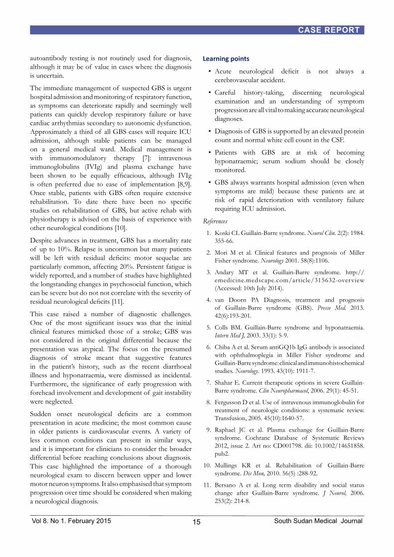
Diagnosis of GBS is supported by an elevated protein • count and normal white cell count in the CSF.
Patients with GBS are at risk of becoming • hyponatraemic; serum sodium should be closely monitored.
GBS always warrants hospital admission (even when • symptoms are mild) because these patients are at risk of rapid deterioration with ventilatory failure requiring ICU admission.
References
Koski CL Guillain-Barre syndrome. 1. Neurol Clin. 2(2): 1984. 355-66.
Mori M et al. Clinical features and prognosis of Miller 2. Fisher syndrome. Neurology 2001. 58(8):1106.
Andary MT et al. Guillain-Barre syndrome. http://3. emedicine.medscape.com/article/315632-overview (Accessed: 10th July 2014).
van Doorn PA Diagnosis, treatment and prognosis 4. of Guillain-Barre syndrome (GBS). Presse Med. 2013. 42(6):193-201.
Colls BM. Guillain-Barre syndrome and hyponatraemia. 5. Intern Med J, 2003. 33(1): 5-9.
Chiba A et al. Serum antiGQ1b IgG antibody is associated 6. with ophthalmoplegia in Miller Fisher syndrome and Guillain-Barre syndrome: clinical and immunohistochemical studies. Neurology. 1993. 43(10): 1911-7.
Shahar E. Current therapeutic options in severe Guillain-7. Barre syndrome. Clin Neuropharmacol, 2006. 29(1): 45-51.
Fergusson D et al. Use of intravenous immunoglobulin for 8. treatment of neurologic conditions: a systematic review. Transfusion, 2005. 45(10):1640-57.
Raphael JC et al. Plasma exchange for Guillain-Barre 9. syndrome. Cochrane Database of Systematic Reviews 2012, issue 2. Art no: CD001798. dii: 10.1002/14651858.pub2.
Mullings KR et al. Rehabilitation of Guillain-Barre 10. syndrome. Dis Mon, 2010. 56(5) :288-92.
Bersano A et al. Long term disability and social status 11. change after Guillain-Barre syndrome. J Neurol, 2006. 253(2): 214-8.

South Sudan Medical Journal Vol 8. No 1. February 2015 16
ShoRt ItEMS
The importance of coughs David Tibbutt
Correspondence: [email protected]
‘Cough’ is so common we sometimes do not realise just how important it can be. It is at best annoying to patients (and families) especially if nocturnal and, at worst, very distressing particularly if associated with dyspnoea, copious sputum and/or pain. It may be associated with many serious diseases including lung cancer and tuberculosis.
The cough mechanism is set off by the stimulation of irritant receptors, which occur in the nose and sinuses, around the vocal cords, carina and the larger airways, and also the eardrums, diaphragm, pericardium and stomach. These receptors have afferent nerves via the Vth, IXth and Xth cranial nerves to the brain. The efferent side of these reflexes leads to:
a. inspiration,
b. glottic closure,
c. diaphragmatic relaxation,
d. intercostal and abdominal muscle tension,
e. rise of intrathoracic pressure up to 200mm Hg.,
f. glottic opening and
g. invagination of the tracheobronchial membrane
h. so narrowing the airway with
i. rapid expulsion of air i.e. a cough
j. carrying mucus with it.
In the light of this mechanism it is clear why so many conditions may be associated with a cough and how the cough mechanism, which is itself often protective to the airway, may be impaired. For example pain following abdominal surgery may reduce abdominal muscle tension, the force of a cough and so reduce mucus clearance predisposing to pulmonary infection.
Causes
There is an important rule to observe when considering the cause of a cough: if the cough has been present for three weeks or more always investigate for pulmonary tuberculosis. The differential diagnosis may be reviewed from knowledge of the cough receptor sites:
1. Upper respiratory tract:
a. Viral infections are the commonest cause of cough which may last up to two months.
b. Sinusitis.
c. Allergic rhinitis
d. Laryngeal lesions including inhalation of irritants.
e. Wax or any foreign body against the tympanic membrane.
2. Pulmonary structures:
a. Bronchial asthma.
b. Inflammation: bronchitis, pneumonia, pertussis (whooping cough), bronchiectasis, smoke irritation, migrating larval stages of parasites (e.g. hookworm), malaria. Tuberculosis.
c. Foreign bodies and tumours.
d. Any cause of increased secretions.
e. Heart failure with pulmonary oedema, pulmonary emboli and secondary tumours.
f. Cystic fibrosis.
g. Sarcoidosis.
h. Any other cause of interstitial lung disease: collagen diseases (e.g. rheumatoid disease, systemic lupus erythematosus, scleroderma), drugs (e.g. nitrofurantoin, methotrexate, anticancer drugs, penicillins, non-steroidal anti-inflammatory drugs, amiodarone).
3. Other:
a. Diaphragmatic, pericardial and gastric receptors are probably of little importance.
b. Gastro-oesophageal reflux may lead to a cough by aspiration. This is probably the most important cause of chronic cough and should be excluded if no other obvious cause can be found. Also a trial of anti-reflux therapy may be worthwhile in any case of chronic persistent cough. It must be remembered that with reflux in patients already taking proton pump inhibitors there may not be heartburn because of the absence of acid but the enzyme pepsin may give rise to the cough.
c. Psychogenic.
Diagnosis
Special points to note in the history
Times when the cough is worse: cough at night may indicate heart failure, bronchial asthma or aspiration from gastro-oesophageal reflux; cough at meal times may suggest aspiration. Also a cough after meals may suggest pharyngeal

Vol 8. No 1. February 2015 South Sudan Medical Journal 17
ShoRt ItEMS
pouch. A cough during a meal that includes a fizzy drink or acid vapours (as in lemon juice) may be due to bronchial hyper-reactivity or asthma.
1. Triggers:
a. Exertion or laughing: interstitial lung disease or bronchial asthma.
b. Environmental change: houses and pets: may suggest an allergic cause or asthma.
c. Medication: angiotensin converting enzyme inhibitors and beta-blockers (ingested and as eye drops). Interstitial lung disease may be caused by many drugs (e.g. as above) and present with cough.
2. Sputum production: Post nasal drip and bronchitis often create morning sputum. Purulent sputum usually suggests bacterial infection but eosinophils in high numbers may give rise to similar appearances. Large volumes of persistent purulent sputum are an indication of probable bronchiectasis.
3. Haemoptysis: Tuberculosis, bronchiectasis, tumours, pulmonary embolism and bronchitis.
4. Sneezing and rhinorrhoea: allergic rhinitis.
5. Dry mouth, from mouth breathing, change or loss of sense of smell: chronic rhinitis.
6.Gastro-oesophagealrefluxsymptomsordysphagia:oesophageal disease.
7. Joint pains and/or swelling: connective tissue disorders.
8.Pointerstoacquiredimmunodeficiency: unexplained cough may be a presentation of Pneumocystis jiroveci (formerly called corinii) pneumonia.
Special points to note on examination.
1. Deep expiration precipitating the cough suggests bronchial asthma.
2. Types of cough:
a. “Wet”: bronchial asthma, bronchitis, bronchiectasis.
b. “Brassy”: tumour.
c. Increasing cough during examination and clearing when the patient is not aware of being observed may suggest a psychogenic cause.
3. Impacted ear wax.
4. Nasal passages: polyps, mucopurulent discharge, signs of inflammation.
5. Tender maxillary sinuses.
6. Goitre and other neck masses.
7. Rales and rhonchi especially if localised. A unilateral fixed rhonchus may indicate a tumour.
8. Finger clubbing: malignancy, lung abscess, bronchiectasis.
9. Central cyanosis and/or anaemia.
Careful clinical assessment will provide a working diagnosis in most cases and special investigations often are not needed.
Sputum examination: Look at it!!! Are there any signs of blood? A rusty looking specimen may suggest a pneumococcal pneumonia. Microscopy will differentiate bacterial infection from eosinophilia: consider TB and fungi.
Chest x-ray: Bear in mind that a normal CXR does not exclude TB, tumour, foreign body or bronchiectasis.
Respiratory function tests: the simplest is to observe the patient exercising e.g. walking or climbing stairs. The inability to complete sentences without added inspirations during ordinary conversation is abnormal. Exercise may induce wheezing in bronchial asthma. Peak expiratory flow (PEF) measurement is helpful but spirometry is better. PEF meters are small, relatively inexpensive and the measurements at least give some quantitative idea of progression. Without such equipment ask the patient to blow out a lighted match from five inches and with the mouth open.
Treatment
It is not the purpose of this article to describe the treatment of all the causes of cough.
1. Treatment should be directed at the specific cause.
2. Removal of an allergen or irritant is usually very effective: cigarette smoking is an important example.
3. “Bronchitis” that does not respond to antibiotics could suggest an obstructing lesion.
4. A distressing cough with an irreversible cause (e.g. metastatic malignancy) should be suppressed using:
a. A simple linctus: honey for children with an acute cough is effective [1].
b. Humidified air (steam inhalation but care to avoid scalding) or
c. Codeine phosphate 30-60mg. 6-8 hourly.
d. Kindness and reassurance: a patient is often more afraid of the cause of the cough than the cough itself.
Reference
1. Oduwole. O, Meremikwu, M.M., Oyo-Ita, A and Udoh, 1. E.E. Honey for acute cough in children. Cochrane Acute respiratory Infections Group. Published Online 14 March 2012. DOI: 10.1002/14651858.CD007094.pub3
South Sudan Medical Journal Vol 8. No 1. February 2015 18
ShoRt ItEMS
A longitudinal study of MUAC as a measure of paediatric malnutrition in Yei, South Sudan: Lessons from a hospital linkAbigail Sharpea and Simon Struthersa
a. Royal Hampshire County Hospital, Romsey Rd, Winchester, SO22 5DG, UK
Correspondence to Dr Abi Sharpe: [email protected]
Background
Paediatric malnutrition is a significant problem in South Sudan, with rates of wasting up to 22% reported in some areas [1]. Severe acute malnutrition (SAM) is associated with a high mortality [2]. Affected patients require thorough assessment and holistic care including appropriate therapeutic feeding, treatment of associated complications and rehabilitation in order to achieve good outcomes.
Since 2009 there has been an NHS Global Link between Yei Civil Hospital (YCH), Central Equatoria Province and Royal Hampshire County Hospital, Winchester, UK. It had been noted that there was very minimal provision for malnourished children in the town (total population in Yei was estimated at 185,000 in 2011). Successive teams from the link therefore assessed the levels of acute paediatric malnutrition in order to supply data to the South Sudanese government and its partners such as UNICEF in order to facilitate decisions as to whether increased provision is warranted.
The measurement used was the Mid-Upper Arm Circumference (MUAC) which is a well-validated indicator of acute malnutrition [3, 4, 5, 6], and is recommended by the World Health Organization (WHO) [7] and the 2009 interim South Sudanese guidelines [8] as a key assessment tool.
Methods
Data were collected during three defined periods between 2012 and 2014:
• Period 1: Oct-Dec 2012 (3months);
• Period 2: June 2013 (10 days);
• Period 3: Oct 2014 (10 days).
MUAC was measured using standardized colour-coded tapes, and by the method described by UNICEF [9] – see Figure 1.
MUAC measurements were collected for all available inpatients and ambulatory care patients aged 6 months to 5 years at YCH, Martha Primary Care Clinic (MPCC), and during mobile clinics (MC) in rural areas over the study periods. The age and sex of each patient was recorded.
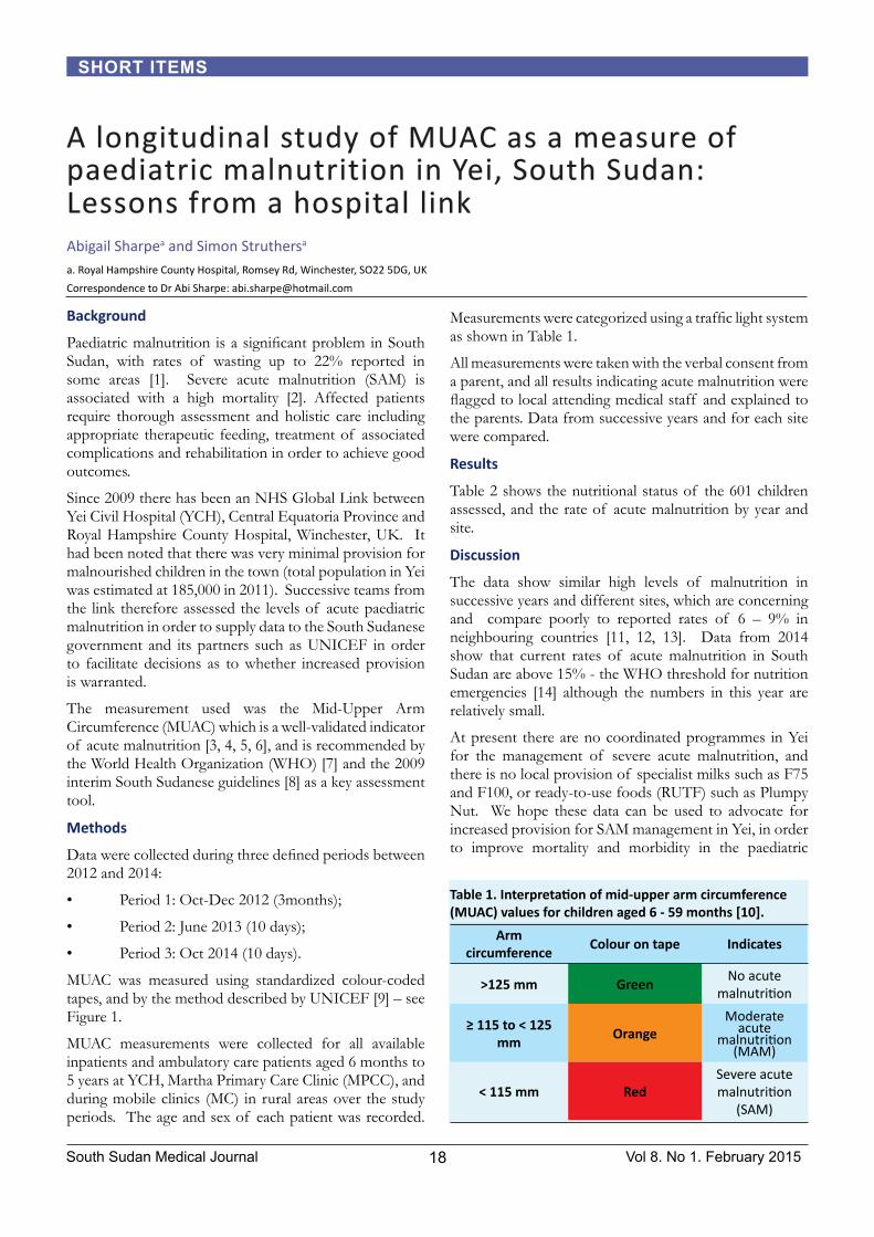
Measurements were categorized using a traffic light system as shown in Table 1.
All measurements were taken with the verbal consent from a parent, and all results indicating acute malnutrition were flagged to local attending medical staff and explained to the parents. Data from successive years and for each site were compared.
Results
Table 2 shows the nutritional status of the 601 children assessed, and the rate of acute malnutrition by year and site.
Discussion
The data show similar high levels of malnutrition in successive years and different sites, which are concerning and compare poorly to reported rates of 6 – 9% in neighbouring countries [11, 12, 13]. Data from 2014 show that current rates of acute malnutrition in South Sudan are above 15% - the WHO threshold for nutrition emergencies [14] although the numbers in this year are relatively small.
At present there are no coordinated programmes in Yei for the management of severe acute malnutrition, and there is no local provision of specialist milks such as F75 and F100, or ready-to-use foods (RUTF) such as Plumpy Nut. We hope these data can be used to advocate for increased provision for SAM management in Yei, in order to improve mortality and morbidity in the paediatric
Arm circumference
Colour on tape Indicates
>125 mm GreenNo acute
malnutrition
≥ 115 to < 125 mm
OrangeModerate
acute malnutrition
(MAM)
< 115 mm RedSevere acute malnutrition
(SAM)
Table 1. Interpretation of mid-upper arm circumference (MUAC) values for children aged 6 - 59 months [10].

Vol 8. No 1. February 2015 South Sudan Medical Journal 19
ShoRt ItEMS
population there.
The authors would like to thank Cathy Williams, Dr Imelda Heyes and Dr Sally Louden for data collection and doctors and staff at YCH including Dr Denis Zachariah, Dr Issam-EldenYousif and Dr Kennedy Samuel, and at MPCC for their support of the project.
References
GOSS MOH, Central Bureau of Statistics (BCS) and 1. Southern Sudan Commission for Census, Statistics and Evaluation (SSCCSE). Sudan Household Health Survey (SHHS) 2006. Juba, Southern Sudan: SSCCSE; FAO and WFP. Crop and Food Supply Assessment Mission to Sudan, 1 February 2007. Rome: FAO and WFP. http://www.irinnews.org/pdf/pn/shhsreport.pdf
Black RE, Allen LH, Bhutta ZA, Caulfield LE, de Onis 2. M, Ezzati M, Mathers C, Rivera J. Maternal and Child Undernutrition Study Group. Maternal and child undernutrition: global and regional exposures and health consequences. Lancet; 2008. 371(9608):243-60. doi: 10.1016/S0140-6736(07)61690-0. http://www.thelancet.com/journals/lancet/article/PIIS0140-6736(07)61690-0/fulltext
WHO Multicentre Growth Reference Study Group. 3. 2006. Reliability of anthropometric measurements in the WHO Multicentre Growth Reference Study. ActaPaediatrica.;Suppl 450: 38/46. http://www.who.int/childgrowth/standards/Reliability_anthro.pdf
Berkley J, Mwangi I, Griffiths K, Ahmed I, Mithwani S, 4. English M, Newton C, Maitland K. Assessment of severe malnutrition among hospitalized children in rural Kenya: comparison of weight for height and mid upper arm circumference. JAMA; 2005. 294(5):591-7. http://www.ncbi.nlm.nih.gov/pubmed/16077053
Briend A, Maire B, Fontaine O, Garenne M. Mid-upper arm 5. circumference and weight-for-height to identify high-risk malnourished under-five children. Matern. Child Nutr. 2012 Jan;8(1):130-3. doi: 10.1111/j.1740-8709.2011.00340.x. Epub 2011 Sep 28. http://onlinelibrary.wiley.com/doi/10.1111/j.1740-8709.2011.00340.x/full
Myatt M, Khara T, Collins S. A review of methods to detect 6. cases of severely malnourished children in the community for their admission into community-based therapeutic care programs. Food Nutrition Bulletin. 2006 Sep;27(3 Suppl):S7-23. http://www.who.int/nutrition/topics/backgroundpapers _A_%20review.pdf
WHO, UNICEF 2009. WHO child growth standards and 7. the identification of severe acute malnutrition in infants and children. A Joint Statement. World Health Organization, Geneva. http://www.who.int/maternal_child_adolescent/documents/9789241598163/en/
Government of South Sudan, Ministry of Health. 8. December 2009. Interim Guidelines on the Integrated Management of Severe Acute Malnutrition. http://www.
Year Site
MUAC Category Total rate of acute
malnutrition (MAM + SAM)
%
Total rate of
SAM%
Green (%)
Orange (%)
Red (%)
2012YCC 223 (88.8) 20 (8.0) 8 (3.2)
11.4 3..3MPCC 175 (88.4) 16 (8.1) 7 (3.5)
2013
YCH 31 (86.1) 4 (11.1) 1 (2.8)
12.6 3.1MPCC 31 (86.1) 4 (11.1) 1 (2.8)
MC 19 (90.5) 1 (4.8) 1 (4.8)
2014YCH 20 (76.9) 3 (11.5)
3 (11.5) 15.6 5.3
MPCC 28 (90.3) 3 (9.7) 0
Table 2. Number and percent of children according to their MUAC category by year and site.
Figure 1. Measuring MUAC. The colour on the tape is orange so the child has moderate acute malnutrition (MAM). Image reproduced courtesy of UNICEF Ethiopia.

South Sudan Medical Journal Vol 8. No 1. February 2015 20
ShoRt ItEMS
southsudanmedicaljournal.com/assets/files/misc/GOSS_IMSAM_Guidelines.pdf
UNICEF, Emergency Nutrition Network. Nutrition in 9. Emergencies. 2009 http://www.unicef.org/nutrition/training/3.1.3/1.html
Nutrition Technical Assistance III Project (FANTA). 2013. 10. Nutrition Assessment, Counselling, and Support (NACS). A User’s Guide. Module 2. Version 1. Nutrition Assessment and Classification. http://www.fantaproject.org/sites/default/files/resources/NACS-Users-Guide-Module2-Dec2013.pdf
Government of Kenya, Ministry of Health. June 2009. 11. National Guideline for Integrated Management of Acute Malnutrition. http://www.cmamforum.org/Pool/Resources/Kenya-MoH-IMAM-Guideline-June-2009.pdf
Kismul H, Schwinger C, Chhagan M, Mapatano M, Van 12. den Broeck J. Incidence and course of child malnutrition according to clinical or anthropometrical assessment: a longitudinal study from rural DR Congo. BMC Pediatrics 2014; 14:22 doi:10.1186/1471-2431-14-22. http://www.ncbi.nlm.nih.gov/pubmed/24467733
The Republic of Uganda, Ministry of Health, December 13. 2010.Integrated management of acute malnutrition guidelines. http://www.unicef.org/uganda/IMAM_Guidelines_final_version.pdf
WHO 2000. The Management of Nutrition in 14. Major Emergencies. World Health Organisation, Geneva. http://www.who.int/nutrition/publications/emergencies/9241545208/en /
Notice of a new bookEnvironmental health and occupational
health & Safety By Samuel Obura Afubwa and Mutuku Alexander Mwanthi
This book is divided into two parts: (i) Environmental Health (ii) Occupational Health & Safety. Environmental Health has been written to cover the pillars of Environmental Health with categories in history, solid waste management, sanitation, pollution, toxicology, ventilation & lighting. Environmental Health Impact Assessment with emphasis on EIA. Food Quality Control that has an international dimension, disaster management and laws covering Environmental Health are also covered.
Part two on Occupational Health and Safety that has evolution with reasons why the concern is a must. Occupational Health Services, toxicology, noise and monitoring of workplace environment are clearly written. Plant and machinery safety is well exemplified. Others are construction and fire safety, electrical safety, ergonomics, accident investigation & prevention, personal protective equipment and safety culture.
This book is summarized to enable professionals perform their tasks efficiently. It will also be of great benefit to learners in professional Colleges and Universities.
The print edition can ordered from Acrodile publishing Ltd through [email protected] . The e-book is available on the publisher’s website www.acrodile.co.ke, amazon.com and at Barnes and Noble.
The above announcement was provided by the publisher and does not necessarily reflect the opinion of SSMJ.
Vol 8. No 1. February 2015 South Sudan Medical Journal 21
LEttER to thE EdItoRNeonatal scalp seborrhoeic dermatitis or
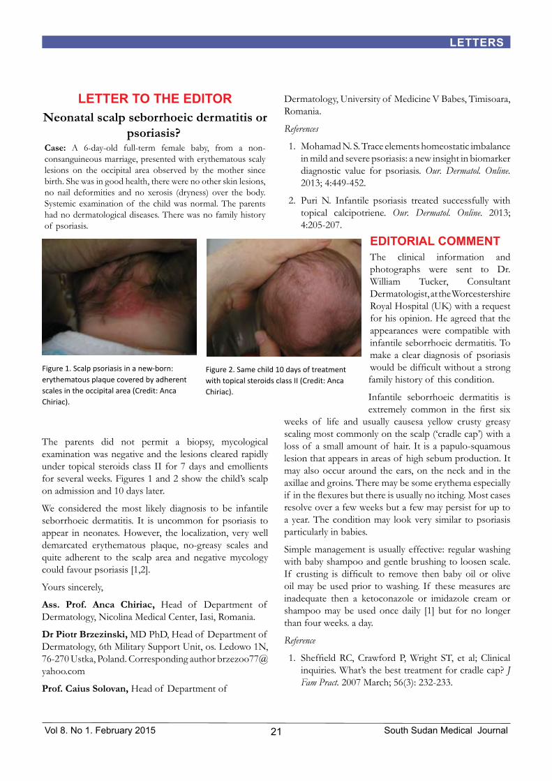
psoriasis? Case: A 6-day-old full-term female baby, from a non-consanguineous marriage, presented with erythematous scaly lesions on the occipital area observed by the mother since birth. She was in good health, there were no other skin lesions, no nail deformities and no xerosis (dryness) over the body. Systemic examination of the child was normal. The parents had no dermatological diseases. There was no family history of psoriasis.
The parents did not permit a biopsy, mycological examination was negative and the lesions cleared rapidly under topical steroids class II for 7 days and emollients for several weeks. Figures 1 and 2 show the child’s scalp on admission and 10 days later.
We considered the most likely diagnosis to be infantile seborrhoeic dermatitis. It is uncommon for psoriasis to appear in neonates. However, the localization, very well demarcated erythematous plaque, no-greasy scales and quite adherent to the scalp area and negative mycology could favour psoriasis [1,2].
Yours sincerely,
Ass. Prof. Anca Chiriac, Head of Department of Dermatology, Nicolina Medical Center, Iasi, Romania.
Dr Piotr Brzezinski, MD PhD, Head of Department of Dermatology, 6th Military Support Unit, os. Ledowo 1N, 76-270 Ustka, Poland. Corresponding author [email protected]
Prof. Caius Solovan, Head of Department of
LEttERS
Dermatology, University of Medicine V Babes, Timisoara, Romania.
References
Mohamad N. S. Trace elements homeostatic imbalance 1. in mild and severe psoriasis: a new insight in biomarker diagnostic value for psoriasis. Our. Dermatol. Online. 2013; 4:449-452.
Puri N. Infantile psoriasis treated successfully with 2. topical calcipotriene. Our. Dermatol. Online. 2013; 4:205-207.
EdItoRIAL CoMMENtThe clinical information and photographs were sent to Dr. William Tucker, Consultant Dermatologist, at the Worcestershire Royal Hospital (UK) with a request for his opinion. He agreed that the appearances were compatible with infantile seborrhoeic dermatitis. To make a clear diagnosis of psoriasis would be difficult without a strong family history of this condition.
Infantile seborrhoeic dermatitis is extremely common in the first six
weeks of life and usually causesa yellow crusty greasy scaling most commonly on the scalp (‘cradle cap’) with a loss of a small amount of hair. It is a papulo-squamous lesion that appears in areas of high sebum production. It may also occur around the ears, on the neck and in the axillae and groins. There may be some erythema especially if in the flexures but there is usually no itching. Most cases resolve over a few weeks but a few may persist for up to a year. The condition may look very similar to psoriasis particularly in babies.
Simple management is usually effective: regular washing with baby shampoo and gentle brushing to loosen scale. If crusting is difficult to remove then baby oil or olive oil may be used prior to washing. If these measures are inadequate then a ketoconazole or imidazole cream or shampoo may be used once daily [1] but for no longer than four weeks. a day.
Reference
Sheffield RC, Crawford P, Wright ST, et al; Clinical 1. inquiries. What’s the best treatment for cradle cap? J Fam Pract. 2007 March; 56(3): 232-233.
Figure 1. Scalp psoriasis in a new-born: erythematous plaque covered by adherent scales in the occipital area (Credit: Anca Chiriac).
Figure 2. Same child 10 days of treatment with topical steroids class II (Credit: Anca Chiriac).

South Sudan Medical Journal Vol 8. No 1. February 2015 22
INFECTION
Ebola Pocket Library
Ebola Pocket Library contains practical information from sources such as the CDC, WHO, Khan Academy and Wikipedia and includes:
Ebola factsheets and basic information, such as • symptoms, treattment, and prevention of Ebola
Training information for medical staff•
Resources for those dealing with grief and loss •
Occupational health and safety guidelines•
Case studies and reports •
General resources for emergency preparedness •
Travel safety•
Posters and other print materials for posting in public • placess
Safe burial practices for the victims of Ebola•
The on-line Internet version of the Ebola Pocket Library can be viewed at: http://www.widernet.org/portals/ebola. The downloadable off-line version can be found here: http://widernet.unc.edu/research/ebolalibrary/downloading-the-ebola-pocket-libraryFrom HIFA2015
Ebola Resource Centre
The Ebola Resource Centre at http://ebola.thelancet.combrings together all Ebola-related content from The Lancet family of journals and is freely accessible.
Ebola Communication Network (ECN)
Ebola Communication Network (ECN), at http://ebolacommunicationnetwork.org/is an online collection of Ebola resources, materials and tools from and for the global health community. It is supported by USAID.RevisedWHOclassificationandtreatmentof childhoodpneumonia at health facilities
The revised guidelines present two major changes to existing guidelines: (A) there are now just 2 categories of pneumonia instead of 3 (“pneumonia” which is treated at home with oral amoxicillin and “severe pneumonia” which requires injectable antibiotics) and (B) oral amoxicillin replaces oral cotrimoxazole as first line treatment, preferably in 250mg dispersible tablet form, twice daily for five days which can be reduced to three days in low HIV settings. This document’s purpose is to assist national child health prgrammes in revising their guidelines to conform to the new recommendations.
Resources
Ref: World Health Organization, 2014. 34pp.364 kB: http://www.ncbi.nlm.nih.gov/books/NBK264162/pdf/TOC.pdf From CHILD2015Malaria treatment rectal artesunate to be developed in India
A TDR multi-country trial among 17 000 patients in Bangladesh, Ghana and Tanzania demonstrated that a single inexpensive artesunate suppository substantially reduced the risk of death or permanent disability. The artesunate was given to children who were so ill they could not take a medication by mouth, giving their families time to get to medical facilities for further care – see http://www.thelancet.com/journals/lancet/article/PIIS0140-6736(08)61734-1/fulltext . Agreements have now been signed between Medicines for Malaria Venture (MMV) and two Indian pharmaceutical companies, Cipla Limited and Strides Arcolab Limited, for the development of rectal artesunate for pre-referral treatment of children with severe malaria - seehttp://www.who.int/tdr/news/2014/malaria-treatment/en/
Praziquanteldoseconfirmedforschistosomiasis
Evidence gathered using different methodologies for combining clinical trial results confirm that 40 mg per kg of body weight, the WHO-recommended dose of praziquantel, works well for all types of schistosomiasis. See details at http://www.who.int/tdr/news/2014/praziquantel-for-schistosomiasis/en/
Guidance for national tuberculosis programmes on the management of tuberculosis in children. 2nd edition
This second edition aims to inform the revision of existing national guidelines and standards for managing TB, many of which include guidance on children. It includes recommendations, based on the best available evidence, for improving the management of children with TB and of children living in families with TB. National and regional TB control programmes may wish to adapt these recommendations according to local circumstances. Note: this publication is priced. http://apps.who.int/bookorders/anglais/detart1.jsp?codlan=1&codcol=93&codcch=299Guidelines on the management of latent tuberculosis infection
These Guidelines on the management of latent tuberculosis infection were developed in accordance to the requirements and recommended process of the WHO Guideline Review Committee, and provide public health approach guidance on evidence-based practices for testing,

Vol 8. No 1. February 2015 South Sudan Medical Journal 23
RESoURCES
treating and managing LTBI in infected individuals with the highest likelihood of progression to active disease. Note: this publication is priced.http://apps.who.int/bookorders/anglais/detart1.jsp?codlan=1&codcol=93&codcch=295
MATERNAL, NEWBORN CARE, CHILDHOOD
Global Health Media Project videos
All Global Health Media Project videos (http://www.healthphone.org ) are free to download and distribute/reproduce. Recent ones are:
• Referring a sick baby- see http://www.healthphone.org/ghmp/referring-a-sick-baby.htm. This film shows the essential features of a referral process. It covers involving the family, arranging transport, preparing the baby for the journey, and writing the referral note. It also includes what to do if referral is not an option. The primary audience are frontline health workers in primary and district level facilities. It was filmed in Nigeria and the Dominican Republic.’
• Newborn care: Inserting an IV - See http://www.healthphone.org/ghmp/inserting-an-iv.htm. This film shows how to insert an IV into the tiny vein of a newborn. It illustrates gathering supplies, finding a good vein, applying a tourniquet, the insertion technique, taping and splinting the IV, and administering medication. The primary audience are frontline health workers in primary and district level facilities. It was filmed in Nigeria and the Dominican Republic. From CHILD2015
The White Ribbon Alliance
See their materials on Respectful Maternity Care at. http://whiteribbonalliance.org/campaigns/respectful-maternity-care/
Early essential newborn care
This WHO Guide aims to provide health professionals with a user-friendly, evidence-based protocol to essential newborn care - focusing on the first hours and days of life. Note: this publication is priced.http://apps.who.int/bookorders/anglais/detart1.jsp?codlan=1&codcol=52&codcch=210Supplement on Integrated Community Case Management in Africa
The Journal of Global Health has published a special supplement entitled “Current scientific evidence and future directions for Integrated Community Case Management in Africa,” whichhighlightes the latest evidence on integrated community case management (iCCM) for childhood illnesses. It lists implementation approaches
that have and have not worked in various settings, along with recommendations for future programming, given the current context of innovation and funding opportunities.See details at http://www.who.int/tdr/news/2014/iccm/en/Note: this publication is priced.Caregivers’ knowledge and home management of fever in children
Fever is a sign that there is an underlying pathologic process, the most common being infection. Many childhood illnesses are accompanied by fever, many of which are treated at home prior to presentation to hospital. Most febrile episodes are benign. Caregivers are the primary contacts to children with fever. Adequate caregivers’ knowledge and proper management of fever at home leads to better management of febrile illnesses and reduces complications. This study at Kenyatta National Hospital questioned 250 caregivers of children under 12 years presenting with fever in 2011.Three quarters of the caregivers’ defined fever correctlyand a majority of them used touch to detect fever. Infection was cited as the leading cause of fever (95.2%). Brain damage (77.6%) and dehydration (65.6%) were viewed as the most common complications. Fever was treated at home by 97.2% of caregivers, most of them used medication. It was concluded that Public Health Education should be implemented in order to enlighten caregivers on fever and advocate for the use of a clinical thermometer to monitor fever at home.
Ref: Caregivers’ Knowledge and Home Management of Fever in Children PJ Koech, FE Onyango, C Jowi E. Afr. med. J. Vol 91, No 5 (2014). Abstract athttp://www.ajol.info/index.php/eamj/article/view/109970
Environmental Enteric Dysfunction
This Technical Brief from the CMAM Forum gives an up-to-date overview of this condition, including impact on health and nutrition, diagnosis, epidemiology, possible aetiology and approaches to treatment. It considers how this can affect management, and highlights current research, future priorities and further reading. It is aimed at stakeholders involved and interested in the reduction of childhood undernutrition, especially those addressing child health in low- and middle-income countries. See the Brief at http://www.cmamforum.org/Pool/Resources/Environmental-Enteric-Dysfunction-CMAM-Forum-techn-brief-Aug-2014.pdf
Ref: Environmental Enteric Dysfunction – an Overview 2014. Crane, Jones and Berkley on CMAM Forumat http://www.cmamforum.org/

South Sudan Medical Journal Vol 8. No 1. February 2015
Every effort has been made to ensure that the information and the drug names and doses quoted in this Journal are correct. However readers are advised to check information and doses before making prescriptions. Unless otherwise
stated the doses quoted are for adults.