SS 1 Filtration Secretion Reabsorption. Circulatory System Circulates Nutrients: glucose, amino...
59
S S 1 Filtration Secretion Reabsorption
-
Upload
job-goodman -
Category
Documents
-
view
223 -
download
0
Transcript of SS 1 Filtration Secretion Reabsorption. Circulatory System Circulates Nutrients: glucose, amino...
S S 1
Filtration
Secretion
Reabsorption
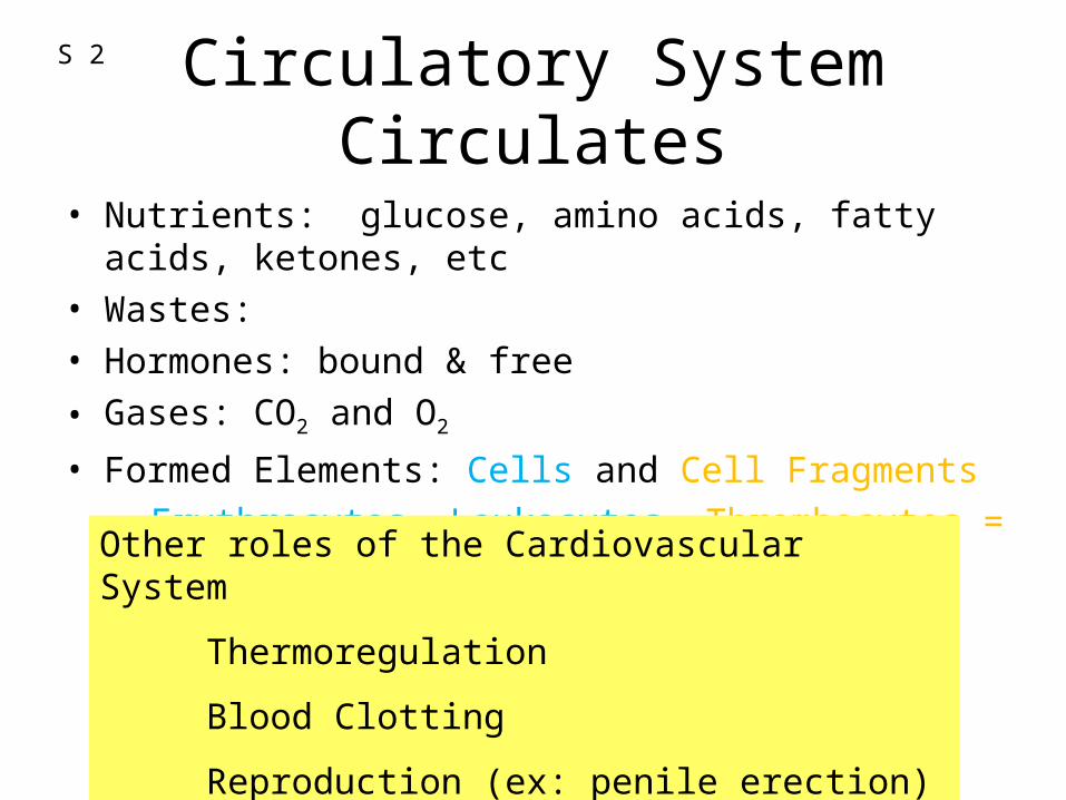
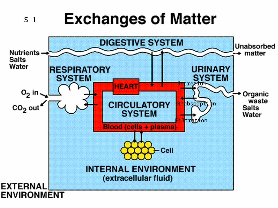
Circulatory System Circulates
• Nutrients: glucose, amino acids, fatty acids, ketones, etc• Wastes:• Hormones: bound & free
• Gases: CO2 and O2
• Formed Elements: Cells and Cell Fragments– Erythrocytes, Leukocytes, Thrombocytes = Platelets
Other roles of the Cardiovascular System
Thermoregulation
Blood Clotting
Reproduction (ex: penile erection)
S 2
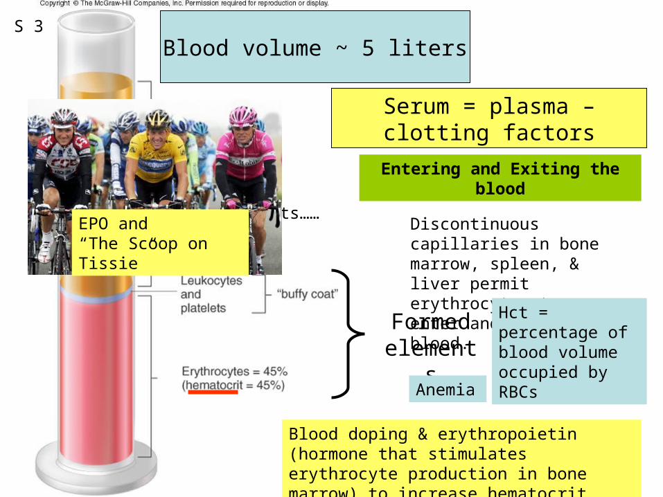
Figure 12.01Blood volume ~ 5 liters
Serum = plasma – clotting factors
Formed elements
Components……
Blood doping & erythropoietin (hormone that stimulates erythrocyte production in bone marrow) to increase hematocrit
Entering and Exiting the blood
Discontinuous capillaries in bone marrow, spleen, & liver permit erythrocytes to enter and exit blood.
Hct = percentage of blood volume occupied by RBCs
Anemia
EPO and “The Scoop on Tissie”
S 3
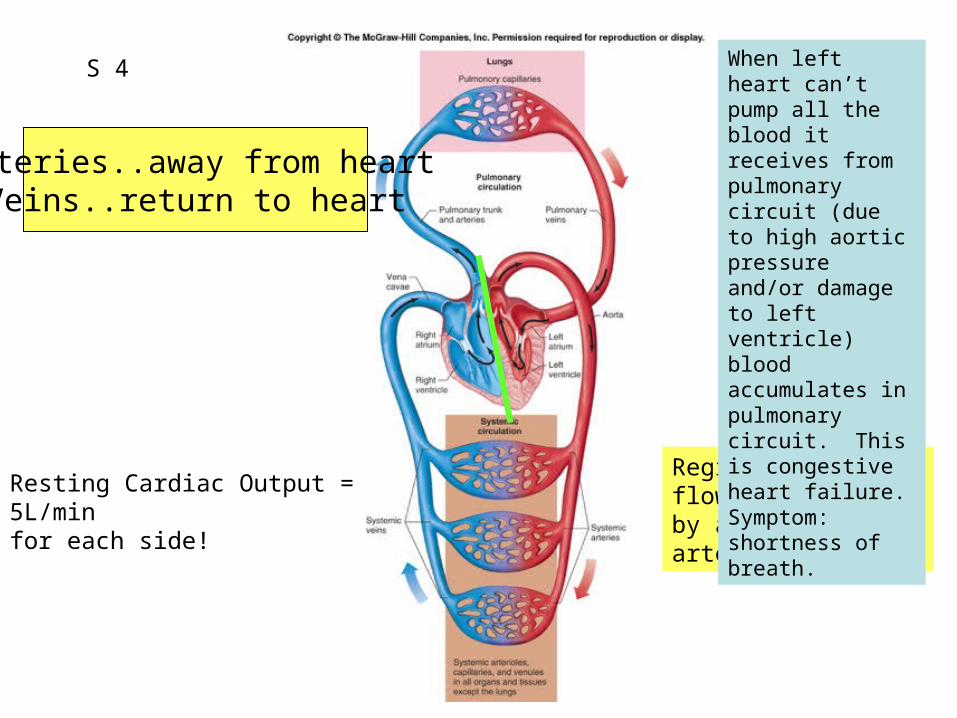
Fig. 12.02Arteries..away from heart
Veins..return to heart
Regional blood flow determined by arteries and arterioles.
Resting Cardiac Output = 5L/min for each side!
When left heart can’t pump all the blood it receives from pulmonary circuit (due to high aortic pressure and/or damage to left ventricle) blood accumulates in pulmonary circuit. This is congestive heart failure. Symptom: shortness of breath.
S 4
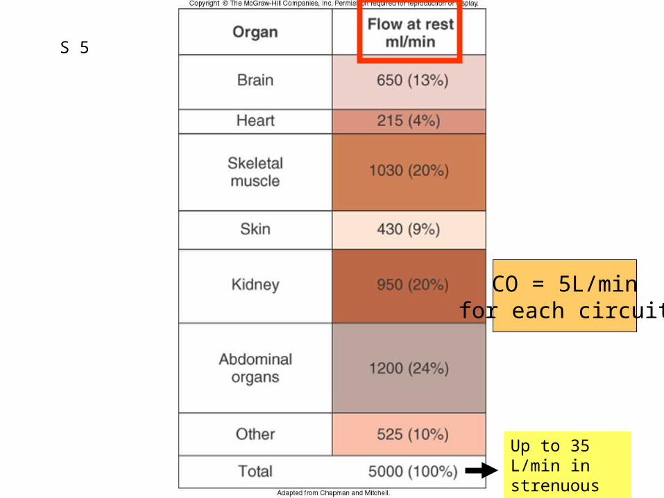
Figure 12.04
CO = 5L/minfor each circuit
Up to 35 L/min in strenuous exercise
S 5
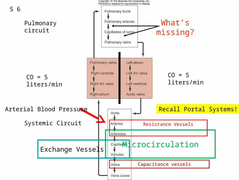
What’s missing?
Microcirculation
Pulmonary circuit
Systemic Circuit
CO = 5 liters/minCO = 5 liters/min
Exchange Vessels
Resistance Vessels
Capacitance vessels
S 6
Recall Portal Systems!Arterial Blood Pressure
Pulmonary circuit
Systemic Circuit
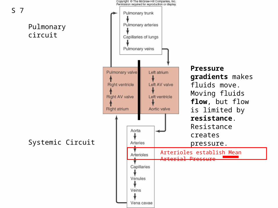
S 7
Pressure gradients makes fluids move. Moving fluids flow, but flow is limited by resistance. Resistance creates pressure.
Arterioles establish Mean Arterial Pressure
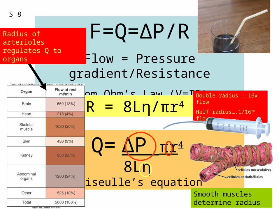
F=Q=ΔP/RFlow = Pressure gradient/Resistance
from Ohm’s Law (V=IR)
R = 8Lη/πr4
Q= ΔP πr4
8LηPoiseulle’s equation
Smooth muscles determine radius
Double radius … 16x flow
Half radius….1/16th flow
Radius of arterioles regulates Q to organs
S 8
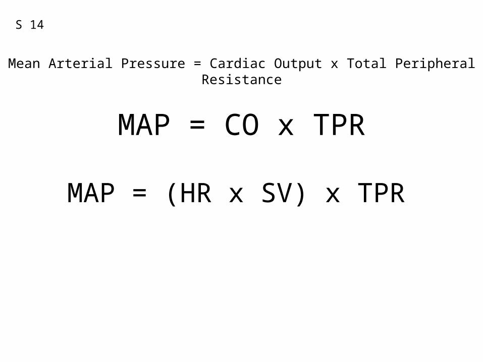
MAP = CO x TPR
Mean Arterial Pressure = Cardiac Output x Total Peripheral Resistance
MAP = (HR x SV) x TPR
S 14
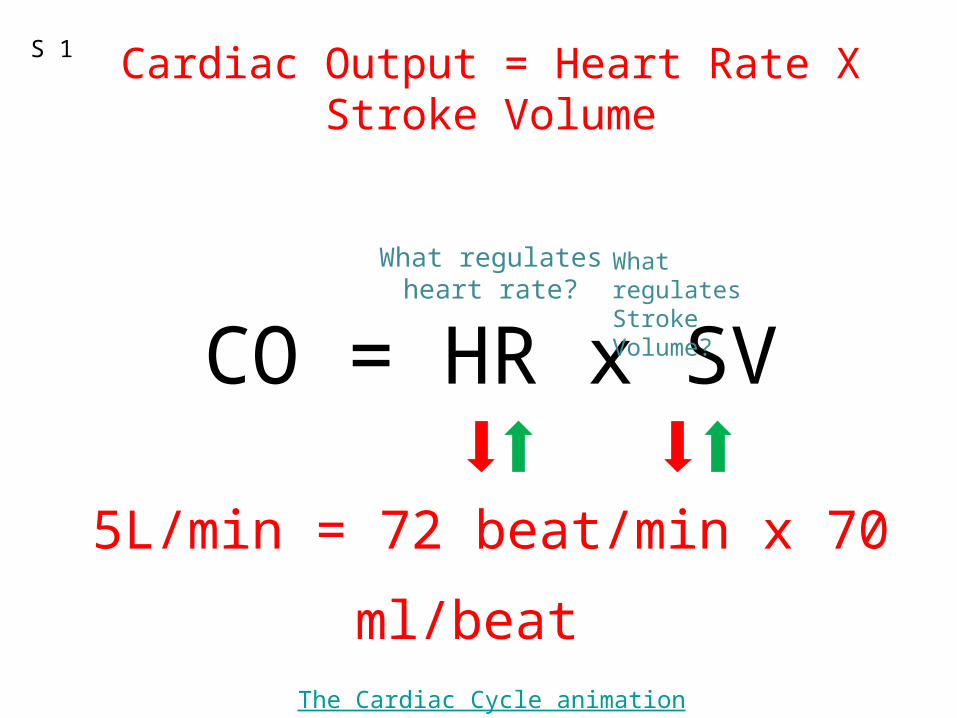
Cardiac Output = Heart Rate X Stroke Volume
What regulatesheart rate?
CO = HR x SV
5L/min = 72 beat/min x 70 ml/beat
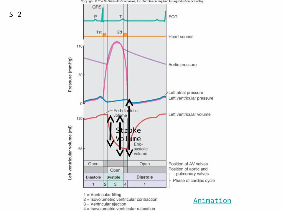
The Cardiac Cycle animation
S 1
What regulatesStroke Volume?
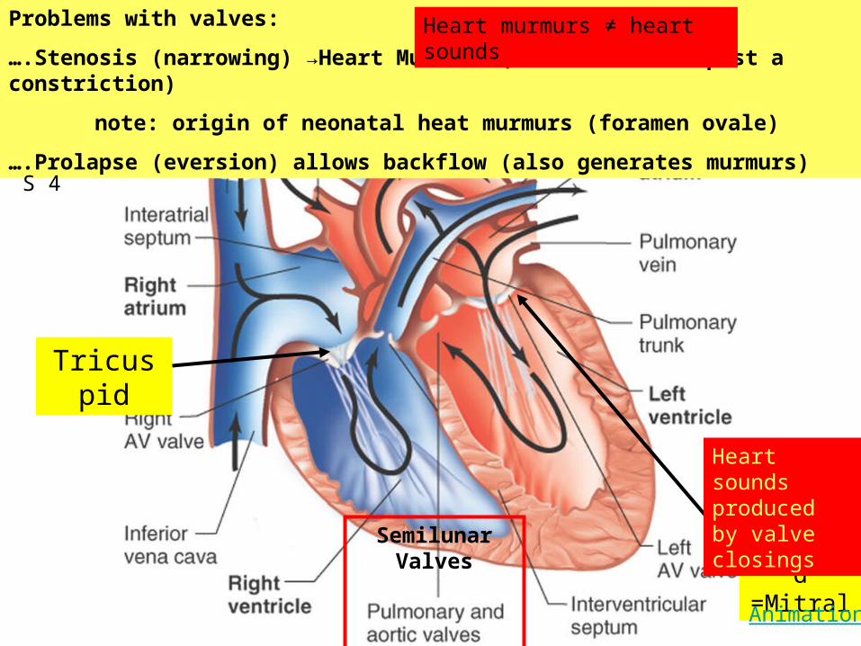
Figure 12.07
Bicuspid=Mitral
Tricuspid
Problems with valves:
….Stenosis (narrowing) →Heart Murmurs (turbulent flow past a constriction)
note: origin of neonatal heat murmurs (foramen ovale)
….Prolapse (eversion) allows backflow (also generates murmurs)
SemilunarValves
Heart sounds produced by valve closings
Animation
Heart murmurs ≠ heart sounds
S 4
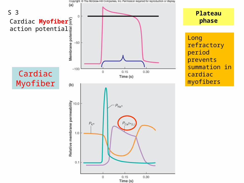
Figure 12.13
CardiacMyofiber
Plateau phaseCardiac Myofiberaction potential
Long refractory period prevents summation in cardiac myofibers
S 3
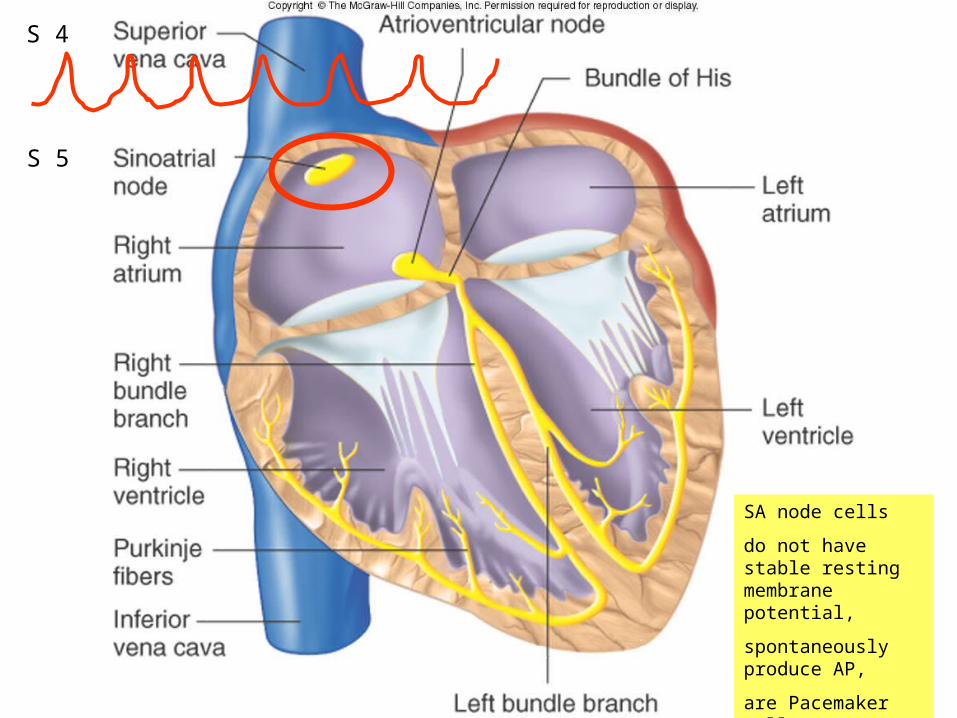
Figure 12.11
SA node cells
do not have stable resting membrane potential,
spontaneously produce AP,
are Pacemaker cells
S 5
S 4
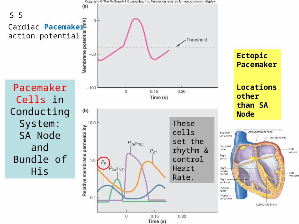
Figure 12.14
Pacemaker Cells in
Conducting System:
SA Node andBundle of His
Ectopic Pacemaker
Locations other than SA Node
S 5
Cardiac Pacemakeraction potential
These cells set the rhythm & control Heart Rate.

1QQ # 14: Answer one.1. A) Which vessels are classified as exchange
vessels?B) Why are they called exchange vessels?
2. A) What produces a heart sound?B) What produces a heart murmur?
3. With all other factors held constant, how would blood flow be affected by a doubling of the pressure gradient?
4. A) Explain how a heart can continue to beat even if the SA node is not functioning.B) Would this heart rate be faster or slower than the rate produced by the SA node?
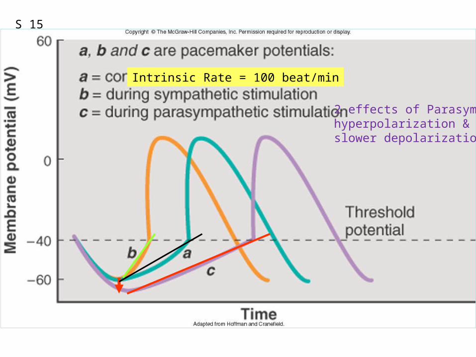
Figure 12.22Intrinsic Rate = 100 beat/min
S 15
2 effects of Parasymp:hyperpolarization &slower depolarization
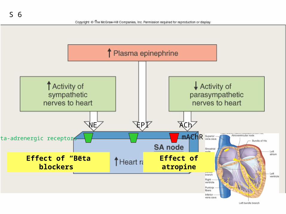
Figure 12.23
Effect of “Beta blockers”
NE EPI ACh
mAChR
Effect of atropine
Beta-adrenergic receptors
S 6
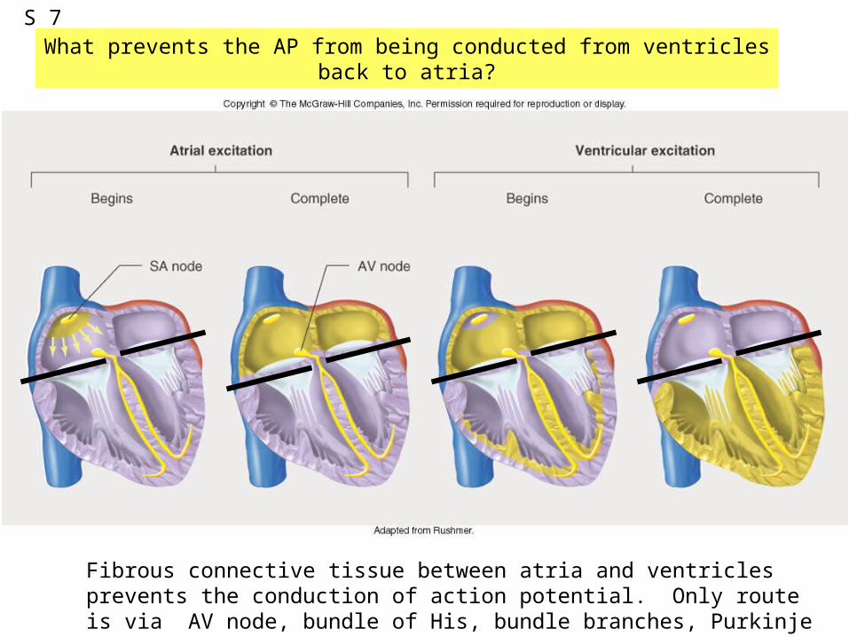
Fibrous connective tissue between atria and ventricles prevents the conduction of action potential. Only route is via AV node, bundle of His, bundle branches, Purkinje fibers, and to ventriclular myofibers.
What prevents the AP from being conducted from ventricles back to atria?S 7
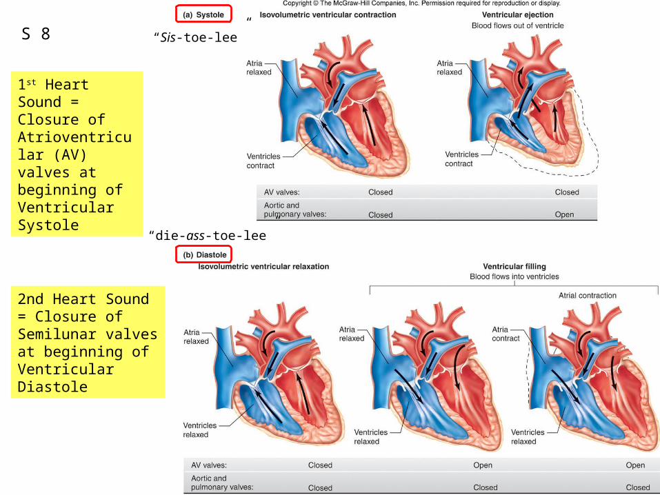
1st Heart Sound = Closure of Atrioventricular (AV) valves at beginning of Ventricular Systole
2nd Heart Sound = Closure of Semilunar valves at beginning of Ventricular Diastole
S 8 “Sis-toe-lee”
“die-ass-toe-lee”
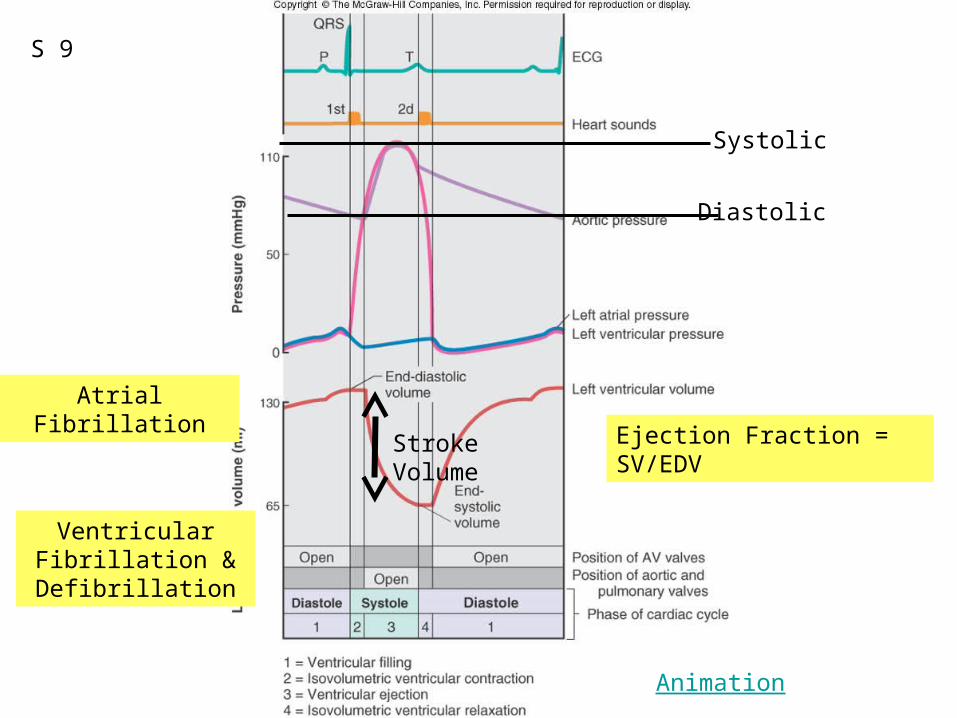
Figure 12.20Systolic
Diastolic
Ejection Fraction = SV/EDV
Atrial Fibrillation
Ventricular Fibrillation & Defibrillation
Stroke Volume
Animation
S 9
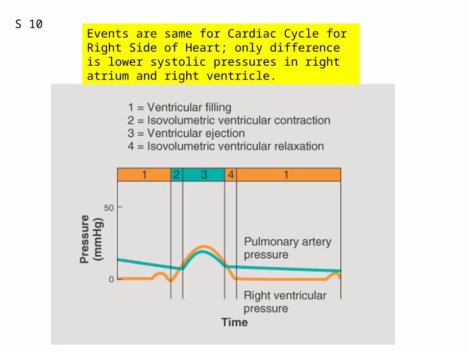
Events are same for Cardiac Cycle for Right Side of Heart; only difference is lower systolic pressures in right atrium and right ventricle.
S 10
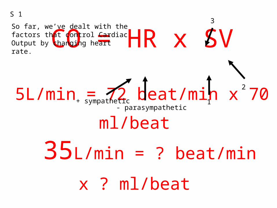
CO = HR x SV
5L/min = 72 beat/min x 70 ml/beat 35L/min = ? beat/min x ? ml/beat
S 1
So far, we’ve dealt with the factors that control Cardiac Output by changing heart rate.
+ sympathetic- parasympathetic
2
1
3
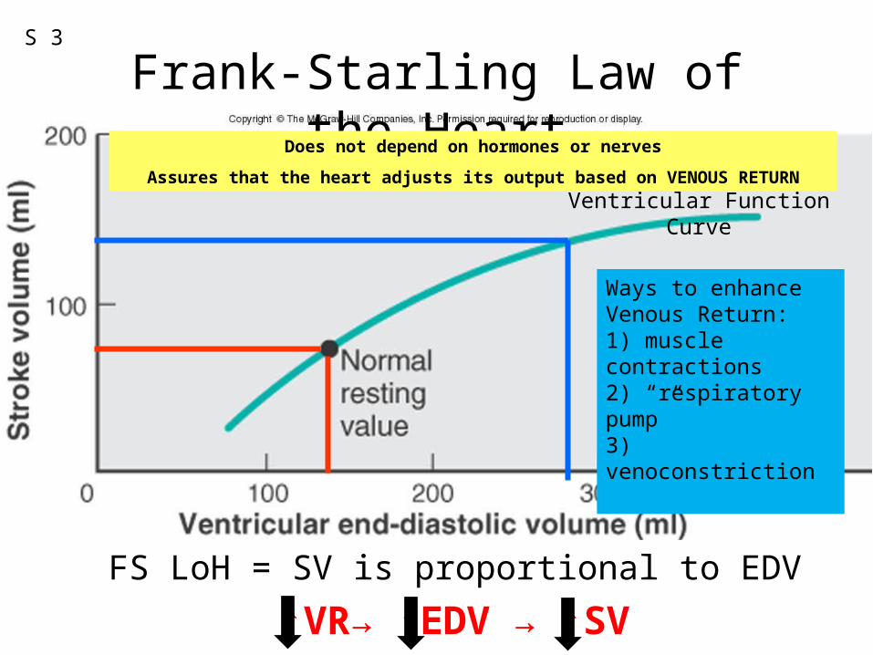
Frank-Starling Law of the Heart
FS LoH = SV is proportional to EDV
Ventricular Function Curve
Does not depend on hormones or nerves
Assures that the heart adjusts its output based on VENOUS RETURN
Ways to enhance Venous Return:1) muscle contractions2) “respiratory pump”3) venoconstriction
S 3
↑VR→ ↑EDV → ↑SV
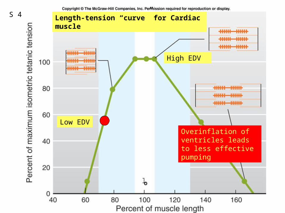
Fig. 09.21
Low EDV
High EDV
Length-tension “curve” for Cardiac muscle
Overinflation of ventricles leads to less effective pumping
S 4
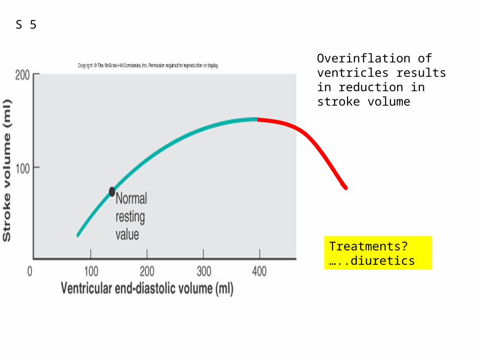
Overinflation of ventricles results in reduction in stroke volume
S 5
Treatments?…..diuretics
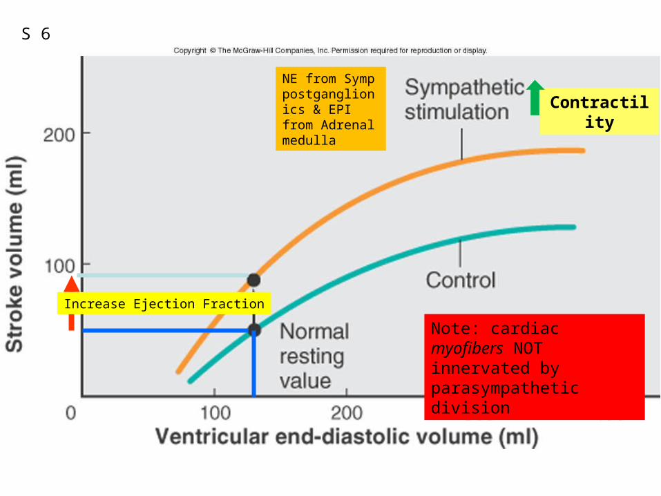
Contractility
NE from Symp postganglionics & EPI from Adrenal medulla
Note: cardiac myofibers NOT innervated by parasympathetic division
Increase Ejection Fraction
S 6
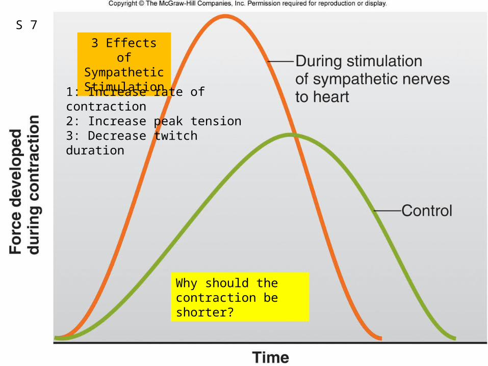
3 Effects of SympatheticStimulation
1: Increase rate of contraction2: Increase peak tension3: Decrease twitch duration
S 7
Why should the contraction be shorter?
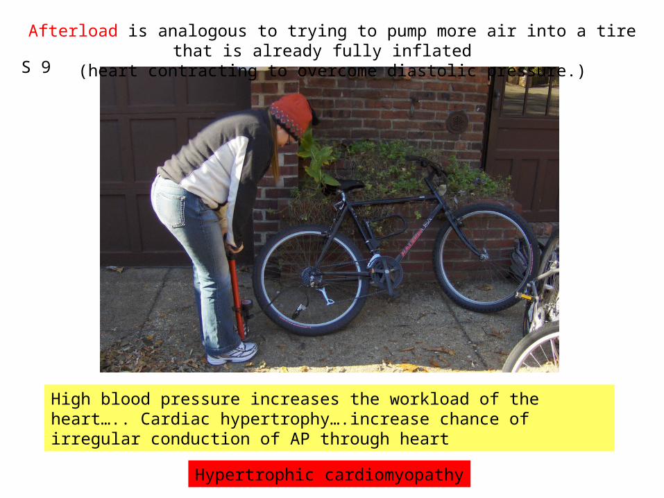
Afterload is analogous to trying to pump more air into a tire that is already fully inflated (heart contracting to overcome diastolic pressure.)
High blood pressure increases the workload of the heart….. Cardiac hypertrophy….increase chance of irregular conduction of AP through heart
S 9
Hypertrophic cardiomyopathy
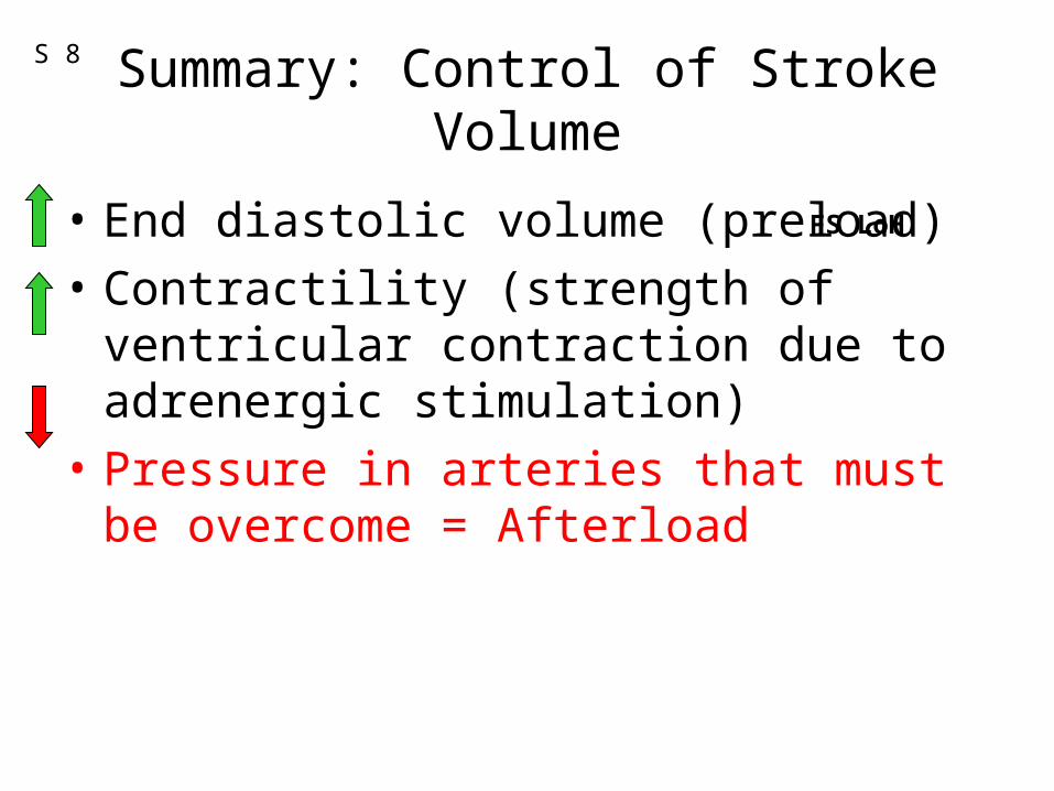
Summary: Control of Stroke Volume
• End diastolic volume (preload)
• Contractility (strength of ventricular contraction due to adrenergic stimulation)
• Pressure in arteries that must be overcome = Afterload
FS LoH
S 8
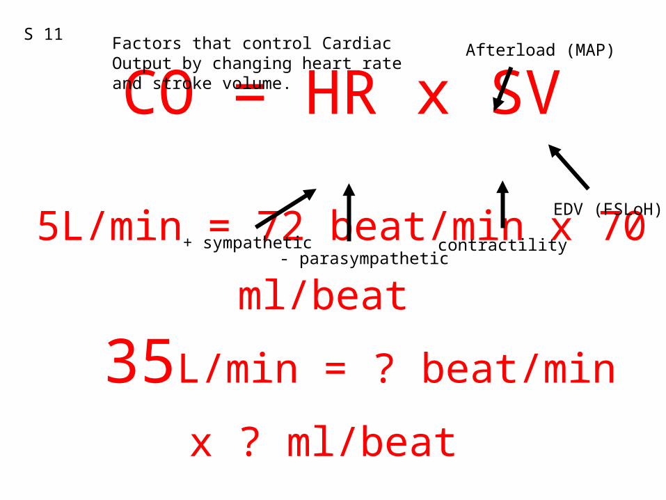
CO = HR x SV
5L/min = 72 beat/min x 70 ml/beat 35L/min = ? beat/min x ? ml/beat
S 11 Factors that control Cardiac Output by changing heart rate and stroke volume.
+ sympathetic- parasympathetic
EDV (FSLoH)
contractility
Afterload (MAP)
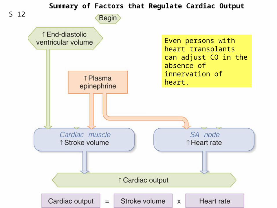
Fig. 12.28Even persons with heart transplants can adjust CO in the absence of innervation of heart.
Summary of Factors that Regulate Cardiac OutputS 12
S 13
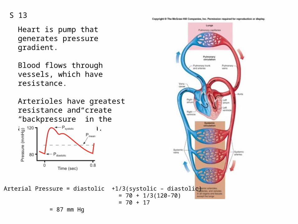
Heart is pump that generates pressure gradient.
Blood flows through vessels, which have resistance.
Arterioles have greatest resistance and create “backpressure” in the arteries and aorta.
Mean Arterial Pressure = diastolic +1/3(systolic – diastolic) = 70 + 1/3(120-70) = 70 + 17
= 87 mm Hg
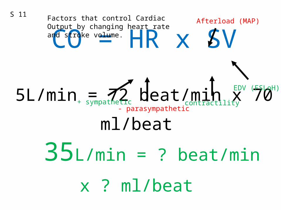
CO = HR x SV
5L/min = 72 beat/min x 70 ml/beat 35L/min = ? beat/min x ? ml/beat
S 11 Factors that control Cardiac Output by changing heart rate and stroke volume.
+ sympathetic- parasympathetic
EDV (FSLoH)
contractility
Afterload (MAP)
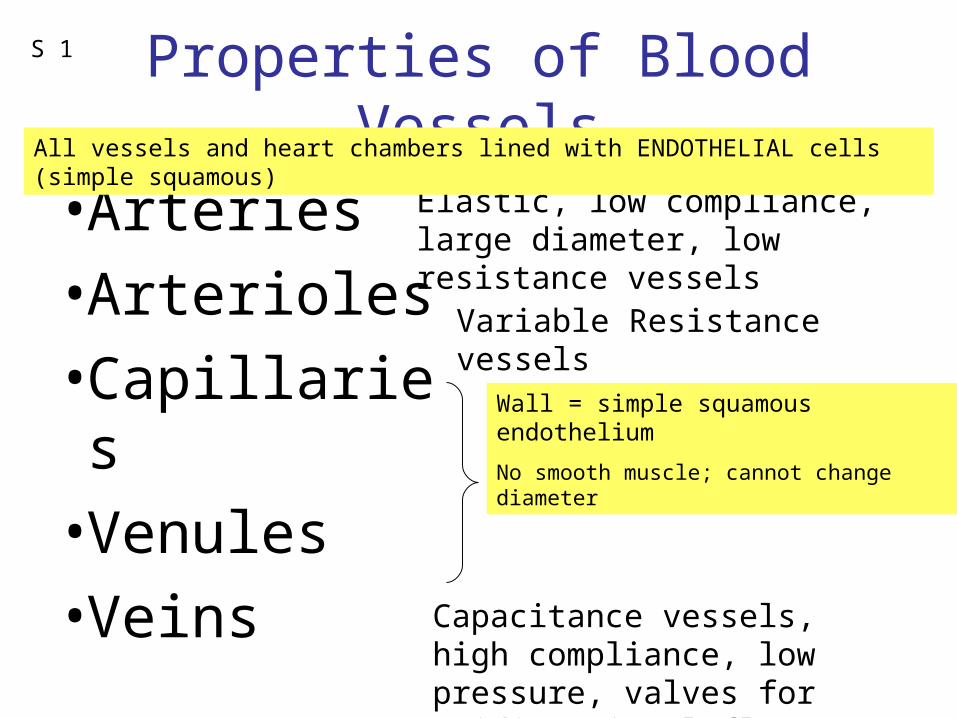
Properties of Blood Vessels
• Arteries
• Arterioles
• Capillaries
• Venules
• Veins
Elastic, low compliance, large diameter, low resistance vessels
Variable Resistance vessels
Exchange
Capacitance vessels, high compliance, low pressure, valves for unidirectional flow
All vessels and heart chambers lined with ENDOTHELIAL cells (simple squamous)
Wall = simple squamous endothelium
No smooth muscle; cannot change diameter
S 1
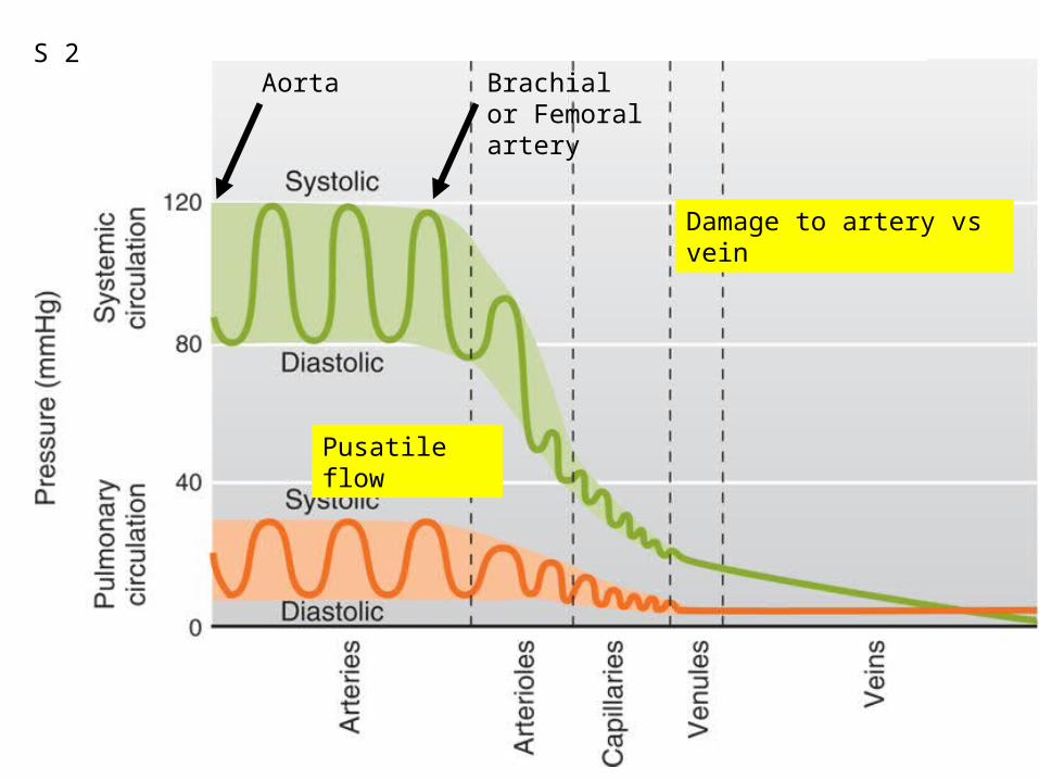
Fig. 12.29Aorta Brachial or Femoral artery
Pusatile flow
Damage to artery vs vein
S 2
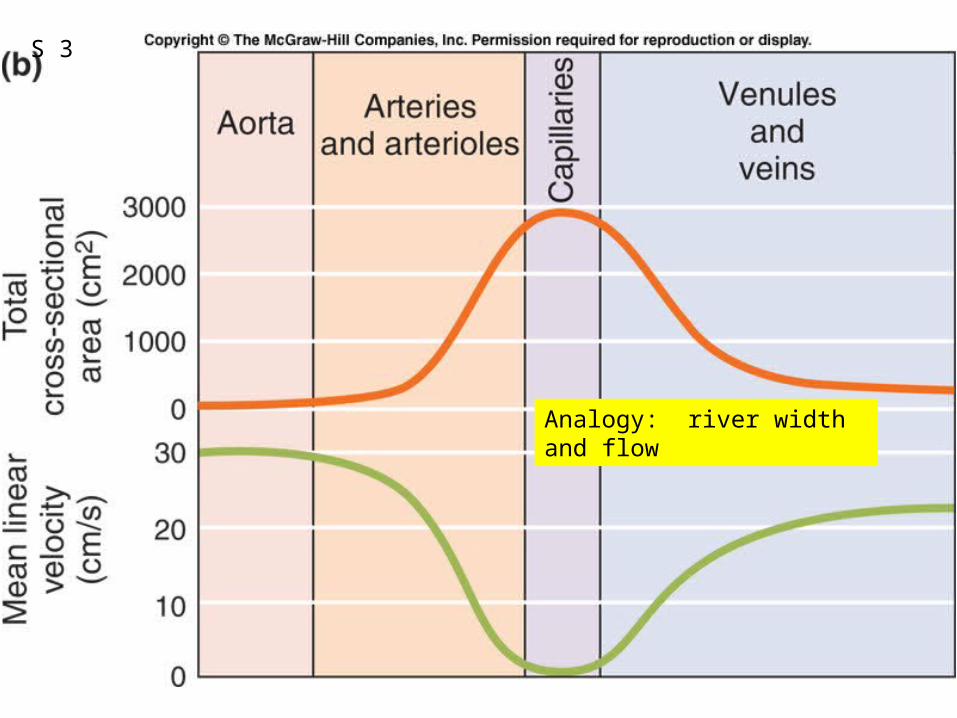
Fig. 12.39b
Analogy: river width and flow
S 3
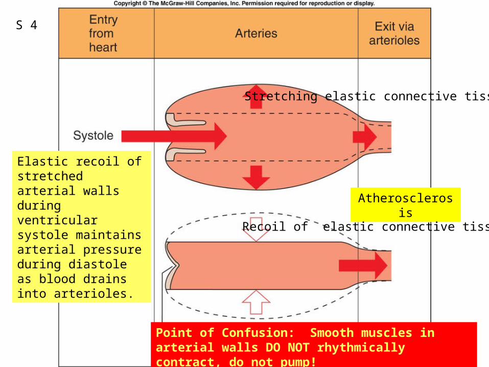
Fig. 12.30
Elastic recoil of stretched arterial walls during ventricular systole maintains arterial pressure during diastole as blood drains into arterioles.
Point of Confusion: Smooth muscles in arterial walls DO NOT rhythmically contract, do not pump!
Atherosclerosis
S 4
Stretching elastic connective tissue
Recoil of elastic connective tissue
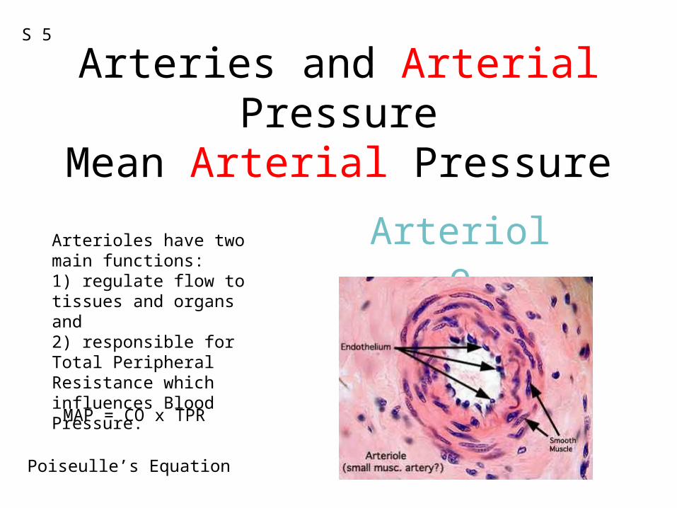
Arteries and Arterial PressureMean Arterial Pressure
ArterioleArterioles have two main functions: 1) regulate flow to tissues and organs and 2) responsible for Total Peripheral Resistance which influences Blood Pressure.
MAP = CO x TPR
Poiseulle’s Equation
S 5
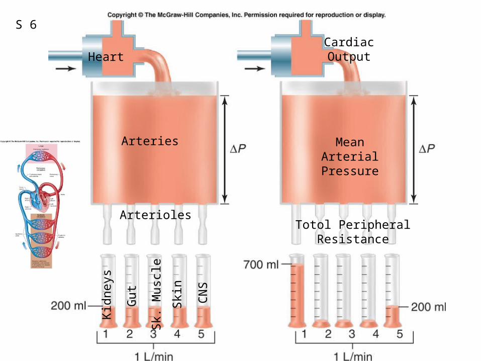
Fig. 12.50
S 6
Heart
Arteries
Arterioles
Kid
neys
Gut
Sk.
Mus
cle
Ski
n
CN
S
Totol Peripheral Resistance
MeanArterial
Pressure
CardiacOutput
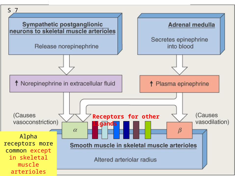
Alpha receptors more common
except in skeletal muscle arterioles which have more
B2 receptors
Receptors for other ligands
S 7
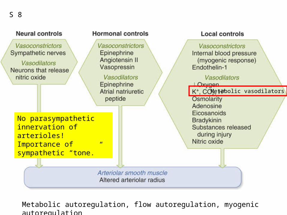
Fig. 12.36
Metabolic autoregulation, flow autoregulation, myogenic autoregulation
No parasympathetic innervation of arterioles!Importance of sympathetic “tone.”
Metabolic vasodilators
S 8
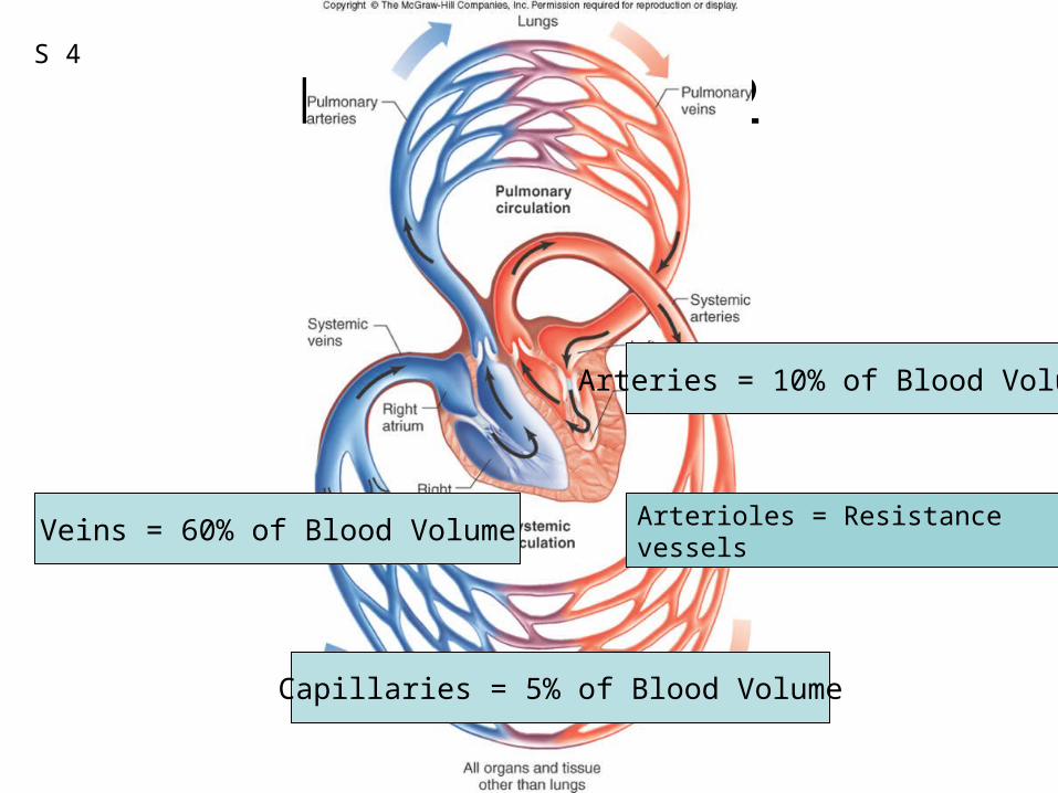
Figure 12.02
Capillaries = 5% of Blood Volume
Veins = 60% of Blood Volume
Arteries = 10% of Blood Volume
Arterioles = Resistance vessels
S 4
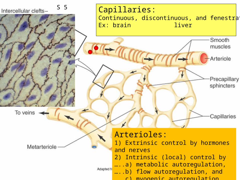
Figure 12.38Capillaries: Continuous, discontinuous, and fenestrated capillaries:Ex: brain liver endocrine glands
Generate vasodilators and vasoconstrictors
Arterioles:1) Extrinsic control by hormones and nerves2) Intrinsic (local) control by …..a) metabolic autoregulation, …..b) flow autoregulation, and …..c) myogenic autoregulation.
S 5
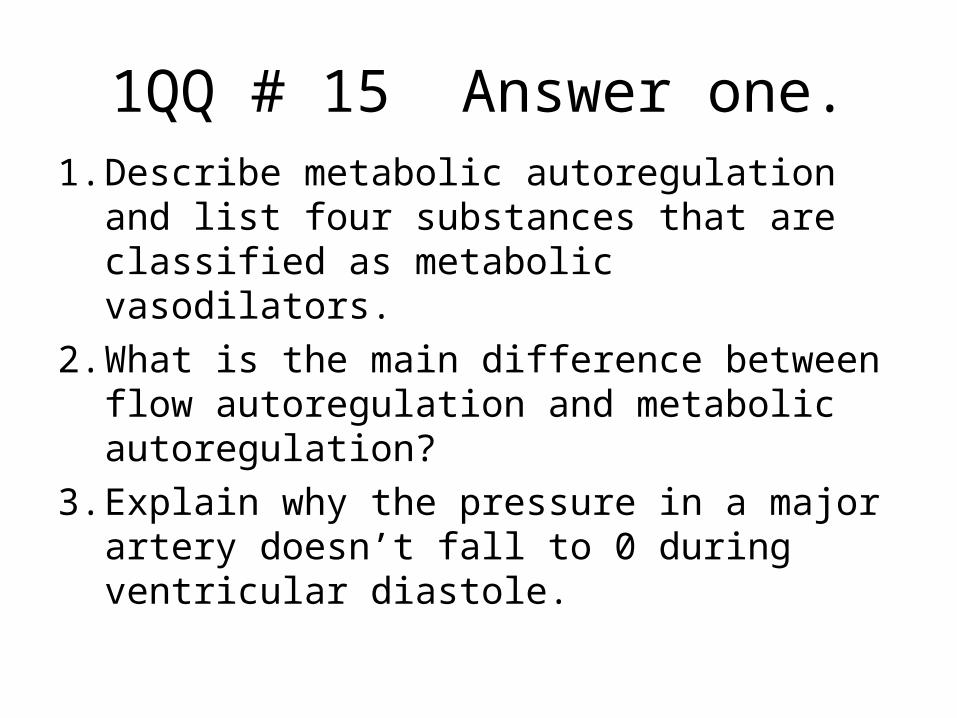
1QQ # 15 Answer one.1. Describe metabolic autoregulation and
list four substances that are classified as metabolic vasodilators.
2. What is the main difference between flow autoregulation and metabolic autoregulation?
3. Explain why the pressure in a major artery doesn’t fall to 0 during ventricular diastole.
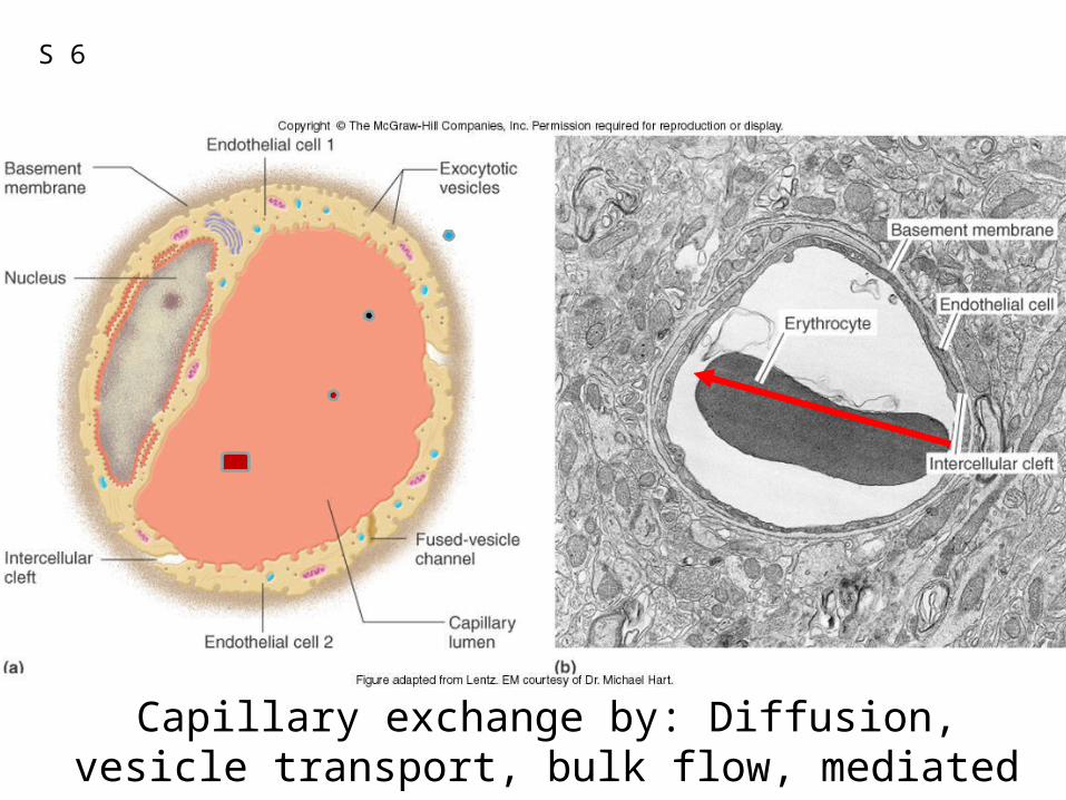
Capillary exchange by: Diffusion, vesicle transport, bulk flow, mediated transport
S 6
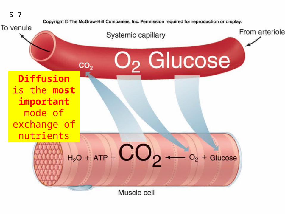
Fig. 12.40
Diffusion is the most important mode of
exchange of nutrients
S 7
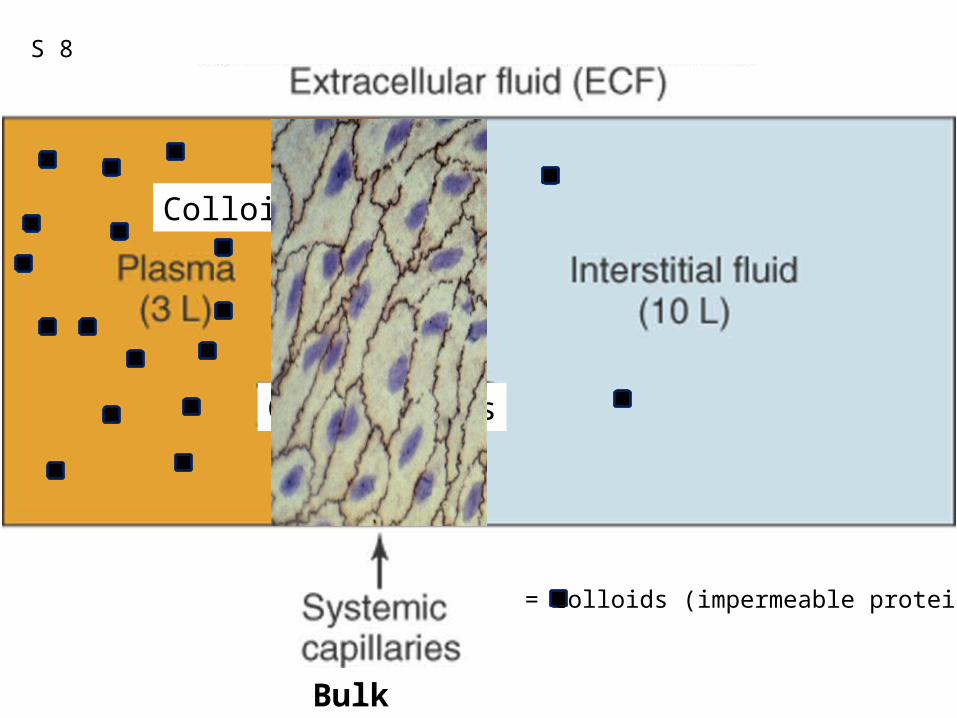
Figure 12.41
Crystalloids
Colloids
Bulk Flow
S 8
= colloids (impermeable proteins)
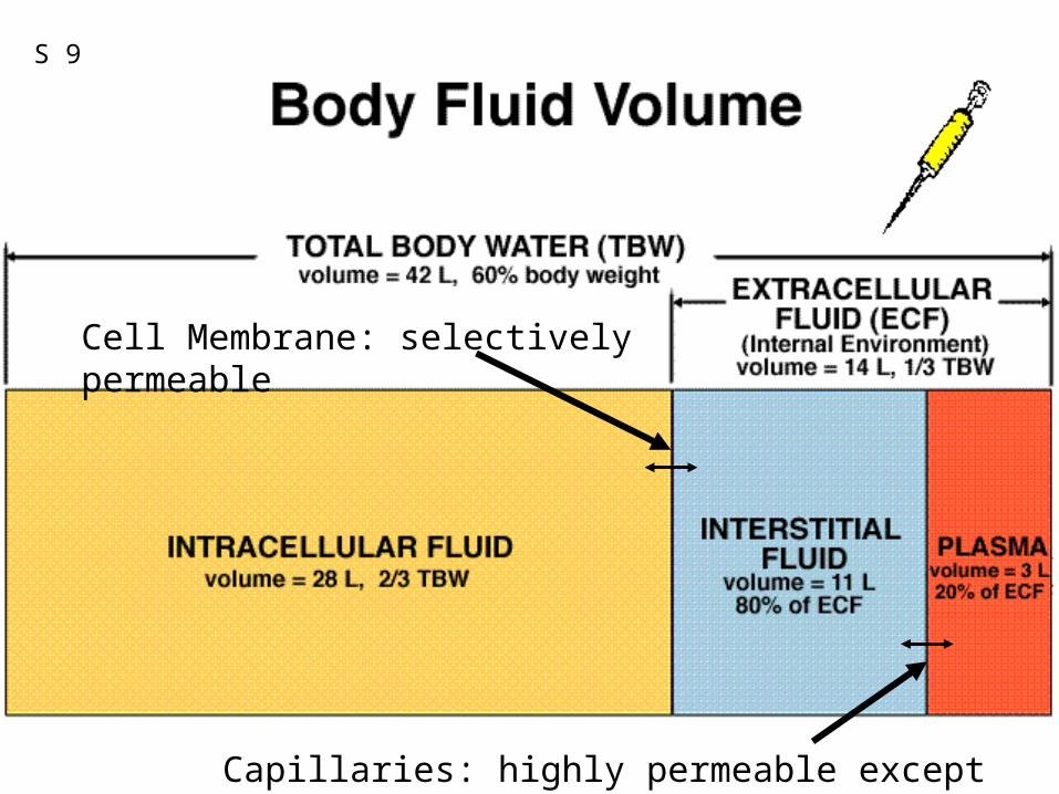
Cell Membrane: selectively permeable
Capillaries: highly permeable except to proteins
S 9
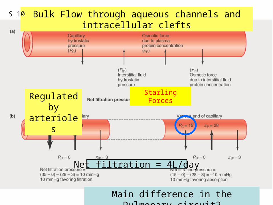
Figure 12.42
Main difference in the Pulmonary circuit?
Net filtration = 4L/day
Bulk Flow through aqueous channels and intracellular clefts
Regulated by arterioles
Starling Forces
S 10
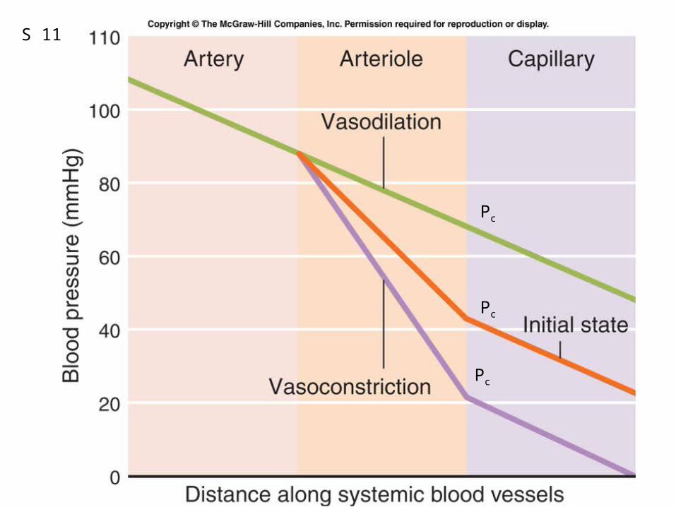
Fig. 12.43
Pc
Pc
Pc
S 11
Who Cares?Aunt Esther
Cancer of the liver;Failure of hepatocytes to produce plasma colloids
S 12
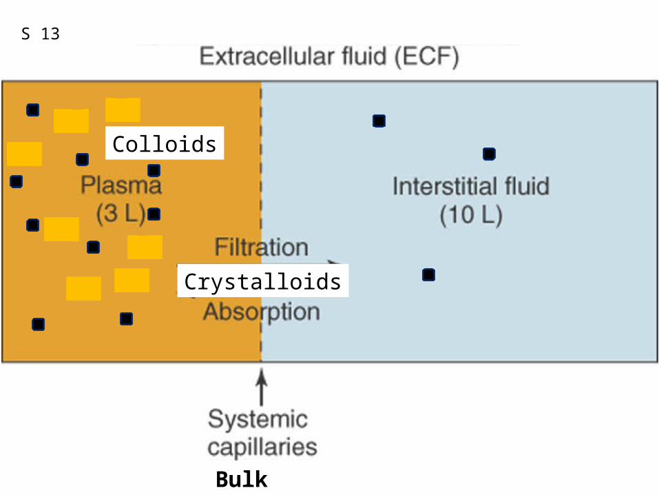
Figure 12.41
Crystalloids
Colloids
Bulk Flow
S 13
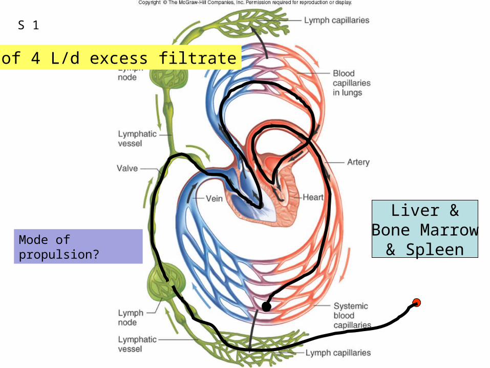
Figure 12.47
Liver &Bone Marrow
& Spleen
Fate of 4 L/d excess filtrate
S 1
Mode of propulsion?
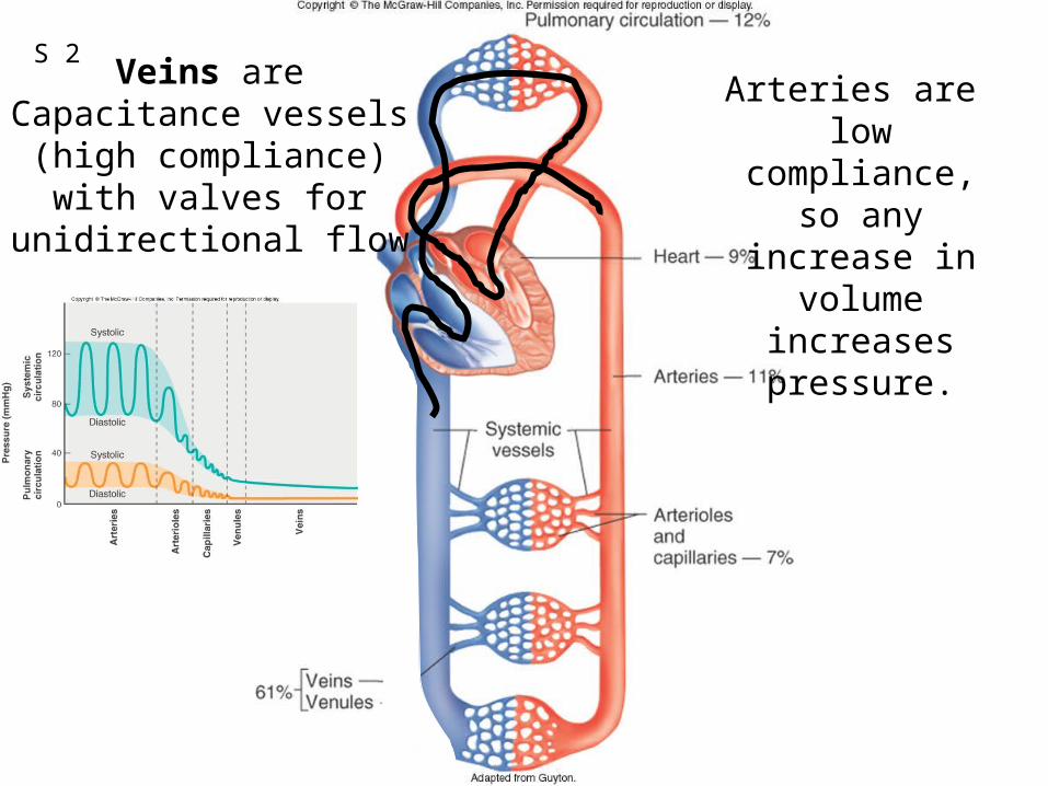
Figure 12.44Veins are
Capacitance vessels(high compliance)
with valves for unidirectional flow
Arteries are low compliance,
so any increase in volume increases
pressure.
S 2
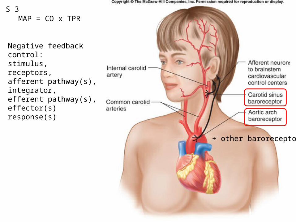
Fig. 12.53
MAP = CO x TPR
Negative feedback control:stimulus, receptors, afferent pathway(s), integrator, efferent pathway(s), effector(s)response(s)
S 3
+ other baroreceptors
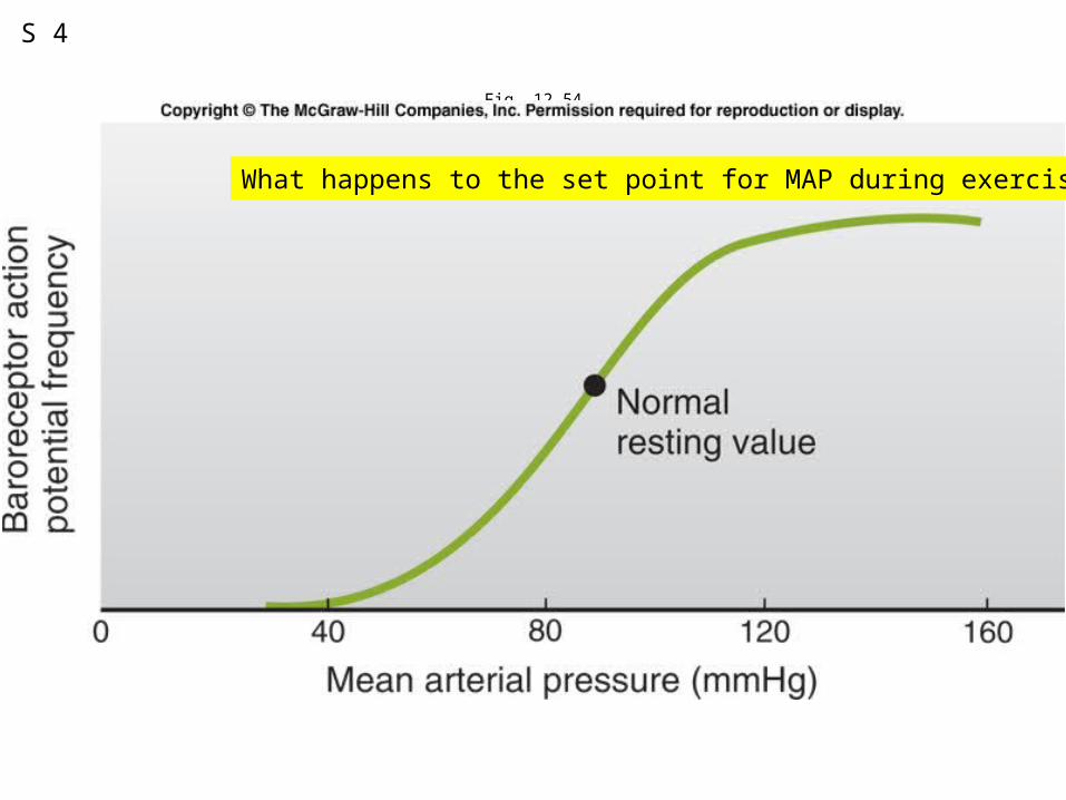
Fig. 12.54
S 4
What happens to the set point for MAP during exercise?
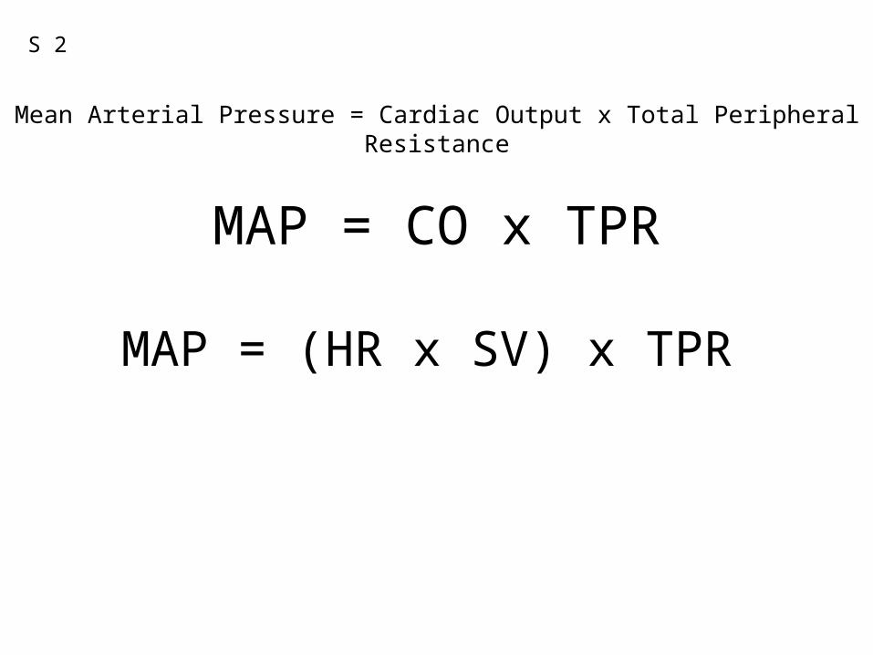
MAP = CO x TPR
Mean Arterial Pressure = Cardiac Output x Total Peripheral Resistance
MAP = (HR x SV) x TPR
S 2
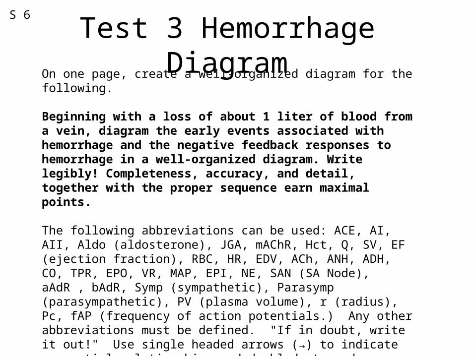
Test 3 Hemorrhage DiagramOn one page, create a well-organized diagram for the following.
Beginning with a loss of about 1 liter of blood from a vein, diagram the early events associated with hemorrhage and the negative feedback responses to hemorrhage in a well-organized diagram. Write legibly! Completeness, accuracy, and detail, together with the proper sequence earn maximal points.
The following abbreviations can be used: ACE, AI, AII, Aldo (aldosterone), JGA, mAChR, Hct, Q, SV, EF (ejection fraction), RBC, HR, EDV, ACh, ANH, ADH, CO, TPR, EPO, VR, MAP, EPI, NE, SAN (SA Node), aAdR , bAdR, Symp (sympathetic), Parasymp (parasympathetic), PV (plasma volume), r (radius), Pc, fAP (frequency of action potentials.) Any other abbreviations must be defined. "If in doubt, write it out!" Use single headed arrows (→) to indicate sequential relationships and doubled-stemmed arrows to indicate increases or decreases.
S 6