Src Family Tyrosine Kinase Signaling in Mouse and...
175
Src Family Tyrosine Kinase Signaling in Mouse and Human Embryonic Stem Cells by Xiong Zhang B.E., Xi’an Jiaotong University, 2007 Submitted to the Graduate Faculty of School of Medicine in partial fulfillment of the requirements for the degree of Doctor of Philosophy University of Pittsburgh 2013
Transcript of Src Family Tyrosine Kinase Signaling in Mouse and...

Src Family Tyrosine Kinase Signaling in Mouse and Human Embryonic Stem Cells
by
Xiong Zhang
B.E., Xi’an Jiaotong University, 2007
Submitted to the Graduate Faculty of
School of Medicine in partial fulfillment
of the requirements for the degree of
Doctor of Philosophy
University of Pittsburgh
2013

ii
UNIVERSITY OF PITTSBURGH
School of Medicine
This thesis was presented
by
Xiong Zhang
It was defended on
June 11th, 2013
and approved by
Thomas E. Smithgall, Ph.D., Major Advisor, Microbiology and Molecular Genetics
J. Richard Chaillet, Ph.D., Thesis Committee Chair, Microbiology and Mocular Genetics
Neil Hukriede, Ph.D., Developmental Biology
Gerald P. Schatten, Ph.D., Obstetrics, Gynecology and Reproductive Sciences
Alan Wells, M.D., D.M.Sc., Pathology

iii
Copyright © by Xiong Zhang
2013
iv
Embryonic stem (ES) cells are derived from the inner cell mass of the blastocyst stage embryo
and are characterized by self-renewal and pluripotency. Previous work has implicated the Src
family of protein-tyrosine kinases (SFKs) in the self-renewal and differentiation of mouse ES
(mES) cells. These kinases display dynamic expression and activity changes during ES cell
differentiation, suggesting distinct functions in the control of developmental fate. To test the
hypothesis that c-Src and its closest phylogenetic relative, c-Yes, act in biological opposition to
one another, I first showed that enforced expression of active c-Yes blocked ES cell
differentiation to embryoid bodies by maintaining pluripotency gene expression. To determine
the interplay of c-Src and c-Yes in mES cell fate determination, I employed a chemical genetics
approach to generate c-Src and c-Yes mutants that are resistant to A-419259, a potent
pyrrolopyrimidine inhibitor of the Src kinase family. This method allowed us to investigate
individual kinase function in the presence of A-419259. I found that c-Src activity alone induces
mES cell differentiation to the ectoderm and endoderm, while c-Yes inhibits this process. These
studies show that even closely related kinases such as c-Src and c-Yes have unique and opposing
functions in the same cell type.
While Src kinase signaling has been investigated in mES cells, the role of this kinase
family in human ES (hES) cells is largely unknown. Using quantitative real-time RT-PCR, I
determined the relative expression profile of individual SFK members in undifferentiated hES
Src Family Tyrosine Kinase Signaling in Mouse and Human Embryonic Stem Cells
Xiong Zhang
University of Pittsburgh, 2013

v
cells vs. embryoid bodies derived from them. Like mES cells, hES cells express multiple SFK
members with dynamic transcription changes during EB differentiation, indicating that
individual members may play non-redundant roles. To assess the role of SFK activity in hES
cells, I treated hES cell cultures with SFK inhibitors. SFK inhibition maintained hES cell colony
morphology and expression of the pluripotency marker Tra-1-60 in differentiation medium.
These observations support a role for Src family kinase signaling in the regulation of hES fate,
and suggest that some parallels may exist in mouse and human ES cells for this intracellular
signaling network.
vi
TABLE OF CONTENTS
ACKNOWLEDGEMENT ....................................................................................................... XIV
1.0 INTRODUCTION ........................................................................................................ 1
1.1 EMBRYONIC STEM CELLS............................................................................ 1
1.1.1 Mouse embryonic stem cells ........................................................................... 2
1.1.1.1 Teratocarcinoma and embryonic carcinoma cells ............................. 2
1.1.1.2 Derivation of embryonic stem cells...................................................... 3
1.1.1.3 ES cell culture conditions ..................................................................... 5
1.1.1.4 ES cell properties: self-renewal and pluripotency ............................. 6
1.1.2 Signaling networks regulating mES cell self-renewal and differentiation . 8
1.1.2.1 Cytokine and growth factor signaling pathways regulating self-
renewal 9
1.1.2.2 Transcription factor networks regulating pluripotency.................. 14
1.1.2.3 Signaling and transcription factors in early development and ES
cell differentiation .............................................................................................. 20
1.1.3 Human Embryonic Stem Cells ..................................................................... 25
1.1.3.1 Human ES cell derivation and culture conditions ........................... 25
1.1.3.2 Human ES cell renewal and pluripotency, EB formation and
teratoma formation ............................................................................................ 27
vii
1.1.3.3 Growth factors and signaling pathways ........................................... 29
1.1.3.4 Core transcription factor regulatory network ................................. 31
1.1.3.5 New different states of pluripotent stem cells: iPS cells, mEpiSCs
and naive human ES cells .................................................................................. 32
1.2 SRC FAMILY NON-RECEPTOR TYROSINE KINASES .......................... 35
1.2.1 Src family kinase structure ........................................................................... 38
1.2.1.1 N-terminal region ................................................................................ 39
1.2.1.2 Unique domain .................................................................................... 40
1.2.1.3 SH3 domain ......................................................................................... 41
1.2.1.4 SH2 domain ......................................................................................... 42
1.2.1.5 Tyrosine kinase domain ...................................................................... 43
1.2.2 SFK regulation ............................................................................................... 45
1.2.2.1 Intramolecular regulation .................................................................. 45
1.2.2.2 Regulation by phosphorylation and dephosphorylation ................. 46
1.2.2.3 Regulation by engagement with binding partners ........................... 48
1.2.3 SFK functions ................................................................................................. 49
1.2.3.1 Phylogenetic relationship of Src family members ............................ 49
1.2.3.2 SFK knockout phenotype and implication for function .................. 51
1.2.4 SFK signaling ................................................................................................. 53
1.2.4.1 Signaling with receptor tyrosine kinases .......................................... 54
1.2.4.2 Signaling with integrin and focal adhesion kinase ........................... 56
1.3 SFK SIGNALING IN MURINE ES CELLS................................................... 57
1.3.1 SFK expression and function in mES cells and EBs................................... 58

viii
1.3.2 Chemical genetics approaches to study the individual functions of SFK
members ...................................................................................................................... 60
1.3.3 A role for Src family kinase c-Yes in ES cell regulation ............................ 62
1.4 HYPOTHESIS AND SPECIFIC AIMS........................................................... 63
1.4.1 Hypothesis ...................................................................................................... 63
1.4.2 Specific Aims .................................................................................................. 64
1.4.2.1 Aim 1: Investigate the contribution of c-Yes to the growth and self-
renewal of murine ES cells and test the opposing roles of c-Src and c-Yes in
ES cell differentiation. ....................................................................................... 64
1.4.2.2 Aim 2: Study SFK signaling in human ES cell self-renewal and
differentiation. .................................................................................................... 66
2.0 THE C-YES TYROSINE KINASE IS A POTENT SUPPRESSOR OF ES CELL
DIFFERENTIATION AND ANTAGONIZES THE ACTION OF ITS CLOSEST
PHYLOGENETIC RELATIVE, C-SRC .................................................................................. 67
2.1 ABSTRACT........................................................................................................ 67
2.2 INTRODUCTION ............................................................................................. 68
2.3 RESULTS ........................................................................................................... 71
2.3.1 Downregulation of c-Yes kinase activity during differentiation of ES cells
to EBs 71
2.3.2 Validation of c-Yes retroviral expression constructs in Rat2 cells ........... 74
2.3.3 Low-level retroviral expression of c-Yes in ES cells does not affect
undifferentiated colony morphology or marker expression................................... 77
2.3.4 ES cells expressing active c-Yes fail to form EBs. ...................................... 79

ix
2.3.5 ES cells expressing active c-Yes kinases express both pluripotency and
differentiation markers during EB formation. ........................................................ 81
2.3.6 Design of c-Yes gatekeeper mutants resistant to the broad spectrum Src-
family kinase inhibitor, A-419259. ............................................................................ 83
2.3.7 Stable expression of inhibitor-resistant c-Yes (Yes-IR) in ES cells does not
affect self-renewal marker expression. ..................................................................... 86
2.3.8 Differentiation of ES cells driven by c-Src is antagonized by c-Yes. ........ 88
2.4 DISCUSSION ..................................................................................................... 93
2.5 MATERIALS AND METHODS ...................................................................... 97
2.5.1 Cell culture ..................................................................................................... 97
2.5.2 Retroviral transduction of mES cells and Rat2 fibroblasts ....................... 98
2.5.3 Embryoid body formation ............................................................................ 99
2.5.4 RT-PCR analysis............................................................................................ 99
2.5.5 Immunoprecipitation and immunoblotting............................................... 100
2.5.6 Rat2 cell soft-agar assay .............................................................................. 101
3.0 SRC FAMIY TYROSINE KINASE SIGNALING IS IMPORTANT FOR
HUMAN EMBRYONIC STEM CELL DIFFERENTIATION ............................................ 102
3.1 ABSTRACT...................................................................................................... 102
3.2 INTRODUCTION ........................................................................................... 103
3.3 RESULTS ......................................................................................................... 106
3.3.1 Human ES cells express multiple Src family kinases ............................... 106
3.3.2 Src family kinase expression during human embryoid body
differentiation ........................................................................................................... 108

x
3.3.3 Src family kinase inhibition blocks hES cell differentiation.................... 113
3.3.4 Human ES cells maintain Tra-1-60 expression following SFK inhibition
118
3.4 DISCUSSION ................................................................................................... 120
3.5 MATERIALS AND METHODS .................................................................... 122
3.5.1 Cell culture ................................................................................................... 122
3.5.2 RT-PCR ........................................................................................................ 123
3.5.3 Protein Blots ................................................................................................. 123
3.5.4 Immunocytochemistry and fluorescence microscopy .............................. 124
4.0 OVERALL DISCUSSION ...................................................................................... 125
4.1 SUMMARY OF FINDINGS AND SIGNIFICANCE ................................... 125
4.1.1 c-Yes is a potent anti-differentiation signal and acts in direct opposition to
c-Src 126
4.1.2 SFK signaling is important for human ES cell differentiation ................ 131
4.2 FUTURE DIRECTIONS................................................................................. 134
4.2.1 Identify signaling pathways downstream of c-Src and c-Yes that account
for the different stem cell fates associated with SFK signaling ............................ 134
4.2.2 Further study of SFK signaling in human ES cells .................................. 136
4.3 CLOSING REMARKS ................................................................................... 139

xi
LIST OF TABLES
Table 1. Functional assays to assess developmental potential of ES cells ..................................... 8
Table 2. Comparison of naïve and primed pluripotent cell states ................................................. 34
Table 3. Summary of expression patterns and mutant phenotypes of SFK members .................. 50

xii
LIST OF FIGURES
Figure 1. Early embryo development stages and ES cell derivation .............................................. 5
Figure 2. Key signaling pathways govern ES cell renewal: LIF-Jak-STAT3, Mek-Erk, BMP-
SMAD-ID and Wnt-GSK-β-Catenin pathways ............................................................................ 10
Figure 3. Core transcription factor network for pluripotency and interconnection with extrinsic
stimuli ........................................................................................................................................... 15
Figure 4. Embryo development lineages and ES cell differentiation markers ............................. 21
Figure 5. Src family tyrosine kinase domain organization and structure ..................................... 38
Figure 6. SFK signaling pathways and function ........................................................................... 55
Figure 7. Model for the regulation of mES cell fate by Src family kinases. ................................ 60
Figure 8. Downregulation of c-Yes kinase activity during EB formation. ................................... 73
Figure 9. Validation of c-Yes retroviral expression constructs in Rat2 cells. .............................. 76
Figure 10. Low-level retroviral expression of c-Yes in mES cells does not affect colony
morphology or pluripotency marker expression. .......................................................................... 78
Figure 11. Mouse ES cells expressing active c-Yes kinases fail to form EBs.............................. 80
Figure 12. EBs formed from mES cells expressing active c-Yes Kinases retain pluripotency
marker expression. ........................................................................................................................ 82
Figure 13. Validation of inhibitor-resistant mutants of c-Yes in Rat-2 fibroblasts. ..................... 85

xiii
Figure 14. Stable expression of inhibitor-resistant c-Yes in ES cells. .......................................... 87
Figure 15. Co-expression of inhibitor-resistant mutants of c-Yes and c-Src in ES cells does not
affect self-renewal or differentiation marker expression in the absence of A-419259 treatment. 91
Figure 16. Differentiation of mES cells driven by c-Src is antagonized by c-Yes. ...................... 93
Figure 17. Src family kinase expression in hES Cells ............................................................... 107
Figure 18. Src family kinase expression during EB formation from H1 Cells. .......................... 110
Figure 19. Src family kinase expression during EB formation from H9 cells. ........................... 111
Figure 20. Src family kinase expression during EB formation from H7 cells. ........................... 112
Figure 21. Inhibition of SFK activity with SKI-1 and PP2 in hES cells. ................................... 115
Figure 22. Global SFK inhibition with A-419259 blocks hES cell differentiation. ................... 117
Figure 23. Both H1 and H7 hES cells retain Tra-1-60 expression following SFK inhibition. ... 119

xiv
ACKNOWLEDGEMENT
Looking back over my experience in graduate school, although there have been ups and downs,
joys and sorrows; it has been a gorgeous journey. I have discovered that life is never a straight
line, yet the beauty lies in the exploration out of the twists and turns. This journey would not
have happened without those who guided me, accompanied me and supported me. I would like
to offer my appreciation.
First, I acknowledge my utmost gratitude to my mentor, Dr. Thomas Smithgall, for his
guidance, inspiration and support. Dr. Smithgall is an outstanding scientist, a thoughtful mentor
and a generous person. No matter how busy he was, he always devoted time to guide me and
generously offered new ideas when I faced challenges and seemly unsolvable problems. I was
often motivated by his contagious enthusiasm and passion for science. Not only did Dr.
Smithgall painstakingly edit and improve my writing, he also supported my exploration for my
career goals according to my own interests.
I owe much gratitude to my dissertation committee members for constructive advice and
constant support. I would like to thank Dr. Richard Chaillet for chairing my committee and his
generosity. I would like to thank Dr. Neil Hukriede, for the commitment, sincere concern and
assistance. I would like to thank Dr. Alan Wells, for his career suggestions and guidance. My
deep appreciation goes to Dr. Gerald Schatten, for the generous support of the human ES cell

xv
project; for allowing me to get hands-on experience with hES cell culture; and inviting me to
participate in the stem cell course at Woods Hole.
I would like to also thank my collaborators from the Schatten group: Carrie Redinger, Dr.
Chas Easley, Stacie Oliver, David Mcfarland, and Jody Mich-Basso. Human ES cell culture is
difficult, yet your help and support made it much better and enjoyable!
Many warm thanks to the current and previous members of the Smithgall laboratory: Dr.
Tony Meyn, Dr. Linda O’Reilly, Dr. Lori Emert-Sedlak, Dr. Jerrod Poe, Dr. Sabine Hellwig, Dr.
Sherry Shu, Dr. Shoghag Panjarian, Dr. Teodora Pene-Dumitrescu, Dr. Jonathan Shaffer, Dr.
Purushottam Narute, Dr. John Jeff Alvarado, Dr. Patty George, Jamie Moroco, Mark Weir,
Prerna Grover, Sreya Tarafdar, Kathleen Makielski, Terri Robinson and Nageswara Alla. It has
been a great pleasure to work with you all. Thank you for all the great suggestions, support and
discussions. Special thanks to Tony and Linda for the guidance, help and suggestions on the stem
cell project.
1
1.0 INTRODUCTION
1.1 EMBRYONIC STEM CELLS
Embryonic stem (ES) cells are pluripotent stem cells derived from the inner cell mass of
blastocyst stage embryo (Evans and Kaufman, 1981; Martin, 1981; Thomson et al., 1998). The
derivation of mouse ES (mES) cells is based on early research on teratomas and teratoma stem
cells to establish the culture conditions and functional assays (Evans, 2011). ES cells are
characterized by two properties: self-renewal, the ability to grow indefinitely without
differentiation, and pluripotency, the developmental potential to generate all cell types from the
embryo and adult body (Nichols and Smith, 2012). In culture, pluripotency is maintained by
extrinsic growth factors, and expression of transcription factors. The transcription factors form
an intricate network to control gene expression and maintain pluripotency. In conditions without
renewal factors, ES cells differentiate, recapitulating the differentiation programs of the
developing embryo (Murry and Keller, 2008). Human ES(hES)cells were derived 17 years
after the establishment of mES cells. Although hES cells and mES cells have different culture
conditions and signaling features, they express the same core transcription factors to maintain
pluripotency (Thomson et al., 1998). Recent establishment of epiblast stem cells indicates that
hES cells and mES cells may represent different states of pluripotent stem cells.
2
1.1.1 Mouse embryonic stem cells
Mouse embryonic stem (mES) cells are self-renewing, pluripotent cells derived from blastocyst
stage mouse embryo (Evans and Kaufman, 1981; Martin, 1981). Early research on teratoma and
embryonic carcinoma (EC) cells defined the feeder cell conditions required to maintain ES cells
in culture, cell-surface markers for self-renewal, and functional assays for pluripotency. Later
work established that mES cell pluripotency requires growth factors including leukemia
inhibitory factor (LIF) and bone morphogenetic proteins (BMPs) (Evans, 2011). ES cells have
the remarkable ability to contribute to embryo development and the germline, which allows
genetic manipulation and production of transgenic animals. These features make ES cells a
powerful tool to study development, genetics and disease (Evans, 2011).
1.1.1.1 Teratocarcinoma and embryonic carcinoma cells
The discovery and establishment of mES cells were largely based on the early research on
teratocarinomas and the pluripotent cells they carry (Chambers and Smith, 2004; Solter, 2006).
Teratoma, a rare tumor in humans, is composed of a mixture of cells from three different germ
layers. The mouse strain 129 was found to have a high incidence of developing spontaneous
teratomas. Teratocarcinoma, the malignant form of teratoma, contains undifferentiated stem cells
that can form secondary tumors after transplantation into another host. These cells, termed
embryonic carcinoma (EC) cells, were subsequently established for cell culture. In EC cell line
derivation, Evans made the discovery that irradiated chick fibroblasts can be used as feeder
layers to support undifferentiated growth of EC cells (Evans, 1972). In suspension culture
without the feeder layer, EC cells differentiate to embryoid bodies, a ball-like structure

3
comprised of cell types derived from all three germ layers, similar to the early stages of
embryonic development in vivo (Martin and Evans, 1975). In addition, alkaline phosphatase
(Berstine et al., 1973) and stage-specific embryonic antigen 1(SSEA-1) (Solter and Knowles,
1978) were identified as the first biological markers for EC cells. When injected into developing
blastocysts, rare EC cells can contribute to chimera mouse formation, including germ line
transmission (Stewart and Mintz, 1981; Stewart and Mintz, 1982). The studies highlighted
above, including the optimized culture conditions for EC cells with feeder fibroblast, in vitro EB
formation assay, cell surface markers, and blastocyst injection of EC cells to assess
developmental potential, laid a strong foundation for the discovery and study of ES cells.
1.1.1.2 Derivation of embryonic stem cells
In addition to spontaneous generation, teratomas can also be formed by transplanting pre-
gastrulation stage embryonic cells into the testis or kidney capsules, which suggests that there
might be a stem cell type that is equivalent to EC cells in the developing embryo. Evans &
Kaufman and Martin independently derived pluripotent embryonic stem cells from the blastocyst
stage of embryos (Evans and Kaufman, 1981; Martin, 1981). Evans and Kaufman induced
embryo arrest (diapause) in pre-implantation stage embryos using ovariectomy and hormone
treatment, and derived ES cells from explants of the intact diapause embryos with serum-
containing medium and division-incompetent STO fibroblast feeders (Stromal cells derived from
Sandoz inbred mouse (S), that are engineered to be resistant to 6-thioguanine(T) and
ouabain(O)). Martin derived ES cell lines from the isolated inner cell mass (ICM) of pre-
implantation embryos with EC cell-conditioned medium.
4
Traditionally, ES cells are derived from out-growth of intact blastocysts or isolated entire
ICMs. ES cells have also been established from single, dissociated epiblast cells (Brook and
Gardner, 1997), which convincingly demonstrated that ESCs are of epiblast origin at embryonic
day 4.5 (E4.5). Mammalian embryonic development begins at the zygote, which undergoes three
rounds of cleavage division to form the 8-cell embryo (E2.5; Figure 1). The blastomeres in the
embryo then undergo compaction, gain polarity and form the morula. A series of subsequent
divisions, accompanied by morphologic changes, result in a fluid-filled ball-like structure called
blastocyst (E3.5), with the inner cell mass, a group of apolar cells, enclosed by an outer layer of
polarized epithelial cells, trophectoderm. The segregation of trophectoderm and ICM is the
hallmark of blastocyst formation, with ICM cells expressing the transcription factor Oct4 and
trophectoderm cells expressing Cdx2. Subsequently, around E4.5, the ICM cells develop to form
the epiblast and primitive endoderm, an epithelium between the epiblast and the blastocyst cavity
(Stephenson et al., 2012). Mouse ES cells can be derived from E3.5 ICM cells, or E4.5 early
epiblast cells (Nichols and Smith, 2012).

5
8-cell~E2.5
Early blastocyst~E3.5
Late blastocyst~E4.5
Trophectoderm
Inner cell mass
Early epiblast
Primitive Endoderm
Figure 1. Early embryo development stages and ES cell derivation. The 8-cell stage embryo (left) undergoes
compaction, polarization and several rounds of division, to form the early blastocyst at around E 3.5. The early
blastocyst (middle) has trophectoderm, the outer polarized layer of epithelium, and the inner cell mass, a group of
apolar cells clustered inside. By E4.5, as the blastocyst further develops, the ICM is segregated into the epiblast and
primitive endoderm (right). The embryo is now ready to implant in the uterus. Adapted from (Stephenson et al.,
2012)
1.1.1.3 ES cell culture conditions
Similar to EC cells, ES cells can be maintained in serum-containing medium with co-culture of
feeder fibroblasts. STO mouse fibroblast cell-conditioned medium was shown to support EC cell
growth and maintenance of pluripotency without feeders (Koopman and Cotton, 1984). Also,
Buffalo rat liver (BRL) cell-conditioned medium can replace the fibroblast requirement for
maintenance of ES cells (Smith and Hooper, 1987). These studies indicate that feeder cells
produce a soluble factor to inhibit EC or ES cell differentiation. Subsequent research identified

6
the cytokine leukemia inhibitory factor (LIF) as the active component of conditioned medium
that maintains the developmental potential of ES cells (Smith et al., 1988; Williams et al., 1988).
In addition, bone morphogenetic proteins (BMPs), known anti-neuronal differentiation factors,
are able to replace serum to support ES cell derivation and maintenance in combination with LIF.
Thus, ES cell culture conditions have evolved from feeder and serum-containing medium, to
feeder-free, LIF and serum containing medium, then to feeder-free, serum-free medium with LIF
and BMPs.
More recently, the Smith group showed that ES cells can be maintained in a chemically-
defined medium with two inhibitors—the MEK inhibitor PD0325901 and GSK3β inhibitor
CHIR99021 (Ying et al., 2008; Silva et al., 2008). This so called ‘2i’ medium, combined with
LIF enabled the derivation of ES cell lines from traditionally refractory mouse strains like
nonobese diabetic (NOD) mice (Hanna et al., 2009), or non-permissive species like the rat
(Buehr et al., 2008).
1.1.1.4 ES cell properties: self-renewal and pluripotency
ES cells have two hallmark properties: Self-renewal and pluripotency. Self-renewal is the ability
of stem cells to produce at least one daughter cell that has the same differentiation capacity as the
parental cells. For ES cells, cell division can be symmetric, producing two daughter cells
identical to the parental cell; or asymmetric, generating one stem cell and one differentiated cell
(Smith, 2001). Pluripotency is the ability to differentiate to cell types from all three germ layers.
In vitro, ES cells can be maintained continuously without differentiation. Self-renewal is
maintained with extracellular signals such as LIF and serum or BMPs, which enforces expression
of core pluripotency transcription factors such as Oct4 and Nanog (discussed in the next section).
7
ES cells have a shortened cell cycle, with a short G1 phase and progression independent of the
retinoblastoma (RB) protein (Burdon et al., 2002). Self-renewal can also be assessed by
clonogenicity assay and expression of renewal markers such as alkaline phosphatase, SSEA-1
and transcription factors such as Oct4 and Nanog (Evans, 2011). However, ES cells are strictly a
cell culture phenomenon: In vivo, the ICM cells only divide symmetrically for a short period of
time and quickly differentiate to more developed lineages (Nichols and Smith, 2012).
Pluripotency can be assessed using a number of different functional criteria, with
increasing stringency, as summarized in Table 1 (Jaenisch and Young, 2008). The most
commonly used and least stringent test of pluripotency is embryoid body formation. Absent of
support from feeder cells or LIF, ES cells are grown in suspension culture in liquid or methyl
cellulose containing medium, where each single ES cell is able to divide to generate a ball-like
structure containing all three germ layers, recapitulating the developmental events of the early
embryo (Keller, 2005). The hallmark of pluripotency of ES cells is their ability to be
incorporated into normal development when injected into developing blastocysts, producing
chimeric mice and being able to contribute to the germ line. Germ line competency is the gold
standard of pluripotency, enabling us to use gene-targeting to manipulate ES cells and create
transgenic animals for study of developmental processes and to model disease (Capecchi, 2005).
Tetraploid complementation is the most stringent test of pluripotency. In this approach, ES cells
are injected into 4n host blastocysts. If the ES cells are truly pluripotent, then all of the somatic
cells of the resulting embyo will be of ES cell origin since the host cells will only support the
extraembryonic lineages.

8
Table 1. Functional assays to assess developmental potential of ES cells
Assay Experimental Approach Limitations
In vitro
differentiation
Cultured cells are induced for differentiation via EB
formation, or attached differentiation and are assayed for
expression of specific linage markers.
No functional assay; multiple
markers is required; hard to
synchronize, heterogeneity
complicates results.
Teratoma
formation
Injection of ES cells into immuno-deficient mice to
induce tumor formation, demonstrating the potential to
generate differentiated cell types from all three germ
layers
Normal development of cells is
not tested.
Chimera
formation
Injection of ES cells into developing blastocyst, to
incorporate to the ICM and contribute to the normal
development, generating chimeric mouse
Non-autonomous defects in test
cells may be blinded by
compensation from the host
derived cells.
Germline
competency
The ability of test cells to generate functional germ cells,
and in the case of genetic manipulation, to generate
transgenic animal.
Does not exclude epigenetic
defects that might interfere with
development.
Tetraploid
complementa-
tion
Injection of test cells to tetraploid host embryo. Since
the 4n host can only contribute to extraembryonic
tissues, the embryo is exclusively derived from the test
cells.
Most Stringent test for
pluripotency; the ability to form
trophectoderm linages is not
tested.
Adapted from (Jaenisch and Young, 2008)
1.1.2 Signaling networks regulating mES cell self-renewal and differentiation
Mouse ES cell pluripotency and differentiation are governed by a complex network of signaling
pathways, which work in concert. Multiple intracellular kinase signaling pathways, including
LIF-Jak-Stat3, BMP-SMAD-ID, MEK/Erk, PI3K-Akt and Wnt-GSK-β-Catenin pathways, play a
dominant role in the regulation of ES cell fate (Liu et al., 2007). These extrinsic signals converge
in an intrinsic transcription factor network controled by Oct4, Sox2 and Nanog to maintain
pluripotency (Jaenisch and Young, 2008). In the absence of factors supporting pluripotency, ES
cells differentiate to specific lineages, recapitulating the developmental process governed by
morphogenesis related signals (Keller, 2005).

9
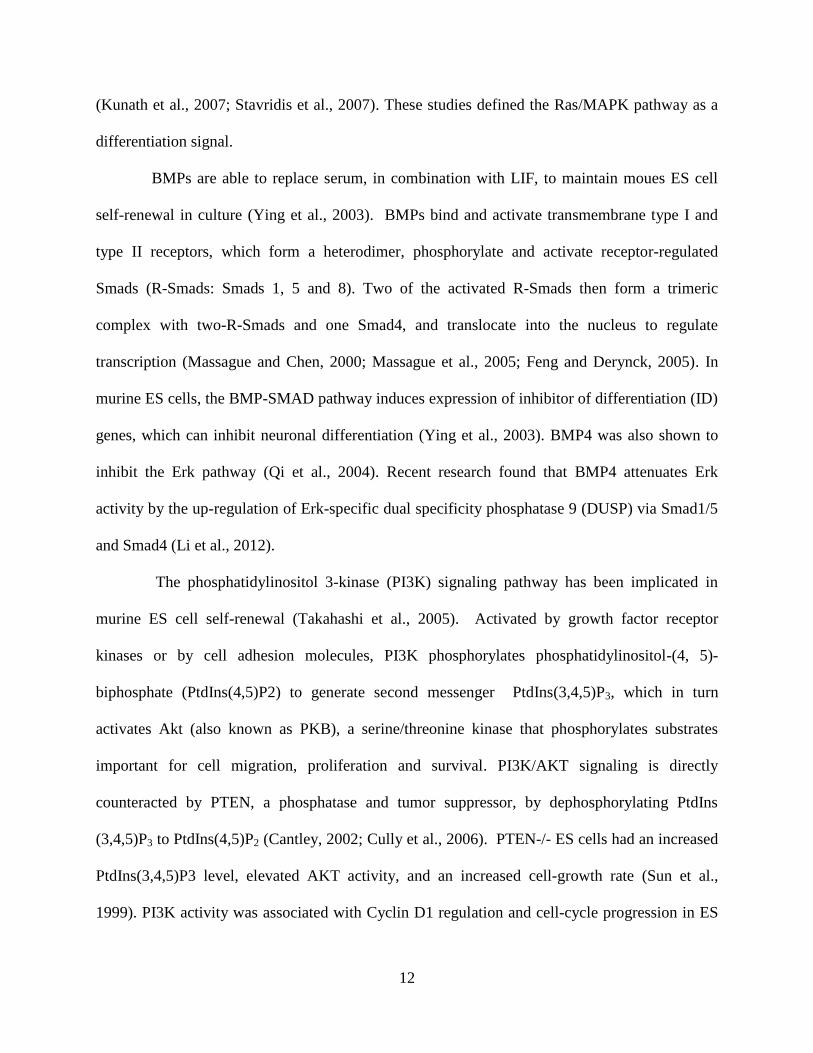
1.1.2.1 Cytokine and growth factor signaling pathways regulating self-renewal
Multiple intracellular kinase signaling pathways play a dominant role in the regulation of ES cell
fate (Liu et al., 2007; Jaenisch and Young, 2008). At least five pathways are important for self-
renewal (Figure 2). LIF signals through Janus-associated tyrosine kinases (Jaks) and signal
transducer and activator of transcription 3 (STAT3), which in turn promotes expression of
renewal factors including the POU domain transcription factor Oct4 (Okamoto et al., 1990) and
the homeobox transcription factor Nanog (Niwa et al., 1998; Burdon et al., 1999a). The Mek/Erk
signaling pathway antagonizes ES cell self-renewal (Burdon et al., 2002). Bone morphogenetic
proteins (BMPs), which are serum components, activate transcription factors of the SMAD
family and inhibit differentiation through induction of inhibitor of differentiation (ID) factors
(Ying et al., 2003). Wnt proteins, which are also found in serum, signal through inhibition of the
kinase GSK3β, leading to β-Catenin accumulation and pluripotency marker gene expression
(Sato et al., 2004; Wray et al., 2011). In addition, the phosphatidylinositol 3’-kinase (PI3K)
signaling pathway promotes ES cell self-renewal partly via regulation of Nanog expression
(Paling et al., 2004; Storm et al., 2007).

10
LIFR
LIF
JAK
STAT3
STAT3STAT3
SHP2
GRB2
RAS
ERK
RAF
MEK
JUNFOS
PI3K
AKT
Cyclin Apoptosis
GSK3Axin
APCβ-Catenin
β-Catenin
β-Catenin
β-CateninTCF3 LEF
Wnt
Frizzled
ID
BMP
BMPR
SMAD4
SMAD5SMAD1
SMAD1/5
DUSP9
Figure 2. Key signaling pathways govern ES cell renewal: LIF-Jak-STAT3, Mek-Erk, BMP-SMAD-ID and Wnt-
GSK-β-Catenin pathways. At least five pathways are important for self-renewal. LIF binds and activates the LIFR
and GP130, which in turn activates three pathways. Firstly, GP130 signal through JAK to phosphorylate STAT3,
which forms a dimer, translocates into the nucleus to regulate gene expression. GP130 also phosphorylates
SHP2/Grb2, which in turn activates the Ras-Raf-Mek-Erk pathway, antagonizing ES cell self-renewal. A third
pathway downstream of LIF involves the activation of PI3K-Akt pathway, which is important for cell-cycle
progression, anti-apoptosis and self-renewal. BMP4, a component of serum, binds to its receptors, which form
dimers and activate Smads. Smads then form a complex to regulate ID gene expression. Wnt binding to its receptor
sequesters the destruction complex including GSK, APC and Axin, protecting β-Catenin from degradation. The
accumulated β-Catenin then translocates into the nucleus and activates Wnt-regulated genes. Extensive crosstalk
occurs between these signaling pathways. Shown in this figure include: Akt, part of PI3K pathway, inhibits GSK3 of
the Wnt pathway; SMAD1/5, part of BMP pathway, inhibits Erk signaling through a phosphatase DUSP9 (see main
text).
11
Mouse ES cells can be maintained and expanded in culture medium contain LIF and
serum, and ES cells differentiate upon withdraw of LIF (Burdon et al., 2002). LIF induces the
hetero-dimerization of LIF receptor and GP130 (Davis et al., 1993; Murakami et al., 1993),
which in term activates JAKs (Stahl et al., 1994). JAKs phosphorylate tyrosine residues on
GP130, which then engages and activates Src homology 2 (SH2) domain-containing signal
transducer and activator of transcription 3 (STAT3). Upon activation, STAT3 forms a dimer,
translocates into the nucleus and targets transcription activation (Niwa et al., 1998; Ihle, 2001).
In addition, STAT3 activation is sufficient to prevent ES cell differentiation in the absence of
LIF (Matsuda et al., 1999), while expression of an inhibitory form of STAT3 causes ES cell
differentiation in the presence of LIF (Niwa et al., 1998), implicating this pathway in ES cell
renewal.
LIF-LIFR-GP130 has another downstream signal, the Ras/mitogen-activated protein
kinases (MAPK, or Erk) pathway, which is associated with ES cell differentiation. Activated
GP130 recruits and phosphorylates the SH2 domain-containing protein-tyrosine phosphatase
SHP2, which complexes with growth-factor-receptor-bound protein 2 (Grb2). SHP2 and Grb2
then activate the small GTPase Ras, which recruits the serine/threonine kinase Raf and
subsequent activation of the MEK-Erk pathway downstream (Burdon et al., 2002). A chimeric
receptor of GP130 without the SHP2 binding site failed to activate the Ras/MAPK pathway, and
enhanced ES cell renewal by reducing the required LIF concentration. Chemical inhibition of
this pathway by a MEK inhibitor had similar effect, indicating that the Ras/MAPK pathway
antagonizes self-renewal (Burdon et al., 1999b). In addition, Erk2-null ES cells are refractory to
differentiation, and FGF stimulation of the Erk pathway is required for ES cells differentiation
12
(Kunath et al., 2007; Stavridis et al., 2007). These studies defined the Ras/MAPK pathway as a
differentiation signal.
BMPs are able to replace serum, in combination with LIF, to maintain moues ES cell
self-renewal in culture (Ying et al., 2003). BMPs bind and activate transmembrane type I and
type II receptors, which form a heterodimer, phosphorylate and activate receptor-regulated
Smads (R-Smads: Smads 1, 5 and 8). Two of the activated R-Smads then form a trimeric
complex with two-R-Smads and one Smad4, and translocate into the nucleus to regulate
transcription (Massague and Chen, 2000; Massague et al., 2005; Feng and Derynck, 2005). In
murine ES cells, the BMP-SMAD pathway induces expression of inhibitor of differentiation (ID)
genes, which can inhibit neuronal differentiation (Ying et al., 2003). BMP4 was also shown to
inhibit the Erk pathway (Qi et al., 2004). Recent research found that BMP4 attenuates Erk
activity by the up-regulation of Erk-specific dual specificity phosphatase 9 (DUSP) via Smad1/5
and Smad4 (Li et al., 2012).
The phosphatidylinositol 3-kinase (PI3K) signaling pathway has been implicated in
murine ES cell self-renewal (Takahashi et al., 2005). Activated by growth factor receptor
kinases or by cell adhesion molecules, PI3K phosphorylates phosphatidylinositol-(4, 5)-
biphosphate (PtdIns(4,5)P2) to generate second messenger PtdIns(3,4,5)P3, which in turn
activates Akt (also known as PKB), a serine/threonine kinase that phosphorylates substrates
important for cell migration, proliferation and survival. PI3K/AKT signaling is directly
counteracted by PTEN, a phosphatase and tumor suppressor, by dephosphorylating PtdIns
(3,4,5)P3 to PtdIns(4,5)P2 (Cantley, 2002; Cully et al., 2006). PTEN-/- ES cells had an increased
PtdIns(3,4,5)P3 level, elevated AKT activity, and an increased cell-growth rate (Sun et al.,
1999). PI3K activity was associated with Cyclin D1 regulation and cell-cycle progression in ES
13
cells (Jirmanova et al., 2002). Chemical inhibition of PI3K or expression of a dominant-negative
form of PI3K induced mES cell differentiation (Paling et al., 2004), while activation of PI3K
signaling by constitutively active AKT maintained ES cell self-renewal without LIF (Watanabe
et al., 2006). In addition, PI3K/AKT was shown to inhibit the MEK-ERK differentiation signal,
and induce Nanog expression by inhibiting GSK-3 activity (Storm et al., 2007). More recently,
PI3K was shown to regulate the transcription factor Tbx3, which in turn regulates Nanog
expression in murine ES cells (Niwa et al., 2009).
Wnt-β-Catenin signaling is a key regulator of early development and embryonic stem
cells (Clevers, 2006; Clevers and Nusse, 2012; Sokol, 2011). In the absence of Wnt, Axin serves
as a scaffold, recruiting glycogen synthase kinase 3(GSK3), casein kinase 1 γ (CK1γ) and the
adenomatosis polyposis coli (APC) protein to form a destruction complex, which phosphorylates
β-Catenin to facilitate its ubiquitination by the E3 ligase β-TrCP and degradation by the
proteasome. Wnt binds to a heterodimeric receptor composed of Frizzled and an LRP5/6 protein,
to induce LRP5/6 phosphorylation, which in turn binds Axin and sequesters the destruction
complex. β-Catenin is not degraded and accumulates in the cytoplasm, then translocates into the
nucleus to regulate target gene expression with T-Cell Factors (TCF) transcription factors
(Clevers and Nusse, 2012). In mES cells, Wnt signaling was implicated in maintenance of self-
renewal and pluripotency: Loss of Wnt signaling is concomitant with ES cell differentiation
(Anton et al., 2007), while null mutation of APC blocked differentiation and promoted self-
renewal (Kielman et al., 2002). Genetic deletion or chemical inhibition of GSK3 promoted self-
renewal in murine ES cells (Sato et al., 2004; Ying et al., 2008). Further, Wnt was able to
prevent ES cell differentiation into epiblast stem cells and can facilitate ES cell renewal with LIF
to replace other defined factors (ten et al., 2011). More recent reports show that β-Catenin

14
interacts with TCF3 to abrogate its repression of gene transcription (Wray et al., 2011) and by
cooperate and enhance the transcriptional activation of Oct4 independent of TCF3 (Kelly et al.,
2011).
To summarize, multiple signaling pathways regulate ES cell self-renewal and
pluripotency. Of note, these signaling pathways form an intricate signaling network, and
crosstalk with each other. For example, LIF activates JAK-Stat3, MEK-Erk, and PI3K pathways
(Burdon et al., 1999a); both BMP-SMAD and PI3K pathways suppresses MEK-Erk pathway (Qi
et al., 2004; Li et al., 2012; Storm et al., 2007); the PI3K pathway regulates Wnt signaling by
inhibiting GSK3β (Storm et al., 2007); and Wnt stimulation upregulates STAT3 (Hao et al.,
2006; Ogawa et al., 2006). Of note, ES cells was able to be maintained in chemically defined
N2B27 medium with two inhibitors—the GSK3 inhibitor CHIR99021 and MEK inhibitor
PD0325901 without LIF, BMP or serum (Ying et al., 2008; Silva et al., 2008). This indicates that
ES cells can be maintained either by providing extrinsic signals (LIF, BMP/ Wnt) or through
inhibition of innate, kinase-dependent differentiation mechanisms (Erk, GSK).
1.1.2.2 Transcription factor networks regulating pluripotency
The ES cell state is maintained by a unique transcription factor network, controlled by the core
co-factors Oct4, Sox2 and Nanog (Niwa, 2007; Jaenisch and Young, 2008; Boyer et al., 2006;
Ng and Surani, 2011).These core factors maintain stemness by regulating their own expression
levels, activating and enhancing the expression of genes characteristic of ES cells, while
repressing expression of genes related to differentiation and lineage commitment(Young,
2011)(Figure 3).

15
.Sox2Oct4
Nanog
Klf2
Tbx3
Gbx2?
Klf4Esrrb
TCF3
Erk
PI3K
Stat3
BMP4 LIF
Β-Cat
GSK3β
Wnt
Figure 3. Core transcription factor network for pluripotency and interconnection with extrinsic stimuli. The core
transcription factors are centered around Oct4 and Sox2, which are indispensable for self-renewal, surrounded by a
circle of other validated facilitating factors including Nanog, Esrrb, Tbx3, Klf4, Klf2 and possibly Gbx2. The factors
in the circle are individually dispensable, but collectively sustain self-renewal by cross-regulating each other,
promoting self-renewal gene expression while suppressing differentiation-related genes. The rectangular boxes
incorporate some key extrinsic signaling pathway components, with green boxes indicating renewal signals while
red boxes indicating signals that antagonize self-renewal. Of note, Klf4 and Gbx2 are both activated by LIF-Stat3;
Esrrb is preferentially regulated by Wnt /Gsk/Tcf3 signaling; and Tbx3 is upregulated by PI3K-Akt signaling. In
contrast, Erk signaling has a negative effect on Nanog, Tbx3 and Klf4. Figure adapted from (Nichols and Smith,
2012), see main text for other references.
Oct4 (Oct3/4, encoded by Pou5f1) is a POU domain transcription factor expressed in the
ICM and epiblast cells of early mouse embryos, and in pluripotent stem cells (Scholer et al.,

16
1990; Nichols et al., 1998). Oct4 is highly expressed in ES cells, and its expression quickly
diminishes when ES cells differentiate. Oct4-deficient embryos can develop to the blastocyst
stage, but the ICM cells are restricted to a trophectoderm fate (Nichols et al., 1998). In ES cells,
Oct4 levels are tightly controlled: Acute repression of Oct4 induces trophectoderm
differentiation; while overexpression causes ES cell differentiation to primitive endoderm and
mesoderm (Niwa et al., 2000).
Sox2 (SRY-related HMG box 2) is an HMG-family protein that co-occupies many gene
targets with Oct4. Sox2 also marks the pluripotent cells of the early embryo, although it is also
expressed in early neuronal lineages. Expression of Sox2 diminishes when ES cells differentiate.
Sox2-null embryos die shortly after implantation with no egg cylinder structure and failed
epiblasts (Avilion et al., 2003). Conditional Sox2 knockouts show that Sox2-null ES cells
differentiate to trophectoderm-like cells. However, those cells are rescued by enforced
expression of Oct4 (Masui et al., 2007). These results indicate that Sox2 stabilizes ES cells in a
pluripotent state by maintaining the requisite level of Oct4 expression.
Nanog, named by Austin Smith for the Celtic land of the ever-young, is a homeobox-
containing transcription factor that is essential for stemness(Chambers et al., 2003; Mitsui et al.,
2003). Nanog is highly expressed in the early embryo and pluripotent cells, and is down-
regulated when cells differentiate. Deletion of Nanog causes early embryonic lethality, with
failed epiblasts containing only primitive endoderm (Mitsui et al., 2003). Overexpression of
Nanog drives ES cell self-renewal independently of LIF and Stat3 activation (Chambers et al.,
2003). Nanog functions by promoting expression of pluripotency markers such as Oct4, Sox2,
and Rex1; and by repressing expression of primitive ectoderm markers Gata4 and Gata6.

17
Accumulating evidence suggests that Oct4, Sox2 and Nanog form the core regulatory
circuitry to control the ES cell pluripotent state (Boyer et al., 2005; Loh et al., 2006). These three
factors bind each other at their own promoters, to positively regulate their own transcription,
forming a positive-feedback autoregulatory loop. In addition, they often co-occupy their target
genes, activating genes important for the ES cell state and pluripotency, while repressing genes
related to lineage-specific differentiation. Oct4, Sox2 and Nanog can recruit multiple
coactivators such as c-Myc, Stat3, Tbx3 and Klf4 to open up chromatin and activate gene
expression. They can also repress linage-specific genes by recruiting chromatin regulators such
as the histone methyltransferase SetDB1 and Polycomb group proteins, and by activating
expression of repressive miRNAs (Jaenisch and Young, 2008; Young, 2011).
Rex1 (Zfp42) is a zinc-finger protein that is primarily expressed in the preimplantation
embryo and ES cells, and downregulated when ES cell differentiate (Hosler et al., 1989; Rogers
et al., 1991). Widely used as a marker for ES cells, Rex1 is regulated by Oct4 (Ben-Shushan et
al., 1998). Rex1-/- ES cells had a greater susceptibility to retinoic acid-induced differentiation,
indicating that Rex1 inhibits ES cell differentiation. However, ES cell derivation and normal
embryonic development were unaffected by Rex1 knockout, demonstrating that Rex1 is
dispensable for ES cell pluripotency (Scotland et al., 2009; Masui et al., 2008). It was therefore
proposed that Rex1 is just a marker of pluripotency but its function is dispensable (Masui et al.,
2008).
Developmental pluripotency-associated gene 4 (Dppa 4), a gene encoding a putative
DNA-binding SAP domain protein, is exclusively expressed in mouse preimplantation embryos
and pluripotent stem cells (Maldonado-Saldivia et al., 2007). Dppa4 overexpression does not
support ES cell self-renewal, but partially inhibits differentiation. Knockdown of Dppa4 with
18
shRNA induced ES cell differentiation to primitive ectoderm. Further, Dppa4 was shown to
localize to active chromatin to inhibit ES cell differentiation (Masaki et al., 2007). In addition,
Dppa4 is a target gene of Oct4 and Sox2 (Chakravarthy et al., 2008). However, a gene knockout
study showed that Dppa4 is dispensable for ES cell identity and germ cell development, but is
essential for embryogenesis (Madan et al., 2009).
Estrogen-related receptor b (Esrrb) is an orphan nuclear receptor that is part of the
pluripotency gene regulatory network (Loh et al., 2006; Ivanova et al., 2006). This protein is
highly expressed in ES cells and the early embryo, and its downregulation causes ES cell
differentiation. Recent research has shown that Esrrb is a pivotal target of Wnt/Gsk3/Tcf3
signaling, and Esrrb is downstream of and able to replace Gsk3 inhibition, in parallel to LIF-
Stat3 signaling (Martello et al., 2012). Esrrb has also been shown to be a direct Nanog target
gene, and is required for LIF-independent self-renewal following Nanog overexpression
(Festuccia et al., 2012). Moreover, Essrb function requires its co-activator, Ncoa3, which bridges
Esrrb to RNA polymerase II complexes and cooperates its gene regulatory effects with the Oct4-
Sox2-Nanog core transcription factor circuitry (Percharde et al., 2012).
Tbx3 is a T-box transcription factor, which is important for pluripotency as RNAi
down-regulation of this factor causes ES cell differentiation (Ivanova et al., 2006). Tbx3 was
shown to be preferentially up-regulated by the PI3K-Akt pathway, down-regulated by the MAPK
pathway and can stimulate Nanog expression. Overexpression of Tbx3 can maintain pluripotency
independent of LIF (Niwa et al., 2009). In addition, Tbx3 can significantly improve the germline
competency of induced pluripotent stem cells (discussed in section 1.1.3.5), by sharing
downstream targets with Oct4, Sox2 and Nanog as well as other pluripotency-related genes and
reprogramming factors (Han et al., 2010).

19
Klf4 is a member of the Kruppel-like family of conserved zinc-finger transcription
factors, and one of the four ‘Yamanaka factors’ originally shown to reprogram fibroblasts to
induced pluripotent stem cells (Takahashi and Yamanaka, 2006). Klf4 is a direct transcriptional
target of Stat3 and is activated by LIF-Jak-Stat3 signaling, with its overexpression sufficient to
maintain pluripotency without LIF (Li et al., 2005; Niwa et al., 2009; Bourillot et al., 2009). Klf4
is negatively regulated by Erk phosphorylation, which promotes proteasome degradation of Klf4
(Kim et al., 2012). Klf4 was shown to directly regulate Nanog expression, and act upstream of
this master pluripotency factor (Zhang et al., 2010). However, knockout of Klf4 does not have a
phenotype. Through RNAi studies, Klf4 was found to be functionally redundant with its closely
related factors Klf2 and Klf5, which also share many common targets with Nanog (Jiang et al.,
2008). Unlike Klf4, which is regulated by LIF-Stat3 signaling, Klf2 is a direct target of Oct4, and
can increase clonogenicity and maintain pluripotency independent of Stat3 (Hall et al., 2009).
Thus, Klf2 and Klf4 are important transcription factors that transduce upstream signals to the
core pluripotency circuitry.
Gbx2 is a homeobox gene that is implicated in pre-gastrulation development and
mid/hindbrain development (Wassarman et al., 1997; Chapman et al., 1997). Gbx2 is highly
expressed in the inner cell mass of the preimplantation embryo, and is down-regulated when ES
cells differentiate (Chapman et al., 1997). Research has shown that Gbx2 is a direct target of
LIF/Stat3 signaling, and when over-expressed, can sustain pluripotency independent of LIF.
Gbx2 can also enhance reprograming efficiency, and alone can reprogram epiblast stem cells to
ES cells (Tai and Ying, 2013). These findings, suggest Gbx2 is a marker of pluripotency and can
integrate signaling cascades to the core pluripotency circuitry.

20
In summary, an intricate, self-regulated transcription factor network controls ES cell
pluripotency (Figure 3). Oct4 and Sox2 sit in the center of this network and while irreplaceable,
are dependent on other facilitating factors, including Nanog, Esrrb, Klf2, Klf4 and Tbx3. These
facilitating factors are individually dispensable, but collectively sustain self-renew by relaying
signals from upstream signaling cascades and by stabilizing the Oct4/Sox2-centered self-renewal
signal (Nichols and Smith, 2012; Ivanova et al., 2006). Assessing the expression profile of this
comprehensive set of transcription factors is a useful tool to study ES cell pluripotency and
differentiation status. For example, EpiSCs express Oct4, Sox2 and Nanog, but not Klf2, Klf4,
Rex1, and Gbx2; this can distinguish between naïve and primed pluripotency as described in
more detail in the sections that follow.
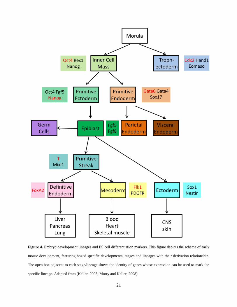
1.1.2.3 Signaling and transcription factors in early development and ES cell differentiation
In vitro differentiation of ES cells includes EB formation, direct differentiation on ECM coated-
plates, or co-culture with stromal cells. These in vitro differentiation protocols recapitulate some
aspects of embryonic development, and the stage and lineage of development can be assessed by
expression of specific lineage markers (Murry and Keller, 2008). Here I discuss the signaling and
lineage markers for the early stages of mouse embryo development, most of which are conserved
in humans as well.
21
Morula
Inner Cell Mass
Troph-ectoderm
PrimitiveEctoderm
Primitive Endoderm
ParietalEndoderm
Visceral Endoderm
Germ Cells
Epiblast
Definitive Endoderm
Mesoderm Ectoderm
.Liver
PancreasLung
.BloodHeart
Skeletal muscle
CNSskin
Oct4 Rex1Nanog
Cdx2 Hand1 Eomeso
Oct4 Fgf5Nanog
Gata6 Gata4 Sox17
Fgf5Fgf8
PrimitiveStreak
T Mixl1
FoxA2Flk1
PDGFRSox1
Nestin
Figure 4. Embryo development lineages and ES cell differentiation markers. This figure depicts the scheme of early
mouse development, featuring boxed specific developmental stages and lineages with their derivation relationship.
The open box adjacent to each stage/lineage shows the identity of genes whose expression can be used to mark the
specific lineage. Adapted from (Keller, 2005; Murry and Keller, 2008)

22
In the early blastocyst, two populations of cells are apparent: the outer polar
trophectoderm cells, and the inner apolar ICM cells. The trophectoderm cells eventually give rise
to trophoblast giant cells and the trophoblast layers of the placenta (Stephenson et al., 2012). In
the outer cells, the transcription factor TEAD4 is activated, which upregulates the caudal-type
homeobox transcription factor Cdx2 and T-box transcription factor Eomeso, driving cells
towards the trophectoderm lineage. In the inner cells, the Hippo pathway is activated in response
to cell-cell interaction, which turns TEAD4 off (Nishioka et al., 2009). Cells of the ICM express
Oct4, which is essential for ICM identity. In the absence of Oct4, a blastocyst-like structure
develops but ICM cells differentiate along the trophectoderm lineage (Nichols et al., 1998). Of
note, trophoblast maintenance requires the inner cell mass, which secretes fibroblast growth
factor 4 (Fgf4) to promote trophoblast proliferation (Nichols et al., 1998).
The early blastocyst then goes through a cavitation process, with trophectoderm cells
transferring fluids into the blastocyst, forming the blastocoel cavity. The ICM then separates into
two cell lineages—the epiblast (EPI, or primitive ectoderm) and primitive endoderm (PE). The
epiblast will eventually form all germ layers of the embryo, while the primitive endoderm cells
form the part of extraembryonic yolk sac (Stephenson et al., 2012). The segregation of EPI with
PE depend on two transcription factors—Nanog and Gata6. These factors are initially co-
expressed in early ICM cells, but gradually become mutually exclusive to determine these two
lineages (Nichols and Smith, 2012). At E3.5 in developing embryo, individual ICM cells are
heterogeneous, with cells expressing either Nanog or Gata6 in a mutually exclusive way.
Lineage restriction starts when cells facing the blastcoel form the PE, and these Gata6-positive
PE cells need a paracrine signal from Nanog-positive epiblast cells and the Grb2-Ras-MAPK

23
signaling pathway (Chazaud et al., 2006). While Fgf4 is expressed in epiblast cells, FGFR is
expressed in the PE (Guo et al., 2010). PE formation is determined by sequential activation of
the transcription factors Gata6, Sox17, Gata4 and Sox7 (Artus et al., 2011). The initial Gata6-
positive PE cells are independent of Nanog-expressing cells, however, later expression of PE
markers such as Sox17 and Gata4 depend on Fgf4 secreted from Nanog-positive cells
(Frankenberg et al., 2011).
After segregation of the EPI and PE layers, the embryo is ready for implantation in the
uterus. Upon implantation, the embryo forms the egg cylinder. During this process, the primitive
endoderm further differentiates to the extraembryonic visceral endoderm and parietal endoderm.
The epiblast cells transform from a cluster of compact cells to an epithelial layer, surrounded by
the visceral endoderm (Stephenson et al., 2012). At this stage, the epiblast cells, or primitive
ectoderm cells, express high levels of Fgf5 (which can serve as a marker for this lineage). EPL
cells retain the ability to form EBs and teratomas. However, unlike ES cells, EPL cells express
very low level of Rex1 and Gbx2, and cannot contribute to chimera formation (Pelton et al.,
2002). Gastrulation follows, with a set of orchestrated morphogenetic movements, cell
proliferation and differentiation to form the ectoderm, mesoderm and endoderm.
The formation of the primitive streak (PS) from epiblast cells marks the beginning of
gastrulation, and then the epiblast cells undergo an epithelial to mesenchymal transition, egress
through the PS, and develop into either mesoderm or definitive endoderm (Tam and Behringer,
1997). The gene Brachyury (T) (Kispert and Herrmann, 1994) and Mixl1 (Hart et al., 2002) are
expressed throughout the PS and can serve as a marker for this transient structure. Foxa2 and
Goosecoid (Gsc) are preferentially expressed in anterior PS regions, and HoxB1 and Evx1 in
posterior regions (Murry and Keller, 2008). The formation of mesoderm and endoderm follow a

24
spatiotemporal development process. First, epiblast cells migrating through the posterior PS
generate extraembryonic mesoderm that eventually develops to parts of the yolk sac.
Subsequently, cells traversing more anterior parts of the PS give rise to mesoderm. Then, cells
moving through the most anterior region of the PS form the definitive endoderm. Unlike
mesoderm or endoderm, the ectoderm is formed from cells in the anterior epiblast that do not
traverse the PS (Murry and Keller, 2008).
Germ layer specification is a complex process tightly controlled by signaling cascades
including the Wnt, Nodal and BMP pathways (Murry and Keller, 2008). The induction of
definitive endoderm is associated with high level of activin/Nodal signaling in the anterior region
of the PS, and FoxA2 can be used as a marker along this lineage (Gadue et al., 2006). Mesoderm
formation is characterized by the expression of Flk-1 and PDGFR (Kataoka et al., 1997). In
contrast, cells of the ectoderm lineage develop from epiblast cells that do not traverse the PS.
Interestingly, ectoderm is the “default” differentiation pathway when ES cells are cultured
without serum or other signals that promote PS differentiation. Sox1 and Nestin can be used as
markers for neuroectoderm. Differentiation along this lineage depends on FGF signaling, which
is inhibited by BMPs (Ying and Smith, 2003; Ying et al., 2003).
In summary, embryonic development involves cell-cell interactions, spatiotemporal
activation of signaling pathways, and expression of specific lineage-related transcription factors.
ES cell differentiation also follows this development process. Based on the signaling pathways
and transcription factors highlighted above, we can use sets of different lineage markers to track
ES cell differentiation stages in vitro (Figure 4).
25
1.1.3 Human Embryonic Stem Cells
Because of species specific ES cell differences and non-optimal human embryo culture, initial
attempts to derive human ES (hES) cells using mES cell culture conditions were unsuccessful.
In fact, 17 years passed from the establishment of the first mES cell line to the derivation of the
first hES cell lines (Yu and Thomson, 2008). Like mES cells, hES cells are pluripotent stem cells
derived from inner cell mass of blastocyst stage human embryos produced by in vitro
fertilization (Thomson et al., 1998). Although hES cells are of the same blastocyst origin as mES
cells, the culture conditions, undifferentiated cell colony morphology, and extrinsic signaling
pathways regulating their fate are very different. Nevertheless, they express the same core
transcription factors to maintain pluripotency. The improvement in culture conditions for hES
cell culture from feeder-based medium to chemically defined medium facilitated genetic
manipulation of hES cells and signaling research. Recent establishment of epiblast stem cells
argues that hES cells and mES cells may represent different states of pluripotent stem cells.
1.1.3.1 Human ES cell derivation and culture conditions
In the pioneering research of hES cell derivation by James Thomson’s group, ICM cells were
isolated by immunosurgery, and plated onto mitotically inactivated MEF cells in serum-
containing medium for extended growth. Five hES cell lines were initially derived by this
approach, with H1, H13 and H14 showing a normal XY karyotype; and H7 and H9 cell showing
a normal XX karyotype. The hES cell lines had a high nucleus to cytoplasm ratio and high levels
of telomerase activity; they expressed cell surfaces markers for pluripotency (alkaline
phosphatase, SSEA-3, SSEA-4, Tra-1-60 and Tra-1-81); and when injected into severe combined
26
immunodeficient (SCID) mice, were able to form teratomas with cell lineages of all three germ
layers (Thomson et al., 1998).
Subsequent work improved the culture conditions for hES cells. Knockout serum
replacement and the addition of basic fibroblast growth factor (bFGF) was shown to support
clonal derivation of hES cells from the original H9 cell line on feeder layers, although with very
low efficiency (Amit et al., 2000). Feeder-free culture was then established by plating hES cells
on Matrigel or laminin coated plates in medium conditioned by MEFs (Xu et al., 2001). At
higher concentrations, bFGF allowed “truly” feeder-free culture of hES cells with unconditioned
medium (Xu et al., 2005a; Levenstein et al., 2006). Noggin, an antagonist of BMP signaling, was
shown to cooperate with bFGF to maintain pluripotency of hES cells (Xu et al., 2005b; Wang et
al., 2005). Last but not the least, chemically defined culture medium was established for hES
culture to facilitate clinical application (Lu et al., 2006; Yao et al., 2006; Ludwig et al., 2006).
One problem with hES cell culture is the poor survival of hES cells after cell dissociation,
which hinders sub-cloning and gene-targeting research. Y-27632, a Rho-associated kinase
(ROCK) inhibitor, was shown to protect dissociated hES cells from apoptosis, and facilitate sub-
cloning (Watanabe et al., 2007). Further mechanistic studies elucidated the protective mechanism
of ROCK inhibitor, where dissociated hES cells lose E-cadherin mediated cell-cell adhesion,
which triggered ROCK-dependent hyper-activation of actin-myosin contraction and apoptosis
(Chen et al., 2010; Ohgushi et al., 2010).
Since the initial derivation of hES cell lines, culture conditions have rapidly evolved from
feeder and serum-based medium, to feeder-free, and finally chemically defined medium. The
introduction of ROCK inhibitor facilitated clonal expansion and sub-cloning of hES cells. These
27
advances have greatly accelerated the pace of hES cell research and facilitated our understanding
of the complex signaling networks that control their fate.
1.1.3.2 Human ES cell renewal and pluripotency, EB formation and teratoma formation
Like mES cells, hES cells have two important properties: self-renewal and pluripotency. Self-
renew is the ability for prolonged proliferation without differentiation. This property can be
assessed as extended passage with a normal karyotype, undifferentiated cell culture morphology,
high activity of telomerase and alkaline phosphatase, expression of cell surface markers such as
SSEA-3, SSEA-4, Tra-1-60 and Tra-1-81, and expression of the pluripotency factors such as
Oct4, Nanog, Sox2 and Rex1.
In hES cells, pluripotency is mainly assessed through embryoid body and teratoma
formation. When cultured in suspension culture without feeder layers, hES cells spontaneously
form aggregates called embryoid bodies as previously described for mES cells (Itskovitz-Eldor et
al., 2000). EB formation is a convenient model for the study of human early embryonic
development in vitro. EB formation can mimic, to some extent, the axis and polarity
reorganization in development, and the temporal/sequential gene expression changes that occur
during the stages of gastrulation and germ layer formation (Dvash et al., 2004). In addition, EB
formation can serve as an initial step for subsequent directed differentiation into specific lineages
with guidance from growth factors and small molecule inhibitors (Murry and Keller, 2008). The
advantage of EB formation is that it provides a three-dimensional model that facilitates and
mimics the complex cell to cell interactions during development. However, it is hard to precisely
control this cell-cell microenvironment to synchronize EB formation. EBs are normally
heterogeneous in size, which can complicate the interpretation of results (Zhu and Huangfu,

28
2013). More recently, the development of microwell culture to control of number of hES cells
per cluster during embryoid body formation improved this limitation of the EB assay (Mohr et al.,
2010).
Teratoma formation is a widely used assay for pluripotency in hES cells, and is
considered the most stringent assay to demonstrate the differentiation potential of hESCs (Zhang
et al., 2008). When transplanted into immunodeficient mice, hES cells give rise to teratomas with
differentiated tumor tissues representing all three germ layers including neuronal tissues from
ectoderm, muscle and blood lineages from mesoderm and gut epithelium and liver tissues from
endoderm. The efficiency and quality of teratoma formation is determined by three factors: cell
quality, cell number and injection site (Zhang et al., 2008). Teratoma formation is most efficient
following subcutaneous or intramuscular implantation, with Matrigel enhancing the efficiency
(Prokhorova et al., 2009; Hentze et al., 2009). Besides teratoma formation, hES cells also
undergo random differentiation when cultured on gelatin-coated plates in serum-containing
medium without bFGF. More importantly, defined differentiation conditions have been
developed to drive hES cells toward specific lineages important for regenerative medicine,
including retinal epithelium, pancreatic progenitor cells, β-cells, cardiomyocytes, and motor
neurons [reviewed in (Zhu and Huangfu, 2013; Murry and Keller, 2008; Cohen and Melton,
2011)].
To summarize, self-renewal and pluripotency are the defining feature of hES cells. EB
formation serves as a standard test for pluripotency and is a useful model for early
embryogenesis, while teratoma formation serves as the gold-standard to test hES cell
differentiation potential. Of note, the more stringent assays in mouse such as chimera formation
and germline competency are not feasible in human for ethical reasons.

29
1.1.3.3 Growth factors and signaling pathways
The self-renewal of hES cells depends largely on the FGF and TGFβ/Nodal/Activin signaling
pathways. This is very different from that of mES cells, which depend on LIF/Jak/Stat3 and
BMP/SMAD/ID signaling. Below I discuss the regulation of FGF and TGFβ signaling in hES
cells (Yu and Thomson, 2008).
FGF signaling is of pivotal importance for hES cell self-renewal. bFGF can maintain hES
cell clonal growth on feeders, and can also support hES cell feeder-free growth at a high
concentration. FGF has multiple signaling roles to maintain hES cell pluripotency. First, FGF
signals through the FGF receptor tyrosine kinase and downstream Erk signaling to inhibit
differentiation into extraembryonic lineages (Li et al., 2007; Dvorak et al., 2005). FGF-2 was
also shown to activate both MEK/Erk and PI3K-Akt signaling pathways, which synergistically
stimulate self-renewal, cell survival and adhesion (Eiselleova et al., 2009). Second, FGF can
modulate TGFβ signaling, upregulating the expression of TGFβ ligands in both feeder cells and
hES cells (Greber et al., 2007). In feeder-free culture, some hES cells self-differentiate into
fibroblast cell-like supporting cells, creating their own “niche”, which responds to FGF in similar
manner as feeder cells (Bendall et al., 2007). Third, FGF signaling can inhibit neuronal induction
in hES cells. FGF/Erk inhibition induced neuroectoderm differentiation in hES cells marked by
Pax6 expression (Greber et al., 2011). Last but not the least, Fgf2 can sustain Nanog expression
through the MEK-ERK pathway, which can switch the cell fate in the context of BMP4-induced
differentiation (Yu et al., 2011).
TGFβ/Nodal/Activin is essential for hES cell self-renewal in culture (James et al., 2005;
Vallier et al., 2005). In undifferentiated hES cells, the TGFβ/Nodal/Activin branch is activated

30
(with downstream signals transduced through SMAD2/3), while the BMP/GDF branch
(SMAD1/5) is largely suppressed (James et al., 2005). Nodal was shown to inhibit hES cell
differentiation to neuroectoderm (Vallier et al., 2004), and inhibition of Activin/Nodal signaling
caused hES cell differentiation even in the presence of FGF (Vallier et al., 2005). In contrast,
BMP caused hESCs to differentiate to trophectoderm in conditioned medium with bFGF (Xu et
al., 2002). Subsequent investigation using defined medium found that both TGFβ-responsive
SMADs (SMAD2/3) and BMP-SMADs (SMAD1/5) bind to the Nanog promoter, with
SMAD2/3 being active in undifferentiated hESCs maintained by TGFβ and FGF. In the absence
of growth factors, BMP-SMADs bind to the Nanog promoter and facilitate differentiation (Xu et
al., 2008).
The FGF and TGFβ/Activin signaling pathways work synergistically to main hES cell
pluripotency. FGF2 induces expression of key TGFβ pathway components including TGFβ1 and
GREM1 while inhibiting BMP4 expression in both feeder cells and hES cells (Greber et al.,
2007). Reciprocally, Activin A induces expression of bFGF to promote self-renewal and inhibit
the BMP signal (Xiao et al., 2006). Thus, TGFβ/Nodal/Activin, in cooperation with bFGF
maintains self-renewal, inhibits the BMP differentiation signal, and promotes proliferation and
survival.
In addition, both Wnt and IGF (insulin-like growth factor) signaling have been implicated
in the maintenance of hES cell pluripotency. BIO, a potent pharmacological inhibitor of GSK-3,
was shown to support self-renewal of hES cells (Sato et al., 2004). IGF1 is secreted from
“feeder-like” cells which spontaneously differentiate from hES cells to support “true” hES cells
(Bendall et al., 2007). However, the role of Wnt signals in self-renewal was challenged when the
Wnt/β-catenin signal was found to be insufficient to maintain pluripotency of hES cells, and

31
associated with differentiation instead (Dravid et al., 2005). IGF1 was also insufficient to support
hES renewal in the absence of bFGF in chemically defined medium (Wang et al., 2009). These
studies point to the heterogeneity of hES cell culture. Further research is needed to elucidate the
roles of Wnt, IGF and other growth factor signaling pathways in hES cells.
1.1.3.4 Core transcription factor regulatory network
In mES cells, Oct4, Sox2 and Nanog form a core regulatory circuitry maintaining self-renewal
and pluripotency (Jaenisch and Young, 2008). These three master transcription factors co-bind
and regulate a large group of target genes, maintaining the expression of pluripotency genes
while suppressing differentiation-related gene expression. Human ES cells express all three
master regulators, suggesting that a similar core transcription factor network exists in hES cells
(Boyer et al., 2005).
A genome-scale location analysis indicated that Oct4, Sox2 and Nanog co-occupy a large
number of their targeted genes in hES cells (Boyer et al., 2005). Around 50% of Oct4 targeted
promoters were co-bound by Sox2, while 90% of the genes co-bound by Oct4 and Sox2 were
also targeted by Nanog, suggesting that these three factors function together in gene regulation.
The genes targeted by all three core factors include both active and inactive genes. The active
targeted genes include Oct4, Sox2 and Nanog themselves, other pluripotency-related
transcription factors such as STAT3 and Dppa4, as well as components of the TGFβ and Wnt
signaling pathways. The inactive genes include many transcription factors that are important in
development and lineage specification, including Pax6 and NeuroD for ectoderm differentiation,
Hand1 for mesoderm differentiation. This study concluded that Oct4, Sox2 and Nanog form an

32
auto-regulated, feed-forward circuitry to maintain self-renewal and pluripotency in hES cells,
like in mES cells.
However, a recent study with shRNA knockdown and targeted overexpression revealed
that in hES cells, Nanog, Oct4 and Sox2 have general and cell-line specific functions (Wang et
al., 2012). Oct4 expression regulates and cooperates with BMP4 to specify hESC cell fate. High
levels of Oct4 sustain self-renewal in the absence of BMP4 but induce mesendoderm
differentiation in the presence of BMP. While low levels of Oct4 induce endoderm formation in
the absence of BMP4, low levels of Oct4 specify extraembryonic differentiation in the presence
of BMP4. Sox2 and Sox3 function redundantly in inhibiting mesendoderm differentiation, while
Nanog mainly represses ectoderm differentiation. This study suggested a modular regulatory
network for three master regulators in hES cells, in contrast to the co-regulatory circuitry in mES
cells.
In summary, hES cells express the core transcription factors Oct4, Nanog, and Sox2,
which regulate target gene expressions to promote self-renewal and pluripotency related genes
while repressing differentiation related genes. Recent research indicates that instead of working
as a trio like in mES cells, these three factors act as a modular regulatory network in hES cells,
with each factors inhibiting a specific differentiation lineage.
1.1.3.5 New different states of pluripotent stem cells: iPS cells, mEpiSCs and naive human
ES cells
While both are derived from ICM of the blastocyst stage embryo, mES cells and hES cells have
different culture conditions and colony morphologies. Mouse ES cells depend on LIF-Jak-Stat3
signaling and BMP-SMAD-ID signaling for self-renewal. In contrast, hES cells depend on bFGF

33
and TGFβ/Activin signaling to remain in the undifferentiated state. LIF does not support self-
renewal of hES cells, while BMP4 induces differentiation. While mES cells grow as domed
colonies and can be passaged with single cell dissociation, hES cells grow as flattened colonies
and can only be passaged in small cell clumps, with single cell dissociation causing hES cell
death. Moreover, mES cells and hES cells express different cell-surface markers. For example,
SSEA-1, a cell surface marker of pluripotency in mES cells, is a marker of differentiation in hES
cells. Instead, hES cells express cell-surface SSEA-3, SSEA-4, TRA-1-60 and TRA-1-81, which
are absent in mES cells. These and other properties of hES cells, when compared with mES cells,
indicates that there are species-specific differences and that they represent different pluripotent
states.
In 2006, the Yamanaka laboratory reported a revolutionary discovery that pluripotent
cells, with similar properties to ES cells, can be generated from mouse fibroblasts by retrovirally
introducing four genes—Oct4, Sox2, Klf4 and c-Myc (Takahashi and Yamanaka, 2006). These
cells, term induced pluripotent stem (iPS) cells, were tested extensively for pluripotency and
passed the most stringent standard of tetraploid complementation (Stadtfeld and Hochedlinger,
2010). Human iPS (hiPS) cells were also generated with the same four factors (Takahashi et al.,
2007) and later with different combinations (Yu et al., 2007). The hiPS cells are shown to be
similar to established hES cells in terms of self-renewal and differentiation potential (Yamanaka,
2012).
The derivation of epiblast stem cells (EpiSCs) from postimplantation mouse embryos
provides the evidence that hES cell-like stem cells can be derived from mouse embryos (Brons et
al., 2007; Tesar et al., 2007). Mouse ES Cells and mEpiSCs represent two different states of
pluripotency, termed naïve and primed states, respectively (Nichols and Smith, 2009)(Table 2).

34
Naïve pluripotent stem cells, exemplified by mES Cells, are responsive to LIF/Stat3 and BMP
signaling, can be passaged with single cell dissociation and show clonal growth, have two active
X chromosomes in female lines, and readily form chimeric mice with germline competence
when injected into developing blastocysts. In contrast, primed pluripotent cells, such as EpiSCs,
depend on bFGF and TGFβ/Activin signaling for maintenance, can only be passaged as clumps,
have one X-chromosome inactivated in female lines, and rarely form chimeras when injected
into host blastocysts but can form teratomas when injected into immunocompromised mice.
Human ES cells share all those features with mouse EpiSCs that are distinct from mES cells.
However, hES Cells are not identical to mEpiSCs, as hES Cells express the ICM marker Rex1
and hES Cell-specific transcription factors such as PRDM14 (Chia et al., 2010), which are absent
in mEpiSCs.
Table 2. Comparison of naïve and primed pluripotent cell states
Property Ground State Primed State
Cultured cells rodent ES Cells rodent EpiSCs; hES Cells
Equivalent embryonic tissue early epiblast egg cylinder or embryonic disc
Response to Lif/Stat3 self-renewal none
Response to Fgf/Erk differentiation self-renew
Response to 2i self-renewal differentiation/death
Response to BMP4 supported renewal differentiation
pluripotency factors Oct4, Nanog, Sox2, Klf2, Klf4 Oct4, Nanog, Sox2
colonly morphology tight, domed colony spread out, flattened
Clonogenicity high low
XX status in female lines XaXa XaXi
Blastocyst chimeras* yes no
Teratomas yes yes
Differentiation bias none variable
* Not applicable to human Adapted from (Nichols and Smith, 2009)

35
With the inhibition of GSK3 and Erk signaling pathways, and exogenous expression of
reprogramming factors Klf4 or c-Myc, naïve ES cells were derived from previously refractory
non-obese diabetic(NOD) mouse strain and from rat (Hanna et al., 2009; Buehr et al., 2008).
These discoveries led to the derivation of mESC-like naïve hES cells (Hanna et al., 2010;
Buecker et al., 2010). Naïve hES cells were achieved by growing hES cells or iPSCs with
ectopic expression of Oct4, Klf4 and Klf2 in 2i medium with LIF, or by growing five-factor iPS
cells under the same conditions. However, those naïve hES cells are very unstable, and require
extended expression of reprogramming factors. These findings suggest that the naïve state of
hES cells is intrinsically unstable, making hES cells least permissive to naïve state (Hanna et al.,
2010).
To summarize, the derivation of mEpiSC lines and establishment of naïve hES cell lines,
compared with traditional mESCs and hESCs, suggest that there are two states of pluripotency in
both mouse and human. However, human pluripotent cells are refractory to culture in the naïve
state, which may due to the species-specific genetic determinants.
1.2 SRC FAMILY NON-RECEPTOR TYROSINE KINASES
A major focus of my thesis research centered on the role of non-receptor protein-tyrosine kinase
signaling pathways in the control of ES cell renewal and differentiation. Before reviewing
previous work related to this theme, I will first provide a brief overview of the structure and
function of the Src kinases, which constitute the largest family of non-receptor tyrosine kinases
in the human kinome. The differential control of ES cell fate by individual members of the Src
kinase family is at the center of my dissertation project.

36
The Src family of non-receptor tyrosine kinases was first discovered in the context of Rous
Sarcoma Virus in the form of v-Src, which is an oncogene with the ability to transform cells in
culture and rapidly induce large sarcomas in chickens (Martin, 2004). This viral oncogene was
later found to be derived from a cellular gene, c-Src, which led to the oncogene hypothesis of
cancer (Stehelin et al., 1976; Brugge and Erikson, 1977). There are 11 members of the Src-
family kinase complement in the human genome (Manning et al., 2002), eight of which have
been studied extensively in mammalian cells: Blk, Fgr, Fyn, Lck, Lyn, Hck, Src and Yes (Engen
et al., 2008). In the mouse, Src, Fyn and Yes are ubiquitously expressed, while Lck, Lyn, Hck,
Blk, and Fgr display more restricted expression to hematopoietic cells (Lowell and Soriano,
1996; Summy and Gallick, 2003a; Summy and Gallick, 2003b). Src family kinases are important
in diverse signal transduction networks that govern cell proliferation, survival, motility and
invasion (Engen et al., 2008; Parsons and Parsons, 1997). They serve as critical signal integrators
or nodes, as their activity is regulated by a multitude of inputs upstream (growth factors,
cytokines, hormones, cell attachment, antigens, to name a few) and they are wired to diverse
signaling pathways downstream, including many that are linked to the regulation of ES cell
growth and differentiation as described in the previous section.

37
SH3 SH2 N-lobe
K295
Y527
N C-lobe
Y416
SH3
SH2
N-lobe
C-lobeTail
α-C
A-loop
SH3
N-lobe
α-C
A-loop
SH2
C-lobeTail
A
B
C
Linker
UD
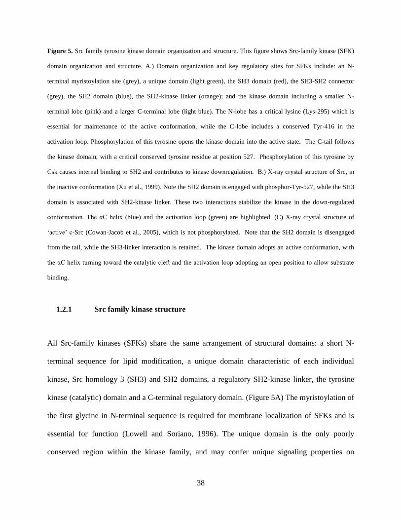
38
Figure 5. Src family tyrosine kinase domain organization and structure. This figure shows Src-family kinase (SFK)
domain organization and structure. A.) Domain organization and key regulatory sites for SFKs include: an N-
terminal myristoylation site (grey), a unique domain (light green), the SH3 domain (red), the SH3-SH2 connector
(grey), the SH2 domain (blue), the SH2-kinase linker (orange); and the kinase domain including a smaller N-
terminal lobe (pink) and a larger C-terminal lobe (light blue). The N-lobe has a critical lysine (Lys-295) which is
essential for maintenance of the active conformation, while the C-lobe includes a conserved Tyr-416 in the
activation loop. Phosphorylation of this tyrosine opens the kinase domain into the active state. The C-tail follows
the kinase domain, with a critical conserved tyrosine residue at position 527. Phosphorylation of this tyrosine by
Csk causes internal binding to SH2 and contributes to kinase downregulation. B.) X-ray crystal structure of Src, in
the inactive conformation (Xu et al., 1999). Note the SH2 domain is engaged with phosphor-Tyr-527, while the SH3
domain is associated with SH2-kinase linker. These two interactions stabilize the kinase in the down-regulated
conformation. The αC helix (blue) and the activation loop (green) are highlighted. (C) X-ray crystal structure of
‘active’ c-Src (Cowan-Jacob et al., 2005), which is not phosphorylated. Note that the SH2 domain is disengaged
from the tail, while the SH3-linker interaction is retained. The kinase domain adopts an active conformation, with
the αC helix turning toward the catalytic cleft and the activation loop adopting an open position to allow substrate
binding.
1.2.1 Src family kinase structure
All Src-family kinases (SFKs) share the same arrangement of structural domains: a short N-
terminal sequence for lipid modification, a unique domain characteristic of each individual
kinase, Src homology 3 (SH3) and SH2 domains, a regulatory SH2-kinase linker, the tyrosine
kinase (catalytic) domain and a C-terminal regulatory domain. (Figure 5A) The myristoylation of
the first glycine in N-terminal sequence is required for membrane localization of SFKs and is
essential for function (Lowell and Soriano, 1996). The unique domain is the only poorly
conserved region within the kinase family, and may confer unique signaling properties on
39
individual members (more below). The SH3 domain binds to proline-rich sequences and
contributes to substrate recruitment and also is essential for kinase activity regulation. The SH2
domain binds to phosphotyrosine-containing peptide sequences, functioning both in protein-
protein interaction and kinase regulation. The kinase domain (also called the SH1 domain), with
a bi-lobed structure comprised of N-terminal and C-terminal lobes, is responsible for
phosphotransfer activity. X-ray crystallography has revealed that two intramolecular interactions
are important for locking the kinase in inactive conformation: the SH3 domain interacts with the
SH2-kinase linker, and the SH2 domain binds to phosphorylated tyrosine in the C-terminal
regulatory tail. Disruption of either of these interactions is sufficient to activate the kinase
(Tatosyan and Mizenina, 2000; Boggon and Eck, 2004; Ingley, 2008; Engen et al., 2008).
1.2.1.1 N-terminal region
The N-terminal domain (also called the SH4 domain) is a region with 15-17 amino acids
which contains lipid-modification signals and is responsible for membrane anchorage of SFKs
(Resh, 1994; Resh, 1999). In all SFKs, the glycine residue right after the initiator methionine is
myristoylated cotranslationally and this modification is required for membrane binding. When v-
Src Gly2 is mutated to Ala, membrane binding and cellular transformation are both abrogated. In
the c-Src N-terminal domain, a module with three alternative lysine residues facilitates
membrane binding in conjunction with myristate, and is also required for kinase function
(Silverman et al., 1993). Most of the other SFKs (except c-Src and Blk) have a cysteine residue
that can be reversibly palmitoylated. Only myristoylated proteins can be palmitoylated, which in
turn can guide the kinase to membrane rafts. In response to different stimuli, reversible
palmitoylation can change SFK membrane localization and signaling events (Resh, 1999). Thus,
40
the N-terminal domain modulates SFK membrane anchorage which is often essential for
biological activity.
1.2.1.2 Unique domain
The unique domain, around 45-75 residues in length, follows the N-terminal domain and
is the only non-conserved region among SFK members. The divergent unique domains are
refractory to structural analysis and are absent from existing structures of SFKs. Although not
required for intramolecular regulation in SFKs, the unique domain is thought to be involved in
specific interactions with binding partners. It can also be phosphorylated at specific residues for
kinase regulation. A well-studied example is the association of Lck with T-cell receptors CD4
and CD8α, which are essential for T-cell maturation (Rudd et al., 1988; Veillette et al., 1988).
Structural analysis showed that two conserved cysteine residues in the Lck unique domain bind
to two cysteine residues from CD4 or CD8α receptors by coordinating a Zn2+
ion (Kim et al.,
2003). In addition, recent research showed that Src can be phosphorylated on Ser/Thr residues in
the unique domain. These phosphorylation events induce a global electrostatic perturbation that
releases c-Src from membrane, and changes subsequent signaling events (Perez et al., 2009).
Hck can also be phosphorylated at a tyrosine in the unique domain, which contributes to its
activation (Johnson et al., 2000). Moreover, in a study of kinase chimeras, replacing the v-Src N-
terminal and unique domain with its c-Yes counterparts abrogated the ability of v-Src to
transform chicken fibroblasts, indicating that the unique domain may confer signaling specificity
between c-Src and c-Yes (Summy et al., 2003a). To summarize, the unique domain is the only
specific region for each SFK protein. It can modulate specific molecular interactions, is regulated
by phosphorylation, and may confer specificity among individual SFKs.
41
1.2.1.3 SH3 domain
The SH3 domain, about 60 amino acids in size, is necessary for SFK kinase regulation
and interaction with adaptor proteins or substrates. Structural analysis revealed that the Src SH3
domain, like all SH3 domains, is a β-barrel comprised of five antiparallel β-strands and two
prominent loops termed the RT-loop and n-Src loop (Musacchio et al., 1992). SH3 domains bind
to target protein sequences that are rich in proline and other hydrophobic residues, especially
proteins with the classic “PxxP” sequence. These sequences usually form a polyproline type II
(PPII) helix which complexes with hydrophobic grooves on the surface of the SH3 domain
(Musacchio et al., 1994). In SFK SH3 domains, PxxP-containing peptides can be docked in two
opposite directions. Additional specificity and binding affinity are conferred by lysine or
arginine residues N- or C- terminal to the PxxP motif (Feng et al., 1994). Class I ligands have an
RxxPxxP motif and bind in an orientation opposite to that of Class II ligands which have an
XpxxPxR motif (Lim et al., 1994). SH3 domains mediate both common and also specific
binding of target protein to SFKs. For example, the Src, Fyn and Lyn SH3 domains have some
common targets such as Shc, p62 and hnRNPK, but also have specific targets. In addition,
extensive structural studies of many SH3 domains have revealed some atypical SH3 domain
binding motifs. These non-consensus SH3 binding motifs do not always form a PPII helix, but
utilize a negatively charged specificity pocket for binding (Saksela and Permi, 2012). An
extreme case is the interactions between Fyn and the immune cell adaptor SAP, where a surface
to surface interaction between Fyn SH3 domain and SAP SH2 domain exists, distinct from
canonical PxxP-modulated biding (Latour et al., 2003; Chan et al., 2003).
Besides these important roles in substrate recruitment, SH3 domains are also critical for
regulation of Src-family kinase activity. Structures of the inactive forms of c-Src and Hck

42
showed that the SH3 domain binds intramolecularly to a PPII helix formed by the SH2-kinase
linker (Xu et al., 1997; Sicheri et al., 1997; Xu et al., 1999; Sicheri et al., 1997). This interaction
pushes the SH3 domain to the back of the N-lobe of the kinase domain and stabilizes the kinase
in the inactive conformation (shown in Figure 5).
1.2.1.4 SH2 domain
The SH2 domain, around 100 residues in length, is another non-catalytic domain
important for SFK substrate recruitment, localization and kinase regulation (Pawson, 2004).
Structurally, SH2 domains contain a central antiparallel β-sheet flanked by two alpha-helices.
These secondary elements form two peptide binding pockets: One accommodates the
phosphotyrosine (pTyr) with a conserved arginine residue in the base of the pocket; the other
recognizes residues C-terminal to the pTyr, providing a specificity determinant (Waksman et al.,
1993; Songyang et al., 1993). Phosphopeptide library screening showed that SFK SH2 domains
preferentially bind to a pYEEI motif, with the pTyr and isoleucine residues occupying the two
pockets, respectively (Songyang et al., 1993). While the pTyr-binding pockets of SH2 domains
are highly conserved and almost invariant, the C-terminal recognition site is more divergent and
can confer specificity among different SH2 domains and corresponding pTyr-containing binding
sequences (Pawson and Nash, 2000). By interacting with pTyr-containing peptide sequences,
SH2 domains relay protein tyrosine kinase signaling to a network of intracellular pathways that
regulate many cellular functions. More recently, a fluorescence polarization-based screen of 50
SH2 domains and 192 physiological pTyr peptides demonstrated that the specificity is based on
SH2 domain recognition of permissive amino acid residues that enhance binding and non-
permissive residues that prohibit binding near the essential pTyr. Thus, subtle differences in the
context of peptide ligands can be distinguished by different SH2 domains (Liu et al., 2010).

43
In addition to substrate binding and modulating signaling partners, the SH2 domain is
also essential for stabilizing SFKs in the inactive conformation. In the downregulated structure of
c-Src, the SH2 domain binds to the kinase tail with a highly conserved tyrosine residue. This
intramolecular interaction locks the kinase in an inactive conformation, with the help from the
SH3-linker interaction (Xu et al., 1997; Sicheri et al., 1997; Xu et al., 1999) (Figure 5).
1.2.1.5 Tyrosine kinase domain
The tyrosine kinase domain, comprised of an N-terminal lobe (N-lobe) and a C-terminal
lobe (C-lobe), is the most conserved region among all SFK members and is structurally
conserved in all protein tyrosine kinases. The smaller N-lobe includes five β-strands and a single
α-helix (α-C-helix). The larger C-lobe is mainly composed of α-helices with only a few β
strands. A short, flexible hinge connects the N-lobe with C-lobe, and catalytic activity occurs in
the cleft between the two lobes (Boggon and Eck, 2004). The active site is formed by the
glycine-rich loop and C-helix from the N-lobe, and the catalytic and activation loops from the C-
lobe. Kinase activity is controlled by the conformation and relative position of these elements,
which are modulated by phosphorylation and by interaction with regulatory domains. The
catalytic loop (residues 385-403 in c-Src) contains highly conserved Asp and Asn residues,
which are important for coordinating the ATP-Mg++
complex. The activation loop (residues 404-
432 in c-Src) is of central importance to kinase activity, with phosphorylation of the highly
conserved Tyr-416 required for full activation.
Several kinase domain elements interact and stabilize the inactive conformation. In the
N-terminal end of activation loop, Asp404 forms an ionic interaction with Lys-295, disrupting
the Lys-295 and Glu-310 interaction which is required for the active state. This Asp-404 and
Lys-295 interaction induces the movement of Phe-424 toward steric clash with the α-C-helix of
44
the N-lobe, forcing the α-C-helix away from the catalytic cleft (Schindler et al., 1999). In
addition, the residues 413-418 of the activation loop form a short α-helix (A-loop helix), which
turns Tyr-416 to the catalytic center of the kinase domain. Tyr-416 forms hydrogen bonds with
Arg-385 and Arg-386, is buried into a hydrophobic pocket and protected from auto
phosphorylation. Thus, the activation loop occludes the catalytic domain and prevents the
binding of peptide substrates (Xu et al., 1999). Moreover, the non-catalytic conformation of the
α-C-helix is stabilized by interactions between the inward-facing surface of the α-C-helix with
the N-terminal end of activation loop, a salt bridge between Glu-310 of the αC-helix and Arg-
409, and by insertion of Trp-260 into the α-C-helix (Xu et al., 1999; Sicheri et al., 1997).
In the active conformation, activation loop Tyr-416 is phosphorylated. This
phosphorylation induces the activation loop to move away from the catalytic cleft and form a
conformation suitable for substrate binding. This conformation is stabilized by the interaction
between pTyr-416, Arg-385 and Arg-409 in Src (Yamaguchi and Hendrickson, 1996; Cowan-
Jacob et al., 2005). The position change of the activation loop and Asp-404 allows the α-C-helix
to turn towards the catalytic cleft and into its active orientation. This liberates Glu-310 in α-C-
helix, which forms a salt bridge with Lys-295, an interaction both critical for ATP binding and
stabilization of the active αC helix conformation (Xu et al., 1999). Note that this Lys:Glu salt
bridge is a highly conserved feature of active kinase domain conformations, and substitution of
the Lys residue is a common approach to creating an inactive kinase mutant.
At the end of the kinase domain, a C-terminal tail (residues 521-535 in c-Src) contains a
tyrosine residue (Tyr-527) that is important for intramolecular regulation. The highly-conserved
Tyr-527 is phosphorylated by regulatory kinases C-terminal Src kinase (Csk) and Csk-
homologous kinase (Chk) (Ingley, 2008; Roskoski, Jr., 2005). This phosphorylation event creates

45
an SH2-binding site and induces intramolecular binding of the tail to SH2 domain, locking the
kinase in an inactive conformation (Xu et al., 1999; Boggon and Eck, 2004). In v-Src, the tail
sequence containing the conserved tyrosine is replaced by an unrelated 12-residue fragment
derived from the virus. The classic SH2-pTyr tail interaction is compromised, making the kinase
constitutively active (Cooper et al., 1986).
1.2.2 SFK regulation
The activity of Src family tyrosine kinases is tightly controlled by intramolecular interactions,
phosphorylation, and engagement with binding partners, all of which are discussed below.
1.2.2.1 Intramolecular regulation
Src family kinase activity is tightly regulated through elegant intramolecular interactions by three
key components, which are called “the latch, the clamp and the switch” (Harrison, 2003). The
short C-terminal tail containing Tyr-527 is the “latch”. Phosphorylation of Tyr-527 by Csk and
Chk enables the tail to interact with the SH2 domain, forming the latch and locking the kinase
domain in an inactive form (Figure 5B). In addition, the SH3 domain interacts with the SH2-
kinase linker through a left-handed PPII helix formed by three proline residues in the linker. This
interaction attaches SH3 domain to the back of the N-lobe of the kinase domain. The SH3 and
SH2 domains assemble to form a “clamp” and cooperate to stabilize the downregulated
conformation of the kinase. The SH3-SH2 clamp packs to the back of the kinase domain in
opposition to the catalytic cleft, indirectly prohibiting the catalytic cleft from opening by
displacing the α-C helix and stabilizing the inactive state (Roskoski, Jr., 2004). Either
dephosphorylation of pTyr-527 or displacement of SH3-SH2 dissociates the clamp and leads to a
46
constitutively active Src (Young et al., 2001). The switch is the activation loop in the kinase
domain, which can switch between active and inactive conformations as a function of Tyr-416
phosphorylation as described above.
Neither the SH2-pTyr-527 interaction nor and SH3-linker interaction is of optimal
affinity: The C-terminal tail has pTyr527 binding to the first SH2 pocket, but has no leucine or
isoleucine in pY+3 position to occupy the second binding pocket. The SH2-kinase domain linker
sequence does not fit a classical PxxP consensus sequence for high-affinity SH3 domain binding.
This flexibility allows binding partners with higher affinity to outcompete these lower affinity
intramoleular interactions, disrupt the assembled, inactive conformation, and expose the Tyr-416
for autophosphorylation and switch on the kinase (Xu et al., 1999). Based on the intramolecular
interaction described above, SFKs can be activated by: unlatching with pTyr527
dephosphorylation and displacement of the tail from SH2 domain, unclamping by SH3 and SH2
ligand binding and switching the enzyme on by phosphorylation on the pY416 (Harrison, 2003).
The examples of these three mechanisms are discussed below.
1.2.2.2 Regulation by phosphorylation and dephosphorylation
As described in the previous section, Src is tightly regulated by Csk-mediated
phosphorylation of Tyr-527 in the C-terminal tail. Purified Src proteins are mostly
phosphorylated at Tyr-527 and had their SH2 domain engaged with the tail to adopt the inactive
conformation, with only 2% tail dephosphorylated and released from the SH2 domain (Wang et
al., 2002). In vivo, 90-95% of Src is Tyr-527 phosphorylated in basal conditions (Roskoski, Jr.,
2005). Mutation of the C-terminal tyrosine to phenylalanine renders SFKs constitutively active
and able to transform Rat2 fibroblasts (Cooper et al., 1986). Csk homology kinase (Chk) also
catalyzes the phosphorylation of Tyr-527 in SFKs. Csk is expressed in all mammalian cells,
47
while Chk is expressed in a more restricted pattern (Roskoski, Jr., 2004). Homozygous knockout
of Csk results in embryonic lethality, with an elevation of overall SFK activity, suggesting that
Csk is a master regulator of all Src Family kinases (Imamoto and Soriano, 1993). These studies
also illustrate the importance of tight control of SFK activity to early embryogenesis.
Phosphorylation of Tyr-416 in the activation loop is the hallmark of SFK activation,
which can override the inhibitory effect of pTyr527 and SH2-tail interaction (Sun et al., 1998).
Tyr-416 phosphorylation is believed to occur mainly via intermolecular auto-phosphorylation
(Martin, 2001). Src family members are also able to phosphorylate one another (Ingley, 2008).
When Tyr-416 is phosphorylated, the kinase is stabilized in an active conformation. Thus, the
pTyr-416 is required for full activation. Phosphospecific antibodies that recognize pTyr-416 in
the context of the activation loop are useful tools for the assessment of SFK activity in cells.
Besides auto-phosphorylation at Tyr-416 and phosphorylation by Csk and Chk at Tyr-
527, Src and SFKs can be phosphorylated at Tyr-213 in SH2 domain, and several serine and
threonine residues (reviewed in (Roskoski, Jr., 2005)). The platelet-derived growth factor
receptor (PDGFR) can phosphorylated Src at Tyr-213, which upregulates kinase activity and can
override negative regulation by pTyr-527 (Stover et al., 1996). In addition, CDK1/cdc2 kinase,
an enzyme important for cell cycle transition, can phosphorylate Src at Thr34, Thr46 and Ser72,
all of which activates Src (Shenoy et al., 1992).
SFK activity is also regulated by dephosphorylation by phosphotyrosine phosphatases.
While dephosphorylation of pTyr-527 is involved in SFK activation, that of pTyr-416 is
associated with kinase inactivation and activity control. For the C-terminal tail pTyr-527, the
phosphatases PEP (proline-enriched tyrosine phosphatase), SHP1 (tandem SH2 domain-
containing protein-tyrosine phosphatase) SHP2 and CD45 (transmembrane receptor like tyrosine

48
phosphatase) have all been shown to dephosphorylate pTyr-527 and upregulate Src activity in
different cell types (reviewed in (Roskoski, Jr., 2005; Ingley, 2008)). Several of these
phosphatases can also dephosphorylate pTyr416 in the activation loop, including CD45, SHP1
and PEP. PEP was shown to dephosphorylate pTyr416 to regulate Src activity. Further, PEP can
recruit Csk to phosphorylated Tyr527 of Src, further down-regulating the kinase (Cloutier and
Veillette, 1996).
1.2.2.3 Regulation by engagement with binding partners
In the assembled, inactive state of Src, the low affinity of SH2-tail and SH3-linker
interactions allows binding partners with higher affinity to outcompete the intramoleular
interactions, and subsequently activate the kinase. Substantial evidence has accumulated
regarding the activation of SFKs by its SH2 and SH3 domain binding partners (Boggon and Eck,
2004). For example, the PDGFR can recruit and activate Src by binding to the Src SH2 domain
through membrane proximate pTyr residues on the receptor (Alonso et al., 1995). Nef, an HIV
accessory protein, binds and activates the macrophage Src-family member Hck. Nef contains a
PxxPxR motif preconfigured in a PPII conformation that binds to the Hck SH3 domain with high
activity (Lee et al., 1996). This SH3-dependent activation of Hck by Nef overrides the inhibitory
tail-SH2 interaction (Lerner and Smithgall, 2002). In addition, Csk binding protein (Cbp) has
been identified as an adaptor for SFK signaling (Kawabuchi et al., 2000; Ingley, 2008). Cbp
contains a SH3 binding sequence, and multiple tyrosines that can engage many SFK molecules.
Cbp can recruit and activate SFK through SH3 binding. The activated SFKs then phosphorylated
multiple tyrosine residues on Cbp. These pTyr residues recruit more SFK molecules by binding
to their SH2 domains, facilitating trans-autophosphorylation in SFK Tyr-416 and signal
amplification. Conversely, Cbp also binds to Csk to mediate SFK inactivation. Cbp can also

49
recruit the suppressor of cytokine signaling 1 (SOCS1) protein by binding to its SH2 domain,
which is involved in ubiquitination and degradation of SFKs (Ingley, 2008). Of note, while the
regulatory domain can alter kinase activity by binding substrates, the state of the kinase domain
can also affect substrate binding affinities of the regulatory domains. For example, binding of
high affinity inhibitors in the kinase domain of Src can reduce substrate binding by its SH2 and
SH3 domains (Krishnamurty et al., 2013).
In addition to binding with SH2 or SH3 domain with classical interactions, substrates can
also bind other domains to activate SFKs. A recent study show that integrin β3 primes and binds
Src, by binding the RGT sequence within the N-Src loop of the SH3 domain (Xiao et al., 2013).
Also, as discussed in the previous section, CD4 and CD8α receptors activate Lck through
interaction with its unique domain (Kim et al., 2003).
1.2.3 SFK functions
1.2.3.1 Phylogenetic relationship of Src family members
SFKs have been implicated in the regulation of many biological processes such as cell
migration, proliferation, differentiation and survival (Thomas and Brugge, 1997). In mammalian
cells, eight ‘classical’ Src family members are expressed, including c-Src, c-Yes, Fyn, Fgr, Hck,
Lck, Lyn and Blk (Yrk is only expressed in chickens and is not included). Three additional
members with very similar sequences to the classical SFKs include Brk, Frk and Srm (Manning
et al., 2002). C-Src, Fyn and c-Yes are expressed ubiquitously; Fgr, Hck, Lck, Lyn and Blk are
expressed primarily in hematopoietic cells; while Frk, Brk and Srm are expressed in epithelial
cells (Table 3) (Thomas and Brugge, 1997; Lowell and Soriano, 1996). Many of the SFKs are
expressed as different isoforms due to alternative splicing. For example, Src has two neuronal
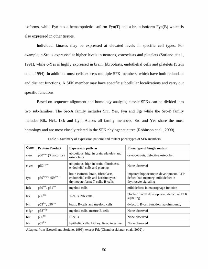
50
isoforms, while Fyn has a hematopoietic isoform Fyn(T) and a brain isoform Fyn(B) which is
also expressed in other tissues.
Individual kinases may be expressed at elevated levels in specific cell types. For
example, c-Src is expressed at higher levels in neurons, osteoclasts and platelets (Soriano et al.,
1991), while c-Yes is highly expressed in brain, fibroblasts, endothelial cells and platelets (Stein
et al., 1994). In addition, most cells express multiple SFK members, which have both redundant
and distinct functions. A SFK member may have specific subcellular localizations and carry out
specific functions.
Based on sequence alignment and homology analysis, classic SFKs can be divided into
two sub-families. The Src-A family includes Src, Yes, Fyn and Fgr while the Src-B family
includes Blk, Hck, Lck and Lyn. Across all family members, Src and Yes share the most
homology and are most closely related in the SFK phylogenetic tree (Robinson et al., 2000).
Table 3. Summary of expression patterns and mutant phenotypes of SFK members
Gene Protein Product Expression pattern Phenotype of Single mutant
c-src p60c-src
(3 isoforms) ubiquitous, high in brain, platelets and
osteoclasts osteopetrosis, defective osteoclast
c-yes p62c-yes
ubiquitous, high in brain, fibroblasts,
endothelial cells and platelets None observed
fyn p59fyn(B)
p59fyn(T)
brain isoform: brain, fibroblasts,
endothelial cells and keritinocytes;
thymocyte form: T-cells, B-cells.
impaired hippocampus development, LTP
defect, bad memory; mild defect in
thymocyte signaling
hck p59hck
, p61hck
myeloid cells mild defects in macrophage function
lck p56lck
T-cells, NK cells blocked T-cell development; defective TCR
signaling
lyn p53lyn
, p56lyn
brain, B-cells and myeloid cells defect in B-cell function, autoimmunity
c-fgr p58c-fgr
myeloid cells, mature B-cells None observed
blk p56blk
B-cells None observed
frk p57frk
Epithelial cells, kidney, liver, intestine None observed
Adapted from (Lowell and Soriano, 1996), except Frk (Chandrasekharan et al., 2002) .

51
1.2.3.2 SFK knockout phenotype and implication for function
Src family kinases have been implicated in many biological processes such as cell
adhesion, migration, proliferation and survival. SFK members are activated by adhesion
receptors like integrins, by receptor tyrosine kinases including the PDGFR, EGFR and IGFR,
and by cytokine receptors including the IL-2 receptor. Active SFKs can activate and cooperate
with the FAK, Ras-Raf-MAPK and PI3K pathways to reorganize the cytoskeleton, to promote
cell cycle progression and to prevent apoptosis (Thomas and Brugge, 1997). These signaling
pathways are interconnected, and involve multiple SFK members. Since cells usually express
multiple SFKs, the same upstream signal can act on multiple SFK members, which function
redundantly. Many studies of SFK function are based on v-Src, the constitutively active, viral
form of the kinase. Although the v-Src function is valuable to predict the physiological function
of c-Src, it is not ideal since SFKs are usually turned on transiently to function. Therefore,
genetic studies using knockout mice are of great value to study the redundant and unique
signaling in SFKs. A summary of phenotypes for individual SFK knockout mice is summarized
in Table 3 (Lowell and Soriano, 1996).
The first SFK disruption study in mice was reported for Src (Soriano et al., 1991).
Despite the high expression of Src in brain, platelets and osteoclasts, only osteoclast function
was defective in Src-null mice. Lack of Src caused osteopetrosis, and most of the homozygous
mice died several weeks after birth. Subsequent studies showed that osteoclasts require Src
activity downstream of integrin signaling to function (Miyazaki et al., 2004). Fyn disruption
mainly resulted in neuronal defects, with defective hippocampal development and long-term
potentiation (Grant et al., 1992). Fyn mutation also had a mild effect on thymocytes by
disrupting their T-cell receptor (TCR) signaling (Stein et al., 1992). Remarkably, no phenotype
52
was observed in mice with disrupted c-Yes, which has a very similar gene expression profile to
c-Src and Fyn (Stein et al., 1994). In the same study, compound knockouts of Src, Yes and Fyn
were created by crossing the single mutant animals. Most of the Src/Fyn or Src/Yes double
mutants died perinatally, while a substantial portion of the Fyn/Yes double mutants were viable.
Histopathological studies of the rare, viable Src/Fyn or Src/Yes double mutant mice failed to
reveal abnormalities other than the osteopetrosis caused by the Src mutation. The surviving
Fyn/Yes double mutant mice developed immune complex glomerulonephritis, a renal defect
(Stein et al., 1994). In another study involving Hck/Src double mutant mice, two thirds died at
birth, and the rest developed a more severe form of osteopetrosis compared with Src mutant
only, indicating that Hck can partially compensate for the absence of Src (Lowell et al., 1996).
In contrast to the relatively mild phenotype of the single and double SFK knockout mice,
triple disruption of Src, Yes and Fyn simultaneously results in embryonic lethality. This triple
knockout leads to embryonic death at E9.5 and is associated with severe developmental defects.
For example, the embryos are not “turned”, with inverted germ layer orientation (Klinghoffer et
al., 1999). In addition, disruption of Csk, a master regulator of SFK activity, also had a severe
phenotype (Nada et al., 1993; Imamoto and Soriano, 1993). Embryos homozygous with Csk
mutant die between E9 and E10, with complex neural tube and other defects. In Csk knockout
embryos, overall SFK activity is greatly enhanced with increased phosphorylation of substrate
proteins.
Gene disruption studies, highlighted above, suggest that SFKs are essential for embryo
development but also may function redundantly. Single or double deletions of Src family
members have a relatively mild phenotype, while triple deletion of Src, Yes, and Fyn, or deletion
of Csk to upregulate all SFK activity, causes severe defects and embryonic lethality. Although

53
the knockout studies reveal some unique functions for individual members (Lck in T-cell
development and Src in osteoclasts, for example), functional redundancy and compensation is
predominant. Since cells express multiple SFKs, the function of one or two SFK members can be
substituted with other members expressed. The surrogate SFK members can be induced for
higher expression, higher activity, or change cellular their localization as possible mechanisms of
compensation. For example, Src has increased solubility in the brain of Fyn knockout mice,
supporting this notion (Stein et al., 1994).
1.2.4 SFK signaling
Src family tyrosine kinases play critical roles in many cell functions by coupling with upstream
receptors and cell-adhesion signaling components. SFKs can be activated by integrin and other
adhesion receptors, receptor tyrosine kinases, cytokine receptors, G-protein coupled receptors
and immune response receptors (Thomas and Brugge, 1997; Parsons and Parsons, 2004). Upon
activation, SFKs phosphorylate substrates in multiple locations of the cell including the cytosol,
peri-plasma membrane, cellular compartments, or cell adhesion sites. SFK-mediated
phosphorylation either directly affects substrate kinase activity, or creates additional binding
sites for more SH2 domain-containing signaling molecules (Martin, 2001). Here, I emphasize
SFK signaling with receptor tyrosine kinases and cell adhesion receptors. These interactions with
signaling partners have been implicated in cancer biology, and similar interactions and functions
are also conserved in ES cells.

54
1.2.4.1 Signaling with receptor tyrosine kinases
SFKs can be activated by many receptor tyrosine kinases, including the platelet-derived
growth factor receptor (PDGFR), epidermal growth factor receptor (EGFR), fibroblast growth
factor receptor (FGFR) and insulin-like growth factor-1 receptor (IGF1R). Activated receptor
tyrosine kinases have been propose to engage SFKs by binding of p-Tyr residues on the active
receptor with the SH2 domain of SFKs, initiating the activation of SFKs through a regulatory
domain displacement model as described above (Bromann et al., 2004). SFKs can then activate
multiple signaling pathways by recruiting and phosphorylating substrates directly, or by creating
binding sites in docking proteins. These events ultimately lead to many biological effects,
including changes in the cytoskeleton and in gene expression (Figure 6).
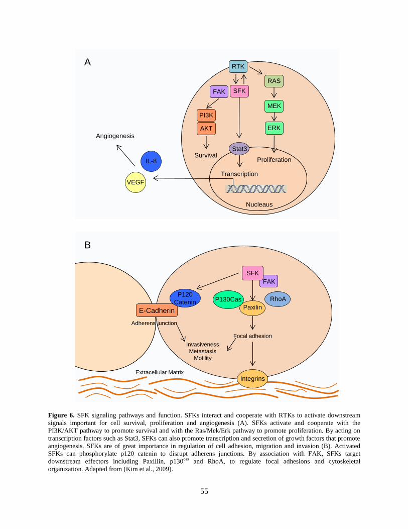
55
A
B
SFK
RTK
FAK
ERK
MEK
RAS
SFK
AKT
Stat3
PI3K
VEGF
IL-8Survival
Proliferation
Transcription
Angiogenesis
Nucleaus
Extracellular Matrix
Paxilin
Integrins
RhoA
FAK
E-Cadherin
P130CasP120
Catenin
Adherens junction
Invasiveness
Metastasis
Motility
Focal adhesion
Figure 6. SFK signaling pathways and function. SFKs interact and cooperate with RTKs to activate downstream
signals important for cell survival, proliferation and angiogenesis (A). SFKs activate and cooperate with the
PI3K/AKT pathway to promote survival and with the Ras/Mek/Erk pathway to promote proliferation. By acting on
transcription factors such as Stat3, SFKs can also promote transcription and secretion of growth factors that promote
angiogenesis. SFKs are of great importance in regulation of cell adhesion, migration and invasion (B). Activated
SFKs can phosphorylate p120 catenin to disrupt adherens junctions. By association with FAK, SFKs target
downstream effectors including Paxillin, p130cas
and RhoA, to regulate focal adhesions and cytoskeletal
organization. Adapted from (Kim et al., 2009).

56
SFKs can activate PI3K by phosphorylation, or up-regulate PI3K signaling by inhibiting
its antagonist—PTEN. PI3K/Akt signaling in turn regulates translation initiation, prevents
apoptosis and promotes cell survival. In addition, Src can activate the Mek/Erk pathway by
recruiting the Grb2-Sos complex or Shc with docking proteins. The Mek/Erk pathway can then
promote proliferation and regulate gene expression. In addition, SFKs can phosphorylate Stat3,
which in turn regulates the expression of transcription factor Myc, contributing to the Myc
regulation of cell growth and proliferation (Kim et al., 2009; Martin, 2001) (Figure 6).
1.2.4.2 Signaling with integrin and focal adhesion kinase
Both focal adhesions and adherens junctions are necessary for cell attachment, motility,
and are important for cell proliferation, survival and differentiation. Focal adhesions are the
interactions of cells with the extracellular matrix (ECM) via integrins or intracellular
transmembrane receptors, while adherens junctions are direct cell-cell connections regulated by
cadherin-catenin mediator complexes. SFKs play an important role in these important adhesive
events which also have a central role in cancer progression (Kim et al., 2009; Ishizawar and
Parsons, 2004; Guarino, 2010).
Integrins can sense changes in the cellular environment, triggering signaling pathways to
adapt the cytoskeletal organization accordingly. SFKs transmit the integrin-dependent signal.
SFKs are directly activated by integrin β, and through engagement with focal adhesion kinase
(FAK), to modulate downstream signals including RhoA and p120 catenin (Huveneers and
Danen, 2009). c-Src and FAK form a complex, with the SH2 domain of Src binding to pTyr397
of FAK. This binding facilitates Src phosphorylation of FAK on multiple tyrosine sites to
57
enhance FAK activity. Active Src-FAK complexes phosphorylate paxillin and p130Cas,
important modulators of cell migration. The Src-FAK comples also activates matrix
metalloproteinases (MMP) 2 and 9, which also contribute to cell migration and invasion. Src also
phosphorylates p120 catenin to disrupt its association with E-cadherin, resulting in enhanced cell
migration (Kim et al., 2009). In addition, active Src drives the activation of multiple small G-
proteins, including the Rho-family GTPases Rac, Cdc42 and Rho, to regulate adhesion and
cytoskeleton organization. In summary, SFKs have important roles in regulating cell adhesion
and migration, which are implicated in cancer progression and invasion. In addition, coordinated
cellular migration and cell:cell contacts are critical to early development, which may explain the
dramatic impact of Csk-knockout on embryogenesis.
1.3 SFK SIGNALING IN MURINE ES CELLS
As summarized in the previous section, SFKs are regulated by many upstream signals and have a
prominent role in the coordinated regulation of cell adhesion, proliferation, growth and survival.
Loss of regulation of these pathways can contribute to the pathogenesis of cancer and also
interfere with early embryogenesis. Previous work from our group and others has shown that
SFKs also play important roles in embryonic stem cell self-renewal and differentiation.

58
1.3.1 SFK expression and function in mES cells and EBs
Several years ago, our group made the unexpected discovery that seven of the eight classical
SFKs—Src, Yes, Fyn, Hck, Lck, Lyn and Fgr—are expressed in mES cells. We also found that
when ES cells were induced to differentiate into EBs, Hck and Lck were quickly silenced
transcriptionally, while Src, Fyn expression was retained (Meyn, III et al., 2005). Around the
same time, the Melton lab reported that c-Yes activity was down-regulated during EB
differentiation, although its expression was maintained at the transcript level (Anneren et al.,
2004). These results indicate that SFK members are differentially regulated in ES cells and EBs,
suggesting that individual SFKs may play discrete, non-redundant roles in mES cells. To study
SFK activity and function in ES cells, small molecule inhibitors were introduced (Meyn, III et
al., 2005). While partial inhibition of endogenous SFK activity with the ATP-competitive
inhibitors SKI-1 and PP2 induced differentiation of ES cells in the presence of LIF, suppression
of all SFK activity using higher concentrations of these inhibitors or with the more potent
inhibitor A-419259 blocked differentiation in the absence of LIF (Meyn, III et al., 2005). The
specificity of these inhibitors against individual SFK members was also explored in this study
using in vitro kinase assays. Interestingly, c-Yes and Hck were found to be more sensitive to
SKI-1 treatment than c-Src and Fyn, suggesting that selective inhibition of Hck and Yes with low
inhibitor concentrations may account for the differentiation observed in ES cells. These
observations led to signaling model in which SFKs play non-redundant and opposing roles in ES
cells (Figure 7A), with Hck and Yes promoting renewal while Fyn and Src control
differentiation (Meyn, III et al., 2005).
This model is also supported by other studies linking both Hck and Yes to ES cell
renewal. Early studies showed that Hck activity increased following LIF treatment of ES cells.
59
Hck was shown to physically associate with gp130, and expression of an active tail mutant of
Hck reduced the LIF requirement for ES cell self-renewal (Ernst et al., 1994). Consistent with
these observations, transcription of Hck was rapidly silenced as ES cells differentiate to EBs
(Meyn, III et al., 2005). Other work linked c-Yes to ES cell renewal. Unlike Hck, c-Yes is
expressed both in pluripotent ES cells and in differentiated EBs (Anneren et al., 2004). While c-
Yes kinase is active in ES cells, where it is regulated by both LIF and serum, its activity is
downregulated during differentiation to EBs. RNAi-mediated knockdown of c-Yes expression
reduces expression of the renewal marker Nanog while increasing expression of the
differentiation marker, GCNF, supporting an inhibitory role for c-Yes in ES cell differentiation.
Transcription of c-Yes in ES cells is regulated by the pluripotency factor Oct4, further
supporting a role for c-Yes in renewal (Zhang et al., 2007). In contrast to Hck and c-Yes, c-Src
and Fyn expression and activity persists when ES cells differentiate, indicating that these two
kinases may function in differentiation (Meyn, III et al., 2005). Using a chemical genetics
approach, our group found that c-Src activity alone is sufficient to induce differentiation of ES
cells to primitive ectoderm (Meyn, III and Smithgall, 2009), further supporting Src as a
differentiation related kinase. Details of this approach are described in the next section.
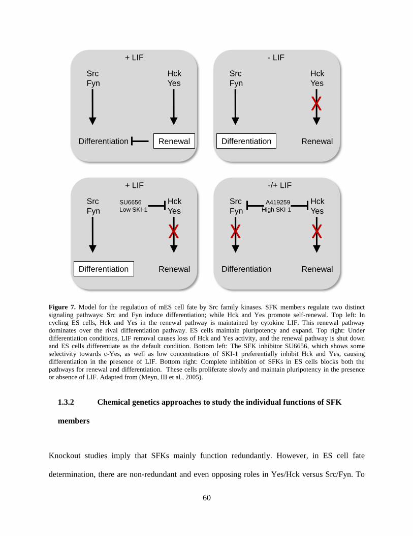
60
Hck
Yes
Src
Fyn
RenewalDifferentiation
+ LIF
Hck
Yes
Src
Fyn
RenewalDifferentiation
- LIF
Hck
Yes
Src
Fyn
RenewalDifferentiation
+ LIF
Hck
Yes
Src
Fyn
RenewalDifferentiation
-/+ LIF
SU6656
Low SKI-1
X
X X X
A419259
High SKI-1
Figure 7. Model for the regulation of mES cell fate by Src family kinases. SFK members regulate two distinct
signaling pathways: Src and Fyn induce differentiation; while Hck and Yes promote self-renewal. Top left: In
cycling ES cells, Hck and Yes in the renewal pathway is maintained by cytokine LIF. This renewal pathway
dominates over the rival differentiation pathway. ES cells maintain pluripotency and expand. Top right: Under
differentiation conditions, LIF removal causes loss of Hck and Yes activity, and the renewal pathway is shut down
and ES cells differentiate as the default condition. Bottom left: The SFK inhibitor SU6656, which shows some
selectivity towards c-Yes, as well as low concentrations of SKI-1 preferentially inhibit Hck and Yes, causing
differentiation in the presence of LIF. Bottom right: Complete inhibition of SFKs in ES cells blocks both the
pathways for renewal and differentiation. These cells proliferate slowly and maintain pluripotency in the presence
or absence of LIF. Adapted from (Meyn, III et al., 2005).
1.3.2 Chemical genetics approaches to study the individual functions of SFK
members
Knockout studies imply that SFKs mainly function redundantly. However, in ES cell fate
determination, there are non-redundant and even opposing roles in Yes/Hck versus Src/Fyn. To
61
elucidate the mechanism, it is necessary to investigate the individual role of each SFK member in
ES cells. One approach is use RNA interference to knockdown one member at a time. For
example, RNAi knockdown of Yes caused ES cells to lose renewal marker expression and an
increase differentiation (Anneren et al., 2004). However, the knockdown approach suffers from
the same limitation as knockout mice, in that functional compensation may occur during the
selection process required to establish the knock-down cell population. In addition, the
knockdown approach, like the genetic knockouts and in contrast to pharmacological inhibition,
eliminates the kinase protein entirely. Another approach is the use of selective kinase inhibitors.
However, because of the highly conserved nature of Src-family kinase domains, truly selective
ATP-competitive inhibitors for individual SFKs are not currently available and may be
impossible to develop. An alternative is to use a chemical genetics approach—combining small
molecule kinase inhibitors with genetic mutations to achieve selective inhibition or resistance for
a specific kinase (Bishop et al., 2000).
The crystal structure of Hck in complex with SFK inhibitor PP1, revealed the structural
basis of PP1 specificity against SFKs (Schindler et al., 1999). In the inactive conformation, the
SFK has a hydrophobic cavity with an opening toward the ATP binding pocket. As an ATP
competitive inhibitor, PP1 binds to the ATP pocket, occupying the site normally bound by the
adenine base of ATP. Several SFK residues are critical for PP1 binding, including Thr-338, Glu-
339, Met-341 and Lys-295, all of which form hydrogen bonds with PP1. These hydrogen bonds
orient PP1 to a position with its 3-substituted pheny group sticking into the hydrophobic pocket.
Accessibility of inhibitors to this hydrophobic pocket confers specificity, and is controlled by a
critical residue Thr-338, often referred to as the “gatekeeper” residue (Liu et al., 1999).
Alignment of the kinase domains of PP1-sensitive SFKs with those of PP1-resistant kinases such

62
as PKA and Jak2 reveals that the PP1-resistant kinases have methionine instead of threonine in
the gatekeeper position (Schindler et al., 1999). This larger methionine residue creates steric
clash with the inhibitor, and limits inhibitor accessibility to the hydrophobic pocket. Therefore,
mutation of the gatekeeper Thr to a bulkier residue such as methionine can confer inhibitor
resistance to SFKs. Note also that the amino acid occupying the gatekeeper position is not
highly conserved across different kinase families, and can be substituted experimentally without
compromising kinase activity.
Using this chemical genetics approach, our group designed gatekeeper mutants of all
SFK members expressed in ES cells to be resistant to the broad-spectrum (non-selective) SFK
inhibitors PP2 and A419259 (Meyn, III and Smithgall, 2009). These mutants were introduced
into mES cells with recombinant retroviruses. This combination of inhibitor resistant (IR)
mutants with inhibitors allowed the investigation of individual SFK function in ES cells. In the
presence of 1 µM of A419259, which was previously shown to inhibit all endogenous SFK
activity, ES cells formed small rounded colonies that were unable to differentiate. In contrast, the
ES cells expressing Src-IR mutants adopted a flattened morphology even in the presence of LIF.
Compared with untreated cells, inhibitor-treated Src-IR cells showed reduced Gbx2 and Rex1
expression and an increased level of Fgf5 expression, resembling primitive ectoderm-like cells.
This study showed that Src activity alone is sufficient to drive murine ES cells towards primitive
ectoderm differentiation (Meyn, III and Smithgall, 2009).
1.3.3 A role for Src family kinase c-Yes in ES cell regulation
In contrast to c-Src as a differentiation-related kinase, c-Yes has been related to self-renewal in
ES cells. c-Yes is highly expressed in both human and mouse ES cells, and is activated by both

63
serum and LIF (Anneren et al., 2004). LIF can induce interaction between gp130 and the Yes
SH2 domain, suggesting a possible mechanism for Yes activation following LIF stimulation
(Tamm et al., 2011). EB differentiation correlated with down-regulation of c-Yes activity in both
human and mouse ES cells. Knockdown of c-Yes with RNAi induced ES cell differentiation,
with reduced expression of Oct4 and Nanog (Anneren et al., 2004). Moreover, Oct4 was shown
to bind to the Yes promoter and to upregulate Yes expression (Zhang et al., 2007). These studies
all support c-Yes as a renewal-related gene in mouse ES cells.
More recent work showed that active c-Yes controls the TEAD2 transcription factor
through the Yes-associated protein, YAP. Active YAP-TEAD2 complexes bind to DNA in the
nucleus and promote transcription of renewal related genes, including the master regulators Oct4
and Nanog (Tamm et al., 2011). Yes, Yap and Tead2 are all highly expressed in ES cells, with
expression down-regulated in response to differentiation. Active c-Yes phosphorylates YAP and
activates YAP-TEAD2 dependent transcription. In addition, activation of this pathway promotes
the expression of Oct4 and Nanog, and its suppression induces ES cell differentiation. Moreover,
suppression of TEAD2 function increased the expression of endoderm specific genes, indicating
that this pathway might suppress endoderm differentiation in ES cells.
1.4 HYPOTHESIS AND SPECIFIC AIMS
1.4.1 Hypothesis
Previous work, highlighted above, has implicated c-Yes in the regulation of ES cell pluripotency.
Whether or not c-Yes kinase activity alone is sufficient to maintain this essential property of ES
64
cells has not been explored. Since c-Yes activity is downregulated in response to EB formation,
and RNAi knockdown of c-Yes induced differentiation in the presence of LIF (Anneren et al.,
2004), I hypothesize that enforced expression of c-Yes may be sufficient to drive self-renewal
and/or block EB differentiation of mES cells. In addition, c-Src, the closest phylogenetic relative
of c-Yes, induces differentiation (Meyn, III and Smithgall, 2009), in direct opposition to c-Yes.
Whether c-Yes is dominant over c-Src, or vice versa is still unknown. In the case of transcription
factors, pluripotent factors are dominant over differentiation factors; it might be the same case in
this pair of opposing kinases. Therefore, I hypothesize that the pro-renewal, or anti-
differentiation signal of c-Yes, is dominant over the differentiation-inducing signal of c-Src.
Finally, while previous research clearly defined the importance of SFKs in mES cells,
their roles in hES cells are largely unknown. Some evidence suggests that although the growth
factor conditions for hES cell growth are disparate from those of mES cells, the downstream
signaling pathways might be similar. SFKs may represent one of those downstream pathways. I
hypothesize that like mES cells, hES cells express multiple SFK members and SFKs may also
play divergent roles in hES cells self-renewal and differentiation.
1.4.2 Specific Aims
1.4.2.1 Aim 1: Investigate the contribution of c-Yes to the growth and self-renewal of
murine ES cells and test the opposing roles of c-Src and c-Yes in ES cell differentiation.
In mES cells, c-Yes has been implicated in proliferation and self-renewal. To better
understand the regulation of c-Yes in mES cell self-renew and differentiation, I first showed that
c-Yes transcript levels were unchanged during ES cell differentiation, while its kinase activity
was downregulated. To determine whether downregulation of c-Yes kinase activity is required
65
for differentiation, I introduced wild-type, inactive and active mutants of c-Yes into mES cells
using recombinant retroviral vectors. I found that enforced expression of active c-Yes did not
change undifferentiated colony morphology or marker expression in the presence of LIF, but
blocked ES cell differentiation to embryoid bodies. This differentiation block was not observed
with kinase-defective c-Yes, indicating a requirement for kinase activity. Further, qPCR analysis
showed that mES cells expressing active forms of c-Yes continued to express pluripotency
markers under conditions for EB formation, suggesting that c-Yes signals for renewal override
the normal differentiation program.
Despite their close phylogenetic similarity, c-Yes and c-Src appear to have opposing
roles in ES cell differentiation. To explore the interplay of c-Src and c-Yes in ES cell renewal
and differentiation, I employed a chemical genetics approach in engineering c-Src and c-Yes
mutants that are resistant to SFK inhibitor A-419259. Previous studies have shown that A-
419259 treatment blocks all SFK activity in ES cells, preventing differentiation while
maintaining pluripotency. Expression of inhibitor-resistant (IR) c-Src alone rescues the A-
419259 differentiation block, resulting in primitive-ectoderm differentiation (Meyn, III and
Smithgall, 2009). Unlike the Src-IR cells, inhibitor treatment of mES cells expressing Yes-IR did
not show changes in lineage marker expression indicative of differentiation. This observation is
consistent with the proposed anti-differentiation function of c-Yes in ES cells. Next, I tested
whether the c-Yes signal for anti-differentiation or the c-Src signal for pro-differentiation is
dominant by expressing both IR-mutants in the same ES cell population. I expanded our previous
study to show that c-Src induced ES cell differentiation to both primitive ectoderm and
endoderm, and promoted epithelial-mesenchymal transition (EMT). Remarkably, c-Yes activity
suppressed c-Src mediated endoderm differentiation and EMT. Together, these results define c-
66
Yes as a potent blocker of ES cell differentiation that can antagonize the primitive endoderm
differentiation induced by c-Src; and show even closely related kinases such as c-Src and c-Yes
have unique and opposing functions in the same cell type.
1.4.2.2 Aim 2: Study SFK signaling in human ES cell self-renewal and differentiation.
While our findings in Aim 1 have clearly defined the importance of Src family kinases in
mES cells, their roles in hES cells are less clear. To investigate SFK signaling in hES cells, I first
used RT-PCR and qPCR to determine the relative expression profile of SFKs in hES cells vs.
EBs derived from them. Expression of six Src-family kinases (Fyn, c-Yes, c-Src, Lyn, Lck and
Hck) was detected in the hES cell lines H1 and H9. During embryoid body formation, Fyn and
Src transcript levels increased slightly, while c-Yes and Lyn levels remained unchanged.
Conversely, Lck and Hck expression levels dropped dramatically. To explore if SFK activity is
required for hES cell maintenance and differentiation, I treated hES cells with the pan-SFK
inhibitor A-419259, and found that the treated cells retained the morphology of pluripotent
colonies and continued to express the pluripotency marker TRA-1-60 under culture conditions
for differentiation. Taken together, these results support an important role for Src family kinase
signaling in the regulation of hES cell fate, and suggest unique functions for individual SFKs in
hES cells as observed previously in mES cells.
67
2.0 THE C-YES TYROSINE KINASE IS A POTENT SUPPRESSOR OF ES CELL
DIFFERENTIATION AND ANTAGONIZES THE ACTION OF ITS CLOSEST
PHYLOGENETIC RELATIVE, C-SRC
2.1 ABSTRACT
ES cells are derived from the inner cell mass of the blastocyst stage embryo and are
characterized by self-renewal and pluripotency. Previous work has implicated the Src family of
protein-tyrosine kinases in the self-renewal and differentiation of mES cells. These kinases
display dynamic expression and activity changes during ES cell differentiation, suggesting
distinct functions in the control of developmental fate. Here we used ES cells to test the
hypothesis that c-Src and its closest phylogenetic relative, c-Yes, act in biological opposition to
one another despite their strong homology. We first showed that enforced expression of active c-
Yes blocked ES cell differentiation to embryoid bodies by maintaining pluripotency gene
expression. To explore the interplay of c-Src and c-Yes in ES cell renewal and differentiation,
we employed a chemical genetics approach. We engineered c-Src and c-Yes mutants that are
resistant to A-419259, a potent pyrrolopyrimidine inhibitor of the Src kinase family. Previous
studies have shown that A-419259 treatment blocks all Src-family kinase activity in ES cells,
preventing differentiation while maintaining pluripotency. Expression of inhibitor-resistant c-Src
but not c-Yes rescued the A-419259 differentiation block, resulting in a cell population with
68
properties of both primitive ectoderm and endoderm. Remarkably, when inhibitor-resistant c-Src
and c-Yes were expressed together in ES cells, c-Yes activity suppressed c-Src mediated
differentiation. These studies show that even closely related kinases such as c-Src and c-Yes
have unique and opposing functions in the same cell type. Selective agonists or inhibitors of c-
Src vs. c-Yes activity may allow more precise pharmacological manipulation of ES cell fate and
have broader applications in other biological systems which express multiple Src family
members such as tumor cells.
2.2 INTRODUCTION
Embryonic stem (ES) cells are derived from the inner cell mass of the developing blastocyst
(Evans and Kaufman, 1981; Martin, 1981). ES cells are characterized by self-renewal, the ability
to multiply indefinitely without differentiation (Suda et al., 1987), and pluripotency, the
developmental potential to generate cell types from all three germ layers (Bradley et al., 1984;
Beddington and Robertson, 1989). In the absence of feeder cell layers, ES cells can be
maintained in an undifferentiated state by culturing them in serum-based medium supplemented
with the cytokine, leukemia inhibitory factor (LIF) (Smith et al., 1988) or in defined medium in
the presence of LIF and bone morphogenetic proteins (BMPs) (Ying et al., 2003). In the absence
of LIF, ES cells differentiate to embryoid bodies (EBs) when cultured under non-adherent
conditions. EB formation mimics the earliest stages of embryonic development, giving rise to all
three germ layers (Keller, 1995; Keller, 2005).
69
Multiple intracellular kinase signaling pathways play a dominant role in the regulation of
ES cell fate (Liu et al., 2007; Jaenisch and Young, 2008), with at least four pathways important
for self-renewal. LIF signals through Janus kinases (JAKs) and signal transducer and activator
of transcription 3 (STAT3). This pathway promotes expression of renewal factors including the
POU domain transcription factor, Oct4 (Okamoto et al., 1990), and the homeobox transcription
factor, Nanog (Niwa et al., 1998; Burdon et al., 1999a). Bone morphogenetic proteins (BMPs),
which are serum components, activate transcription factors of the SMAD family and inhibit
differentiation through induction of inhibitor of differentiation (ID) factors (Ying et al., 2003).
Wnt proteins, which are also found in serum, inhibit glycogen synthase kinase-3β activity,
leading to β-Catenin accumulation and pluripotency marker gene expression (Sato et al., 2004;
Wray et al., 2011). In addition, the phosphatidylinositol 3’-kinase (PI3K) signaling pathway
promotes ES cell self-renewal partly via regulation of Nanog expression (Paling et al., 2004;
Storm et al., 2007).
Previous work has implicated the Src family of non-receptor protein tyrosine kinases in
self-renewal and differentiation of murine ES cells as well(Anneren et al., 2004; Meyn, III et al.,
2005). Seven of the eight mammalian Src family members are expressed in murine ES cells, and
several family members are active in cycling ES cells cultured in the presence of LIF and serum
(c-Src, c-Yes, Fyn, and Hck). Accumulating evidence supports the hypothesis that individual
members of this kinase family may play distinct roles in regulating ES cell fate. For example,
early studies showed that expression of an active mutant of the Src-family kinase Hck reduces
the LIF requirement for ES cell self-renewal, implicating Hck in the suppression of
differentiation (Ernst et al., 1994). More recent studies from our group showed that transcription
of Hck is rapidly silenced as ES cells differentiate to EBs, consistent with this idea (Meyn, III et
70
al., 2005). In contrast to Hck, c-Src is expressed in both ES cells and differentiated EBs.
Moreover, when c-Src remains active in the absence of all other Src-family kinase activity, it is
sufficient to induce differentiation of ES cells (Meyn, III and Smithgall, 2009).
Other work has linked c-Yes, the closest phylogenetic relative of c-Src, to the
suppression of ES cell differentiation. Like c-Src, c-Yes is expressed in both pluripotent ES cells
and in differentiated EBs (Anneren et al., 2004). While the c-Yes kinase is active in self-
renewing ES cells, where it is regulated by both LIF and serum, its activity is downregulated
during differentiation. RNAi-mediated knockdown of c-Yes expression reduces expression of the
renewal factor Nanog while increasing expression of the differentiation marker, GCNF.
Transcription of c-Yes in ES cells is regulated by the pluripotency factor Oct4, supporting a role
for c-Yes in self-renewal (Zhang et al., 2007). Recent work shows that active c-Yes controls the
TEAD2 transcription factor through the Yes-associated protein, YAP (Tamm et al., 2011).
Active YAP-TEAD2 complexes bind Oct4 promoters, supporting a positive feedback loop
between c-Yes and Oct4 in self-renewal.
In this study we examined the biological interplay of c-Yes and c-Src, closely
homologous kinases previously shown to act in direct opposition to one another in ES cells.
First, we expressed c-Yes in mES cells using a retroviral vector system that drives low-level
protein expression in transduced ES cell populations (Meyn, III and Smithgall, 2009). EB
maturation was completely blocked in ES cells expressing active c-Yes, while EBs formed by ES
cells expressing a kinase-inactive c-Yes mutant were unaffected. EBs that formed from the c-
Yes-transduced ES cell population expressed both pluripotency and differentiation markers,
suggesting that c-Yes kinase activity prevents differentiation by maintaining expression of the
self-renewal program. Using a chemical genetics approach that permits only c-Yes and c-Src
71
signaling in ES cells in the absence of all other SFK signaling, we found that c-Yes interfered
with the induction of differentiation previously observed with c-Src in this system (Meyn, III and
Smithgall, 2009). In addition, we found that c-Yes also suppressed the induction of the
epithelial-mesenchymal transition (EMT) by c-Src. Together, these observations show that
despite their high degree of sequence similarity, c-Yes and c-Src have opposing roles in the
regulation of ES cell fate. Our results have important implications not only for the regulation of
ES cell differentiation, but many other cellular contexts in which multiple members of this
kinase family are expressed.
2.3 RESULTS
2.3.1 Downregulation of c-Yes kinase activity during differentiation of ES cells to
EBs
To monitor changes in c-Yes expression and activity during ES cell self-renewal and
differentiation, we compared endogenous c-Yes RNA levels, protein abundance and kinase
activity in self-renewing ES cells vs. EBs following 6 days in differentiation culture (6-day EBs).
For these experiments, we used the mES cell line D3, which readily forms EBs when plated
under non-adherent conditions in the absence of LIF (Figure 8A) (Meyn, III et al., 2005). Total
RNA was isolated from self-renewing ES cells and 6-day EBs, followed by quantitative real-time
RT-PCR (qPCR) analysis of c-Yes and c-Src transcript levels as well as markers of self-renewal
and differentiation. Differentiation to EBs resulted in negligible changes in the expression of c-

72
Yes and c-Src (Figure 8B). Expression of the self-renewal markers Oct4 and Nanog were
significantly down-regulated while the lineage-specific differentiation markers Gata4
(endoderm), Fgf5 (ectoderm) and T (mesoderm) were up-regulated in the 6-day EBs, consistent
with the onset of differentiation in the developing EBs.
To investigate c-Yes protein stability and activity as a function of differentiation, c-Yes
was immunoprecipitated from ES cells and 6-day EBs, followed by immunoblotting with a
phosphospecific antibody against the c-Yes activation loop tyrosine. As shown in Figure 8C,
activation loop tyrosine phosphorylation was greatly diminished in EBs relative to ES cells
despite equivalent recovery of c-Yes protein. Immunoblots also showed a dramatic reduction in
the level of the pluripotency marker, Oct4, consistent with differentiation. This result shows that
c-Yes kinase activity is downregulated during EB formation, consistent with a role for this Src-
family member in maintenance of pluripotency as originally proposed by Anneren et al.
(Anneren et al., 2004).

73
mES cells 6 Day EBs
ES
C
EB
s
pYes
Yes
Oct4
Actin
A
CB
Ex
pre
ss
ion
fo
ld c
ha
ng
e
rela
tiv
e t
o E
S c
ell
s
Yes
Src
Oct4
Nanog
GA
TA
4
Fgf5 T
2 -4
2 1
2 6
* *
*
*
Figure 8. Downregulation of c-Yes kinase activity during EB formation. A) Bright-field images of the mES cell line
D3 cultured under conditions for self-renewal (left) or differentiation to embryoid bodies (EBs) for 6 days (right).
Magnification, 100X. B) Src-family kinase and ES cell marker gene expression was determined by qPCR in self-
renewing ES cells and 6-day EBs. This panel of markers includes the self-renewal regulators Oct4 and Nanog as
well as the differentiation genes Gata4 (endoderm), Fgf5 (ectoderm/epiblast) and T (mesoderm). Analyses were
conducted using three biological replicates and the results are expressed as the average of the Log2 fold-change in
each marker level in EBs relative to ES cells ± S.E.M. (*P < 0.05, Pair-wise Fixed Reallocation Randomization
Test.) C) Lysates were prepared from ES cells and 6-day EBs, and endogenous c-Yes protein was
immunoprecipitated and blotted with a phosphospecific antibody for the active form of the kinase (pYes) as well as
the c-Yes protein. Cell lysates were also blotted for Oct4 as marker of ES cell self-renewal status and actin as a
loading control.

74
2.3.2 Validation of c-Yes retroviral expression constructs in Rat2 cells
This Results presented in the previous section support a role for c-Yes as a positive
regulator of ES cell pluripotency and suggest that downregulation of c-Yes kinase activity may
be required for ES cells to exit the self-renewal program. To test this hypothesis, we engineered a
series of c-Yes retroviral expression constructs based on a murine stem cell virus (MSCV)
promoter which we have previously shown to remain active in both self-renewing ES cells and
differentiated EBs (Meyn, III and Smithgall, 2009). In addition, the c-Yes cDNA clones were
coupled to a G418 selection marker via an internal ribosome entry site (IRES) to ensure stable
expression in the transduced cell populations.
Like all Src kinases, c-Yes consists of an N-terminal unique region, followed by SH3 and
SH2 domains, the kinase domain, and a negative regulatory tail (Engen et al., 2008). To create an
inactive mutant of c-Yes, we substituted Lys295 (numbering based on the c-Src crystal structure
(Xu et al., 1999)) in the kinase domain with Arg. In addition, we created a constitutively active
form of c-Yes by replacement of the negative regulatory tail tyrosine (Tyr527) with
phenylalanine (Figure 9A). To validate the retroviral expression vectors carrying wild-type and
mutant forms of c-Yes, we used Rat2 fibroblasts as a model system. These cells provide a
convenient assay that relates Src-family kinase activity with biological function (transformed
colony formation (Pene-Dumitrescu et al., 2008; Pene-Dumitrescu and Smithgall, 2010)).
Rat2 fibroblasts were infected with recombinant retroviruses carrying wild-type, kinase-
dead and kinase-active forms of c-Yes, followed by G418 selection. Each of the transduced Rat2
cell populations was then plated in soft agar colony assays for anchorage independent growth as
a measure of c-Yes biological function. As shown in Figures 9B and 9C, cells expressing the
active mutant of c-Yes produced a large number of transformed colonies, while no colonies were

75
observed with cells expressing wild-type c-Yes or the kinase-dead mutant. Expression of c-Yes
from the retroviral vector was confirmed by RT-PCR with total RNA isolated from each cell
population and a primer pair specific for the c-Yes transgene (Figure 9D).
To assay for c-Yes kinase activity in each of the Rat2 cell populations, c-Yes was
immunoprecipitated followed by immunoblotting for protein recovery and activation loop
tyrosine phosphorylation. Figure 9E shows a small increase in the quantity of c-Yes protein
recovered from cells expressing the c-Yes cDNAs relative to the vector controls. The tail mutant
of c-Yes reacted strongly with the phosphospecific antibody, consistent with elevated kinase
activity and transforming function. In contrast, c-Yes recovered from cells expressing the wild-
type protein showed only a modest increase in reactivity with the activation loop antibody
relative to endogenous c-Yes. This result demonstrates that the level of wild-type c-Yes over-
expression achieved with the MSCV-based retrovirus was not sufficient to cause kinase
upregulation, consistent with the lack of transformed colony formation.
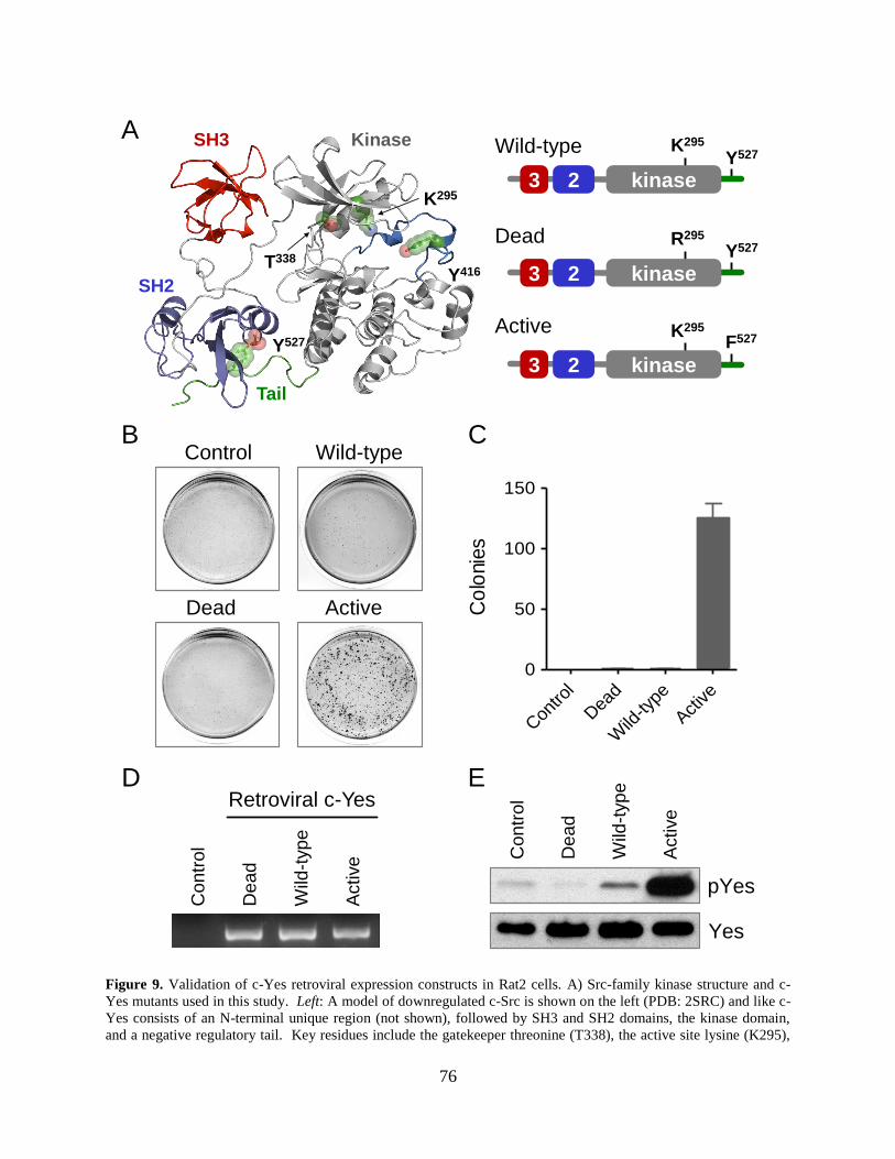
76
Wild-type
Dead
Active
Control
Dead
Wild-type
Active
Yes
pYes
Co
lonie
s
Con
trol
Dea
d
Wild
-type
Active
0
50
100
150
A
E
B C
3 2 kinase
Retroviral c-Yes
Co
ntr
ol
Wild
-typ
e
Active
De
ad
D
Co
ntr
ol
Wild
-typ
e
Active
De
ad
SH3
SH2
Tail
Y416
Kinase
K295
T338
Y527
3 2 kinase
3 2 kinase
K295
Y527
R295
Y527
K295
F527
Figure 9. Validation of c-Yes retroviral expression constructs in Rat2 cells. A) Src-family kinase structure and c-
Yes mutants used in this study. Left: A model of downregulated c-Src is shown on the left (PDB: 2SRC) and like c-
Yes consists of an N-terminal unique region (not shown), followed by SH3 and SH2 domains, the kinase domain,
and a negative regulatory tail. Key residues include the gatekeeper threonine (T338), the active site lysine (K295),
77
the activation loop tyrosine (Tyr416), and the tail tyrosine (Y527). Right: Substitution of Lys295 in the kinase
domain with arginine renders the kinase inactive (Dead), while replacement of the tail tyrosine (Y527) with
phenylalanine produces a constitutively active mutant (Active). B, C) The wild-type and mutant forms of c-Yes
shown in part A were introduced into Rat2 fibroblasts using recombinant retroviruses. Following selection with
G418, control and c-Yes-transduced fibroblasts were cultured in soft-agar colony assays for the transformed
phenotype. Images of representative plates are shown in part B, and the average number of transformed colonies
formed from each cell population is shown in Part C ± S.D.; n=3. D) RT-PCR indicates virus-specific c-Yes
expression. RNA was isolated from each of the four Rat2 cell populations shown in part B, and RT-PCR was
performed with a primer pair specific for the c-Yes transgene. A scanned image of the resulting agarose gel shows
the expected 735 base-pair product. E) Kinase activity. c-Yes proteins were immunoprecipitated from each of the
Rat2 cell populations and blotted with a phosphospecific antibody for the active form of the kinase (pYes) as well as
the c-Yes protein.
2.3.3 Low-level retroviral expression of c-Yes in ES cells does not affect
undifferentiated colony morphology or marker expression.
To test the effect of these c-Yes constructs on self-renewal, cultures of ES cells were transduced
with recombinant retroviruses carrying wild-type, kinase-active and kinase-dead forms of c-Yes
as described in the preceding section. Following selection with G418, each ES cell population
formed undifferentiated colonies indistinguishable from control cells transduced with the empty
vector (Figure 10A). RT-PCR was performed using total RNA isolated from each cell
population and a primer pair specific for the c-Yes transgene. Figure 10B shows that the c-Yes
retroviral transgene is expressed in each case but was not observed in RNA isolated from control
cells transduced with the empty retroviral vector. The presence of the kinase domain mutations
was confirmed in the RT-PCR products from the transduced cell lines by DNA sequence analysis
(data not shown). The relative growth rates of the c-Yes-transduced ES cell populations were
indistinguishable from that of the control cells (data not shown). Finally, qPCR analysis showed
that the expression levels of the pluripotency markers Rex1, Nanog and Oct4, as well as the
differentiation markers Fgf5 (ectoderm) and T (mesoderm) were essentially unchanged in each
of the c-Yes-transduced ES cell populations relative to control ES cells (Figure 10C). Taken

78
together, these results indicate that retroviral transduction with c-Yes expression vectors did not
effect, de novo, the regulation of self-renewal in cycling mES cells.
Control Dead Wild-type ActiveP
lasm
id
Co
ntr
ol
Yes
HPRT
Retroviral Yes
Wild
-typ
e
De
ad
Active
A
B CE
xp
res
sio
n f
old
ch
an
ge
rela
tiv
e t
o c
on
tro
l E
S c
ell
s
R e x1 N a no g O c t4 F g f5 T
2 -2
2 -1
2 0
2 1
2 2
D e a d W ild - ty p e A c tiv e
Figure 10. Low-level retroviral expression of c-Yes in mES cells does not affect colony morphology or pluripotency
marker expression. A) Cultures of mES cells were transduced with the wild-type, kinase-active and kinase-dead c-
Yes retroviruses shown in Figure 2, or with an empty vector as negative control. Infected cell populations were
selected with G418, and retained undifferentiated colony morphology when cultured in presence of LIF and serum.
Magnification, 100X. B) RT-PCR indicates virus-specific c-Yes expression. RNA was isolated from each of the ES
cell populations shown in part A, and RT-PCR was performed with a primer pair specific for the c-Yes transgene. A
scanned image of the resulting agarose gel shows the expected 735 base-pair product. The retroviral plasmid with
the c-Yes insert served as the amplification template in a positive control reaction (Plasmid). C) ES cell marker gene
expression. Quantitative RT-PCR analysis was performed on each of the c-Yes-transduced cell populations as well
as the negative control cells for expression of the pluripotency markers Rex1, Oct4, and Nanog as well as the
differentiation markers Fgf5 (ectoderm) and T (mesoderm). Results are expressed as fold change of each c-Yes
transduced cell population relative to the vector control ES cells. All five markers examined varied by less than
two-fold in each of the transduced cell populations (shaded grey area). This experiment was repeated twice with
comparable results; a representative example is shown.

79
2.3.4 ES cells expressing active c-Yes fail to form EBs.
As described in the preceding section, ectopic expression of wild type c-Yes, kinase dead c-Yes,
or constitutively active c-Yes in ES cells did not affect undifferentiated colony morphology,
selected pluripotency and differentiation marker expression (Figure 11) or the rate of cell
proliferation (data not shown) under conditions of self-renewal. These observations are
consistent with previous knock down experiments that support a role for c-Yes in the
maintenance of mES cell self-renewal(Anneren et al., 2004; Tamm et al., 2011). One prediction
of this hypothesis is that the presence of unregulated c-Yes activity during EB formation will
interfere with the loss of pluripotency that accompanies differentiation. To test this idea, ES cells
expressing wild-type, kinase dead, or constitutively active c-Yes were plated in suspension
culture without LIF to induce EB formation along with vector control cells. As shown in Figure
4, control ES cells and cells expressing kinase-dead c-Yes formed spherical clusters of cells after
6 days, consistent with normal differentiation to EBs. In contrast, ES cells expressing either
wild-type c-Yes or the kinase-active mutant grew as much smaller irregularly shaped groups of
cells. To allow for a quantitative size comparison, the cultures were stained with DAPI and EB
size was estimated from the area of two-dimensional projections of confocal images. As shown
in Figure 11, this size analysis revealed that the EBs derived from ES cells expressing the wild-
type or active forms of c-Yes were significantly smaller (p < 0.05) than those derived from
control ES cells or cells expressing kinase-dead c-Yes. Furthermore, these results show that the
small increase in c-Yes activity resulting from expression of the wild type c-Yes construct is
sufficient to interrupt normal EB development (See Figure 9E). This suggests that endogenous c-
Yes activity must be tightly regulated to ensure normal ES cell differentiation.

80
Control Dead Wild-type Active
Are
a,
m2
Con
trol
Dea
d
Wild
-type
Active
2 x 10³
2 x 10⁴
2 x 10⁵
Figure 11. Mouse ES cells expressing active c-Yes kinases fail to form EBs. ES cell populations expressing wild-
type, active and dead forms of c-Yes as well as the vector control were plated in EB formation assays, and
representative images were taken 6 days later (top row). EBs were stained with DAPI and imaged by confocal
microscopy (bottom row; scale bar 100 μm). EB size was estimated from the area of a 2D projection of these
confocal images using the Olympus FV10-ASW 2.1 software. Size estimates for 50 EBs derived from each mES cell
population are shown with the median size indicated by the black bar. This experiment was repeated twice with
comparable results; a representative example is shown. Statistical comparisons (unpaired Student’s t-tests) showed
no difference in the average sizes of the EBs formed from the control vs. kinase-dead cell populations or between
the wild-type vs. active c-Yes populations. However, both the wild-type and active c-Yes cell populations formed
EBs statistically smaller than the control population (p < 0.0001).
81
2.3.5 ES cells expressing active c-Yes kinases express both pluripotency and
differentiation markers during EB formation.
ES cells transduced with active c-Yes were significantly impaired in their ability to form EBs of
similar size to those derived from control cells. We hypothesized that this defect in EB
development may result from a failure of these cultures to properly execute differentiation
pathways. To investigate this possibility, pluripotency and differentiation marker expression
profiles were compared in 6-day EBs formed from each of the undifferentiated ES cell
populations. As shown in Figure 12A, levels of the pluripotency markers Rex1, Dppa4, Esrrb,
Klf2, Klf4, Oct4 and Nanog were significantly higher in EBs derived from ES cells expressing
either wild-type or the active mutant of c-Yes compared with those from control ES cells and
from cells expressing kinase-dead c-Yes. These results link c-Yes kinase activity to the
expression of genes directly involved in self-renewal. The failure of these cells to downregulate
pluripotency gene expression may account for their failure to form EBs.
We next explored changes in differentiation marker expression in the EBs derived from
the same four ES cell populations. As shown in Figure 12B, Sox17/Gata4 (endoderm), Pax6,
Fgf5 (ectoderm), Fgf8 (epiblast) and T (mesoderm) were expressed at similar levels in all four
cell populations following six days of EB culture conditions. These observations support the idea
that c-Yes kinase activity does not directly inhibit differentiation marker expression but instead
maintains the expression of self-renewal genes which in turn prevents normal EB development.
Similar results were seen with cells expressing either the wild-type or the constitutively active c-
Yes proteins, providing further evidence that even a small increase in c-Yes kinase activity is
sufficient to disrupt mES cell differentiation. Finally, the similarity of the results obtained with
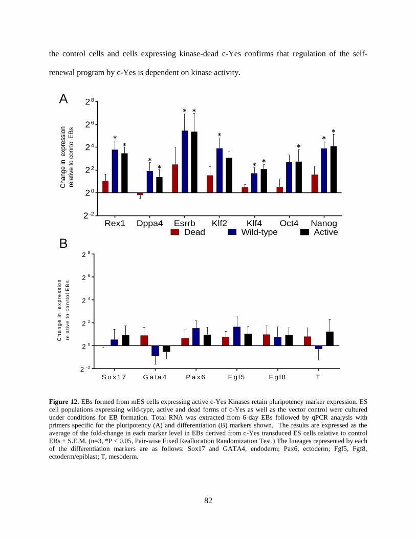
82
the control cells and cells expressing kinase-dead c-Yes confirms that regulation of the self-
renewal program by c-Yes is dependent on kinase activity.
Change in
exp
ress
ion
rela
tive to c
onrt
ol E
Bs
Rex1 Dppa4 Esrrb Klf2 Klf4 Oct4 Nanog2 -2
20
22
24
26
28
Dead Wild-type Active
Ch
an
ge
in
e
xp
res
sio
n
rela
tiv
e t
o c
on
rto
l E
Bs
S o x1 7 G a ta 4 P a x6 F g f5 F g f8 T
2 -2
2 0
2 2
2 4
2 6
2 8
A
B
**
**
* *
*
* *
**
*
Figure 12. EBs formed from mES cells expressing active c-Yes Kinases retain pluripotency marker expression. ES
cell populations expressing wild-type, active and dead forms of c-Yes as well as the vector control were cultured
under conditions for EB formation. Total RNA was extracted from 6-day EBs followed by qPCR analysis with
primers specific for the pluripotency (A) and differentiation (B) markers shown. The results are expressed as the
average of the fold-change in each marker level in EBs derived from c-Yes transduced ES cells relative to control
EBs ± S.E.M. (n=3, *P < 0.05, Pair-wise Fixed Reallocation Randomization Test.) The lineages represented by each
of the differentiation markers are as follows: Sox17 and GATA4, endoderm; Pax6, ectoderm; Fgf5, Fgf8,
ectoderm/epiblast; T, mesoderm.
83
2.3.6 Design of c-Yes gatekeeper mutants resistant to the broad spectrum Src-
family kinase inhibitor, A-419259.
Selective kinase inhibitors represent valuable probes for biological function. Unlike genetic
knock-outs or RNAi-based approaches, inhibitor treatment has immediate biological impact, and
does not allow the system to respond with compensatory changes in gene expression. However,
isoform-selective inhibitors of c-Yes or other members of the Src-kinase family are currently
unavailable, due to the close sequence and structural similarity of the individual family members.
To circumvent this issue for c-Yes in ES cells, we turned to a chemical genetics approach
previously developed in our laboratory to demonstrate a role for c-Src kinase activity in ES cell
differentiation to primitive ectoderm (Meyn, III and Smithgall, 2009). This method paired the
broad-spectrum Src-family kinase inhibitor A-419259 with a c-Src variant engineered to be
resistant to this compound. Introduction of the inhibitor-resistant (Src-IR) mutant into ES cells
had no effect in the absence of inhibitor treatment. However, ES cells expressing Src-IR
differentiated upon addition of A-419259, indicating that c-Src kinase activity, in the absence of
all endogenous SFK activity, was sufficient to induce this differentiation response.
To create analogous IR variants of c-Yes, we substituted the threonine residue at the c-
Yes kinase domain gatekeeper position (T338) with methionine (Figure 13A) in the context of
the tail-activated (Y527F) form of c-Yes described above. Tail-activated mutants of c-Yes with
wild-type and IR kinase domains were then expressed in Rat2 cells, followed by soft-agar colony
assays in the presence of A-419259. Figure 13B shows that both populations of Rat2 cells
formed transformed colonies. Colony formation by Rat2 cells expressing tail-activated c-Yes
with a wild-type kinase domain was very sensitive to A-419259 treatment, with more than a 90%
reduction in colony number at an inhibitor concentration of 100 nM. In contrast, colony
84
formation by Rat2 cells expressing the tail-activated c-Yes mutant with the IR kinase domain
was unaffected by A-419259 treatment even at a concentration of 1 µM. To correlate
transforming activity with kinase function, c-Yes proteins were immunoprecipitated from the
transformed cell populations and immunoblotted with a phosphospecific antibody to the c-Yes
activation loop tyrosine as described earlier. As shown in Figure 13C, c-Yes from cells
transformed by active Yes with a wild-type kinase domain was sensitive to A-419259 treatment,
while c-Yes-IR remained active at all concentrations of A-419259 tested, consistent with colony
formation data. These experiments show that methionine substitution of the c-Yes gatekeeper
threonine results in inhibitor resistance in the same manner as observed previously with c-Src.

85
Wild-type
Active
Active-IR
A
B
3 2 kinase
T338
3 2 kinase
3 2 kinase
T338
Y527
T338
F527
M338
F527
A-419259 (M)
Colo
ny C
ou
nt
0 0.1
0.3
1.0 0 0.
10.
31.
0
0
100
200
300
400
Yes
pYes
0 0.1 0.3 1.0
ActiveC
Yes
pYes
0 0.1 0.3 1.0
Active-IR
Active Active-IR
Figure 13. Validation of inhibitor-resistant mutants of c-Yes in Rat-2 fibroblasts. A) Right: Close-up view of the
active site of the Src-family kinase Hck highlighting the juxtaposition of the kinase domain gatekeeper threonine
(T338) and an analog of the broad spectrum Src-family kinase inhibitor, A-419259 (model rendered using the PDB
file 2C0I; inhibitor carbon atoms in yellow). For fibroblast transformation experiments, the gatekeeper threonine
was replaced with methionine and combined with an activating tail mutation to create the Active-IR mutant shown
on the left. B) Rat2 fibroblasts were infected with recombinant retroviruses expressing tail-activated mutants of c-
Yes with wild-type and IR kinase domains and assayed for soft-agar colony formation in the presence of the A-
419259 at the concentrations shown. Transformed colonies were counted 12 days later from triplicate assays and
the data are shown as the average colony count ± S.D. C) Rat2 cells expressing the Active and Active-IR forms of
c-Yes were treated in the presence or absence of A-419259 for 48 hours. c-Yes proteins were immunoprecipitated
from cell lysates and blotted with a phosphospecific antibody (pYes) and for c-Yes protein recovery.

86
2.3.7 Stable expression of inhibitor-resistant c-Yes (Yes-IR) in ES cells does not
affect self-renewal marker expression.
We next infected ES cells with the wild-type or IR c-Yes retroviral vectors or with an empty
vector as a control, followed by selection with G418. In the absence of A-419259 treatment, all
three ES cell populations grew as colonies characteristic of undifferentiated ES cells (Figure
14A). In the presence of A-419259, cells expressing wild-type c-Yes as well as the vector control
cells formed small tight colonies devoid of flattened, differentiated cells, consistent with our
previous results with ES cells grown in the presence of this inhibitor(Meyn, III and Smithgall,
2009; Meyn, III et al., 2005). In contrast, the Yes-IR ES cells formed larger colonies compared
to control ES cells, with some flattened colony morphology when cultured in the presence of the
inhibitor. Active c-Yes was readily detected in Yes-IR expressing cells treated with A-419259,
but not in control cells or cells expressing Yes-WT, verifying that Yes-IR remains active in A-
419259 treated cells (Figure 14B). We next examined the effect of inhibitor treatment on the
expression of self-renewal and differentiation markers in Yes-IR cells (Figure 14C). No
significant expression changes were observed following qPCR analysis of the pluripotency
markers Rex1, Nanog and Oct4 or the differentiation markers T (mesoderm) and Fgf5
(ectoderm). These experiments suggest that under conditions where c-Yes is the lone active Src
family member present, no major changes in ES cell pluripotency or differentiation occur.
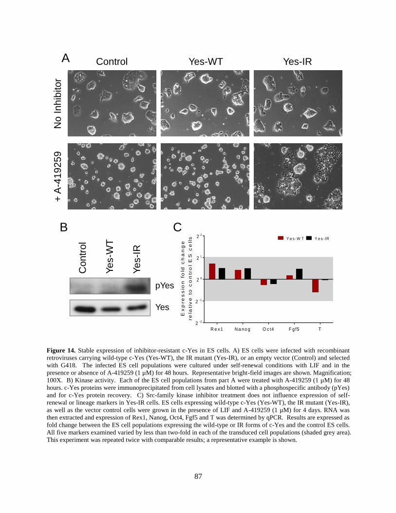
87
A
CB
Control Yes-WT Yes-IRN
o I
nh
ibito
r+
A-4
19
25
9
Co
ntr
ol
Yes-W
T
Ye
s-I
R
pYes
Yes
Ex
pre
ss
ion
fo
ld c
ha
ng
e
re
lati
ve
to
co
ntr
ol
ES
ce
lls
R e x1 N a no g O c t4 F g f5 T
2 -2
2 -1
2 0
2 1
2 2
Y e s -W T Y e s -IR
Figure 14. Stable expression of inhibitor-resistant c-Yes in ES cells. A) ES cells were infected with recombinant
retroviruses carrying wild-type c-Yes (Yes-WT), the IR mutant (Yes-IR), or an empty vector (Control) and selected
with G418. The infected ES cell populations were cultured under self-renewal conditions with LIF and in the
presence or absence of A-419259 (1 µM) for 48 hours. Representative bright-field images are shown. Magnification;
100X. B) Kinase activity. Each of the ES cell populations from part A were treated with A-419259 (1 µM) for 48
hours. c-Yes proteins were immunoprecipitated from cell lysates and blotted with a phosphospecific antibody (pYes)
and for c-Yes protein recovery. C) Src-family kinase inhibitor treatment does not influence expression of self-
renewal or lineage markers in Yes-IR cells. ES cells expressing wild-type c-Yes (Yes-WT), the IR mutant (Yes-IR),
as well as the vector control cells were grown in the presence of LIF and A-419259 (1 µM) for 4 days. RNA was
then extracted and expression of Rex1, Nanog, Oct4, Fgf5 and T was determined by qPCR. Results are expressed as
fold change between the ES cell populations expressing the wild-type or IR forms of c-Yes and the control ES cells.
All five markers examined varied by less than two-fold in each of the transduced cell populations (shaded grey area).
This experiment was repeated twice with comparable results; a representative example is shown.
88
2.3.8 Differentiation of ES cells driven by c-Src is antagonized by c-Yes.
Previous work from our group demonstrated that c-Src activity alone is sufficient to drive
differentiation of ES cells using an analogous inhibitor-resistant allele of c-Src and A-419259
(Meyn, III and Smithgall, 2009). In contrast, work presented here demonstrates that despite close
structural homology to c-Src, c-Yes activity inhibits rather than promotes ES cell differentiation.
To determine which of these two opposing signals is epistatic to the other, we introduced the IR
forms of both c-Yes and c-Src into ES cells. This was accomplished using c-Yes-IRES-Puro and
c-Src-IRES-Neo retroviral vectors, followed by double selection with puromycin and neomycin.
When cultured under standard conditions in the absence of A-419259, the resulting four cell
populations grew with similar undifferentiated colony morphology (Figure 15A). Ectopic
expression of the c-Yes and c-Src transgenes was confirmed by RT-PCR with vector-specific
primers (Figure 15B). The presence of the c-Src and C-yes gatekeeper mutations (T338M) was
confirmed in the PCR products from the transduced cell lines by nucleotide sequencing (data not
shown). Selected pluripotency and differentiation markers were also examined by qPCR (Figure
15C). No changes in marker expression were observed in cells expressing c-Src-IR, c-Yes-IR or
both IR mutants in the absence of A-419259 treatment.
We next examined the effect of A-419259 treatment on cell morphology, pluripotency
and differentiation marker expression in ES cells expressing Src-IR or Yes-IR either alone or in
combination. Cells were cultured in the presence of A-419259 and LIF for 4 days and images of
the resulting colonies are shown in Figure 16A. With A-419259 treatment, control ES cells
formed tight, small colonies, while colonies formed from the c-Yes-IR cells showed a more
flattened morphology as described earlier. In contrast, cells expressing c-Src-IR or both
inhibitor-resistant alleles grew as flat sheets in the presence of the inhibitor. We then assayed
89
relative expression levels of pluripotency (Tbx3, Rex1, Nanog, Oct4 and Gbx2) and
differentiation markers for endoderm (Sox17, Gata4), ectoderm (Fgf5, Sox1, Nestin), mesoderm
(T), epiblast (Fgf8) and trophectoderm (Hand1) by qPCR. The data are presented in Figures 16B
and C as changes in expression relative to the control cell population. Inhibitor treatment of ES
cells expressing c-Src-IR alone resulted in the upregulation of Gbx2, Sox17, Gata4, Gata6, Fgf5
and Hand1 expression, indicative of differentiation to endoderm and trophectoderm in addition
to primitive ectoderm-like cells as reported previously by our group (Meyn, III and Smithgall,
2009). This finding suggests that c-Src selectively regulates a braoder range of differentiation
responses that originally reported. In contrast to c-Src, the c-Yes-IR cells did not express any of
these differentiation markers in response to A-419259 treatment. This observation is consistent
with the idea that c-Yes signals help to maintain ES cells in an undifferentiated state.
Expression of both inhibitor resistant alleles in the same cell population revealed
dominance of c-Yes over c-Src in terms of a subset of differentiation marker expression. As
shown in Figure 16C, induction of the endoderm markers Sox17, Gata4 and Gata6 by c-Src was
significantly repressed in cells expressing IR alleles of both c-Src and c-Yes vs. c-Src alone. In
contrast, expression of Gbx2, Fgf5 and Hand1 remained the same when both IR alleles were
expressed, raising the possibility that other renewal-related SFKs (e.g., Hck) may influence these
pathways. This observation demonstrates that the c-Yes anti-differentiation signal is epistatic to
the c-Src signal for endoderm differentiation, despite the close structural homology of these two
Src family kinases.
Recent studies have shown that c-Src promotes the EMT (Li et al., 2011), a process
essential for lineage specification during development (Thiery et al., 2009). To investigate a
possible connection of the EMT to Src-induced differentiation of ES cells, we assayed the

90
relative expression levels of EMT markers previously linked to c-Src, including Igf2, SIP1, Ncad,
Snail1, Twist1 and Eomes. We also assessed changes in the expression of matrix
metalloproteinases (MMPs), which play an important role in the EMT. As shown in Figure 16D,
A-419259 treatment of ES cells expressing c-Src-IR alone resulted in up-regulation of all of
these EMT markers, consistent with a role of the EMT in c-Src-mediated differentiation of ES
cells. In contrast, c-Yes-IR cells did not express any of the EMT markers tested in response to
A419259 treatment. Interestingly, co-expression of c-Yes-IR suppressed c-Src-mediated
induction of Snail1, Twist1 and MMP14 expression, with a partial reversal of MMP9 expression
in response to inhibitor treatment of ES cells expressing both IR alleles. These results show that
c-Src activity alone is sufficient to promote EMT marker expression and differentiation, while c-
Yes activity inhibits both c-Src mediated EMT and endodermal differentiation.
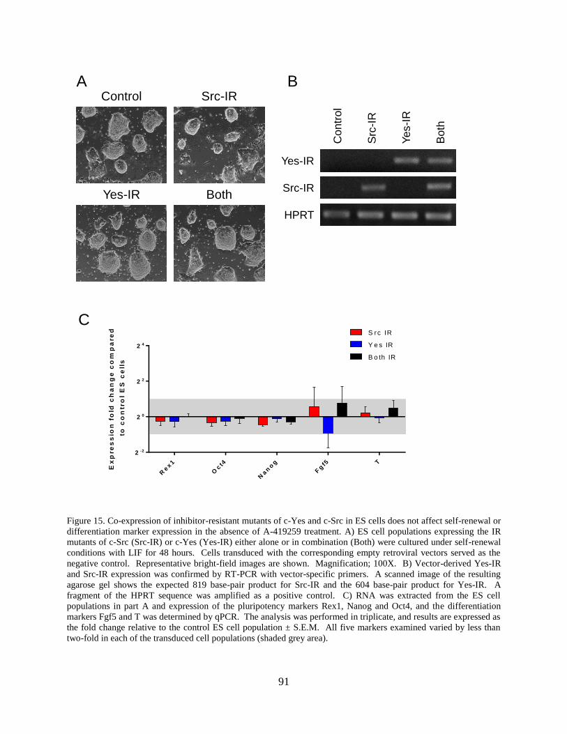
91
A BControl
Yes-IR
Src-IR
Both
Yes-IR
Co
ntr
ol
Src-IR
HPRT
Src
-IR
Yes-I
R
Bo
th
C
Ex
pre
ss
ion
fo
ld c
ha
ng
e c
om
pa
re
d
to
co
ntr
ol
ES
ce
lls
Rex1
Oct4
Nan
og
Fg
f5 T
2 -2
2 0
2 2
2 4
S rc IR
Y e s IR
B o th IR
Figure 15. Co-expression of inhibitor-resistant mutants of c-Yes and c-Src in ES cells does not affect self-renewal or
differentiation marker expression in the absence of A-419259 treatment. A) ES cell populations expressing the IR
mutants of c-Src (Src-IR) or c-Yes (Yes-IR) either alone or in combination (Both) were cultured under self-renewal
conditions with LIF for 48 hours. Cells transduced with the corresponding empty retroviral vectors served as the
negative control. Representative bright-field images are shown. Magnification; 100X. B) Vector-derived Yes-IR
and Src-IR expression was confirmed by RT-PCR with vector-specific primers. A scanned image of the resulting
agarose gel shows the expected 819 base-pair product for Src-IR and the 604 base-pair product for Yes-IR. A
fragment of the HPRT sequence was amplified as a positive control. C) RNA was extracted from the ES cell
populations in part A and expression of the pluripotency markers Rex1, Nanog and Oct4, and the differentiation
markers Fgf5 and T was determined by qPCR. The analysis was performed in triplicate, and results are expressed as
the fold change relative to the control ES cell population ± S.E.M. All five markers examined varied by less than
two-fold in each of the transduced cell populations (shaded grey area).

92

93
Figure 16. Differentiation of mES cells driven by c-Src is antagonized by c-Yes. A) ES cell populations expressing
inhibitor-resistant mutants of c-Src (Src-IR) or c-Yes (Yes-IR) either alone or in combination (Both) were cultured
in the presence of LIF and A-419259 (1 μM). Cells transduced with the corresponding empty retroviral vectors
served as the negative control. Representative bright-field images were recorded 4 days later. Magnification; 100X.
B, C, D) RNA was extracted from the A-419259-treated ES cell populations shown in part A and qPCR analysis
was performed with primers specific for markers of pluripotency (B), differentiation (C) and EMT (D). The analysis
was performed on four replicates from two independently derived sets of ES cell populations, and results are
expressed as the fold change relative to the control ES cell population ± S.E.M. (*P < 0.05 compared with control
ES cells; **P < 0.05 compared with Src-IR cells; Pairwise Fixed Reallocation Randomization Test.) Note that
expression of Src-IR alone induced differentiation in the presence of A-419259 treatment, with up-regulation of the
endoderm markers Sox17, Gata4 and Gata6, the primitive ectoderm marker Fgf5, the trophectoderm marker Hand1,
and the EMT markers Igf2, Snail1, Twist1, MMP9 and MMP14. Endoderm differentiation marker expression was
blocked in cells co-expressing both Src-IR and Yes-IR and in the presence of the inhibitor. This antagonistic effect
of Yes-IR correlates with the down-regulation of EMT markers Snail1, Twist1, MMP9 and MMP14.
2.4 DISCUSSION
Work presented here shows that the Src-family kinase c-Yes generates a potent anti-
differentiation signal in mES cells. Enforced expression of either wild-type or an active form of
c-Yes at modest levels completely inhibited differentiation of ES cells to EBs. This suppressive
effect was not observed with kinase-defective c-Yes, indicating a requirement for c-Yes kinase
activity. Interestingly, expression of both wild-type c-Yes as well as a kinase-active ‘tail’ mutant
inhibited EB formation to the same extent, showing that ES cells are very sensitive to the c-Yes
anti-differentiation signal. Our observations are consistent with prior studies showing that c-Yes
kinase activity is stimulated by the self-renewal cytokine LIF and that RNAi-mediated
knockdown of c-Yes induces ES cell differentiation (Anneren et al., 2004). Although c-Yes
kinase activity interferes with EB formation, it cannot sustain self-renewal following LIF
withdrawal. Indeed, ES cells expressing the active forms of c-Yes undergo morphological
differentiation when cultured under adherent conditions in the absence of LIF. Furthermore,
qPCR analysis showed that the small EBs formed from ES/c-Yes cells express the same

94
differentiation markers as control EBs (Figure 12). However, unlike control EBs, these cell
clusters also continued to express pluripotency factors, including Oct4 and Nanog. Thus the
presence of active c-Yes appears to prevent EB formation by interfering with the repression of
pluripotency genes as opposed to blocking the differentiation program.
Previous studies have shown that individual members of the Src kinase family display
distinct patterns of expression and activity during ES cell self-renewal and differentiation (see
Introduction). Seven of the eight mammalian Src family members are expressed simultaneously
in self-renewing mES cells (Meyn, III et al., 2005), making investigation of their individual
contributions to self-renewal and differentiation a challenge. To address this problem, we
developed a chemical genetics approach based on the broad-spectrum Src-family kinase
inhibitor, A-419259. Treatment of ES cells with this inhibitor blocks all endogenous Src-family
kinase activity in ES cells, locking them in an undifferentiated state (Meyn, III et al., 2005).
Expression of a mutant of c-Src with engineered resistance to this inhibitor caused the ES cells to
differentiate to primitive ectoderm-like cells (Meyn, III and Smithgall, 2009). This chemical
genetics approach allowed us to demonstrate a role for c-Src in the earliest stages of ES cell
differentiation for the first time. In the present study, we expanded our marker analysis and
found c-Src activity alone is able to induce ES cell differentiation to cells with characteristics of
both primitive ectoderm and endoderm, as exemplified by Fgf5, Sox17, Gata4 and Gata6
expression. Moreover, we discovered that c-Src activity alone drives the expression of EMT
markers, consistent with a previous report of Src-mediated EMT during induction of ES cell
differentiation through the Calcineurin-NFAT pathway (Li et al., 2011).
Here we also describe an analogous 'gatekeeper' mutant of c-Yes that is resistant to A-
419259. Unlike c-Src, ES cells expressing this c-Yes mutant did not differentiate in response to
95
inhibitor treatment, and instead retained the same pluripotency gene expression pattern as control
ES cells. Interestingly, when inhibitor-resistant mutants of both c-Yes and c-Src were co-
expressed in ES cells, the presence of c-Yes activity prevented the induction of both endoderm
and EMT marker expression by c-Src. These observations support a previous model of the
regulation of endogenous c-Src and c-Yes kinase activity during ES cell renewal and
differentiation (Meyn, III et al., 2005). In presence of LIF, both c-Yes and c-Src are active, but
the presence of active c-Yes overrides the c-Src signal for differentiation. Upon LIF withdrawal,
c-Yes activity is shut off, allowing active c-Src to drive differentiation. More broadly, our work
suggests that although ES cells can be maintained in a perpetual state of self-renewal, they are
poised to differentiate. Other studies have established that key differentiation genes are
transcriptionally initiated in self-renewing ES cells, but are silenced by the master transcriptional
regulators Oct4, Nanog and Sox2 (Jaenisch and Young, 2008). The ability of c-Yes kinase
activity to prevent the transcriptional silencing of these master regulators of self-renewal as
shown here may explain its potent suppressive effect on ES cell differentiation.
Our result is surprising given that c-Src, c-Yes and Fyn have been shown to play
redundant roles and are able to compensate for one another in knockout mouse models (Stein et
al., 1994; Lowell and Soriano, 1996; Klinghoffer et al., 1999). Although c-Yes and c-Src are
similar in structure and are regulated by shared upstream signals, they are also have specific
signaling roles (Sancier et al., 2011). The chemical genetics approach used here may be
advantageous over gene knockdown in that it employs an inhibitor that is fast acting and
reversible, ruling out the effects of compensation. Taking advantage of this system, we are
investigating downstream targets that are differentially affected by Src-IR and Yes-IR. Our
initial experiments suggest that SFK signaling may cross-talk with the Wnt signaling pathway.
96
Preliminary experiments show that c-Src may phosphorylate and activate GSK3β, thereby
promoting β-catenin degradation. This outcome is consistent with a role for c-Src in
differentiation, as β-catenin promotes renewal. In contrast, other work suggests that c-Yes may
have a protective effect on β-catenin degradation. In HT29 colon cancer cell lines, c-Yes activity
was associated with β-catenin localization and β-catenin target gene expression (Sancier et al.,
2011).
In addition, SFKs have been shown to be required for integrin-FAK signaling and
important for cytoskeletal organization, cell shape and adhesion (Klinghoffer et al., 1999). Here
we show that A-419259 causes ES cells to adopt a small and tight morphology, devoid of
flattened, differentiated cells. This indicates that SFK activity is important for flattened cellular
morphology. However, either Yes-IR or Src-IR expression rescued this phenotype, indicating the
redundant role of c-Src and c-Yes in maintaining cell shape. This might due to SFK interaction
with the cytoskeletal and cell-cell interaction proteins. Since differentiation is associated with
flattened cellular morphology, pan-SFK inhibition eliminates flattened cells and promotes self-
renewal. Interestingly, SFK inhibition is found to promote direct reprograming of somatic cells
to iPS cells by replacing Sox2 (Staerk et al., 2011).
In conclusion, we report that low-level, sustained expression of active c-Yes maintains
ES cell renewal marker expression following LIF withdrawal, resulting in a profound block to
EB formation. Using a chemical genetics approach, we demonstrated that the c-Yes signal
favoring self-renewal is indirect opposition to the c-Src differentiation signal, despite the close
structural homology between these kinases. A long-standing tenet of the Src-family kinase field
is that individual family members have similar, if not redundant, biological functions. Our results
clearly demonstrate that this is not always the case, and provide an important caveat to the use of

97
broad-spectrum, small molecule inhibitors of the entire Src kinase family to make conclusions
about the biological activity of individual family members or the family as a whole. Finally, our
results provide a strong rationale for the development of selective compounds to control c-Src vs.
c-Yes activity; such compounds may allow more precise pharmacological manipulation of ES
cell renewal and differentiation. Indeed, a recent unbiased chemical library screen identified
broad-spectrum Src-family kinase inhibitors as potent enhancers of somatic cell reprogramming
to an ES cell-like state (iPS cells) (Staerk et al., 2011). Our work predicts that a Src-selective
inhibitor (or a c-Yes agonist) may provide an even greater enhancement in reprogramming
efficiency.
2.5 MATERIALS AND METHODS
2.5.1 Cell culture
The mES cell line D3 was obtained from the ATCC and maintained in Dulbecco’s modified
Eagle’s medium (DMEM) supplemented with 15% fetal bovine serum (FBS), 1% antibiotic-
antimycotic, 2 mM L-glutamine, 1% nonessential amino acids, 1 mM sodium pyruvate, 0.1 mM
2-mercaptoethanol, and 1000 U/ml LIF as described previously (Meyn, III et al., 2005). Rat2
fibroblasts were obtained from the ATCC and maintained in DMEM supplemented with 10%
FBS and 1% antibiotic-antimitotic as described elsewhere (Pene-Dumitrescu and Smithgall,
2010). For inhibitor treatment, ES cells (106) were plated on gelatin-coated 60 mm plates for 24
h. A-419259 (Santa Cruz Biotechnology) was added to the culture medium to a final
98
concentration of 1 µM using DMSO as the carrier solvent (0.1% final) (Meyn, III and Smithgall,
2009).
2.5.2 Retroviral transduction of mES cells and Rat2 fibroblasts
A cDNA encoding murine c-Yes was purchased from Thermo Scientific in the mammalian
expression vector pSPORT6. Kinase-dead (Lys295 to Arg), active (Yes-Tyr527 to Phe; Yes-
YF), and inhibitor-resistant (Thr338 to Met; Yes-IR) mutants of c-Yes were generated by site-
directed mutagenesis (QuickChange XL method; Stratagene). Residues are numbered according
to the crystal structure of c-Src (PDB: 2SRC) (Xu et al., 1999). The c-Yes cDNAs were
subcloned into the retroviral expression vectors, pMSCV-IRES-Neo and pMSCV-IRES-Puro
(Clontech) using the In-Fusion cloning method (Clontech). Analogous retroviral expression
vectors for c-Src have been described elsewhere (Meyn, III and Smithgall, 2009). Retroviral
stocks were produced by co-transfecting 293T cells with the retroviral vectors and an ectropic
packaging plasmid and the resulting viral supernatants were used to infect ES cells as described
(Cherry et al., 2000; Meyn, III and Smithgall, 2009). Transduced ES cell populations were
selected and maintained with 250 µg/ml G418 or 1.5 µg/ml puromycin or both for double
selection. Rat2 cells were infected as described previously (Pene-Dumitrescu and Smithgall,
2010), followed by selection with 800 µg/ml G418 for 14 days and maintenance in 400 µg/ml
G418.

99
2.5.3 Embryoid body formation
Embryoid bodies were cultured as previously described (Meyn, III et al., 2005). Briefly,
pluripotent ES cell colonies were trypsinized to single cells and plated at low density (5000
cells/ml) without LIF in 10 cm bacterial grade petri dishes to prevent attachment. EBs were fixed
in 4% paraformaldehyde 6 d later, permeabilized with 0.1% Triton-X-100 in PBS, and stained
with DAPI (Sigma). EBs were imaged by confocal microscopy and their size was estimated
from the area of a 2D projection of the confocal images using the Olympus FV10-ASW 2.1
software as previously described (O'Reilly et al., 2011).
2.5.4 RT-PCR analysis
Total RNA was isolated from ES cell cultures with the RNeasy Plus Mini Kit (Qiagen) and
cDNA was synthesized from 2 µg total RNA with a random decamer as primer according to the
manufacturers’ protocols (Ambion). For RT-PCR to confirm c-Yes and c-Src transgene
expression, one-twentieth of each RT reaction was used in a 50 µl PCR reaction, with primers (5
µM) specific for detection of transcripts from the transduced retroviruses. PCR reaction
products were resolved on 1% agarose gels and stained with ethidium bromide. For quantitative
RT-PCR (qPCR) analysis, 1 µl of a 1:50 dilution of the cDNA reaction, primers (5 µM) and RT2
SYBR green qPCR master mix (Qiagen) were used for each reaction. Primer sets were obtained
from Qiagen (Quantitative Primer Assays), and results were normalized using GAPDH as the
reference gene. Quantitative PCR data were analyzed using the pairwise Fixed Reallocation
Randomization Test and the REST 2009 software (Pfaffl et al., 2002). Primer efficiency was set
100
to a value of 1 and 5,000 iterations were used to calculate changes in expression and statistical
significance. Results are reported as fold change in expression ± S.E.M.
2.5.5 Immunoprecipitation and immunoblotting
Cells were washed with phosphate-buffered saline (PBS) and lysed with RIPA buffer
supplemented with phosphatase and protease inhibitors as described (Meyn, III et al., 2005). Cell
lysates were clarified by centrifugation and 1 mg of protein extract was incubated with 1 µg of a
c-Yes antibody (BD Biosciences, Cat. # BD610375) for 1 h. Protein G-Sepharose (20 µl of a
50% w/v slurry; Invitrogen, 10-1243) was added followed by incubation for an additional 3 h.
Samples were washed three times with RIPA buffer, mixed with SDS-PAGE loading buffer,
heated to 95 °C for 5 min and separated on SDS polyacrylamide gels. Electrophoresis samples
were prepared in a similar fashion for cell lysates, followed by SDS-PAGE and membrane
transfer.
For immunoblotting, proteins were transferred to PVDF membranes and probed with
antibodies to the c-Yes activation loop, the c-Yes protein, Oct4, and actin as a loading control.
The following primary antibodies were used at a 1:1000 dilution: active Src pY418 (Invitrogen
44660G), rabbit anti-Yes (Cell Signaling Technology, AB13954), mouse anti-Oct4 (Santa Cruz,
SC-5279), and mouse anti-Actin (Millipore, MAB1501).

101
2.5.6 Rat2 cell soft-agar assay
Oncogenic transformation of Rat2 cells was assessed as colony forming activity as described
previously (Pene-Dumitrescu and Smithgall, 2010). In brief, Rat2 cells transduced with various
retroviral expression vectors were assayed in triplicate in 35 mm Petri dishes (BD Biosciences)
using SeaPlaque agarose (Lonza). The bottom layer of 0.5% agarose was prepared in growth
medium (1.0 ml) containing the DMSO carrier solvent alone (0.2%) or twice the final
concentration of A-419259 where indicated. The top layer (1.0 ml) consisted of 0.3% agarose in
growth medium contain 10,000 Rat2 fibroblasts. Colonies were stained with 3-(4,5-
dimethylthiazol-2-yl)-2,5-diphenyltetrazolium bromide (Sigma) ten days later and counted from
scanned images using Quantity One colony counting software (BioRad). For experiments with
the Src-family kinase inhibitor, A-419259, the compound was added to the bottom agarose layer
at twice the final concentration with DMSO as carrier solvent at 0.1% final concentration.

102
3.0 SRC FAMIY TYROSINE KINASE SIGNALING IS IMPORTANT FOR HUMAN
EMBRYONIC STEM CELL DIFFERENTIATION
3.1 ABSTRACT
Embryonic stem (ES) cells are characterized by pluripotency, the developmental potential to
generate cell lineages derived from all three primary germ layers. In the past decade, great
progress has been made on the cell culture conditions, transcription factor programs and
intracellular signaling pathways that control murine and human ES cells. Although both are
derived from the inner cell mass of the blastocyst stage embryo, human ES (hES) cells and
mouse ES (mES) cells have distinct culture conditions, and respond to different receptor tyrosine
kinase signaling pathways. For example, FGF signaling is important to maintain hES cell
pluripotency, yet induces differentiation in mES cells. Previous work from our group has
implicated the Src family of non-receptor protein-tyrosine kinases in mES cell self-renewal and
differentiation. Multiple members of the Src kinase family are expressed in mES cells, and
individual family members appear to play distinct roles in regulating their developmental fate.
Both Hck and Yes are important in self-renewal, while c-Src activity alone is sufficient to induce
differentiation. While these findings implicate Src kinase signaling in mES cell renewal and
differentiation, the role of this kinase family in hES cells is largely unknown. Here, I explored
Src family kinase signaling in hES cells. First, quantitative real-time RT-PCR was used to
103
determine the relative expression profile of individual Src family members in undifferentiated
hES cells vs. EBs derived from them. Of the eleven Src-related kinases in the human genome,
Fyn, c-Yes, c-Src, Lyn, Lck and Hck were expressed in H1, H7 and H9 hES cells, while Fgr,
Blk, Srm, Brk, and Frk transcripts were not detected. Of these, c-Yes, Lyn, and Hck transcript
levels remained constant in self-renewing hES cells and differentiated EBs, while c-Src and Fyn
showed a modest increase in expression as a function of differentiation. In contrast, Lck
expression levels dropped dramatically as a function of EB differentiation. In addition, to assess
the role of Src family kinase activity hES cells, we treated hES cell cultures with inhibitors
specific for the Src kinase family, including Src Kinase Inhibitor 1, PP2 and A-419259.
Untreated control hES cultures grown in mTeSR1 medium formed typical pluripotent colonies
circumscribed by differentiated cells. Transfer of hES cells to differentiation medium resulted in
flattened colony morphology that correlated with a loss of cell-surface staining for the hES cell
pluripotency marker, Tra-1-60. Dramatically, hES cells maintained in the presence of 1 µM A-
419259 retained the morphology of domed, pluripotent colonies and maintained TRA-1-60
expression in differentiation medium. Similar morphological changes were observed in SKI-1
treated cells. Taken together, these observations support a role for Src family kinase signaling in
the regulation of hES fate, and suggest that some parallels may exist in mouse and hES cells for
this intracellular signaling network.
3.2 INTRODUCTION
Human ES (hES) cells are pluripotent stem cells derived from the inner cell mass of blastocyst
stage human embryos produced by in vitro fertilization (Thomson et al., 1998). Although hES
104
cells are of the same blastocyst origin as mES cells, they respond to different growth factors for
maintenance in culture and depend on distinct receptor tyrosine kinase signaling pathways. For
example, hES cells depend on bFGF and TGFβ/Activin signaling to maintain the
undifferentiated state. While factors essential for mES cell renewal, LIF and BMPs, have no
effect, or induce differentiation in hES cells, respectively (Yu and Thomson, 2008). FGF signals
through the FGF receptor to activate MEK/Erk signaling to inhibit differentiation and PI3K-Akt
signaling to promote survival (Dvorak et al., 2005; Li et al., 2007). In addition, the
TGFβ/Nodal/Activin signaling inhibits neuronal differentiation, and works synergistically with
FGF to maintain hES cell pluripotency (Vallier et al., 2005). Despite these and other differences
between mES and hES cells, the transcription factors governing pluripotency are similar, with
both mES and hES cells expressing the master pluripotency factors, Oct4, Nanog and Sox2
(Boyer et al., 2005). Moreover, hES cells are pluripotent, as they can from embryoid bodies in
vitro and teratomas in vivo when injected into immunodeficient mice. While the growth factor
conditions, receptor kinase signaling, and transcription factor networks have been investigated in
hES cells, the signaling pathways downstream of receptor tyrosine kinases have not been fully
explored.
As critical signal integrators, the Src family of non-receptor tyrosine kinases has been
linked to many receptor tyrosine kinases including the FGFR to regulate cell adhesion,
proliferation, growth and survival (Thomas and Brugge, 1997). There are 11 Src-related kinases
in the human genome (Manning et al., 2002), eight of which have been studied extensively in
mammalian cells: Blk, Fgr, Fyn, Lck, Lyn, Hck, c-Src and c-Yes. In the mouse, c-Src, Fyn and c-
Yes are ubiquitously expressed, while Lck, Lyn, Hck, Blk and Fgr display more restricted
expression to hematopoietic cells (Lowell and Soriano, 1996). Surprisingly, at least seven
105
members of the Src kinase family are expressed in mES cells, and individual family members
appear to play distinct roles in regulating their developmental fate (Meyn, III et al., 2005).
Expression of Hck is rapidly silenced as mES cells differentiate to embryoid bodies (EBs),
suggesting a role in self-renewal. In contrast, c-Src activity alone is sufficient to induce
differentiation of mES cells to primitive ectoderm (Meyn, III et al., 2005; Meyn, III and
Smithgall, 2009). Other studies have shown that both Hck and c-Yes are important for mES cell
self-renewal downstream of LIF (Ernst et al., 1994; Anneren et al., 2004; Tamm et al., 2011).
While these findings implicate Src kinase signaling in mES cell renewal and differentiation, the
role of this kinase family in hES cells is largely unknown.
To explore whether SFK signaling is conserved in hES cell renewal and pluripotency, I
first showed that three hES cell lines, H1, H7 and H9, all express comparable levels of the SFK
members c-Src, c-Yes, Fyn, Lck, Lyn and Hck. Next, I investigated changes in expression levels
of each SFK during differentiation of hES cells to EBs. As hES cells differentiate, c-Src and Fyn
transcript levels increased slightly, while Lck expression dropped dramatically. To assess the
role of Src family kinase activity in hES cells, I treated hES cell cultures with the SFK-specific
inhibitors. Human ES cells treated with the potent pyrrolopyrimidine SFK inhibitor A-419259,
previously shown to block all endogenous SFK activity in mES cells, retained the morphology of
pluripotent colonies and continued to express the cell-surface renewal marker TRA-1-60 under
culture conditions for differentiation. These observations support a role for SFK signaling in the
regulation of hES cell fate, and suggest that unique function for individual SFKs may exist in
hES cells as observed previously in mES cells.
106
3.3 RESULTS
3.3.1 Human ES cells express multiple Src family kinases
Of the eleven Src-related kinases in the human genome, only c-Yes has been studied in hES
cells. c-Yes kinase activity is downregulated as hES cells differentiate to EBs, while the c-Yes
protein is expressed in both hES cells and differentiated EBs (Anneren et al., 2004). To study
SFK signaling in hES cells, we first determined which SFK members are expressed in hES cells
using RT-PCR. Human ES cell lines (H1, H7 and H9) were maintained in chemically defined
mTeSR medium, under-feeder free conditions on Matrigel-coated plates (Ludwig et al., 2006).
The cells grow as slightly domed colonies with some random differentiation at the edge of each
colony (Figure 17A). RT-PCR analysis revealed that H1 hES cells express six SFKs, including c-
Src, c-Yes, Fyn, Lck, Hck and Lyn. Of these, c-Src, c-Yes, Fyn and Lck are most highly
expressed (Figure 17B). The housekeeping gene glyceraldehyde 3-phosphate dehydrogenase
(GAPDH), and pluripotency marker, Pou-domain transcription factor Oct4 were used as positive
control.
We next determined the relative expression levels of these six SFKs in comparison to the
pluripotency markers Oct4 and Nanog, in the H1, H7 and H9 hES cell lines using quantitative
real-time PCR (qPCR, Figure 17C). Compared with H1 hES cells, both H7 and H9 cells
expressed comparable levels of the pluripotency marker Oct4 as well as the SFKs Fyn, c-Src, c-
Yes, Lck and Lyn, with less than two-fold differences in relative expression levels. We also
observed that H7 cells express higher levels of the pluripotency marker Nanog and the SFK Hck
compared with H1 cells, while H9 cells express lower levels of Nanog, indicative of
heterogeneity between these hES cell lines. In summary, these results show that hES cells

107
express multiple SFK members, and expression levels of SFKs c-Src, c-Yes, Fyn and Lck are
largely consistent among the three hES cell lines tested.
Oct4SrmFyn Hck Lck Lyn Src YesBrkBlkGAPDH Fgr Frk
H1 hESC H7 hESC
A.
B.
C.
M a rk e rs a n d S F K m e m b e rs
Ex
pre
ss
ion
fo
ld c
ha
ng
e
co
mp
are
d t
o H
1 h
um
an
ES
C
Oct4
Nan
og
Fyn
Hck
Lck
Lyn
Src
Yes
2 -4
2 -2
2 0
2 2
2 4 H 7 E S C
H 9 E S C
H9 hESC
*
*
*
Figure 17. Src family kinase expression in hES Cells (A) Human ES cell lines (H1, H7 and H9) were maintained
under feeder-free conditions on Matrigel-coated plates using mTeSR1 medium. Cell culture images were taken 5
days after passage, before harvesting for RNA isolation (magnification: 100×). (B) SFK expression was determined
by RT-PCR using specific primers for each kinase with RNA isolated from H1 cells. Fyn, c-Yes, c-Src, Lyn, Lck
and Hck were detected in H1 cells. (C) Relative SFK and Oct4 expression levels were determined by Q-PCR in H1,
H7 and H9 cells. For each hES cell line, this experiment was repeated three times in different passages of cells. The
results are presented as average expression fold change compared to that in H1 hES cells ± S.E.M (*P < 0.05,
Pairwise Fixed Reallocation Randomization Test), with GAPDH as internal control. Oct4 and most of the SFK
expression varied by less than two-fold (shaded grey area).

108
3.3.2 Src family kinase expression during human embryoid body differentiation
Embryoid body (EB) formation is a convenient model to assess the pluripotency of hES cells.
When cultured in suspension culture without feeder layers, hES cells spontaneously form
aggregates or EBs as previously described for mES cells (Itskovitz-Eldor et al., 2000).
Developing EBs turn on expression of differentiation markers, contain cell types characterizing
all three germ layers, and mimic the early developmental stages of gastrulation and germ layer
formation (Murry and Keller, 2008).
Previous research from our group found that SFK members have dynamic changes of
expression in response to murine ES cell differentiation (Meyn, III et al., 2005). To investigate
whether SFK members are also differentially regulated during the differentiation of hES cells
into EBs, I initiated EB formation from cultured H1, H7 and H9 cells. Aggrewell microplates
were used to initiate formation of EBs that are homogenous in size and cell number (2000 hES
cells/ EB), to allow direct and reproducible comparison (Mohr et al., 2010). H1 and H9 EBs were
maintained in a commercial differentiation medium (Aggrewell medium, Stemcell technologies),
while H7 EBs were maintained in hES cell differentiation medium. EBs were harvested 3, 6 and
12 days after EB initiation. Total RNA was extracted from EBs derived from all three hES cell
lines, followed by qRT-PCR analysis of SFK expression as well as markers of self-renewal and
differentiation (Figures 18, 19 and 20).
In all three hES cell lines tested, Lck expression levels were downregulated in response
to EB differentiation, suggesting that Lck might be important for hES cell renewal. In contrast to
Lck, c-Src and Fyn expression were increased approximately 2-fold in 12 day EBs compared
with corresponding hES cells, indicating that c-Src and Fyn might be related to differentiation. In

109
addition, the expression levels of pluripotency markers Oct4 and Nanog were consistently
downregulated, suggesting that the human EBs were differentiated.
In addition we observed some cell-line specific expression changes. In H1 hES cells
(Figure 18), both Lyn and Hck were downregulated in 12 day EBs compared with renewing H1
ES cells, while only Sox1 was upregulated among the differentiation markers analyzed. The
differentiation markers showed a trend of increased expression in response to EB formation.
However, the heterogeneous nature of hES H1 cell line may express high levels of these
differentiation markers as well, masking the increase in response to EB formation. In H9 hES
cells (Figure 19), Hck expression levels were down-regulated, while most differentiation markers
analyzed were upregulated during EB differentiation. In contrast, in H7 hES cells (Figure 20),
expression of Hck was increased in response to EB differentiation, while the differentiation
markers T and Gata4 were both upregulated. These results indicate that hES cell lines are
heterogeneous with respect to differentiation marker and SFK expression changes during EB
differentiation.
Despite the heterogeneity mentioned above, Lck expression was consistently
downregulated while c-Src and Fyn expression were up-regulated during EB differentiation of
H1, H7 and H9 hES cells. These dynamic changes in individual SFK expression support distinct
functions for SFKs in the regulation of hES cell self-renewal and differentiation, similar to what
we have already defined in murine ES cells.

110
3 Day EBs
6 Day EBs 12 Day EBs
H1 hESC
Fo
ld c
ha
ng
e
rela
tiv
e t
o H
1 h
ES
C
Oct4
Nan
og T
Pax6
So
x1
Msx1
So
x17
2 -5
2 0
2 5
E S c e ll m a rk e r e x p re s s io n
Fo
ld c
han
ge
rela
tive t
o H
1 h
ES
C
Fyn SrcYes Lyn
Hck Lck
2 -4
2 -2
20
22
24
Day 3
Day 6
Day12
Src Family Kinase Expression
A.
B.
C.
Figure 18. Src family kinase expression during EB formation from H1 Cells. A.) EB formation was initiated from
H1 ES cells using Aggrewell plates (StemCell Technologies), with 2000 cells/EB. EBs were maintained in
Aggrewell medium in suspension culture. EB images were taken 3, 6 and 12 days later and before harvest for RNA
and qPCR analysis (Magnification 100×). B.) RNA was extracted from 3 day, 6 day and 12 day EBs. Expression of
self-renewal (Oct4, Nanog) and differentiation markers (T, mesoderm; Pax6, Sox1, Msx1, ectoderm; and Sox17,
endoderm) was determined by Q-PCR relative to control hES cells maintained in mTeSR medium. C.) Expression of
the SFKs Fyn, c-Src, c-Yes, Lyn, Hck and Lck was determined by Q-PCR. This experiment was performed twice
with similar results; one representative set of data is shown.

111
3 Day EBsH9 hESC
6 Day EBs 12 Day EBs
Fo
ld c
ha
ng
e
rela
tiv
e t
o H
9 h
ES
C
Oct4
Nan
og T
Pax6
So
x1
Msx1
So
x17
2 -5
2 0
2 5
E S c e ll m a rk e r e x p re s s io n
Fo
ld c
ha
ng
e
rela
tiv
e t
o H
9 h
ES
C
Fyn
Src
Yes
Lyn
Hck
Lck
2 -5
2 0
2 5
D a y 3
D a y 6
D a y 1 2S rc F a m ily K in a s e s
A.
B.
C.
Figure 19. Src family kinase expression during EB formation from H9 cells. A.) EB formation was initiated from
H9 ES cells using Aggrewell plates (StemCell Technologies), with 2000 cells/EB. EBs were maintained in
Aggrewell medium in suspension culture. EB images were taken 3, 6 and 12 days later and before harvest for RNA
and qPCR analysis (Magnification 100×). B.) RNA was extracted from 3 day, 6 day and 12 day EBs. Expression of
self-renewal markers (Oct4, Nanog) and differentiation markers (T, mesoderm; Pax6, Sox1, Msx1, ectoderm; and
Sox17, endoderm) was determined by Q-PCR relative to control hES cells maintained in mTeSR medium. C.)
Expression of SFKs Fyn, c-Src, c-Yes, Lyn, Hck and Lck was determined by Q-PCR. This experiment was
performed twice with similar results, and one representative set of data is shown.

112
Fo
ld c
ha
ng
e
rela
tiv
e t
o H
7 h
ES
C
Oct4
Nan
og T
Gata
4
2 -2
2 0
2 2
E S c e ll m a rk e r e x p re s s io n
Fo
ld c
ha
ng
e
rela
tiv
e t
o H
7 h
ES
C
Fyn
Src
Yes
Lyn
Hck
Lck
2 -4
2 -2
2 0
2 2
2 4
D a y 3
D a y 6
D a y 1 2S rc F a m ily K in a s e s
A.
B.
Figure 20. Src family kinase expression during EB formation from H7 cells. EB formation was initiated from H7 ES
cells using Aggrewell plates (StemCell Technologies), with 2000 cells/EB. EBs were maintained in hESC
differentiation medium in suspension culture. A.) RNA was extracted from 3 day, 6 day and 12 day EBs. Expression
of self-renewal markers (Oct4, Nanog) and differentiation markers (T, mesoderm and Gata4, endoderm) was
determined by Q-PCR relative to control hES cells maintained in mTeSR medium. B.) Expression of SFKs Fyn, c-
Src, c-Yes, Lyn, Hck and Lck was determined by Q-PCR. This experiment was performed twice with similar results,
and one representative set of data is shown.
113
3.3.3 Src family kinase inhibition blocks hES cell differentiation
The dynamic changes in SFK expression that occur during EB formation suggest that the activity
of this kinase family might be important in early development. To test whether the SFK activity
is required for hES cell maintenance and differentiation, we treated hES cells in mTeSR renewal
medium or differentiation medium with small molecule inhibitors that are specific against the
Src family as a whole. Three different SFK inhibitors were used: Src Kinase Inhibitor-I (SKI-1),
PP2 and A-419259. Previous studies from our lab with these three inhibitors showed that partial
suppression of overall SFK activity with lower concentrations of these inhibitors led to mES cell
differentiation, while complete suppression of all SFK activity suppressed mES cell growth and
delayed differentiation (Meyn, III et al., 2005). Using the inhibitor concentrations determined in
the previous study with mES cells, we tested whether the same effects are observed in hES cells.
H1 hES cells were passaged onto Matrigel-coated plates and grown in mTeSR medium for three
days. The culture medium was then replaced with differentiation medium (DM) or fresh renewal
medium (mTeSR) with or without the SFK inhibitors for an additional three days. The
morphology of the resulting hES cell cultures was then recorded.
SKI-1, a 4-anilinoquinazoline with moderate potency against SFKs, was first examined.
As shown in Figure 21A, untreated control H1 cells formed slightly domed, pluripotent colonies
circumscribed by some differentiated cells in mTeSR medium, while transfer of hES cells to
differentiation medium resulted in flattened colony morphology. Incubation with SKI-1 at a
concentration of 5 µM had no effect on colony morphology in cells either in mTeSR or in
differentiation medium. However, treatment of hES cells with a higher concentration (10 µM) of
SKI-1 resulted in maintenance of typical undifferentiated hES cell colony morphology, despite

114
the switch to differentiation medium. These results suggest that inhibition of SFK activity with
SKI-I blocked hES cell differentiation.

115
mTeSR mTeSR 5µM SKI-1 mTeSR 10µM SKI-1
DM DM 5µM SKI-1 DM 10µM SKI-1
A.
B.
mTeSR mTeSR 10µM PP2 mTeSR 20µM PP2
DM DM 10µM PP2 DM 20µM PP2
Figure 21. Inhibition of SFK activity with SKI-1 and PP2 in hES cells. H1hES cells were maintained in mTeSR
medium or switched to differentiation medium (DM; DMEM/F-12 with 5% knockout serum replacement and 15%
Fetal Bovine Serum) with or without the SFK inhibitor SKI-1 (A) and PP2 (B) at the concentrations indicated.
Images were recorded three days later (Magnification 100×).
PP2, a pyrazolo-pyrimidine SFK inhibitor, was tested next. As shown in Figure 21B, PP2
treatment at 10 µM maintained hES cell colony morphology in differentiation medium.
However, PP2 also reduced the overall number and size of the attached hES cell colonies in both
mTeSR and differentiation medium, suggesting that PP2 may be toxic to hES cells at this
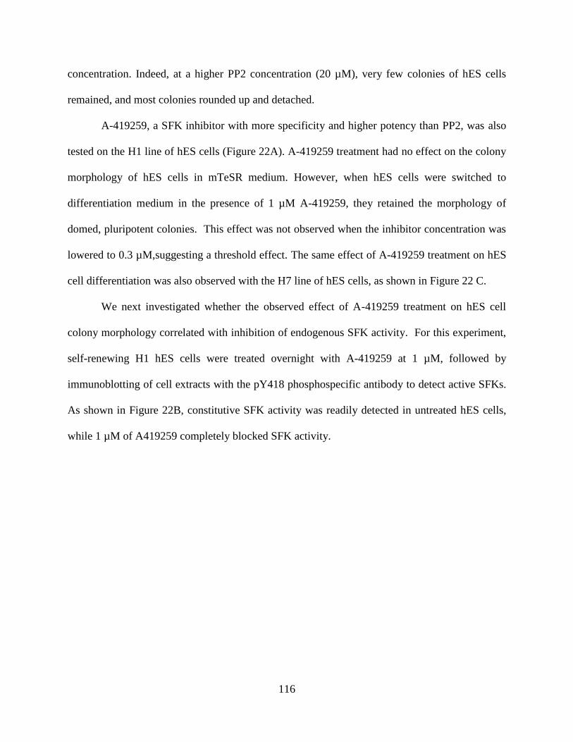
116
concentration. Indeed, at a higher PP2 concentration (20 µM), very few colonies of hES cells
remained, and most colonies rounded up and detached.
A-419259, a SFK inhibitor with more specificity and higher potency than PP2, was also
tested on the H1 line of hES cells (Figure 22A). A-419259 treatment had no effect on the colony
morphology of hES cells in mTeSR medium. However, when hES cells were switched to
differentiation medium in the presence of 1 µM A-419259, they retained the morphology of
domed, pluripotent colonies. This effect was not observed when the inhibitor concentration was
lowered to 0.3 µM,suggesting a threshold effect. The same effect of A-419259 treatment on hES
cell differentiation was also observed with the H7 line of hES cells, as shown in Figure 22 C.
We next investigated whether the observed effect of A-419259 treatment on hES cell
colony morphology correlated with inhibition of endogenous SFK activity. For this experiment,
self-renewing H1 hES cells were treated overnight with A-419259 at 1 µM, followed by
immunoblotting of cell extracts with the pY418 phosphospecific antibody to detect active SFKs.
As shown in Figure 22B, constitutive SFK activity was readily detected in untreated hES cells,
while 1 µM of A419259 completely blocked SFK activity.
117
mTeSRmTeSR 0.3µM
A419259
mTeSR 1µM
A419259
DM DM 0.3µM
A419259
DM 1µM
A419259
A.
pSFK
Oct4
Actin
+
-
+
+
mTeSR
1µM A419259
B.
mTeSRmTeSR 0.3µM
A419259
mTeSR 1µM
A419259
DM DM 0.3µM
A419259
DM 1µM
A419259
C.
Figure 22. Global SFK inhibition with A-419259 blocks hES cell differentiation. A) H1 hES cells were grown in
mTeSR renewal medium or switched to differentiation medium with or without the pan-SFK inhibitor, A-419259.
Note that inhibitor treatment results in the maintenance of colonies with rounded, domed morphology characteristic
of undifferentiated hES cells. (B) H1 hES cells growing in mTeSR medium were treated with or without A-419259
at a concentration of 1 µM overnight. Global SFK activity was determined by probing cell lysates with
phosphospecific antibodies directed against the activation loop phosphotyrosine as a measure of kinase activity.
Oct4 and Actin protein levels in the cell lysates were also examined as control. C) H7 hES cells were treated with A-
419259 under the same conditions as in part A.
118
3.3.4 Human ES cells maintain Tra-1-60 expression following SFK inhibition
Human ES cells express several markers for pluripotency, including cell surface markers SSEA-
3, SSEA-4, Tra-1-60 and Tra-1-81, as well as nuclear pluripotency factors such as Oct4, Nanog
and Sox2 (Thomson et al., 1998; Boyer et al., 2005). To determine whether SFK inhibition has
an effect in marker expression in hES cells, we determined Tra-1-60 expression in H7 cells
following treatment with A-419259. Tra-1-60 is a cell-surface protein that has been recognized
as one of the most stringent markers for hES cell pluripotency (Chan et al., 2009).
H1 hES cells were passaged and grown in mTeSR renewal medium for 3 days, followed
by continued growth on mTeSR or differentiation medium (DM) in the presence or absence of 1
µM A-419259 for 3 additional days. Cells were then fixed and stained for cell-surface marker
Tra-1-60 (Figure 23A). Untreated hESC cultures maintain typical, round hES cell morphology
and stained strongly with Tra-1-60 in mTeSR medium. The switch to DM caused the cells to
adopt the flattened colony morphology and lose Tra-1-60 expression, indicative of
differentiation. However, hES cells maintained in 1 µM A-419259 retained the morphology of
domed, pluripotent colonies and continued to express TRA-1-60 despite the switch to
differentiation culture conditions. Treatment of the H7 line of hES cells with A-419259 produced
and identical result, as shown in Figure 23B.
Together, these experiments show that global inhibition of endogenous SFK activity in
hES cells with A-419259 is sufficient to sustain pluripotent hES cell colony morphology and
Tra-1-60 expression under culture conditions for differentiation.

119
Tra
-1-6
0D
AP
IM
erg
e
mTeSR mTeSR 1µM DM DM+1µM
B.
Tra
-1-6
0D
AP
IM
erg
e
mTeSR DM DM+0.3µM DM+1µM
A.
Figure 23. Both H1 and H7 hES cells retain Tra-1-60 expression following SFK inhibition. A.) H1 hES cells were
grown on mTeSR medium for 3 days, then switched to either mTeSR medium or differentiation medium (DM;
DMEM/F-12 with 5% KSR, 15% FBS) with 0.3 or 1 µM A419259 or left untreated. Cells were fixed and stained for
the cell-surface pluripotency marker Tra-1-60 three days later (Magnification 400×). Untreated hESC cultures stain
strongly with Tra-1-60, while the switch to DM causes the cells to adopt flattened colony morphology and lose Tra-
1-60 expression. However, hES cells maintained in 1 µM A-419259 retained the morphology of domed, pluripotent
colonies and continued to express TRA-1-60 in DM. B.) H7 hES cells were grown on mTeSR medium for 3 days,
then switched to either mTeSR medium or differentiation medium with 1 µM A419259 or left untreated. Cells were
fixed and stained for the Tra-1-60 3 days later (Magnification 100×).
120
3.4 DISCUSSION
In this study, we found that hES cells express multiple SFK members with c-Src, c-Yes, Fyn and
Lck most highly expressed at the transcript level. SFK members showed dynamic changes in
expression levels during the differentiation process of hES cells to EBs: Lck expression dropped
dramatically as hES cells differentiate while c-Src and Fyn expression were up-regulated slightly
during this process. These expression changes suggest that Lck may be associated with self-
renewal, while c-Src and Fyn may be related to differentiation. In mES cells, both c-Src and Fyn
kinases are expressed. c-Src activity alone induces mES cell to differentiate into primitive
ectoderm (Meyn, III and Smithgall, 2009). c-Src may play similar role in hES cell as well.
Human ES cells express a high level of Lck, which is surprising given that Lck
expression is restricted to T-lymphocytes in adults. Lck has also been identified as a highly
expressed gene in a microarray study comparing gene expression profiles in pluripotent hES
cells with their differentiated counterparts (Cao et al., 2008). In addition, in a global
phosphoproteomic study of hES cells, Lck was identified as a signaling protein with more
phosphorylation sites in pluripotent hES cells than in differentiated hES cell derivatives (Brill et
al., 2009). These results are consistent with our findings, showing that Lck is regulated through
transcriptional regulation as well as phosphorylation, implicating this kinase in hES cell self-
renewal and/or suppression of differentiation. In addition to Lck, another SFK member, Hck,
was shown to be highly expressed in mES cells and was rapidly down-regulated in mouse EBs.
However, we only detected a low levels of Hck transcripts in hES cells. Interestingly, two
pluripotent states have recently been proposed: the naïve and primed states (Nichols and Smith,

121
2009) (see Introduction). Mouse EpiSCs and hES cells exemplify primed pluripotency while
mES cells exemplify naïve pluripotency. We have preliminary results suggesting that Hck is
highly expressed in mES cells but down-regulated in moues EpiSCs. Thus, Hck may be a marker
of naïve pluripotency, which explains why it is strongly expressed in mES cells yet only
expressed in hES cells at very low levels.
We also show that global inhibition of SFK activity blocks hES cell differentiation,
implicating SFK signaling in hES cell differentiation. This is similar to what we observed in
mES cells. Treatment of hES cells with the pan-SFK inhibitors PP2, SKI-1 and A-419259 all
suppressed hES cell differentiation and maintained hES cell colony morphology in
differentiation medium. Moreover, hES cells treated with A-419259, the most potent and
selective of these inhibitors, continued to express Tra-1-60 under culture conditions for
differentiation, consistent with the maintenance of undifferentiated colony morphology.
Finally, we found that the renewal marker Nanog and the Src-family member Hck are
differentially expressed among different hES cell lines. Also, in the process of EB formation,
some SFK members such as Hck and Lyn, and some differentiation markers such as Pax6 and
Msx1 are differentially up-regulated or down-regulated. These observations demonstrate the
heterogeneity between hES cell lines. Human ES cell lines have functional and molecular
heterogeneity, with cell lines requiring different pluripotency regulators and showing
differentiation bias (Wang et al., 2012; Bock et al., 2011). This cell line-specific heterogeneity
may be associated with the intrinsically unstable state of “primed” pluripotency (Nichols and
Smith, 2009). For future studies, it would be interesting to isolate purer hES populations from
these lines for gene expression analysis.

122
In summary, hES cells express multiple SFK members, and individual members are
dynamically regulated in different ways during EB differentiation of hES cells, indicating that
individual SFK members may play distinct role in hES cell self-renewal and differentiation.
Global inhibition of SFK activity blocked hES cell differentiation, and maintained pluripotency
marker expression. Our results support a role for SFK signaling in the regulation of hES fate, and
suggest that some parallels may exist in mouse and hES cells for this intracellular signaling
network.
3.5 MATERIALS AND METHODS
3.5.1 Cell culture
Human ES cell culture. H1, H7 and H9 hES cells (WiCell WA01, WA07, WA09) were
maintained in feeder-free culture conditions with mTeSR medium (Stemcell Technologies) on 6-
well plates coated with hESC-qualified matrigel (BD Biosciences). Medium was changed daily,
and cells were passaged every 6 days using Dispase (Stemcell Technologies) as per the
manufacturer’s instructions.
EB formation. ES cells were dissociated into single cells with Accutase (Stemcell
Technologies), pelleted and re-suspended in mTeSR medium containing 10 µM of the ROCK
inhibitor Y27632. Viable cells were counted using Trypan Blue (Invitrogen) and equal numbers
of cells were loaded into each well of AggreWell plates (Stemcell Technologies). For each hES
cell line used, each EB was started with aggregates of 2000 cells. EB aggregation of dissociated
ES cells were then initiated as per manufacturer’s instructions. EBs were harvested 24 hours

123
later, re-suspended in Aggrewell medium (Stemcell Technologies) or differentiation medium
(described below) , and maintained on ultra-low attachment plates (Corning).
Inhibitor treatment. ES cells were passaged onto Matrigel-coated 6-well plates and maintained
in mTeSR medium for three days. Culture medium was then changed to differentiation medium
or mTeSR with or without different concentrations of SFK inhibitors for an additional three
days. Differentiation medium was composed of 5% knockout serum replacement, 15% fetal
bovine serum, 1% non-essential amino acid, 1% L-glutamine, and 1% Pen/Strep in DMEM/F-12
medium (Life Technologies). SKI-1 and PP2 were purchased from Calbiochem while A-419259
was purchased from Santa Cruz Biotechnology.
3.5.2 RT-PCR
RNA isolation, RT-PCR, quantitative real-time RT-PCR, and data analysis were performed as
described in section 2.5.4. Human EBs were harvested 3, 9 and 12 days after initiation and were
washed using a cell strainer (BD Biosciences) to remove single cells before lysis.
3.5.3 Protein Blots
Cells were washed with phosphate-buffered saline (PBS) and lysed with RIPA buffer
supplemented with phosphatase and protease inhibitors as described (Meyn, III et al., 2005). Cell
lysates were clarified by centrifugation and equal amounts of total protein were mixed with SDS-
loading buffer, heated and separated on SDS polyacrylamide gels. Proteins were transferred to
PVDF membranes and probed with antibodies to the SFK activation loop, Oct4, and actin as a
loading control. The following primary antibodies were used at a 1:1000 dilution: active Src
124
pY418 (Invitrogen 44660G), mouse anti-Oct4 (Santa Cruz, SC-5279), and mouse anti-Actin
(Millipore, MAB1501).
3.5.4 Immunocytochemistry and fluorescence microscopy
Cells were plated onto Matrigel-coated coverslips in 6-well plates and maintained in mTeSR
medium for three days. The cell culture medium was then switched to differentiation medium or
mTeSR with or without inhibitors for three additional days. Cells were then washed with PBS,
fixed in 4% paraformaldehyde (Sigma) for 15 minutes, and blocked with PBS containing 5%
bovine serum albumin (BSA) (Fisher Sicientific) and 5% normal goat serum (Invitrogen) for 1
hour at room temperature. Cells were then incubated with primary antibodies overnight at 4 oC in
blocking buffer, followed by 3 washes in PBS. Secondary antibody (Invitrogen, 1:1000)
incubation was performed at 37 o
C for 1 hour followed by 3 washes. Samples are then mounted
using mounting medium containing DAPI (Vector Laboratories). The primary antibody used
was mouse anti-Tra-1-60 1:200 (Santa Cruz, SC-21705).

125
4.0 OVERALL DISCUSSION
4.1 SUMMARY OF FINDINGS AND SIGNIFICANCE
Embryonic stem cells have to choose between two fates in every cell cycle, to renew, or to
differentiate. This fate determination program is intrinsic to stem cells and is a tightly controlled
process. While the growth factor conditions and transcription factor networks governing stem
cell fates have been studied extensively, the signaling pathways connecting these extrinsic
signals to intrinsic gene transcription programs are not fully explored. Src family tyrosine
kinases are such modules relaying extrinsic signals to biological functions, and have been
implicated in mES cell fate determination. Here, I tested the hypothesis that c-Src and its closest
phylogenetic relative, c-Yes, act in biological opposition to one another in the context of ES cell
fate determination. I first discovered that enforced expression of active c-Yes blocked ES cell
differentiation to embryoid bodies by maintaining pluripotency gene expression. To determine
the interplay of c-Src and c-Yes in mES cell fate determination, I employed a chemical genetics
approach to investigate individual kinase function. I found that c-Src activity alone induces mES
cell differentiation to the ectoderm and endoderm, while c-Yes inhibits the endoderm
differentiation, acting in direct opposite to c-Src. These studies are the first to directly show that
even closely related kinases such as c-Src and c-Yes have unique and opposing functions in the
same cell type. In addition, my work shows that c-Src-induced differentiation correlates with
126
upregulation of EMT-related genes, incorporating the c-Src signal, EMT and ES cell
differentiation. Elucidating the connection between these mechanisms and developing specific
small molecule modulators of c-Src activity vs. c-Yes or vice versa may promote cellular
programing or differentiation to specific lineages. My second aim investigated the largely
unknown role of SFKs in human ES (hES) cells. I determined the relative expression profile of
individual Src family members in undifferentiated hES cells vs. embryoid bodies derived from
them. We found that hES cells express multiple SFK members, some of which display dynamic
transcription changes during EB differentiation, indicating that individual members may play
non-redundant roles. To assess the role of Src family kinase activity hES cells, I treated hES cell
cultures with a panel of chemically distinct SFK inhibitors. SFK inhibition maintained human ES
cell colony morphology and expression of the pluripotency marker Tra-1-60 in differentiation
medium. These observations support a role for Src family kinase signaling in the regulation of
hES fate, and suggest that some parallels may exist in mouse and human ES cells for this
intracellular signaling network. My research lays a foundation for further investigation of SFK
signaling in hES cells, which may shed light on hES cell maintenance and differentiation,
essential steps for regenerative medicine.
4.1.1 c-Yes is a potent anti-differentiation signal and acts in direct opposition to c-Src
In the first chapter, I showed that c-Yes is a potent suppressor of mES cell differentiation.
Enforced expression of either wild-type or an active mutant of c-Yes at modest levels blocked
ES cell differentiation to embryoid bodies. This suppressive effect requires kinase activity, since
it is not observed with kinase-defective c-Yes. Interestingly, EB formation is very sensitive to c-
Yes signaling, as both wild-type c-Yes and a kinase-active c-Yes mutant inhibited this process,

127
resulting in the formation of only small cell clusters. In addition, we found that c-Yes activity is
associated with the maintenance of pluripotency marker expression. Unlike control EBs, where
pluripotency factors are all down-regulated, the small cell clusters formed from ES/c-Yes cells
continue to express pluripotency factors, such as Oct4, Nanog, Klf4 and Esrrb. This finding is
consistent with the reported role of c-Yes as a kinase associated with mES cell self-renewal
(Anneren et al., 2004), where c-Yes is downstream of LIF and signals through Yes associated
protein (YAP) and transcription factor TEAD2 to activate Nanog and Oct4 expression (Tamm et
al., 2011).
Next, to study the role of Src family kinases individually, we employed a chemical
genetics approach and designed inhibitor resistant (IR) mutants of c-Src and c-Yes. Treatment of
ES cells with SFK inhibitor A-419259 blocks all endogenous Src-family kinase activity in ES
cells, locking them in an undifferentiated state (Meyn, III et al., 2005). Expression of IR mutants
in mES cells allowed us to study the role of c-Src or c-Yes activity individually, in the presence
of inhibitor concentrations that block all endogenous SFK activity. Expression of c-Yes IR
mutant did not rescue the differentiation block associated with A-419259 treatment, but
pluripotency marker expression was retained. This is consistent with the role of c-Yes as a
renewal-related kinase. In contrast, inhibitor-treated cells expressing Src-IR mutants adopted a
flattened, differentiated morphology, consistent with our previous finding that that c-Src activity
alone promotes ES cell differentiation to primitive ectoderm-like cells (Meyn, III and Smithgall,
2009).
By expanding the qPCR marker analysis, I showed that c-Src activity alone induces mES
cell differentiation into both primitive ectoderm and endoderm lineages, as exemplified by Fgf5,
Sox17, Gata4 and Gata6 expression. Further, I found c-Src is a potent activator of the epithelial-

128
mesenchymal transition (EMT), with significant upregulation of EMT related genes such as
Snail1, Twist1, Igf2, MMP9 and MMP14. The activation of EMT-associated genes is
concomitant with the up-regulation of cell-lineage differentiation markers. The EMT regulates
multiple important tissue remodeling processes in early embryonic development and has also
been implicated in cancer invasion and metastasis (Thiery et al., 2009). EMT has also been
associated with differentiation in both mouse and human ES cells (Eastham et al., 2007; Spencer
et al., 2007). Conversely, inhibition of this process promoted the nuclear reprograming of mouse
fibroblast cells to induced pluripotent stem cells (iPSCs) (Li et al., 2011). In addition, the EMT is
proposed to be an essential and early step for lineage commitment in ES cells, since elevation of
EMT markers preceded activation of lineage markers in response to differentiation signals (Li et
al., 2011). Our results directly link c-Src kinase activity with the EMT, implicating this
fundamental process in c-Src-induced differentiation of mES cells.
The mechanism of how c-Src activity regulates EMT progression is still unknown. EMT
is a developmental program tightly controlled by the TGFβ/Nodal/BMP, Wnt/β-Catenin, Notch
and FGF signaling pathways (Yang and Weinberg, 2008; Thiery et al., 2009). Src may modulate
the EMT downstream of these growth factors. In addition, c-Src has been implicated in cell
adhesion, migration and cytoskeletal reorganization—key processes associated with the EMT as
well. For example, c-Src phosphorylates p120 catenin to disrupt its association with E-cadherin;
c-Src couples with FAK to activate MMP2 and MMP9 and promote migration; and c-Src
activates the Rho-family GTPases Rac, Cdc42 and Rho, to regulate adhesion and the F-actin
cytoskeleton (Guarino, 2010; Kim et al., 2009). Interestingly, functional loss of E-cadherin in
epithelial cells is a hallmark for the EMT, while both MMPs and Rho-family GTPases are major
players in EMT (Yang and Weinberg, 2008). These findings indicate that c-Src may play a major
129
role in the EMT process, by cooperating with and modulating multiple proteins associated with
cell-cell adhesion, cell-matrix adhesion and migration.
Moreover, we made the discovery that c-Yes acts in direct opposition to c-Src. We found
that the c-Yes signal suppresses c-Src-induced differentiation and the EMT. Remarkably, when
both c-Yes and c-Src IR mutants were introduced into the same cell population and treated with
the inhibitor, c-Yes activity prevented the induction of endoderm differentiation and EMT by c-
Src. Of note, suppression of endoderm differentiation might be a characteristic of the Yes-YAP-
TEAD2 signaling pathway. A significant increase in endoderm-specific gene expression was
observed when TEAD2 activity is downregulated in mES cells (Tamm et al., 2011), consistent
with our findings described here.
Our observations help to define c-Yes as a suppressor of differentiation. We show that c-
Yes activity is sufficient to maintain expression of the core transcription factors that govern
pluripotency, and is dominant over the differentiation signals induced by c-Src. These findings
support a previous model of the regulation of endogenous c-Src and c-Yes kinase activity during
ES cell renewal and differentiation (Meyn, III et al., 2005). In the presence of LIF, both c-Yes
and c-Src are active, but the presence of active c-Yes overrides the c-Src signal for
differentiation. Upon LIF withdrawal, c-Yes activity is shut off, allowing active c-Src to drive
differentiation. More broadly, our work suggests that although ES cells can be maintained in a
perpetual state of self-renewal, they are poised to differentiate. However, our data also show that
c-Yes activity alone cannot sustain ES cell self-renewal. This is point is supported by at least
three lines of evidence. 1) ES cells expressing active c-Yes adopted a flattened, differentiated
morphology in the absence of LIF in adherent culture. 2) Although ES cells expressing active c-
Yes only form abnormal, small aggregates under culture conditions for EB formation,
130
differentiation markers were expressed. 3) Complete inhibition of SFK activity does not result in
differentiation; rather, the cells remain pluripotent (Meyn, III et al., 2005). Taken together, these
results show that c-Yes is an important suppressor of differentiation but is not sufficient to
maintain ES cell self-renewal. In addition, our results provide a strong rationale for the
development of selective compounds to control c-Src vs. c-Yes activity; such compounds may
allow more precise pharmacological manipulation of ES cell renewal and differentiation. Indeed,
a recent unbiased chemical library screen identified broad-spectrum Src-family kinase inhibitors
as potent enhancers of somatic cell reprogramming to an ES cell-like state (iPS cells) (Staerk et
al., 2011). Our work predicts that a Src-selective inhibitor (or a c-Yes agonist) may provide an
even greater enhancement in reprogramming efficiency.
A long-standing tenet of the Src-family kinase field is that individual family members
have similar, if not redundant, biological functions. Our results clearly demonstrate that this is
not always the case, and provide an important caveat to the use of broad-spectrum, small
molecule inhibitors of the entire Src kinase family to make conclusions about the biological
activity of individual family members or the family as a whole. However, the mechanisms of
how these two closely related kinases act in opposition to one another are still not clear. Several
factors might account for this difference: 1) c-Src and c-Yes may bind different adaptor or
substrate proteins through their SH3 or SH2 domains or even through their unique domains. For
example, c-Yes protects and maintains tight junctions by interacting with occludins; in contrast,
c-Src activity is associated with occludin downregulation and tight junction dissociation by
activating Raf-1 (Summy et al., 2003b). 2) c-Yes and c-Src may have different sub-cellular
localizations. c-Yes can be both myristoylated and palmitoylated, which target it to lipid rafts,
131
while c-Src has no palmitoylation site (Thomas and Brugge, 1997). This localization difference
may also account for recruitment of distinct binding partners.
In summary, we defined c-Yes as a potent suppressor of mES cell differentiation,
showing that low-level, sustained expression of active c-Yes maintains ES cell renewal marker
expression following LIF withdrawal, resulting in a profound block to EB formation. In addition,
using a chemical genetics approach, we discovered that c-Yes acts in direct opposition to c-Src,
to inhibit c-Src-induced endoderm differentiation and EMT marker expression. These findings
reveal a key regulatory mechanism in the dynamic control of murine ES cell pluripotency and
differentiation. More broadly, our results are the first to show that closely related kinases like c-
Src and c-Yes are not necessarily functioning redundantly, but may act in direct opposition to
each other in the same cell type.
4.1.2 SFK signaling is important for human ES cell differentiation
While our group and others have implicated SFK signaling in mES cells, the role of this kinase
family is not clear in human ES cells. In my second aim, I explored Src family kinase signaling
in hES cells.
We found that human ES cells express multiple SFK members: Of the eleven Src-related
kinases in the human genome, Fyn, c-Yes, c-Src, Lyn, Lck and Hck were expressed in H1, H7
and H9 hES cells, while Fgr, Blk, Srm, Brk, and Frk transcripts were not detected. During hES
cell differentiation into EBs, c-Yes, Lyn, and Hck transcript levels remained constant in self-
renewing hES cells and differentiated EBs, while c-Src and Fyn had a modest increase in
expression. In contrast, Lck expression levels dropped dramatically as a function of EB
differentiation. The dynamic expression changes of SFK members during hES cell differentiation
132
into EBs indicate that they play different roles in this process. Lck, which is expressed only in
the T-cell compartment in adults, is highly expressed in human ES cells, yet down-regulated
during differentiation. This suggests that Lck might be important for the self-renew program. On
the other hand, c-Src and Fyn, which are highly expressed in hES cells, are further upregulated
during differentiation, suggesting that they may be involved in this process.
Our surprising finding that Lck is strongly expressed in human ES cells and is
transcriptionally silenced during differentiation to EBs supports a unique function in the control
of hES cell renewal. This idea is supported by previous gene expression profiling studies
showing that Lck transcript levels are high in renewing hES cells but down-regulated in
differentiated cells, and a phosphoproteomic analysis showing Lck proteins are preferentially
phosphorylated at Tyr-179 in undifferentiated H1 cells (Brill et al., 2009; Cao et al., 2008).
However, the questions of how Lck activity regulates the hES cell self-renewal program is still
unknown. SFKs are involved in multiple signaling pathways, including receptor tyrosine kinase
signaling and integrin modulated cell-adhesion signaling (Thomas and Brugge, 1997; Ingley,
2008). In human ES cells, SFK may signal downstream of FGF/FGFR, and cross talk with
MEK/Erk, PI3K/Akt and Wnt/β-Catenin signaling pathways, all of which have been implicated
in hES cell self-renew and differentiation.
In addition, we report that SFK inhibition maintains hES cell colony morphology and
expression of the pluripotency marker Tra-1-60. To assess the role of Src family kinase activity
hES cells, we treated hES cell cultures with inhibitors specific for the Src kinase family,
including Src Kinase Inhibitor 1 (SKI-1), PP2 and A-419259. Untreated control hES cultures
grown in mTeSR1 medium formed typical pluripotent colonies circumscribed by differentiated
cells. Transfer of hES cells to differentiation medium resulted in flattened colony morphology

133
that correlated with a loss of cell-surface staining for the hES cell pluripotency marker, Tra-1-60.
Dramatically, hES cells maintained in the presence of 1 µM A-419259 retained the morphology
of domed, pluripotent colonies and maintained TRA-1-60 expression in differentiation medium.
Similar morphological changes were observed in SKI-1 treated cells.
These inhibitor studies suggest an important role for SFKs in hES cell differentiation.
Based on our previous work in mES cells, we speculate the c-Src may have an important role in
the differentiation process. As suggested in Chapter 2, c-Src induces EMT and differentiation in
murine ES cells. Since EMT is also correlates with human ES cell differentiation (Eastham et al.,
2007), Src may induce EMT and promote hES cell differentiation as well. While we were able to
show that A-419259 treatment inhibits all endogenous SFK activity in hES cells in a manner
very similar to mES cells, we were not able to identify the specific family members that are
constitutively active in hES cells. Future studies may approach this problem using the chemical
genetic strategy that was successfully applied to mES cells.
In summary, the observations reported in chapter 3 support a role for Src family kinase
signaling in the regulation of hES fate, and suggest that some parallels may exist in mouse and
human ES cells for this intracellular signaling network. This is the first report to
comprehensively investigating SFKs in human ES cells, and these results will help elucidate the
mechanism of this important family of kinases in human ES cells.
134
4.2 FUTURE DIRECTIONS
4.2.1 Identify signaling pathways downstream of c-Src and c-Yes that account for the
different stem cell fates associated with SFK signaling
My thesis research led to the surprising discovery that c-Src and c-Yes directly oppose each
other in mES cells. The signaling pathways downstream of c-Src that are responsible for ES cell
differentiation are not known. While c-Yes has been shown to phosphorylate YAP and activate
YAP-TEAD2-dependent transcription (Tamm et al., 2011), other pathways may also contribute
to the role of c-Yes as suppressor of differentiation in mES cells. The opposing roles of c-Yes
and c-Src may be related to the distinct signaling partners they recruit, or their subcellular
localizations. The SH2 and SH3 domains of c-Src and c-Yes may have different specificities for
binding partners. For example, the c-Src SH3 domain readily binds actin filament associated
protein (AFAP) while the c-Yes SH3 domain does not (Summy et al., 2003b). This may partially
explain why c-Yes fails to control actin filament rearrangement, and thus cannot compensate for
c-Src function in osteoclast cells from c-Src knockout mice (Soriano et al., 1991; Summy et al.,
2003b). Although not able to bind AFAP, c-Yes SH3 domain can uniquely modulate interaction
with YAP (Clump et al., 2005).
To narrow down the signaling molecules most closely related to ES cell fate
determination, we could combine the Src-IR or Yes-IR mutants with SH3 and SH2 null mutants,
to probe which function domain mutation abrogates the biological function. For example, Src-IR
could be combined with mutations that inactivate the function of the Src SH3 domain. The
resulting Src-IR/SH3 mutant could then be expressed in ES cells, and tested for differentiation in
response to A-419259 treatment as described in Chapter 2. If the Src-IR/SH3 null mutant fails to

135
induce ES cell differentiation, then SH3-binding partners may be involved in this biological
process. Subsequently, Src SH3 domain can be immobilized and used as baits for capture of SH3
binding partners from ES cell extracts, followed by identification through standard MS-based
proteomics. This technology has been successfully used by our group to identify a circadian
rhythm protein, Timeless, as a substrate of Src that is essential for ES cell differentiation to EBs
(O'Reilly et al., 2011).
To interrogate signaling pathways downstream of c-Src that induce ES cell
differentiation, we could use Src-IR in combination with A-419259 as a model system for small
molecule screening. Since a number of signaling pathways active in ES cells might act
downstream of Src, small molecule inhibitors of these pathways can be introduced, and those
inhibitors which abrogate Src-induced differentiation could be selected for further biochemical
analysis. Candidate pathways include the MEK/Erk, PI3K/AKT, FAK, Wnt/β-Catenin and EMT
signaling pathways. As described in earlier sections, each of these pathways has been linked to c-
Src in other biological contexts.
The ultimate tool to identify downstream signaling pathways involves the
phosphoproteomics approach of stable isotope labeling of amino acids in cell culture (SILAC)
(Pimienta et al., 2009). Control ES cells with all SFK activity blocked by A-419259 will be
grown in normal medium, while Src-IR ES cells with A419259 treatment will be grown in heavy
medium with all arginine labeled with 13
C. Therefore, all proteins from the Src-IR cells will
generate heavier peptides than their counterparts in control cells. These two cell populations can
then be combined in equal ratios, trypsinized, and analyzed together by Liquid Chromatography-
Mass Spectrometry. In the resulting mass spectra, a pair of light and heavy peptides will show up
together as a doublet because of the isotope labeling. The peak intensities in those doublets will

136
reflect the abundance of the actual protein. Proteins giving rise to mass peaks with high intensity
in Src-IR cells but low intensity in control cells will be implicated in Src mediated
differentiation. A pilot experiment was performed in our lab, and we identified several target
proteins that were differentially phosphorylated in cells as a function of A-419259 treatment for
complete SFK inhibition. These targets included key components of the Wnt/β-Catenin pathway,
the pluripotency factor Sox2 and zinc-finger transcription factor Sal-like protein 4 (SAL4).
Another interesting question is how c-Src activity promotes the EMT in the context of ES
cells. ES cell colonies expressing Src-IR cells undergo a dramatic morphological change
following A-419259 treatment, adopting a flattened morphology. One could speculate that part
of this morphological shift is due to the EMT, as cell:cell contacts are lost and the cells become
more autonomous. It would be interesting to investigate how c-Src activity connects with the
hallmark EMT transcription factors such as Snail1 and Twist, which may contribute to these
morphological changes. This will shed light on the connection between the EMT and ES cell
differentiation. The EMT has also been implicated in cancer progression, and c-Src-induced
changes related to the EMT in ES cells may also have a crucial role in cancer metastasis.
4.2.2 Further study of SFK signaling in human ES cells
We found that Src family kinase signaling is important in the regulation of hES fate, and that
some parallels exist between mouse and human ES cells for this intracellular signaling network.
Further studies to elucidate SFK signaling in hES cells are crucial.
First, are SFKs responsive to hES cell growth factors? Since Lck has been identified as a
self-renewal related kinase, it would be important to determine if it is responsive to bFGF, and

137
activated by FGFR, because this growth factor is a key regulator of hES cell pluripotency. Also,
the downstream effectors of SFKs are not clear in human ES cells.
In addition, what will happen to hES cells overexpressing SFKs? Can overexpression of
Lck block human ES cell differentiation, in a manner similar to that observed with c-Yes in
mouse ES cells? Conversely, can overexpression of c-Src or Fyn induce differentiation? We
could also combine kinase active mutants, or employ the chemical genetics approach to study
specific SFK signaling in hES cells.
Another interesting point relates to the absence of Hck expression in hES cells despite the
fact that it is highly expressed in renewing mES cells but undergoes rapid transcriptional
silenceing as mES cells differentiate. These observations suggest that Hck expression and
activity may be specifically associated with naïve as opposed to primed pluripotency. A
comparative analysis of SFK expression and activity profiles in mEpiSCs vs. mESCs, and in
hESCs vs. naïve hESCs, would help to clarify this important issue.
Moreover, we hypothesize that c-Src and Fyn may be involved in hES cell
differentiation. Previous studies have shown that the EMT is associated with hESC
differentiation (Eastham et al., 2007), and in our mES cell study, c-Src activates EMT-associated
gene expression. Both c-Src and Fyn may have similar function in hES cells, and parallel
pathways may exist in human ES cells.
One major difficulty of hES cell research is the heterogeneity of cell culture. Unlike
mES cells, hES cultures represent a mixture of pluripotent and differentiated cell types, even
under self-renewal conditions, which complicates the analysis of fate marker expression. Also,
hES cells are sensitive to single cell dissociation, which hinders clonal passage and genetic
manipulation. The newly developed naïve human ES cells culture and human ES cell lines with

138
selective reporters for pluripotency (for example, Oct4-GFP hESCs) may facilitate future
signaling studies with SFKs (Hanna et al., 2010; Buehr et al., 2008).

139
4.3 CLOSING REMARKS
ES cells have the remarkable property of self-renewal and pluripotency, the ability to
generate all cell types of the adult body. ES cells provide an in vitro model system of cell
differentiation that mimics early embryonic development. When combined with genetic
manipulation, mES cells provide an unprecedented gateway to in vivo models in the form of
transgenic animals (Evans, 2011). The development of human ES cells (Thomson et al., 1998),
and the newer human induced pluripotent stem cells (Takahashi et al., 2007; Yu et al., 2007)
opened a new era for disease modeling and hold great potential for cell therapy, regenerative
medicine, and drug discovery. Cell fate determination is of central importance to stem cell
biology and the development of stem cell therapy. Multiple signaling pathways contribute to fate
determination of stem cells. In this dissertation reseach project, I provided new evidence that
Src-family tyrosine kinases are important for the growth and fate specification of both human
and mouse embryonic stem cells. In mES cells, I provided direct evidence that c-Yes is a potent
inhibitor of differentiation and opposes the action of c-Src to drive differentiation. This result
provides a strong challenge to the idea that individual Src family members play redundant
biological roles. In addition, this study suggests that selective inhibitors of individual members
of the Src family may promote reprograming efficiency, or drive differentiation to specific
lineages. Moreover, our results connected stem cell differentiation, Src activity, with the
fundamental developmental program of the EMT. Since the EMT is also associated with cancer
progression and the formation of cancer stem cells, my results in the ES cell context may also
have significance for tumor biology.

140
BIBLIOGRAPHY
Alonso,G., Koegl,M., Mazurenko,N., and Courtneidge,S.A. (1995). Sequence requirements for
binding of Src family tyrosine kinases to activated growth factor receptors. J. Biol. Chem. 270,
9840-9848.
Amit,M., Carpenter,M.K., Inokuma,M.S., Chiu,C.P., Harris,C.P., Waknitz,M.A., Itskovitz-
Eldor,J., and Thomson,J.A. (2000). Clonally derived human embryonic stem cell lines maintain
pluripotency and proliferative potential for prolonged periods of culture. Dev. Biol. 227, 271-
278.
Anneren,C., Cowan,C.A., and Melton,D.A. (2004). The Src family of tyrosine kinases is
important for embryonic stem cell self-renewal. J. Biol. Chem. 279, 31590-31598.
Anton,R., Kestler,H.A., and Kuhl,M. (2007). Beta-catenin signaling contributes to stemness and
regulates early differentiation in murine embryonic stem cells. FEBS Lett. 581, 5247-5254.
Artus,J., Piliszek,A., and Hadjantonakis,A.K. (2011). The primitive endoderm lineage of the
mouse blastocyst: sequential transcription factor activation and regulation of differentiation by
Sox17. Dev. Biol. 350, 393-404.
Avilion,A.A., Nicolis,S.K., Pevny,L.H., Perez,L., Vivian,N., and Lovell-Badge,R. (2003).
Multipotent cell lineages in early mouse development depend on SOX2 function. Genes Dev. 17,
126-140.
Beddington,R.S. and Robertson,E.J. (1989). An assessment of the developmental potential of
embryonic stem cells in the midgestation mouse embryo. Development 105, 733-737.
Ben-Shushan,E., Thompson,J.R., Gudas,L.J., and Bergman,Y. (1998). Rex-1, a gene encoding a
transcription factor expressed in the early embryo, is regulated via Oct-3/4 and Oct-6 binding to
an octamer site and a novel protein, Rox-1, binding to an adjacent site. Mol. Cell Biol. 18, 1866-
1878.
Bendall,S.C., Stewart,M.H., Menendez,P., George,D., Vijayaragavan,K., Werbowetski-
Ogilvie,T., Ramos-Mejia,V., Rouleau,A., Yang,J., Bosse,M., Lajoie,G., and Bhatia,M. (2007).
IGF and FGF cooperatively establish the regulatory stem cell niche of pluripotent human cells in
vitro. Nature 448, 1015-1021.
Berstine,E.G., Hooper,M.L., Grandchamp,S., and Ephrussi,B. (1973). Alkaline phosphatase
activity in mouse teratoma. Proc. Natl. Acad. Sci. U. S. A 70, 3899-3903.
Bishop,A.C., Ubersax,J.A., Petsch,D.T., Matheos,D.P., Gray,N.S., Blethrow,J., Shimizu,E.,
Tsien,J.Z., Schultz,P.G., Rose,M.D., Wood,J.L., Morgan,D.O., and Shokat,K.M. (2000). A
chemical switch for inhibitor-sensitive alleles of any protein kinase. Nature 407, 395-401.

141
Bock,C., Kiskinis,E., Verstappen,G., Gu,H., Boulting,G., Smith,Z.D., Ziller,M., Croft,G.F.,
Amoroso,M.W., Oakley,D.H., Gnirke,A., Eggan,K., and Meissner,A. (2011). Reference Maps of
human ES and iPS cell variation enable high-throughput characterization of pluripotent cell
lines. Cell 144, 439-452.
Boggon,T.J. and Eck,M.J. (2004). Structure and regulation of Src family kinases. Oncogene 23,
7918-7927.
Bourillot,P.Y., Aksoy,I., Schreiber,V., Wianny,F., Schulz,H., Hummel,O., Hubner,N., and
Savatier,P. (2009). Novel STAT3 target genes exert distinct roles in the inhibition of mesoderm
and endoderm differentiation in cooperation with Nanog. Stem Cells 27, 1760-1771.
Boyer,L.A., Lee,T.I., Cole,M.F., Johnstone,S.E., Levine,S.S., Zucker,J.P., Guenther,M.G.,
Kumar,R.M., Murray,H.L., Jenner,R.G., Gifford,D.K., Melton,D.A., Jaenisch,R., and
Young,R.A. (2005). Core transcriptional regulatory circuitry in human embryonic stem cells.
Cell 122, 947-956.
Boyer,L.A., Mathur,D., and Jaenisch,R. (2006). Molecular control of pluripotency. Curr. Opin.
Genet. Dev. 16, 455-462.
Bradley,A., Evans,M., Kaufman,M.H., and Robertson,E. (1984). Formation of germ-line
chimaeras from embryo-derived teratocarcinoma cell lines. Nature 309, 255-256.
Brill,L.M., Xiong,W., Lee,K.B., Ficarro,S.B., Crain,A., Xu,Y., Terskikh,A., Snyder,E.Y., and
Ding,S. (2009). Phosphoproteomic analysis of human embryonic stem cells. Cell Stem Cell 5,
204-213.
Bromann,P.A., Korkaya,H., and Courtneidge,S.A. (2004). The interplay between Src family
kinases and receptor tyrosine kinases. Oncogene 23, 7957-7968.
Brons,I.G., Smithers,L.E., Trotter,M.W., Rugg-Gunn,P., Sun,B., Chuva de Sousa Lopes SM,
Howlett,S.K., Clarkson,A., Ahrlund-Richter,L., Pedersen,R.A., and Vallier,L. (2007). Derivation
of pluripotent epiblast stem cells from mammalian embryos. Nature 448, 191-195.
Brook,F.A. and Gardner,R.L. (1997). The origin and efficient derivation of embryonic stem cells
in the mouse. Proc. Natl. Acad. Sci. U. S. A 94, 5709-5712.
Brugge,J.S. and Erikson,R.L. (1977). Identification of a transformation-specific antigen induced
by an avian sarcoma virus. Nature 269, 346-348.
Buecker,C., Chen,H.H., Polo,J.M., Daheron,L., Bu,L., Barakat,T.S., Okwieka,P., Porter,A.,
Gribnau,J., Hochedlinger,K., and Geijsen,N. (2010). A murine ESC-like state facilitates
transgenesis and homologous recombination in human pluripotent stem cells. Cell Stem Cell 6,
535-546.
Buehr,M., Meek,S., Blair,K., Yang,J., Ure,J., Silva,J., McLay,R., Hall,J., Ying,Q.L., and
Smith,A. (2008). Capture of authentic embryonic stem cells from rat blastocysts. Cell 135, 1287-
1298.

142
Burdon,T., Chambers,I., Stracey,C., Niwa,H., and Smith,A. (1999a). Signaling mechanisms
regulating self-renewal and differentiation of pluripotent embryonic stem cells. Cells Tissues.
Organs 165, 131-143.
Burdon,T., Smith,A., and Savatier,P. (2002). Signalling, cell cycle and pluripotency in
embryonic stem cells. Trends Cell Biol. 12, 432-438.
Burdon,T., Stracey,C., Chambers,I., Nichols,J., and Smith,A. (1999b). Suppression of SHP-2 and
ERK signalling promotes self-renewal of mouse embryonic stem cells. Dev. Biol. 210, 30-43.
Cantley,L.C. (2002). The phosphoinositide 3-kinase pathway. Science 296, 1655-1657.
Cao,F., Wagner,R.A., Wilson,K.D., Xie,X., Fu,J.D., Drukker,M., Lee,A., Li,R.A., Gambhir,S.S.,
Weissman,I.L., Robbins,R.C., and Wu,J.C. (2008). Transcriptional and functional profiling of
human embryonic stem cell-derived cardiomyocytes. PLoS. One. 3, e3474.
Capecchi,M.R. (2005). Gene targeting in mice: functional analysis of the mammalian genome for
the twenty-first century. Nat. Rev. Genet. 6, 507-512.
Chakravarthy,H., Boer,B., Desler,M., Mallanna,S.K., McKeithan,T.W., and Rizzino,A. (2008).
Identification of DPPA4 and other genes as putative Sox2:Oct-3/4 target genes using a
combination of in silico analysis and transcription-based assays. J. Cell Physiol 216, 651-662.
Chambers,I., Colby,D., Robertson,M., Nichols,J., Lee,S., Tweedie,S., and Smith,A. (2003).
Functional expression cloning of Nanog, a pluripotency sustaining factor in embryonic stem
cells. Cell 113, 643-655.
Chambers,I. and Smith,A. (2004). Self-renewal of teratocarcinoma and embryonic stem cells.
Oncogene 23, 7150-7160.
Chan,B., Lanyi,A., Song,H.K., Griesbach,J., Simarro-Grande,M., Poy,F., Howie,D., Sumegi,J.,
Terhorst,C., and Eck,M.J. (2003). SAP couples Fyn to SLAM immune receptors. Nat. Cell Biol.
5, 155-160.
Chan,E.M., Ratanasirintrawoot,S., Park,I.H., Manos,P.D., Loh,Y.H., Huo,H., Miller,J.D.,
Hartung,O., Rho,J., Ince,T.A., Daley,G.Q., and Schlaeger,T.M. (2009). Live cell imaging
distinguishes bona fide human iPS cells from partially reprogrammed cells. Nat. Biotechnol. 27,
1033-1037.
Chandrasekharan,S., Qiu,T.H., Alkharouf,N., Brantley,K., Mitchell,J.B., and Liu,E.T. (2002).
Characterization of mice deficient in the Src family nonreceptor tyrosine kinase Frk/rak. Mol.
Cell Biol. 22, 5235-5247.
Chapman,G., Remiszewski,J.L., Webb,G.C., Schulz,T.C., Bottema,C.D., and Rathjen,P.D.
(1997). The mouse homeobox gene, Gbx2: genomic organization and expression in pluripotent
cells in vitro and in vivo. Genomics 46, 223-233.

143
Chazaud,C., Yamanaka,Y., Pawson,T., and Rossant,J. (2006). Early lineage segregation between
epiblast and primitive endoderm in mouse blastocysts through the Grb2-MAPK pathway. Dev.
Cell 10, 615-624.
Chen,G., Hou,Z., Gulbranson,D.R., and Thomson,J.A. (2010). Actin-myosin contractility is
responsible for the reduced viability of dissociated human embryonic stem cells. Cell Stem Cell
7, 240-248.
Cherry,S.R., Biniszkiewicz,D., van,P.L., Baltimore,D., and Jaenisch,R. (2000). Retroviral
expression in embryonic stem cells and hematopoietic stem cells. Mol. Cell Biol. 20, 7419-7426.
Chia,N.Y., Chan,Y.S., Feng,B., Lu,X., Orlov,Y.L., Moreau,D., Kumar,P., Yang,L., Jiang,J.,
Lau,M.S., Huss,M., Soh,B.S., Kraus,P., Li,P., Lufkin,T., Lim,B., Clarke,N.D., Bard,F., and
Ng,H.H. (2010). A genome-wide RNAi screen reveals determinants of human embryonic stem
cell identity. Nature 468, 316-320.
Clevers,H. (2006). Wnt/beta-catenin signaling in development and disease. Cell 127, 469-480.
Clevers,H. and Nusse,R. (2012). Wnt/beta-catenin signaling and disease. Cell 149, 1192-1205.
Cloutier,J.F. and Veillette,A. (1996). Association of inhibitory tyrosine protein kinase p50csk
with protein tyrosine phosphatase PEP in T cells and other hemopoietic cells. EMBO J. 15,
4909-4918.
Clump,D.A., Qazi,I.H., Sudol,M., and Flynn,D.C. (2005). c-Yes response to growth factor
activation. Growth Factors 23, 263-272.
Cohen,D.E. and Melton,D. (2011). Turning straw into gold: directing cell fate for regenerative
medicine. Nat. Rev. Genet. 12, 243-252.
Cooper,J.A., Gould,K.L., Cartwright,C.A., and Hunter,T. (1986). Tyr527 is phosphorylated in
pp60c-src: implications for regulation. Science 231, 1431-1434.
Cowan-Jacob,S.W., Fendrich,G., Manley,P.W., Jahnke,W., Fabbro,D., Liebetanz,J., and
Meyer,T. (2005). The Crystal Structure of a c-Src Complex in an Active Conformation Suggests
Possible Steps in c-Src Activation. Structure. (Camb. ) 13, 861-871.
Cully,M., You,H., Levine,A.J., and Mak,T.W. (2006). Beyond PTEN mutations: the PI3K
pathway as an integrator of multiple inputs during tumorigenesis. Nat. Rev. Cancer 6, 184-192.
Davis,S., Aldrich,T.H., Stahl,N., Pan,L., Taga,T., Kishimoto,T., Ip,N.Y., and Yancopoulos,G.D.
(1993). LIFR beta and gp130 as heterodimerizing signal transducers of the tripartite CNTF
receptor. Science 260, 1805-1808.
Dravid,G., Ye,Z., Hammond,H., Chen,G., Pyle,A., Donovan,P., Yu,X., and Cheng,L. (2005).
Defining the role of Wnt/beta-catenin signaling in the survival, proliferation, and self-renewal of
human embryonic stem cells. Stem Cells 23, 1489-1501.

144
Dvash,T., Mayshar,Y., Darr,H., McElhaney,M., Barker,D., Yanuka,O., Kotkow,K.J., Rubin,L.L.,
Benvenisty,N., and Eiges,R. (2004). Temporal gene expression during differentiation of human
embryonic stem cells and embryoid bodies. Hum. Reprod. 19, 2875-2883.
Dvorak,P., Dvorakova,D., Koskova,S., Vodinska,M., Najvirtova,M., Krekac,D., and Hampl,A.
(2005). Expression and potential role of fibroblast growth factor 2 and its receptors in human
embryonic stem cells. Stem Cells 23, 1200-1211.
Eastham,A.M., Spencer,H., Soncin,F., Ritson,S., Merry,C.L., Stern,P.L., and Ward,C.M. (2007).
Epithelial-mesenchymal transition events during human embryonic stem cell differentiation.
Cancer Res. 67, 11254-11262.
Engen,J.R., Wales,T.E., Hochrein,J.M., Meyn,M.A., III, Banu,O.S., Bahar,I., and Smithgall,T.E.
(2008). Structure and dynamic regulation of Src-family kinases. Cell Mol. Life Sci. 65, 3058-
3073.
Ernst,M., Gearing,D.P., and Dunn,A.R. (1994). Functional and biochemical association of Hck
with the LIF/IL- 6 receptor signal transducing subunit gp130 in embryonic stem cells. EMBO J.
13, 1574-1584.
Evans,M. (2011). Discovering pluripotency: 30 years of mouse embryonic stem cells. Nat. Rev.
Mol. Cell Biol. 12, 680-686.
Evans,M.J. (1972). The isolation and properties of a clonal tissue culture strain of pluripotent
mouse teratoma cells. J. Embryol. Exp. Morphol. 28, 163-176.
Evans,M.J. and Kaufman,M.H. (1981). Establishment in culture of pluripotential cells from
mouse embryos. Nature 292, 154-156.
Feng,S., Chen,J.K., Yu,H., Simon,J.A., and Schreiber,S.L. (1994). Two binding orientations for
peptides to the Src SH3 domain: Development of a general model for SH3-ligand interactions.
Science 266, 1241-1247.
Feng,X.H. and Derynck,R. (2005). Specificity and versatility in tgf-beta signaling through
Smads. Annu. Rev. Cell Dev. Biol. 21, 659-693.
Festuccia,N., Osorno,R., Halbritter,F., Karwacki-Neisius,V., Navarro,P., Colby,D., Wong,F.,
Yates,A., Tomlinson,S.R., and Chambers,I. (2012). Esrrb is a direct Nanog target gene that can
substitute for Nanog function in pluripotent cells. Cell Stem Cell 11, 477-490.
Frankenberg,S., Gerbe,F., Bessonnard,S., Belville,C., Pouchin,P., Bardot,O., and Chazaud,C.
(2011). Primitive endoderm differentiates via a three-step mechanism involving Nanog and RTK
signaling. Dev. Cell 21, 1005-1013.
Gadue,P., Huber,T.L., Paddison,P.J., and Keller,G.M. (2006). Wnt and TGF-beta signaling are
required for the induction of an in vitro model of primitive streak formation using embryonic
stem cells. Proc. Natl. Acad. Sci. U. S. A 103, 16806-16811.
145
Grant,S.G., O'Dell,T.J., Karl,K.A., Stein,P.L., Soriano,P., and Kandel,E.R. (1992). Impaired
long-term potentiation, spatial learning, and hippocampal development in fyn mutant mice.
Science 258, 1903-1910.
Greber,B., Coulon,P., Zhang,M., Moritz,S., Frank,S., Muller-Molina,A.J., Arauzo-Bravo,M.J.,
Han,D.W., Pape,H.C., and Scholer,H.R. (2011). FGF signalling inhibits neural induction in
human embryonic stem cells. EMBO J. 30, 4874-4884.
Greber,B., Lehrach,H., and Adjaye,J. (2007). Fibroblast growth factor 2 modulates transforming
growth factor beta signaling in mouse embryonic fibroblasts and human ESCs (hESCs) to
support hESC self-renewal. Stem Cells 25, 455-464.
Guarino,M. (2010). Src signaling in cancer invasion. J. Cell Physiol 223, 14-26.
Guo,G., Huss,M., Tong,G.Q., Wang,C., Li,S.L., Clarke,N.D., and Robson,P. (2010). Resolution
of cell fate decisions revealed by single-cell gene expression analysis from zygote to blastocyst.
Dev. Cell 18, 675-685.
Hall,J., Guo,G., Wray,J., Eyres,I., Nichols,J., Grotewold,L., Morfopoulou,S., Humphreys,P.,
Mansfield,W., Walker,R., Tomlinson,S., and Smith,A. (2009). Oct4 and LIF/Stat3 additively
induce Kruppel factors to sustain embryonic stem cell self-renewal. Cell Stem Cell 5, 597-609.
Han,J., Yuan,P., Yang,H., Zhang,J., Soh,B.S., Li,P., Lim,S.L., Cao,S., Tay,J., Orlov,Y.L.,
Lufkin,T., Ng,H.H., Tam,W.L., and Lim,B. (2010). Tbx3 improves the germ-line competency of
induced pluripotent stem cells. Nature 463, 1096-1100.
Hanna,J., Cheng,A.W., Saha,K., Kim,J., Lengner,C.J., Soldner,F., Cassady,J.P., Muffat,J.,
Carey,B.W., and Jaenisch,R. (2010). Human embryonic stem cells with biological and epigenetic
characteristics similar to those of mouse ESCs. Proc. Natl. Acad. Sci. U. S. A 107, 9222-9227.
Hanna,J., Markoulaki,S., Mitalipova,M., Cheng,A.W., Cassady,J.P., Staerk,J., Carey,B.W.,
Lengner,C.J., Foreman,R., Love,J., Gao,Q., Kim,J., and Jaenisch,R. (2009). Metastable
pluripotent states in NOD-mouse-derived ESCs. Cell Stem Cell 4, 513-524.
Hao,J., Li,T.G., Qi,X., Zhao,D.F., and Zhao,G.Q. (2006). WNT/beta-catenin pathway up-
regulates Stat3 and converges on LIF to prevent differentiation of mouse embryonic stem cells.
Dev. Biol. 290, 81-91.
Harrison,S.C. (2003). Variation on an Src-like theme. Cell 112, 737-740.
Hart,A.H., Hartley,L., Sourris,K., Stadler,E.S., Li,R., Stanley,E.G., Tam,P.P., Elefanty,A.G., and
Robb,L. (2002). Mixl1 is required for axial mesendoderm morphogenesis and patterning in the
murine embryo. Development 129, 3597-3608.
Hentze,H., Soong,P.L., Wang,S.T., Phillips,B.W., Putti,T.C., and Dunn,N.R. (2009). Teratoma
formation by human embryonic stem cells: evaluation of essential parameters for future safety
studies. Stem Cell Res. 2, 198-210.
146
Hosler,B.A., LaRosa,G.J., Grippo,J.F., and Gudas,L.J. (1989). Expression of REX-1, a gene
containing zinc finger motifs, is rapidly reduced by retinoic acid in F9 teratocarcinoma cells.
Mol. Cell Biol. 9, 5623-5629.
Huveneers,S. and Danen,E.H. (2009). Adhesion signaling - crosstalk between integrins, Src and
Rho. J. Cell Sci. 122, 1059-1069.
Ihle,J.N. (2001). The Stat family in cytokine signaling. Curr. Opin. Cell Biol. 13, 211-217.
Imamoto,A. and Soriano,P. (1993). Disruption of the csk gene, encoding a negative regulator of
Src family tyrosine kinases, leads to neural tube defects and embryonic lethality in mice. Cell 73,
1117-1124.
Ingley,E. (2008). Src family kinases: regulation of their activities, levels and identification of
new pathways. Biochim. Biophys. Acta 1784, 56-65.
Ishizawar,R. and Parsons,S.J. (2004). c-Src and cooperating partners in human cancer. Cancer
Cell 6, 209-214.
Itskovitz-Eldor,J., Schuldiner,M., Karsenti,D., Eden,A., Yanuka,O., Amit,M., Soreq,H., and
Benvenisty,N. (2000). Differentiation of human embryonic stem cells into embryoid bodies
compromising the three embryonic germ layers. Mol. Med. 6, 88-95.
Ivanova,N., Dobrin,R., Lu,R., Kotenko,I., Levorse,J., DeCoste,C., Schafer,X., Lun,Y., and
Lemischka,I.R. (2006). Dissecting self-renewal in stem cells with RNA interference. Nature 442,
533-538.
Jaenisch,R. and Young,R. (2008). Stem cells, the molecular circuitry of pluripotency and nuclear
reprogramming. Cell 132, 567-582.
James,D., Levine,A.J., Besser,D., and Hemmati-Brivanlou,A. (2005). TGFbeta/activin/nodal
signaling is necessary for the maintenance of pluripotency in human embryonic stem cells.
Development 132, 1273-1282.
Jiang,J., Chan,Y.S., Loh,Y.H., Cai,J., Tong,G.Q., Lim,C.A., Robson,P., Zhong,S., and Ng,H.H.
(2008). A core Klf circuitry regulates self-renewal of embryonic stem cells. Nat. Cell Biol. 10,
353-360.
Jirmanova,L., Afanassieff,M., Gobert-Gosse,S., Markossian,S., and Savatier,P. (2002).
Differential contributions of ERK and PI3-kinase to the regulation of cyclin D1 expression and
to the control of the G1/S transition in mouse embryonic stem cells. Oncogene 21, 5515-5528.
Johnson,T.M., Williamson,N.A., Scholz,G., Jaworowski,A., Wettenhall,R.E., Dunn,A.R., and
Cheng,H.C. (2000). Modulation of the catalytic activity of the Src family tyrosine kinase Hck by
autophosphorylation at a novel site in the unique domain. J. Biol. Chem. 275, 33353-33364.
Kataoka,H., Takakura,N., Nishikawa,S., Tsuchida,K., Kodama,H., Kunisada,T., Risau,W.,
Kita,T., and Nishikawa,S.I. (1997). Expressions of PDGF receptor alpha, c-Kit and Flk1 genes

147
clustering in mouse chromosome 5 define distinct subsets of nascent mesodermal cells. Dev.
Growth Differ. 39, 729-740.
Kawabuchi,M., Satomi,Y., Takao,T., Shimonishi,Y., Nada,S., Nagai,K., Tarakhovsky,A., and
Okada,M. (2000). Transmembrane phosphoprotein Cbp regulates the activities of Src-family
tyrosine kinases. Nature 404, 999-1003.
Keller,G. (2005). Embryonic stem cell differentiation: Emergence of a new era in biology and
medicine. Genes and Development 19, 1129-1155.
Keller,G.M. (1995). In vitro differentiation of embryonic stem cells. Curr. Opin. Cell Biol. 7,
862-869.
Kelly,K.F., Ng,D.Y., Jayakumaran,G., Wood,G.A., Koide,H., and Doble,B.W. (2011). beta-
catenin enhances Oct-4 activity and reinforces pluripotency through a TCF-independent
mechanism. Cell Stem Cell 8, 214-227.
Kielman,M.F., Rindapaa,M., Gaspar,C., van,P.N., Breukel,C., van,L.S., Taketo,M.M.,
Roberts,S., Smits,R., and Fodde,R. (2002). Apc modulates embryonic stem-cell differentiation
by controlling the dosage of beta-catenin signaling. Nat. Genet. 32, 594-605.
Kim,L.C., Song,L., and Haura,E.B. (2009). Src kinases as therapeutic targets for cancer. Nat.
Rev. Clin. Oncol. 6, 587-595.
Kim,M.O., Kim,S.H., Cho,Y.Y., Nadas,J., Jeong,C.H., Yao,K., Kim,D.J., Yu,D.H., Keum,Y.S.,
Lee,K.Y., Huang,Z., Bode,A.M., and Dong,Z. (2012). ERK1 and ERK2 regulate embryonic stem
cell self-renewal through phosphorylation of Klf4. Nat. Struct. Mol. Biol. 19, 283-290.
Kim,P.W., Sun,Z.Y., Blacklow,S.C., Wagner,G., and Eck,M.J. (2003). A zinc clasp structure
tethers Lck to T cell coreceptors CD4 and CD8. Science 301, 1725-1728.
Kispert,A. and Herrmann,B.G. (1994). Immunohistochemical analysis of the Brachyury protein
in wild-type and mutant mouse embryos. Dev. Biol. 161, 179-193.
Klinghoffer,R.A., Sachsenmaier,C., Cooper,J.A., and Soriano,P. (1999). Src family kinases are
required for integrin but not PDGFR signal transduction. EMBO J. 18, 2459-2471.
Koopman,P. and Cotton,R.G. (1984). A factor produced by feeder cells which inhibits
embryonal carcinoma cell differentiation. Characterization and partial purification. Exp. Cell
Res. 154, 233-242.
Krishnamurty,R., Brigham,J.L., Leonard,S.E., Ranjitkar,P., Larson,E.T., Dale,E.J., Merritt,E.A.,
and Maly,D.J. (2013). Active site profiling reveals coupling between domains in SRC-family
kinases. Nat. Chem. Biol. 9, 43-50.
Kunath,T., Saba-El-Leil,M.K., Almousailleakh,M., Wray,J., Meloche,S., and Smith,A. (2007).
FGF stimulation of the Erk1/2 signalling cascade triggers transition of pluripotent embryonic
stem cells from self-renewal to lineage commitment. Development 134, 2895-2902.

148
Latour,S., Roncagalli,R., Chen,R., Bakinowski,M., Shi,X., Schwartzberg,P.L., Davidson,D., and
Veillette,A. (2003). Binding of SAP SH2 domain to FynT SH3 domain reveals a novel
mechanism of receptor signalling in immune regulation. Nat. Cell Biol. 5, 149-154.
Lee,C.-H., Saksela,K., Mirza,U.A., Chait,B.T., and Kuriyan,J. (1996). Crystal structure of the
conserved core of HIV-1 Nef complexed with a Src family SH3 domain. Cell 85, 931-942.
Lerner,E.C. and Smithgall,T.E. (2002). SH3-dependent stimulation of Src-family kinase
autophosphorylation without tail release from the SH2 domain in vivo. Nat. Struct. Biol. 9, 365-
369.
Levenstein,M.E., Ludwig,T.E., Xu,R.H., Llanas,R.A., VanDenHeuvel-Kramer,K., Manning,D.,
and Thomson,J.A. (2006). Basic fibroblast growth factor support of human embryonic stem cell
self-renewal. Stem Cells 24, 568-574.
Li,J., Wang,G., Wang,C., Zhao,Y., Zhang,H., Tan,Z., Song,Z., Ding,M., and Deng,H. (2007).
MEK/ERK signaling contributes to the maintenance of human embryonic stem cell self-renewal.
Differentiation 75, 299-307.
Li,X., Zhu,L., Yang,A., Lin,J., Tang,F., Jin,S., Wei,Z., Li,J., and Jin,Y. (2011). Calcineurin-
NFAT signaling critically regulates early lineage specification in mouse embryonic stem cells
and embryos. Cell Stem Cell 8, 46-58.
Li,Y., McClintick,J., Zhong,L., Edenberg,H.J., Yoder,M.C., and Chan,R.J. (2005). Murine
embryonic stem cell differentiation is promoted by SOCS-3 and inhibited by the zinc finger
transcription factor Klf4. Blood 105, 635-637.
Li,Z., Fei,T., Zhang,J., Zhu,G., Wang,L., Lu,D., Chi,X., Teng,Y., Hou,N., Yang,X., Zhang,H.,
Han,J.D., and Chen,Y.G. (2012). BMP4 Signaling Acts via dual-specificity phosphatase 9 to
control ERK activity in mouse embryonic stem cells. Cell Stem Cell 10, 171-182.
Lim,W.A., Richards,F.M., and Fox,R.O. (1994). Structural determinants of peptide-binding
orientation and of sequence specificity in SH3 domains. Nature 372, 375-379.
Liu,B.A., Jablonowski,K., Shah,E.E., Engelmann,B.W., Jones,R.B., and Nash,P.D. (2010). SH2
domains recognize contextual peptide sequence information to determine selectivity. Mol. Cell
Proteomics. 9, 2391-2404.
Liu,N., Lu,M., Tian,X., and Han,Z. (2007). Molecular mechanisms involved in self-renewal and
pluripotency of embryonic stem cells. J. Cell Physiol 211, 279-286.
Liu,Y., Bishop,A., Witucki,L., Kraybill,B., Shimizu,E., Tsien,J., Ubersax,J., Blethrow,J.,
Morgan,D.O., and Shokat,K.M. (1999). Structural basis for selective inhibition of Src family
kinases by PP1. Chem. Biol. 6, 671-678.
Loh,Y.H., Wu,Q., Chew,J.L., Vega,V.B., Zhang,W., Chen,X., Bourque,G., George,J., Leong,B.,
Liu,J., Wong,K.Y., Sung,K.W., Lee,C.W., Zhao,X.D., Chiu,K.P., Lipovich,L., Kuznetsov,V.A.,
Robson,P., Stanton,L.W., Wei,C.L., Ruan,Y., Lim,B., and Ng,H.H. (2006). The Oct4 and Nanog

149
transcription network regulates pluripotency in mouse embryonic stem cells. Nat. Genet. 38,
431-440.
Lowell,C.A., Niwa,M., Soriano,P., and Varmus,H.E. (1996). Deficiency of the Hck and Src
tyrosine kinases results in extreme levels of extramedullary hematopoiesis. Blood 87, 1780-1792.
Lowell,C.A. and Soriano,P. (1996). Knockouts of Src-family kinases: stiff bones, wimpy T cells,
and bad memories. Genes Dev. 10, 1845-1857.
Lu,J., Hou,R., Booth,C.J., Yang,S.H., and Snyder,M. (2006). Defined culture conditions of
human embryonic stem cells. Proc. Natl. Acad. Sci. U. S. A 103, 5688-5693.
Ludwig,T.E., Levenstein,M.E., Jones,J.M., Berggren,W.T., Mitchen,E.R., Frane,J.L.,
Crandall,L.J., Daigh,C.A., Conard,K.R., Piekarczyk,M.S., Llanas,R.A., and Thomson,J.A.
(2006). Derivation of human embryonic stem cells in defined conditions. Nat. Biotechnol. 24,
185-187.
Madan,B., Madan,V., Weber,O., Tropel,P., Blum,C., Kieffer,E., Viville,S., and Fehling,H.J.
(2009). The pluripotency-associated gene Dppa4 is dispensable for embryonic stem cell identity
and germ cell development but essential for embryogenesis. Mol. Cell Biol. 29, 3186-3203.
Maldonado-Saldivia,J., van den Bergen,J., Krouskos,M., Gilchrist,M., Lee,C., Li,R.,
Sinclair,A.H., Surani,M.A., and Western,P.S. (2007). Dppa2 and Dppa4 are closely linked SAP
motif genes restricted to pluripotent cells and the germ line. Stem Cells 25, 19-28.
Manning,G., Whyte,D.B., Martinez,R., Hunter,T., and Sudarsanam,S. (2002). The protein kinase
complement of the human genome. Science 298, 1912-1934.
Martello,G., Sugimoto,T., Diamanti,E., Joshi,A., Hannah,R., Ohtsuka,S., Gottgens,B., Niwa,H.,
and Smith,A. (2012). Esrrb is a pivotal target of the Gsk3/Tcf3 axis regulating embryonic stem
cell self-renewal. Cell Stem Cell 11, 491-504.
Martin,G.R. (1981). Isolation of a pluripotent cell line from early mouse embryos cultured in
medium conditioned by teratocarcinoma stem cells. Proc. Natl. Acad. Sci. U. S. A 78, 7634-
7638.
Martin,G.R. and Evans,M.J. (1975). Differentiation of clonal lines of teratocarcinoma cells:
formation of embryoid bodies in vitro. Proc. Natl. Acad. Sci. U. S. A 72, 1441-1445.
Martin,G.S. (2001). The hunting of the Src. Nat. Rev. Mol. Cell Biol. 2, 467-475.
Martin,G.S. (2004). The road to Src. Oncogene 23, 7910-7917.
Masaki,H., Nishida,T., Kitajima,S., Asahina,K., and Teraoka,H. (2007). Developmental
pluripotency-associated 4 (DPPA4) localized in active chromatin inhibits mouse embryonic stem
cell differentiation into a primitive ectoderm lineage. J. Biol. Chem. 282, 33034-33042.
Massague,J. and Chen,Y.G. (2000). Controlling TGF-beta signaling. Genes Dev. 14, 627-644.

150
Massague,J., Seoane,J., and Wotton,D. (2005). Smad transcription factors. Genes Dev. 19, 2783-
2810.
Masui,S., Nakatake,Y., Toyooka,Y., Shimosato,D., Yagi,R., Takahashi,K., Okochi,H.,
Okuda,A., Matoba,R., Sharov,A.A., Ko,M.S., and Niwa,H. (2007). Pluripotency governed by
Sox2 via regulation of Oct3/4 expression in mouse embryonic stem cells. Nat. Cell Biol. 9, 625-
635.
Masui,S., Ohtsuka,S., Yagi,R., Takahashi,K., Ko,M.S., and Niwa,H. (2008). Rex1/Zfp42 is
dispensable for pluripotency in mouse ES cells. BMC. Dev. Biol. 8, 45.
Matsuda,T., Nakamura,T., Nakao,K., Arai,T., Katsuki,M., Heike,T., and Yokota,T. (1999).
STAT3 activation is sufficient to maintain an undifferentiated state of mouse embryonic stem
cells. EMBO J. 18, 4261-4269.
Meyn,M.A., III, Schreiner,S.J., Dumitrescu,T.P., Nau,G.J., and Smithgall,T.E. (2005). SRC
family kinase activity is required for murine embryonic stem cell growth and differentiation.
Mol. Pharmacol. 68, 1320-1330.
Meyn,M.A., III and Smithgall,T.E. (2009). Chemical genetics identifies c-Src as an activator of
primitive ectoderm formation in murine embryonic stem cells. Sci. Signal. 2, ra64.
Mitsui,K., Tokuzawa,Y., Itoh,H., Segawa,K., Murakami,M., Takahashi,K., Maruyama,M.,
Maeda,M., and Yamanaka,S. (2003). The homeoprotein Nanog is required for maintenance of
pluripotency in mouse epiblast and ES cells. Cell 113, 631-642.
Miyazaki,T., Sanjay,A., Neff,L., Tanaka,S., Horne,W.C., and Baron,R. (2004). Src kinase
activity is essential for osteoclast function. J. Biol. Chem. 279, 17660-17666.
Mohr,J.C., Zhang,J., Azarin,S.M., Soerens,A.G., de Pablo,J.J., Thomson,J.A., Lyons,G.E.,
Palecek,S.P., and Kamp,T.J. (2010). The microwell control of embryoid body size in order to
regulate cardiac differentiation of human embryonic stem cells. Biomaterials 31, 1885-1893.
Murakami,M., Hibi,M., Nakagawa,N., Nakagawa,T., Yasukawa,K., Yamanishi,K., Taga,T., and
Kishimoto,T. (1993). IL-6-induced homodimerization of gp130 and associated activation of a
tyrosine kinase. Science 260, 1808-1810.
Murry,C.E. and Keller,G. (2008). Differentiation of embryonic stem cells to clinically relevant
populations: lessons from embryonic development. Cell 132, 661-680.
Musacchio,A., Noble,M., Pauptit,R., Wierenga,R., and Saraste,M. (1992). Crystal structure of a
Src-homology 3 (SH3) domain. Nature 359, 851-855.
Musacchio,A., Saraste,M., and Wilmanns,M. (1994). High-resolution crystal structures of
tyrosine kinase SH3 domains complexed with proline-rich peptides. Nat. Struct. Biol. 1, 546-
551.

151
Nada,S., Yagi,T., Takeda,H., Tokunaga,T., Nakagawa,H., Ikawa,Y., Okada,M., and Aizawa,S.
(1993). Constitutive activation of Src family kinases in mouse embryos that lack Csk. Cell 73,
1125-1135.
Ng,H.H. and Surani,M.A. (2011). The transcriptional and signalling networks of pluripotency.
Nat. Cell Biol. 13, 490-496.
Nichols,J. and Smith,A. (2009). Naive and primed pluripotent states. Cell Stem Cell 4, 487-492.
Nichols,J. and Smith,A. (2012). Pluripotency in the embryo and in culture. Cold Spring Harb.
Perspect. Biol. 4, a008128.
Nichols,J., Zevnik,B., Anastassiadis,K., Niwa,H., Klewe-Nebenius,D., Chambers,I., Scholer,H.,
and Smith,A. (1998). Formation of pluripotent stem cells in the mammalian embryo depends on
the POU transcription factor Oct4. Cell 95, 379-391.
Nishioka,N., Inoue,K., Adachi,K., Kiyonari,H., Ota,M., Ralston,A., Yabuta,N., Hirahara,S.,
Stephenson,R.O., Ogonuki,N., Makita,R., Kurihara,H., Morin-Kensicki,E.M., Nojima,H.,
Rossant,J., Nakao,K., Niwa,H., and Sasaki,H. (2009). The Hippo signaling pathway components
Lats and Yap pattern Tead4 activity to distinguish mouse trophectoderm from inner cell mass.
Dev. Cell 16, 398-410.
Niwa,H. (2007). How is pluripotency determined and maintained? Development 134, 635-646.
Niwa,H., Burdon,T., Chambers,I., and Smith,A. (1998). Self-renewal of pluripotent embryonic
stem cells is mediated via activation of STAT3. Genes Dev. 12, 2048-2060.
Niwa,H., Miyazaki,J., and Smith,A.G. (2000). Quantitative expression of Oct-3/4 defines
differentiation, dedifferentiation or self-renewal of ES cells. Nat. Genet. 24, 372-376.
Niwa,H., Ogawa,K., Shimosato,D., and Adachi,K. (2009). A parallel circuit of LIF signalling
pathways maintains pluripotency of mouse ES cells. Nature 460, 118-122.
O'Reilly,L.P., Watkins,S.C., and Smithgall,T.E. (2011). An unexpected role for the clock protein
timeless in developmental apoptosis. PLoS. One. 6, e17157.
Ogawa,K., Nishinakamura,R., Iwamatsu,Y., Shimosato,D., and Niwa,H. (2006). Synergistic
action of Wnt and LIF in maintaining pluripotency of mouse ES cells. Biochem. Biophys. Res.
Commun. 343, 159-166.
Ohgushi,M., Matsumura,M., Eiraku,M., Murakami,K., Aramaki,T., Nishiyama,A.,
Muguruma,K., Nakano,T., Suga,H., Ueno,M., Ishizaki,T., Suemori,H., Narumiya,S., Niwa,H.,
and Sasai,Y. (2010). Molecular pathway and cell state responsible for dissociation-induced
apoptosis in human pluripotent stem cells. Cell Stem Cell 7, 225-239.
Okamoto,K., Okazawa,H., Okuda,A., Sakai,M., Muramatsu,M., and Hamada,H. (1990). A novel
octamer binding transcription factor is differentially expressed in mouse embryonic cells. Cell
60, 461-472.
152
Paling,N.R., Wheadon,H., Bone,H.K., and Welham,M.J. (2004). Regulation of embryonic stem
cell self-renewal by phosphoinositide 3-kinase-dependent signaling. J. Biol. Chem. 279, 48063-
48070.
Parsons,J.T. and Parsons,S.J. (1997). Src family protein tyrosine kinases: Cooperating with
growth factor and adhesion signaling pathways. Curr. Opin. Cell Biol. 9, 187-192.
Pawson,T. (2004). Specificity in signal transduction: from phosphotyrosine-SH2 domain
interactions to complex cellular systems. Cell 116, 191-203.
Pawson,T. and Nash,P. (2000). Protein-protein interactions define specificity in signal
transduction. Genes Dev. 14, 1027-1047.
Pelton,T.A., Sharma,S., Schulz,T.C., Rathjen,J., and Rathjen,P.D. (2002). Transient pluripotent
cell populations during primitive ectoderm formation: correlation of in vivo and in vitro
pluripotent cell development. J. Cell Sci. 115, 329-339.
Pene-Dumitrescu,T., Peterson,L.F., Donato,N.J., and Smithgall,T.E. (2008). An inhibitor-
resistant mutant of Hck protects CML cells against the antiproliferative and apoptotic effects of
the broad-spectrum Src family kinase inhibitor A-419259. Oncogene 27, 7055-7069.
Pene-Dumitrescu,T. and Smithgall,T.E. (2010). Expression of a Src family kinase in chronic
myelogenous leukemia cells induces resistance to imatinib in a kinase-dependent manner. J.
Biol. Chem. 285, 21446-21457.
Percharde,M., Lavial,F., Ng,J.H., Kumar,V., Tomaz,R.A., Martin,N., Yeo,J.C., Gil,J.,
Prabhakar,S., Ng,H.H., Parker,M.G., and Azuara,V. (2012). Ncoa3 functions as an essential
Esrrb coactivator to sustain embryonic stem cell self-renewal and reprogramming. Genes Dev.
26, 2286-2298.
Perez,Y., Gairi,M., Pons,M., and Bernado,P. (2009). Structural characterization of the natively
unfolded N-terminal domain of human c-Src kinase: insights into the role of phosphorylation of
the unique domain. J. Mol. Biol. 391, 136-148.
Pfaffl,M.W., Horgan,G.W., and Dempfle,L. (2002). Relative expression software tool (REST)
for group-wise comparison and statistical analysis of relative expression results in real-time
PCR. Nucleic Acids Res. 30, e36.
Pimienta,G., Chaerkady,R., and Pandey,A. (2009). SILAC for global phosphoproteomic
analysis. Methods Mol. Biol. 527, 107-16, x.
Prokhorova,T.A., Harkness,L.M., Frandsen,U., Ditzel,N., Schroder,H.D., Burns,J.S., and
Kassem,M. (2009). Teratoma formation by human embryonic stem cells is site dependent and
enhanced by the presence of Matrigel. Stem Cells Dev. 18, 47-54.
Qi,X., Li,T.G., Hao,J., Hu,J., Wang,J., Simmons,H., Miura,S., Mishina,Y., and Zhao,G.Q.
(2004). BMP4 supports self-renewal of embryonic stem cells by inhibiting mitogen-activated
protein kinase pathways. Proc. Natl. Acad. Sci. U. S. A 101, 6027-6032.

153
Resh,M.D. (1994). Myristylation and palmitylation of Src family members: The fats of the
matter. Cell 76, 411-413.
Resh,M.D. (1999). Fatty acylation of proteins: new insights into membrane targeting of
myristoylated and palmitoylated proteins. Biochim. Biophys. Acta 1451, 1-16.
Robinson,D.R., Wu,Y.M., and Lin,S.F. (2000). The protein tyrosine kinase family of the human
genome. Oncogene 19, 5548-5557.
Rogers,M.B., Hosler,B.A., and Gudas,L.J. (1991). Specific expression of a retinoic acid-
regulated, zinc-finger gene, Rex-1, in preimplantation embryos, trophoblast and spermatocytes.
Development 113, 815-824.
Roskoski,R., Jr. (2004). Src protein-tyrosine kinase structure and regulation. Biochem. Biophys.
Res. Commun. 324, 1155-1164.
Roskoski,R., Jr. (2005). Src kinase regulation by phosphorylation and dephosphorylation.
Biochem. Biophys. Res. Commun. 331, 1-14.
Rudd,C.E., Trevillyan,J.M., Dasgupta,J.D., Wong,L.L., and Schlossman,S.F. (1988). The CD4
receptor is complexed in detergent lysates to a protein-tyrosine kinase (pp58) from human T
lymphocytes. Proc. Natl. Acad. Sci. U. S. A 85, 5190-5194.
Saksela,K. and Permi,P. (2012). SH3 domain ligand binding: What's the consensus and where's
the specificity? FEBS Lett. 586, 2609-2614.
Sancier,F., Dumont,A., Sirvent,A., Paquay de,P.L., Edmonds,T., David,G., Jan,M., de,M.C.,
Coge,F., Leonce,S., Burbridge,M., Bruno,A., Boutin,J.A., Lockhart,B., Roche,S., and
Cruzalegui,F. (2011). Specific oncogenic activity of the Src-family tyrosine kinase c-Yes in
colon carcinoma cells. PLoS. One. 6, e17237.
Sato,N., Meijer,L., Skaltsounis,L., Greengard,P., and Brivanlou,A.H. (2004). Maintenance of
pluripotency in human and mouse embryonic stem cells through activation of Wnt signaling by a
pharmacological GSK-3-specific inhibitor. Nat. Med. 10, 55-63.
Schindler,T., Sicheri,F., Pico,A., Gazit,A., Levitzki,A., and Kuriyan,J. (1999). Crystal structure
of Hck in complex with a Src family-selective tyrosine kinase inhibitor. Mol. Cell 3, 639-648.
Scholer,H.R., Ruppert,S., Suzuki,N., Chowdhury,K., and Gruss,P. (1990). New type of POU
domain in germ line-specific protein Oct-4. Nature 344, 435-439.
Scotland,K.B., Chen,S., Sylvester,R., and Gudas,L.J. (2009). Analysis of Rex1 (zfp42) function
in embryonic stem cell differentiation. Dev. Dyn. 238, 1863-1877.
Shenoy,S., Chackalaparampil,I., Bagrodia,S., Lin,P.H., and Shalloway,D. (1992). Role of
p34cdc2-mediated phosphorylations in two-step activation of pp60c-src during mitosis. Proc.
Natl. Acad. Sci. U. S. A 89, 7237-7241.
154
Sicheri,F., Moarefi,I., and Kuriyan,J. (1997). Crystal structure of the Src family tyrosine kinase
Hck. Nature 385, 602-609.
Silva,J., Barrandon,O., Nichols,J., Kawaguchi,J., Theunissen,T.W., and Smith,A. (2008).
Promotion of reprogramming to ground state pluripotency by signal inhibition. PLoS. Biol. 6,
e253.
Silverman,L., Sudol,M., and Resh,M.D. (1993). Members of the src family of nonreceptor
tyrosine kinases share a common mechanism for membrane binding. Cell Growth Differ. 4, 475-
482.
Smith,A.G. (2001). Embryo-derived stem cells: of mice and men. Annu. Rev. Cell Dev. Biol. 17,
435-462.
Smith,A.G., Heath,J.K., Donaldson,D.D., Wong,G.G., Moreau,J., Stahl,M., and Rogers,D.
(1988). Inhibition of pluripotential embryonic stem cell differentiation by purified polypeptides.
Nature 336, 688-690.
Smith,A.G. and Hooper,M.L. (1987). Buffalo rat liver cells produce a diffusible activity which
inhibits the differentiation of murine embryonal carcinoma and embryonic stem cells. Dev. Biol.
121, 1-9.
Sokol,S.Y. (2011). Maintaining embryonic stem cell pluripotency with Wnt signaling.
Development 138, 4341-4350.
Solter,D. (2006). From teratocarcinomas to embryonic stem cells and beyond: a history of
embryonic stem cell research. Nat. Rev. Genet. 7, 319-327.
Solter,D. and Knowles,B.B. (1978). Monoclonal antibody defining a stage-specific mouse
embryonic antigen (SSEA-1). Proc. Natl. Acad. Sci. U. S. A 75, 5565-5569.
Songyang,Z., Shoelson,S.E., Chaudhuri,M., Gish,G., Pawson,T., Haser,W.G., King,F.,
Roberts,T., Ratnofsky,S., Lechleider,R.J., Neel,B.G., Birge,R.B., Fajardo,J.E., Chou,M.M.,
Hanafusa,H., Schaffhausen,B., and Cantley,L.C. (1993). SH2 domains recognize specific
phosphopeptide sequences. Cell 72, 767-778.
Soriano,P., Montgomery,C., Geske,R., and Bradley,A. (1991). Targeted disruption of the c-src
proto-oncogene leads to osteopetrosis in mice. Cell 64, 693-702.
Spencer,H.L., Eastham,A.M., Merry,C.L., Southgate,T.D., Perez-Campo,F., Soncin,F., Ritson,S.,
Kemler,R., Stern,P.L., and Ward,C.M. (2007). E-cadherin inhibits cell surface localization of the
pro-migratory 5T4 oncofetal antigen in mouse embryonic stem cells. Mol. Biol. Cell 18, 2838-
2851.
Stadtfeld,M. and Hochedlinger,K. (2010). Induced pluripotency: history, mechanisms, and
applications. Genes Dev. 24, 2239-2263.

155
Staerk,J., Lyssiotis,C.A., Medeiro,L.A., Bollong,M., Foreman,R.K., Zhu,S., Garcia,M., Gao,Q.,
Bouchez,L.C., Lairson,L.L., Charette,B.D., Supekova,L., Janes,J., Brinker,A., Cho,C.Y.,
Jaenisch,R., and Schultz,P.G. (2011). Pan-Src Family Kinase Inhibitors Replace Sox2 during the
Direct Reprogramming of Somatic Cells. Angew. Chem. Int. Ed Engl.
Stahl,N., Boulton,T.G., Farruggella,T., Ip,N.Y., Davis,S., Witthuhn,B.A., Quelle,F.W.,
Silvennoinen,O., Barbieri,G., Pellegrini,S., and . (1994). Association and activation of Jak-Tyk
kinases by CNTF-LIF-OSM-IL-6 beta receptor components. Science 263, 92-95.
Stavridis,M.P., Lunn,J.S., Collins,B.J., and Storey,K.G. (2007). A discrete period of FGF-
induced Erk1/2 signalling is required for vertebrate neural specification. Development 134,
2889-2894.
Stehelin,D., Varmus,H.E., Bishop,J.M., and Vogt,P.K. (1976). DNA related to the transforming
gene(s) of avian sarcoma viruses is present in normal avian DNA. Nature 260, 170-173.
Stein,P.L., Lee,H.M., Rich,S., and Soriano,P. (1992). pp59fyn mutant mice display differential
signaling in thymocytes and peripheral T cells. Cell 70, 741-750.
Stein,P.L., Vogel,H., and Soriano,P. (1994). Combined deficiencies of Src, Fyn, and Yes
tyrosine kinases in mutant mice. Genes Dev. 8, 1999-2007.
Stephenson,R.O., Rossant,J., and Tam,P.P. (2012). Intercellular interactions, position, and
polarity in establishing blastocyst cell lineages and embryonic axes. Cold Spring Harb. Perspect.
Biol. 4.
Stewart,T.A. and Mintz,B. (1981). Successive generations of mice produced from an established
culture line of euploid teratocarcinoma cells. Proc. Natl. Acad. Sci. U. S. A 78, 6314-6318.
Stewart,T.A. and Mintz,B. (1982). Recurrent germ-line transmission of the teratocarcinoma
genome from the METT-1 culture line to progeny in vivo. J. Exp. Zool. 224, 465-469.
Storm,M.P., Bone,H.K., Beck,C.G., Bourillot,P.Y., Schreiber,V., Damiano,T., Nelson,A.,
Savatier,P., and Welham,M.J. (2007). Regulation of Nanog expression by phosphoinositide 3-
kinase-dependent signaling in murine embryonic stem cells. J. Biol. Chem. 282, 6265-6273.
Stover,D.R., Furet,P., and Lydon,N.B. (1996). Modulation of the SH2 binding specificity and
kinase activity of Src by tyrosine phosphorylation within its SH2 domain. J. Biol. Chem. 271,
12481-12487.
Suda,Y., Suzuki,M., Ikawa,Y., and Aizawa,S. (1987). Mouse embryonic stem cells exhibit
indefinite proliferative potential. J. Cell Physiol 133, 197-201.
Summy,J.M. and Gallick,G.E. (2003a). Src family kinases in tumor progression and metastasis.
Cancer Metastasis Rev. 22, 337-358.
Summy,J.M. and Gallick,G.E. (2003b). Src family kinases in tumor progression and metastasis.
Cancer Metastasis Rev. 22, 337-358.

156
Summy,J.M., Qian,Y., Jiang,B.H., Guappone-Koay,A., Gatesman,A., Shi,X., and Flynn,D.C.
(2003a). The SH4-Unique-SH3-SH2 domains dictate specificity in signaling that differentiate c-
Yes from c-Src. J. Cell Sci. 116, 2585-2598.
Summy,J.M., Sudol,M., Eck,M.J., Monteiro,A.N., Gatesman,A., and Flynn,D.C. (2003b).
Specificity in signaling by c-Yes. Front Biosci. 8, s185-s205.
Sun,G., Sharma,A.K., and Budde,R.J. (1998). Autophosphorylation of Src and Yes blocks their
inactivation by Csk phosphorylation. Oncogene 17, 1587-1595.
Sun,H., Lesche,R., Li,D.M., Liliental,J., Zhang,H., Gao,J., Gavrilova,N., Mueller,B., Liu,X., and
Wu,H. (1999). PTEN modulates cell cycle progression and cell survival by regulating
phosphatidylinositol 3,4,5,-trisphosphate and Akt/protein kinase B signaling pathway. Proc. Natl.
Acad. Sci. U. S. A 96, 6199-6204.
Tai,C.I. and Ying,Q.L. (2013). Gbx2, a LIF/Stat3 target, promotes reprogramming to and
retention of the pluripotent ground state. J. Cell Sci. 126, 1093-1098.
Takahashi,K., Murakami,M., and Yamanaka,S. (2005). Role of the phosphoinositide 3-kinase
pathway in mouse embryonic stem (ES) cells. Biochem. Soc. Trans. 33, 1522-1525.
Takahashi,K., Tanabe,K., Ohnuki,M., Narita,M., Ichisaka,T., Tomoda,K., and Yamanaka,S.
(2007). Induction of pluripotent stem cells from adult human fibroblasts by defined factors. Cell
131, 861-872.
Takahashi,K. and Yamanaka,S. (2006). Induction of pluripotent stem cells from mouse
embryonic and adult fibroblast cultures by defined factors. Cell 126, 663-676.
Tam,P.P. and Behringer,R.R. (1997). Mouse gastrulation: the formation of a mammalian body
plan. Mech. Dev. 68, 3-25.
Tamm,C., Bower,N., and Anneren,C. (2011). Regulation of mouse embryonic stem cell self-
renewal by a Yes-YAP-TEAD2 signaling pathway downstream of LIF. J. Cell Sci. 124, 1136-
1144.
Tatosyan,A.G. and Mizenina,O.A. (2000). Kinases of the Src family: structure and functions.
Biochemistry (Mosc. ) 65, 49-58.
ten,B.D., Kurek,D., Blauwkamp,T., Koole,W., Maas,A., Eroglu,E., Siu,R.K., and Nusse,R.
(2011). Embryonic stem cells require Wnt proteins to prevent differentiation to epiblast stem
cells. Nat. Cell Biol. 13, 1070-1075.
Tesar,P.J., Chenoweth,J.G., Brook,F.A., Davies,T.J., Evans,E.P., Mack,D.L., Gardner,R.L., and
McKay,R.D. (2007). New cell lines from mouse epiblast share defining features with human
embryonic stem cells. Nature 448, 196-199.
Thiery,J.P., Acloque,H., Huang,R.Y., and Nieto,M.A. (2009). Epithelial-mesenchymal
transitions in development and disease. Cell 139, 871-890.

157
Thomas,S.M. and Brugge,J.S. (1997). Cellular functions regulated by Src family kinases. Annu.
Rev. Cell Dev. Biol. 13, 513-609.
Thomson,J.A., Itskovitz-Eldor,J., Shapiro,S.S., Waknitz,M.A., Swiergiel,J.J., Marshall,V.S., and
Jones,J.M. (1998). Embryonic stem cell lines derived from human blastocysts. Science 282,
1145-1147.
Vallier,L., Alexander,M., and Pedersen,R.A. (2005). Activin/Nodal and FGF pathways cooperate
to maintain pluripotency of human embryonic stem cells. J. Cell Sci. 118, 4495-4509.
Vallier,L., Reynolds,D., and Pedersen,R.A. (2004). Nodal inhibits differentiation of human
embryonic stem cells along the neuroectodermal default pathway. Dev. Biol. 275, 403-421.
Veillette,A., Bookman,M.A., Horak,E.M., and Bolen,J.B. (1988). The CD4 and CD8 T cell
surface antigens are associated with the internal membrane tyrosine-protein kinase p56lck. Cell
55, 301-308.
Waksman,G., Shoelson,S.E., Pant,N., Cowburn,D., and Kuriyan,J. (1993). Binding of a high
affinity phosphotyrosyl peptide to the Src SH2 domain: Crystal structures of the complexed and
peptide-free forms. Cell 72, 779-790.
Wang,D., Esselman,W.J., and Cole,P.A. (2002). Substrate conformational restriction and CD45-
catalyzed dephosphorylation of tail tyrosine-phosphorylated Src protein. J. Biol. Chem. 277,
40428-40433.
Wang,G., Zhang,H., Zhao,Y., Li,J., Cai,J., Wang,P., Meng,S., Feng,J., Miao,C., Ding,M., Li,D.,
and Deng,H. (2005). Noggin and bFGF cooperate to maintain the pluripotency of human
embryonic stem cells in the absence of feeder layers. Biochem. Biophys. Res. Commun. 330,
934-942.
Wang,X., Lin,G., Martins-Taylor,K., Zeng,H., and Xu,R.H. (2009). Inhibition of caspase-
mediated anoikis is critical for basic fibroblast growth factor-sustained culture of human
pluripotent stem cells. J. Biol. Chem. 284, 34054-34064.
Wang,Z., Oron,E., Nelson,B., Razis,S., and Ivanova,N. (2012). Distinct lineage specification
roles for NANOG, OCT4, and SOX2 in human embryonic stem cells. Cell Stem Cell 10, 440-
454.
Wassarman,K.M., Lewandoski,M., Campbell,K., Joyner,A.L., Rubenstein,J.L., Martinez,S., and
Martin,G.R. (1997). Specification of the anterior hindbrain and establishment of a normal
mid/hindbrain organizer is dependent on Gbx2 gene function. Development 124, 2923-2934.
Watanabe,K., Ueno,M., Kamiya,D., Nishiyama,A., Matsumura,M., Wataya,T., Takahashi,J.B.,
Nishikawa,S., Nishikawa,S., Muguruma,K., and Sasai,Y. (2007). A ROCK inhibitor permits
survival of dissociated human embryonic stem cells. Nat. Biotechnol. 25, 681-686.

158
Watanabe,S., Umehara,H., Murayama,K., Okabe,M., Kimura,T., and Nakano,T. (2006).
Activation of Akt signaling is sufficient to maintain pluripotency in mouse and primate
embryonic stem cells. Oncogene 25, 2697-2707.
Williams,R.L., Hilton,D.J., Pease,S., Willson,T.A., Stewart,C.L., Gearing,D.P., Wagner,E.F.,
Metcalf,D., Nicola,N.A., and Gough,N.M. (1988). Myeloid leukaemia inhibitory factor
maintains the developmental potential of embryonic stem cells. Nature 336, 684-687.
Wray,J., Kalkan,T., Gomez-Lopez,S., Eckardt,D., Cook,A., Kemler,R., and Smith,A. (2011).
Inhibition of glycogen synthase kinase-3 alleviates Tcf3 repression of the pluripotency network
and increases embryonic stem cell resistance to differentiation. Nat. Cell Biol. 13, 838-845.
Xiao,L., Yuan,X., and Sharkis,S.J. (2006). Activin A maintains self-renewal and regulates
fibroblast growth factor, Wnt, and bone morphogenic protein pathways in human embryonic
stem cells. Stem Cells 24, 1476-1486.
Xiao,R., Xi,X.D., Chen,Z., Chen,S.J., and Meng,G. (2013). Structural framework of c-Src
activation by integrin beta3. Blood 121, 700-706.
Xu,C., Inokuma,M.S., Denham,J., Golds,K., Kundu,P., Gold,J.D., and Carpenter,M.K. (2001).
Feeder-free growth of undifferentiated human embryonic stem cells. Nat. Biotechnol. 19, 971-
974.
Xu,C., Rosler,E., Jiang,J., Lebkowski,J.S., Gold,J.D., O'Sullivan,C., Delavan-Boorsma,K.,
Mok,M., Bronstein,A., and Carpenter,M.K. (2005a). Basic fibroblast growth factor supports
undifferentiated human embryonic stem cell growth without conditioned medium. Stem Cells 23,
315-323.
Xu,R.H., Chen,X., Li,D.S., Li,R., Addicks,G.C., Glennon,C., Zwaka,T.P., and Thomson,J.A.
(2002). BMP4 initiates human embryonic stem cell differentiation to trophoblast. Nat.
Biotechnol. 20, 1261-1264.
Xu,R.H., Peck,R.M., Li,D.S., Feng,X., Ludwig,T., and Thomson,J.A. (2005b). Basic FGF and
suppression of BMP signaling sustain undifferentiated proliferation of human ES cells. Nat.
Methods 2, 185-190.
Xu,R.H., Sampsell-Barron,T.L., Gu,F., Root,S., Peck,R.M., Pan,G., Yu,J., Antosiewicz-
Bourget,J., Tian,S., Stewart,R., and Thomson,J.A. (2008). NANOG is a direct target of
TGFbeta/activin-mediated SMAD signaling in human ESCs. Cell Stem Cell 3, 196-206.
Xu,W., Doshi,A., Lei,M., Eck,M.J., and Harrison,S.C. (1999). Crystal structures of c-Src reveal
features of its autoinhibitory mechanism. Mol. Cell 3, 629-638.
Xu,W., Harrison,S.C., and Eck,M.J. (1997). Three-dimensional structure of the tyrosine kinase c-
Src. Nature 385, 595-602.
Yamaguchi,H. and Hendrickson,W.A. (1996). Structural basis for activation of human
lymphocyte kinase Lck upon tyrosine phosphorylation. Nature 384, 484-489.

159
Yamanaka,S. (2012). Induced pluripotent stem cells: past, present, and future. Cell Stem Cell 10,
678-684.
Yang,J. and Weinberg,R.A. (2008). Epithelial-mesenchymal transition: at the crossroads of
development and tumor metastasis. Dev. Cell 14, 818-829.
Yao,S., Chen,S., Clark,J., Hao,E., Beattie,G.M., Hayek,A., and Ding,S. (2006). Long-term self-
renewal and directed differentiation of human embryonic stem cells in chemically defined
conditions. Proc. Natl. Acad. Sci. U. S. A 103, 6907-6912.
Ying,Q.L., Nichols,J., Chambers,I., and Smith,A. (2003). BMP induction of Id proteins
suppresses differentiation and sustains embryonic stem cell self-renewal in collaboration with
STAT3. Cell 115, 281-292.
Ying,Q.L. and Smith,A.G. (2003). Defined conditions for neural commitment and
differentiation. Methods Enzymol. 365, 327-341.
Ying,Q.L., Wray,J., Nichols,J., Batlle-Morera,L., Doble,B., Woodgett,J., Cohen,P., and Smith,A.
(2008). The ground state of embryonic stem cell self-renewal. Nature 453, 519-523.
Young,M.A., Gonfloni,S., Superti-Furga,G., Roux,B., and Kuriyan,J. (2001). Dynamic coupling
between the SH2 and SH3 domains of c-Src and Hck underlies their inactivation by C-terminal
tyrosine phosphorylation. Cell 105, 115-126.
Young,R.A. (2011). Control of the embryonic stem cell state. Cell 144, 940-954.
Yu,J. and Thomson,J.A. (2008). Pluripotent stem cell lines. Genes Dev. 22, 1987-1997.
Yu,J., Vodyanik,M.A., Smuga-Otto,K., ntosiewicz-Bourget,J., Frane,J.L., Tian,S., Nie,J.,
Jonsdottir,G.A., Ruotti,V., Stewart,R., Slukvin,I.I., and Thomson,J.A. (2007). Induced
pluripotent stem cell lines derived from human somatic cells. Science 318, 1917-1920.
Yu,P., Pan,G., Yu,J., and Thomson,J.A. (2011). FGF2 sustains NANOG and switches the
outcome of BMP4-induced human embryonic stem cell differentiation. Cell Stem Cell 8, 326-
334.
Zhang,P., Andrianakos,R., Yang,Y., Liu,C., and Lu,W. (2010). Kruppel-like factor 4 (Klf4)
prevents embryonic stem (ES) cell differentiation by regulating Nanog gene expression. J. Biol.
Chem. 285, 9180-9189.
Zhang,W.Y., de Almeida,P.E., and Wu,J.C. (2008). Teratoma formation: A tool for monitoring
pluripotency in stem cell research.
Zhang,Z., Liao,B., Xu,M., and Jin,Y. (2007). Post-translational modification of POU domain
transcription factor Oct-4 by SUMO-1. FASEB J. 21, 3042-3051.
Zhu,Z. and Huangfu,D. (2013). Human pluripotent stem cells: an emerging model in
developmental biology. Development 140, 705-717.

160