Spores and Germination WORCESTER … number: tcc-0104 3 figure 1 bacillius subtilis in the spore...
48
Project Number: TCC-0104 Spores and Germination A Major Qualifying Project Report: submitted to the Faculty of the WORCESTER POLYTECHNIC INSTITUTE in partial fulfillment of the requirements for the Degree of Bachelor of Science by Vanessa E. Doto Date: April 28, 2005 Approved: Professor Theodore Crusberg, PhD, Major Advisor 1. Spores 2. Disinfectants 3. Germination
Transcript of Spores and Germination WORCESTER … number: tcc-0104 3 figure 1 bacillius subtilis in the spore...

Project Number: TCC-0104
Spores and Germination
A Major Qualifying Project Report:
submitted to the Faculty
of the
WORCESTER POLYTECHNIC INSTITUTE
in partial fulfillment of the requirements for the
Degree of Bachelor of Science
by
Vanessa E. Doto
Date: April 28, 2005
Approved:
Professor Theodore Crusberg, PhD, Major Advisor
1. Spores
2. Disinfectants
3. Germination

Project Number: TCC-0104
2
ABSTRACT............................................................................................................................................ 4
ACKNOWLEDGMENTS ...................................................................................................................... 5
INTRODUCTION.................................................................................................................................. 6
BACKGROUND ON GERMINATION................................................................................................. 7
THE SPORE........................................................................................................................................... 7 GERMINATION ...................................................................................................................................... 9 GERMINATION PROCESS: .................................................................................................................... 12 REAGENTS ......................................................................................................................................... 13
Hydrogen Peroxide ....................................................................................................................... 13 Hypochlorite ................................................................................................................................. 14 UV ................................................................................................................................................ 15
METHODOLOGY............................................................................................................................... 16
WAYS TO OBSERVE GERMINATION ........................................................................................... 16 MATERIALS USED: .............................................................................................................................. 17
TECHNIQUES AND VOLUMES FOR EACH OF THE EXPERIMENTS....................................... 18
SPORES + H20 + L-ALANINE ................................................................................................................ 18 SPORES + H2O2................................................................................................................................... 18 SPORES + H2O2, AND FIXER ................................................................................................................. 18 SPORES + HYPOCHLORITE ................................................................................................................... 19 SPORES IRRADIATED IN UV LIGHT ....................................................................................................... 19
RESULTS............................................................................................................................................. 21
SPORES AND L-ALANINE................................................................................................................... 21 SPORES AND WATER........................................................................................................................... 23 HYDROGEN PEROXIDE TEST RUNS- H2O2 (INCUBATED FOR X MIN) + FIXER ........................................... 25 HYDROGEN PEROXIDE (30MINS INCUBATION) + FIXER.......................................................... 27 HYDROGEN PEROXIDE (10 MINS INCUBATION) + FIXER........................................................... 29 HDYDROGEN PEROXIDE + SPORES............................................................................................. 31 HYPOCHLORITE............................................................................................................................. 33 UV WITH WATER ........................................................................................................................... 36 UV AND L-ALANINE........................................................................................................................... 38
DISCUSSION/CONCLUSIONS .......................................................................................................... 40
OVER ALL CONCLUSIONS .................................................................................................................... 42
FUTURE RESEARCH......................................................................................................................... 44
REFERENCES..................................................................................................................................... 45
Project Number: TCC-0104
3
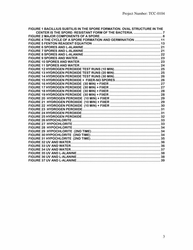
FIGURE 1 BACILLIUS SUBTILIS IN THE SPORE FORMATION. OVAL STRUCTURE IN THE
CENTER IS THE SPORE- RESISTANT FORM OF THE BACTERIA. ............................. 7 FIGURE 2 MAJOR COMPONENTS OF A SPORE. .............................................................. 8 FIGURE 4 THE CYCLE OF A SPORE FORMATION AND GERMINATION .......................... 11 FIGURE 5 FENTON REAGENT EQUATION ..................................................................... 13 FIGURE 6 SPORES AND L-ALANINE.............................................................................. 21 FIGURE 7 SPORES AND L-ALANINE.............................................................................. 21 FIGURE 8 SPORES AND L-ALANINE.............................................................................. 22 FIGURE 9 SPORES AND WATER.................................................................................... 23 FIGURE 10 SPORES AND WATER.................................................................................. 23 FIGURE 11 SPORES AND WATER.................................................................................. 24 FIGURE 12 HYDROGEN PEROXIDE TEST RUNS (10 MIN)............................................... 25 FIGURE 13 HYDROGEN PEROXIDE TEST RUNS (20 MIN)............................................... 25 FIGURE 14 HYDROGEN PEROXIDE TEST RUNS (30 MIN)............................................... 26 FIGURE 15 HYDROGEN PEROXIDE + FIXER-NO SPORES.............................................. 26 FIGURE 16 HYDROGEN PEROXIDE (30 MIN) + FIXER .................................................... 27 FIGURE 17 HYDROGEN PEROXIDE (30 MIN) + FIXER .................................................... 27 FIGURE 18 HYDROGEN PEROXIDE (30 MIN) + FIXER .................................................... 28 FIGURE 19 HYDROGEN PEROXIDE (30 MIN) + FIXER .................................................... 28 FIGURE 20 HYDROGEN PEROXIDE (10 MIN) + FIXER ................................................... 29 FIGURE 21 HYDROGEN PEROXIDE (10 MIN) + FIXER ................................................... 29 FIGURE 22 HYDROGEN PEROXIDE (10 MIN) + FIXER ................................................... 30 FIGURE 23 HYDROGEN PEROXIDE............................................................................... 31 FIGURE 24 HYDROGEN PEROXIDE................................................................................ 31 FIGURE 25 HYDROGEN PEROXIDE................................................................................ 32 FIGURE 26 HYPOCHLORITE.......................................................................................... 33 FIGURE 27 HYPOCHLORITE ......................................................................................... 33 FIGURE 28 HYPOCHLORITE ......................................................................................... 34 FIGURE 29 HYPOCHLORITE (2ND TIME)...................................................................... 34 FIGURE 30 HYPOCHLORITE (2ND TIME)....................................................................... 34 FIGURE 31 HYPOCHLORITE (2ND TIME)....................................................................... 35 FIGURE 32 UV AND WATER........................................................................................... 36 FIGURE 33 UV AND WATER........................................................................................... 36 FIGURE 34 UV AND WATER........................................................................................... 37 FIGURE 35 UV AND L-ALANINE..................................................................................... 38 FIGURE 36 UV AND L-ALANINE..................................................................................... 38 FIGURE 37 UV AND L-ALANINE..................................................................................... 39

Project Number: TCC-0104
4
ABSTRACT:
In order for spores, which are metabolically dormant, to return to life they must
go through the process of germination. Germination is a complex and vital step in the
generation of growing cells from spores. It is known that spore germination is initiated by
specific nutrients. The purpose of this Major Qualifying Project was to identify certain
initiators that will begin the germination process. Hydrogen Peroxide, uv treated spores
and hypochlorite were used to investigate the effect of these lethal agents in the
germination process.

Project Number: TCC-0104
5
Acknowledgments
I would like to thank Professor Crusburg Ph.D, for all of his guidance and
assistance throughout the duration of this project.

Project Number: TCC-0104
6
INTRODUCTION:
Spore germination is an extremely complex process. Following germination
spores become vegetatively growing cells, which are much less resistant to chemical and
physical agents. Due to the increasing terrorist threats using biological weapons as attack
methods, it is becoming more of an issue to find ways to stop spores before they have the
opportunity to cause any damage. It is important to know the biochemical signals that
start the germination process which then allow us to figure a way to end this complex
process.
In this MQP I tested three agents that are known to have an effect on the
germination of spores. To carry out the experiments, mixtures of spores along with
different reagents were tested using a spectrophotometric assay to measure the
absorbance of the spore suspensions over a one to two hour time period.
The scattering of light from vegetative cells versus germinating spores was used
to view the differences in absorbance between the spore and the germinating spore.
Spores have a higher absorbance compared to vegetative cells, since they scatter light
more effectively than vegetative cells. The absorbance will decrease when the spore is
germinating or changing from its typical spore form. These recordings allowed us to
conclude which of the experiments have the greatest effect on spores/cells.
Reagents that were used in this MQP to test for their affect on the spore
germination process were hydrogen peroxide, hypochlorite and UV treated spores. The
aim was to see if the spores still had the ability to germinate after such treatments. These
experiments will help identify solutions that will stop the spores from germinating into
viable vegetative cells and then being able to multiply.

Project Number: TCC-0104
7
BACKGROUND ON GERMINATION
The Spore
The Bacillus family’s distinguishing feature is the production of endospores,
which are highly refractile resting structures formed within the bacterial cells. The genus
Bacillus is distinguished from the other endospore-forming bacteria on the basis of being
a strict or facultative aerobe, rod-shaped, and catalase-positive [9].
Figure 1 Bacillius subtilis in the spore formation. Oval Structure in the center is the spore- resistant
form of the bacteria.
[12] http://europa.eu.int/comm/research/success/en/pur/0291e.html
The bacterial endospore is a highly-evolved structure capable of maintaining the
bacterial genome in a protected, viable state for extended periods [1]. Bacterial spores
can survive in the environment for incredibly long time periods even at physical extremes
for any other life forms on earth [2]. Their ability to survive is mainly due to their tough
outer coat which protects them from any outside harm. This is one of the key reasons that
makes it such a desirable weapon for biological warfare attacks.

Project Number: TCC-0104
8
Figure 2 Major components of a spore.
[9] http://textbookofbacteriology.net/Bacillus.html
Many characteristics aid in the spore’s ability to withstand extreme conditions.
Spores contain a protoplast, protoplast membrane, cortex and three coat layers. The coat
is a particularly thick protein coat. It is a highly ordered structure consisting of the
following three distinct layers: an electron-dense outer coat, a thinner inner
coat, and an
electron-diffuse undercoat. This outer coat helps protect the dormant spore from
enzymes, such as lysozyme, and from mechanical disruption. Resistance to organic
solvents and heat seems to be a function of the peptidoglycan cortex which underlies the
spores’ coat [6].
The spore coat also protects the spore from some chemicals, such as hydrogen
peroxide. We observed this process in the experiment that was conducted for this MQP.
Hydrogen peroxide, along with UV treated spores and hypochlorite were all used to

Project Number: TCC-0104
9
observe the spores’ germination process using the spectrophotometer. Since the coat is
protecting the spore we observed whether or not the absorbance would decrease after the
spore had been exposed to this reagent, indicating a change in the spore’s composition.
All these layers of the coat acting together aid in the spores’ ability to linger
around long enough to then be used as biological weapons to inflict harm. Spores can be
left alone for an extremely long period of time and then under ideal germination
conditions they can begin to grow and produce the harmful, sometimes deadly proteins,
which cause a number of infections and sicknesses. [7]
The formation of a spore involves the original cell replicating its genetic material,
and then one copy of this becomes surrounded by the tough coating. The outer cell then
disintegrates, releasing the spore which is now well protected against a variety of trauma,
including extremes of heat and cold, and an absence of nutrients, water, or air.[4]
Under these protective layers of the outer cell lies the spore’s core. It is inside the
core where dipicolinic acid, salt calcium dipicolinate are all found. These materials are
what greatly aid in the spores’ longevity [7]. Once the spore’s outer layers begin to break
down in the presents of certain disinfectants, such as the ones being tested in this MQP,
it is these inner components of the core that will spill out of the spore and alter the
solution’s make-up; thus allowing us to see a change in absorbance.
It is of much interest to try and extract all the coat proteins of the spore’s core, but
it is an extremely hard process to try and obtain these proteins from an intact spore. One
spore specific organic chemical, dipicolinic acid (DPA) which is only found in spores, is
synthesized via the lysine biosynthetic pathway. [5] The major role of the calcium DPA
complex seems to be the removal of water during the germination process. Calcium
dipicolinate also contributes to about 17% of a spore’s dry weight.[7] Not only does the
DPA help aid in the spore’s indestructibility but it is also only observed in endospores,
making it an indicator of the spore. [8].
Germination
Germination of a spore is the process by which a dormant spore goes through a
number of degradative events in order to become a viable cell. This is a process of

Project Number: TCC-0104
10
interrelated biochemical events occurs in the spore. Spore germination has been difficult
to study because of the extremely rapid physiological responses in a cell whose structure
is biochemically intractable [14].
A number of nutrients such as, amino acids, sugars, dodecylamine, exogenous
Ca2+
-DPA among others are all inducers of the germination process.
Once a spore is influenced by these extra-cellular compounds it will then trigger
the production of an intracellular signal, which signals the next step in germination.
When the spores are exposed to the ideal conditions or specific nutrients they break the
dormancy period and begin to take on all the features associated with a vegetative cell
[15] Throughout this MQP I observed spores that were undergoing a number of changes
induced by the agents I was adding to the spore suspension. This allowed me to view a
change in the absorbance indicating that the agent had some affect on a part of the
germination process.
Heating spores to a designed temperature, around 70oC, can also affect the
germination of the spore. Since DPA is such a big part of the spores’ make-up, if the
spore is heated to a certain point the DPA will be lost and the spore will no longer
germinate. This MQP was designed to find out which of the several disinfectants had the
most effect on the components involved in the germination process. The absorbance we
observed was continuously changing when the spore became affected by the reagent.
This is due to the fact that spores scatter light more effectively then the cells do. A
change in the absorbance indicates that there is a change in the enzymatic activity in the
spore.
The germination process triggers many nutrient-receptor interactions, including
the release of DPA as well as Ca2+
. It is the release of DPA which allows the uptake of
water into the spores’ core. The spore then swells to a much larger size than it is while it
is germinating. [10] This indicates the spore is coming out of its dormancy stage. For this
MQP it meant a lower absorbency would be observed since the larger the cell the less
scattering of light, indicating a vegetative cell is being formed and the spore is coming
out of its distinctive spore form.

Project Number: TCC-0104
11
Figure 3 The cycle of a spore formation and germination
[13]
http://www.bmb.leeds.ac.uk/mbiology/ug/ugteach/icu8/introduction/bacteria.html
In one study that was conducted to examine which enzymes have the most
importance in the germination process, it was concluded that agents had one of two
possible effects on the spore. Exogenous compounds either repressed the response to one
of the nutrient receptors, or these exogenous components blocked the response to several
of the nutrient receptors. [11] Knowing these interactions then can allow one to control
the germination process by altering the treatment of the spores using one of the agents
that were used in this MQP. For the purpose of this MQP the interactions allowed us to
measure the amount of light being scattered or not being scattered when the spore
suspension was treated with one of three known initiators of germination.
Once the spores had been pre-treated by one of the methods their light absorbance
were measured at 600nm and will be plotted versus time. Loss of rigidness of the spores
reduces their light-scattering properties. This then permits more light to pass through the
spore suspension and reach the detector, giving us a lower optical density. After
germination, the spore core, polar tubes and other contents of a spore are all free in the
suspension which reduces the optical density.
Project Number: TCC-0104
12
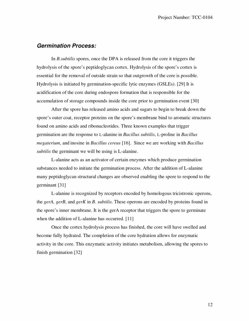
Germination Process:
In B.subtilis spores, once the DPA is released from the core it triggers the
hydrolysis of the spore’s peptidoglycan cortex. Hydrolysis of the spore’s cortex is
essential for the removal of outside strain so that outgrowth of the core is possible.
Hydrolysis is initiated by germination-specific lytic enzymes (GSLEs). [29] It is
acidification of the core during endospore formation that is responsible for the
accumulation of storage compounds inside the core prior to germination event [30]
After the spore has released amino acids and sugars to begin to break down the
spore’s outer coat, receptor proteins on the spore’s membrane bind to aromatic structures
found on amino acids and ribonucleotides. Three known examples that trigger
germination are the response to L-alanine in Bacillus subtilis, L-proline in Bacillus
megaterium, and inosine in Bacillus cereus [16]. Since we are working with Bacillus
subtilis the germinant we will be using is L-alanine.
L-alanine acts as an activator of certain enzymes which produce germination
substances needed to initiate the germination process. After the addition of L-alanine
many peptidoglycan structural changes are observed enabling the spore to respond to the
germinant [31]
L-alanine is recognized by receptors encoded by homologous tricistronic operons,
the gerA, gerB, and gerK in B. subtilis. These operons are encoded by proteins found in
the spore’s inner membrane. It is the gerA receptor that triggers the spore to germinate
when the addition of L-alanine has occurred. [11]
Once the cortex hydrolysis process has finished, the core will have swelled and
become fully hydrated. The completion of the core hydration allows for enzymatic
activity in the core. This enzymatic activity initiates metabolism, allowing the spores to
finish germination [32]

Project Number: TCC-0104
13
Reagents
Hydrogen Peroxide
Fenton's Reagent is a solution of hydrogen peroxide and an iron catalyst that is
used to oxidize contaminants.[17] Ionic catalysts plus the hydrogen peroxide, which
involves the conversion of hydrogen peroxide to highly reactive hydroxyl radicals. It is
the dissolving of O2 that results in a lethal action. [18] The reactivity of hydroxyl radicals
means that they are most effective when they are produced in the immediate area of their
target, often times DNA. [17]
Fe 2+
+ H2O2 ----> Fe 3+
+ OH - +
. OH
Fe 3+
+ H2O2 ----> Fe 2+
+ . OOH + H
+
Figure 4 Fenton Reagent equation
[22] http://www.h2o2.com/applications/industrialwastewater/fentonsreagent.html
Hydroxyl radical is one of the strongest oxidants known. Since it is highly
reactive it attacks membrane lipids, DNA, and other essential cell components which is
why it is often used in water waste clean-up because of its ability to kill many microbes.
Transition metals are often added to catalyze the formation of the hydroxyl radicals,
which is why in the Fenton reagent iron increases the efficacy of hydrogen peroxide [20]
The optimal concentration of H2O2 to germinate spores is usually between 2 to
5% but varies between species. [19] For my experiments we used a solution that was 3%
H2O2.
Hydrogen peroxide is used widely as a method of sterilization. H2O2 is both
bactericidal and sporicidal but does not leave toxic residues that could adversely affect
the product being sterilized. It is well known that hydrogen peroxide’s strength increases
noticeably with increasing temperatures. At very high concentrations, H2O2 can cause a
major break up of the spores coat structure, cortex as well as the core. At lower
concentrations, H2O2 can kill spores as well, just without evident cytological changes.

Project Number: TCC-0104
14
This shows that low concentrations are sufficient to kill spores and that lytic responses
are secondary to the initial killing. [ 24]
The Fenton reagent combines an iron catalyst with hydrogen peroxide to kill
spores of Bacillus species. This killing is increased by the addition of a surfactant which
will allow the liquid to directly come in contact with many more of the spores.
There are many different hypotheses of why the Fenton Reagent kills spores. One
of the many ways is that this reagent kills spores by DNA damage. Another idea is that it
acts in the spore core or on an external spore layer to destroy the spore. [17]
Many toxic chemicals or events, such as heat, oxidizing agents, and UV and
gamma radiation that are usually used to damage spore DNA are not quite successful.
This is due to the fact that the DNA is highly protected by the spore coats, the low
permeability of the spore's inner membrane, the core's low water content,
and the
saturation of spore DNA. The DNA is generally not damaged when the spore is killed by
many of these toxic chemicals.[17]
In one study that was done to see the killing time of hydrogen peroxide, it was
found that it has a CT time of about five logs in ten minutes. This means virtually all of
the spores would be killed (not viable) after just ten minutes.[33] Knowing this
information allowed me to conclude that if in that time period hydrogen peroxide kills
bacteria it will have an effect on the germination process of spores, and how great of an
effect.
Hypochlorite
Sodium hypochlorite (NaOCl) is a compound that is often times used in water
purification and tends to be an unstable compound. Sodium hypochlorite is available
commercially as a liquid and comes in different concentrations depending on its usage.
For domestic use, the commercial products usually contain 5% sodium hypochlorite and
have a pH around 11. For more industrial usages it is more concentrated and contains 10-
15% sodium hypochlorite with a pH of 13. [23]
Sodium hypochlorite is mainly used as a disinfectant and is effective against
bacteria, viruses and fungi. Often times after an anthrax contamination/scare, healthcare
workers use standard precautions to thoroughly disinfect all instruments used in an

Project Number: TCC-0104
15
invasive procedure or autopsy. This involves the use of hypochlorite as a sporicidal
agent. Besides anthrax, hypochlorite is also effective in destroying Brucellosis, cholera,
as well as inactivating, Ricin-Toxin.[25]
In one study that was conducted to find the mechanisms of killing B.subtilis
spores by use of hypochlorite it was discovered that hypochlorite does not kill B. subtilis
spores by DNA damage. This is because of the protective spore coat, which is major
factor in a spore’s resistance to hypochlorite. Spore killing by hypochlorite appears to
make the germination process defective, which could be due to the severe damage to the
spore's inner membrane which was observed in the study published in the Journal of
Applied Microbiology. [26]
The concentration of disinfectant x time(in minutes); is the CT, to achieve a
certain level of kill for hypochlorite was measured to be four logs in about seventeen
minutes.[34] This also confirms the fact mentioned above, that the line of attack
hypochlorite follows on killing spores does in fact make the germination process
defective.
UV
Ultraviolet (UV) radiation is often used as a method for inactivating viruses,
mycoplasma, bacteria and fungi. It is also successfully used in the destruction of airborne
microorganisms. [27]
One study showed the combination of UV and hydrogen peroxide used to kill
spores synergistically. Spores that are sensitized to ultraviolet killing by H2O2 treatment
followed by drying are much less able to absorb and then degrade H2O2. The combined
effort requires that both agents be present at the same time. The interactions between UV
and H2O2 most likely occurred close to or within the spore body and enhanced production
of hydroxyl radical. [28]

Project Number: TCC-0104
16
METHODOLOGY
WAYS TO OBSERVE GERMINATION:
When heat activated B. subtilis spores lose their dipicolinic acid and become
vegetative cells they will not scatter light as efficiently as do spores. As a suspension of
spores undergoes germination, an increase in the transmission of light and a decrease in
the absorbance is observed. This allowed us to know that all the spores had become
vegetative cells and were no longer germinating or vice versa, depending on the results
we observed.
Germination of spores can be monitored in a number of other ways as well. In this
MQP we observed the increase or decrease in the OD for suspensions of germinating
spores.
After conducting these three experiments to record the absorbance I was able to
see what effect each of the disinfectants had on the germination process. By viewing a
change in the absorbance over time, it is evident that some metabolically related process
was occurring in the spore, causing it to change from the characteristic spore shape into
another form that does not scatter light as effectively.
The experiments included (a) native spores in water only, (b) spores treated with
3% concentration of just H2O2 and same incubation time, (c) spores treated to
hypochlorite as well as spore exposed to UV light. (See below for treatment methods and
volumes of each used)

Project Number: TCC-0104
17
Materials used:
To conduct the spectrophotometirc assay the spectrophotomer used was Milton
Roy-Spectronic 601. Set to 600nm throughout the whole experiment.
Bacillus subtilis spores were used from a stock solution of 3.56x10⁸ mL.
Agents used were H2O2 at a 3% concentration. The sulfur fixer (Rapid Fixer A) was also
used in some of the experiments. L-alanine (Sigma) was used to induce germination in
the spores. Sodium hypochlorite (commercial bleach) was used in a 1% diluted form to
mix into the spore suspention.
A UV light system was used in the UV experiments. UV-transparent cuvettes
were used to mix and illuminate the spores.

Project Number: TCC-0104
18
Techniques and volumes for each of the experiments:
Spores + H20 + L-alanine
1. Pipet 30 uL of B. subtilis spore suspension (3.56 x 10
8/mL) into 1.00 mL of dH20 into
a cuvette.
2. Add 1.25 mL of 20 mM L-alanine to get a final concentration of 10 mM. (for water
and spores only do not add L-alanine)
3. Next place the in the spectrophotometer which was set to 600 nm. Zero using a
cuvette filled with dH2O. This is the initial reading.
4. Once the first reading is taken place the cuvette in the 40oC heat block and incubate
over a two hour period; taking readings every 10/20 minutes.
5. Repeat the above steps 3 times.
6. Dispose cuvettes in the Biohazards disposal.
(See Figures 6-11- for results)
Spores + H2O2
• 35uL spores
• 2.267 H2O
• 33.3uL H2O2
1. Pipeted 35 uL of B. subtilis spore suspension (3.56 x 10⁸/mL) into 2.267 mL of dH20
into a cuvette.
2. Add 33.3 uL of 30% H2O2.
3. Heat for 10 minutes in the 40oC heat block.
4. Place the cuvette in the spectrophotometer, which was set to 600 nm and take a
reading. (Always zero using a cuvette filled with dH2O)
5. Now incubate in the heat block and take readings every 10/20 minutes over a two hour
period.
6. Repeat the above steps three times.
7. Dispose cuvettes in the Biohazards disposal.
(See Figures 23-25 for results)
Spores + H2O2, and fixer
• 35uL spores
• 967 dH2O
• 33.3uL of 3% H2O2

Project Number: TCC-0104
19
1. Pipet 35 uL of B. subtilis spore suspension (3.56 x 10⁸/mL) into .967 mL of dH20 into
a cuvette.
2. Add 33.3 uL of 30% H2O2.
3. Heat for 30 minutes in the 40oC heat block.
3a. Heat for 10minutes.
4. Now add in 1.3mL of Rapid Fixer A to the cuvette. Place this in the
spectrophotometer, which was set to 600 nm and take a reading. (Always zero using a
cuvette filled with dH2O)
5. After this initial reading incubate in the heat block and take readings every 10/20
minutes.
6. Repeat the above steps three times. Do for both of the times stated in step 3.
7. For a control: using the Fixer and H202 do the same steps except do not add spores.
(This was to see the reaction between the two and if the sulfur precipitate will affect
absorbance). So in a cuvette only mix 1.3 fixer + 1.0 dH2O + 33.3uL H2O2 and then
incubate in the heat block and take readings every 10/20 minutes for a two hour period.
8. Dispose cuvettes in the Biohazards disposal.
(See Figures 16-22 for results)
Spores + hypochlorite
• 1.15 H2O
• 35uL spores
• 23uL NaOCl
1. Pipet 35 uL of B. subtilis spore suspension (3.56 x 10⁸/mL) into 1.15 mL of dH20 into
a cuvette.
2. Add 23 uL of NaOCl.
3. Heat for 10 minutes in the 40oC heat block.
4. To the heated mixture, now add 1.15 L-alanine.
5. Place the cuvette in the spectrophotometer, which was set to 600 nm and take a
reading. (Always zero using a cuvette filled with dH2O)
6. Now incubate cuvette in the heat block and take readings every 10/20 minutes for a
two hour interlude.
7. Repeat the above steps three times.
8. Dispose cuvettes in the Biohazards disposal.
(See Figures 26-31 for results)
Spores irradiated in UV light
• 35uL spores
• 1.25mL dH20
• Irradiated for 20 mins
• For the control add 1.25mL dH2O

Project Number: TCC-0104
20
Or add in 1.25 mL L-alanine
1. Pipet 35 uL of B. subtilis spore suspension (3.56 x 10⁸/mL) into 1.25 mL of dH20 into
a cuvette.
2. Place on the UV block for 20 min.
3. Add 1.25 mL of dH2O (for control)
3a. Add 1.25mL of L-alanine for another experiment.
4. Place the cuvette in the spectrophotometer, which was set to 600 nm and take a
reading. (Always zero using a cuvette filled with dH2O)
5. Now incubate in the heat block and take readings every 10/20 minutes for a two hour
interlude.
6. Repeat the above steps three times for each mixture 3 and 3a.
7. Dispose cuvettes in the Biohazards disposal.
(See table 32-37 for results)

Project Number: TCC-0104
21
Results
SPORES and L-alanine
Figure 5 Spores and L-alanine
Germination of Spores in water + L-alanine
0.90.92
0.94
0.960.98
1
0 20 40 60 80 100 120 140 160 180
time (min)
OD
@ 6
00n
m
Figure 6 Spores and L-alanine
Germination in water + L-alanine
0.860.88
0.90.920.940.960.98
1
0 20 40 60 80 100 120 140 160 180
time (min)
OD
@ 6
00
nm
Time Absorbance
0 0.96
10 0.95
20 0.94
30 0.936
45 0.931
60 0.925
75 0.929
90 0.927
105 0.931
120 0.931
135 0.93
Time Absrobance
0 0.958
10 0.968
20 0.959
30 0.947
45 0.945
60 0.934
75 0.933
90 0.937
105 0.939
120 0.946
135 0.944

Project Number: TCC-0104
22
Figure 7 Spores and L-alanine
Germination in water + L-alanine
1
1.02
1.04
1.06
0 20 40 60 80 100 120 140 160 180
time (min)O
D a
t 600n
m
Time Absorbance
0 1.051
10 1.047
20 1.035
30 1.036
45 1.027
60 1.023
75 1.02
90 1.017
105 1.017
120 1.023
135 1.027

Project Number: TCC-0104
23
Spores and Water
Figure 8 Spores and water
Germination in water
1
1.2
1.4
0 20 40 60 80 100 120 140 160 180
time (min)
OD
@6
00
nm
Figure 9 Spores and water
Germination in water
0.8
1
1.2
0 20 40 60 80 100 120 140 160 180time (min)
OD
@ 6
00
nm
Time Absorbance
0 1.215
10 1.198
20 1.189
40 1.182
60 1.176
80 1.167
100 1.173
Time Absorbance
0 1.024
10 0.995
20 0.984
40 0.982
60 0.972
80 0.967
100 0.954

Project Number: TCC-0104
24
Figure 10 Spores and water
Germination in water
0
0.2
0.4
0.6
0.8
1
1.2
0 20 40 60 80 100 120 140 160 180
time (min)
OD
@ 6
00 n
m
Time Absorbance
0 0
10 0.973
20 0.986
40 0.962
60 0.955
80 0.945
100 0.939

Project Number: TCC-0104
25
Hydrogen Peroxide Test runs- H2O2 (incubated for X min) + Fixer
Figure 11 Hydrogen Peroxide test runs (10 min)
Germination w/H2O2 (incubated for 10min)
00.5
11.5
22.5
33.5
0 20 40 60 80 100 120 140 160 180time (min)
OD
@ 6
00 n
m
Figure 12 Hydrogen Peroxide test runs (20 min)
Germination w/H2O2 (incubated for 20min)
00.5
11.5
22.5
33.5
0 20 40 60 80 100 120 140 160 180
time (min)
OD
@ 6
00
nm
Time Absorbance
0(prefixer) 1.7
10 2.907
20 2.726
40 2.050
60 1.469
80 1.117
100 .976
120 .861
Time Absorbance
0(prefixer) 1.69
10 2.685
20 2.149
40 1.591
60 1.286
80 1.230
100 1.110
120 .861

Project Number: TCC-0104
26
Figure 13 Hydrogen Peroxide test runs (30 min)
Germination w/H2O2 incubated for 30min
00.5
11.5
22.5
3
0 20 40 60 80 100 120 140 160 180
time (min)
OD
@ 6
00 n
m
Figure 14 Hydrogen Peroxide + fixer-no spores
Germination with H2O2 +fixer; no spores
00.5
11.5
22.5
33.5
0 20 40 60 80 100 120 140 160 180
time (min)
OD
@ 6
00n
m
Time Absorbance
0(prefixer) 1.698
10 2.695
20 2.402
40 1.649
60 1.385
80 1.232
100 1.040
120 577
Time Absorbance
0(prefixer)
10 2.883
20 2.688
40 2.230
60 1.914
80 1.342
100 1.034
120 .731

Project Number: TCC-0104
27
HYDROGEN PEROXIDE (30MINS INCUBATION) + FIXER
Figure 15 Hydrogen Peroxide (30 min) + Fixer
Germination w/ H2O2 incubated for 30mins +fixer
00.5
11.5
22.5
33.5
0 20 40 60 80 100 120 140 160 180
time (min)
OD
@ 6
00
nm
Figure 16 Hydrogen Peroxide (30 min) + Fixer
Germination w/ H2O2 incubated for 30mins + fixer
00.5
11.5
22.5
33.5
0 20 40 60 80 100 120 140 160 180
time (min)
OD
@ 6
00 n
m
Time Absorbance
0 1.678
10 2.889
30 2.757
50 2.208
70 1.569
90 0.862
110 0.744
Time Absorbance
0 1.643
10 2.937
30 2.765
50 2.27
70 1.649
90 0.941
110 0.753

Project Number: TCC-0104
28
Figure 17 Hydrogen Peroxide (30 min) + Fixer
Germination w/H2O2 incubated for 30min + fixer
00.5
11.5
22.5
33.5
0 20 40 60 80 100 120 140 160 180
time (min)O
D @
600 n
m
Figure 18 Hydrogen Peroxide (30 min) + Fixer
Fixer and H2O2 incubated for 30min + fixer
00.5
11.5
22.5
33.5
0 20 40 60 80 100 120 140 160 180
time (min)
OD
@ 6
00n
m
Time Absorbance
0 1.722
10 2.867
30 2.763
50 2.095
70 1.165
90 0.78
110 0.636
Time Absorbance
0
10 2.738
30 2.593
50 2.48
70 2.34
90 2.13
110 1.914
130 1.388

Project Number: TCC-0104
29
HYDROGEN PEROXIDE (10 MINS INCUBATION) + Fixer
Figure 19 Hydrogen Peroxide (10 min) + Fixer
Germination w/H2O2 incubated for 10min + fixer
00.5
11.5
22.5
33.5
0 20 40 60 80 100 120 140 160 180
time (min)
OD
@ 6
00
nm
Figure 20 Hydrogen Peroxide (10 min) + Fixer
Germination w/H2O2 incubated for 10min + fixer
00.5
11.5
22.5
33.5
0 20 40 60 80 100 120 140 160 180
time (min)
OD
@ 6
00
nm
Time Absorbance
0 1.845
10 2.967
20 2.877
40 2.432
60 1.6
80 1.224
100 1.202
120 1.145
200 1.41
Time Absorbance
0 1.704
10 2.921
20 2.831
40 2.593
60 1.915
80 1.343
100 1.094
120 0.881
200 1.378

Project Number: TCC-0104
30
Figure 21 Hydrogen Peroxide (10 min) + Fixer
Germination w/H2O2 incubated for 10min + fixer
00.5
11.5
22.5
33.5
0 20 40 60 80 100 120 140 160 180
time (min)
OD
@ 6
00
nm
Time Absorbance
0 1.729
10 2.949
20 2.89
40 2.816
60 2.664
80 2.441
100 2.127
120 1.801
200 0.803

Project Number: TCC-0104
31
HDYDROGEN PEROXIDE + SPORES
Figure 22 Hydrogen Peroxide
Germination with H2O2
1.024
1.028
1.032
1.036
0 20 40 60 80 100 120 140 160 180
time (min)
OD
@ 6
00
nm
Figure 23 Hydrogen Peroxide
Germination with H2O2
0.955
0.96
0.965
0.97
0.975
0 20 40 60 80 100 120 140 160 180
time (min)
OD
@ 6
00
nm
Time Absorbance
0 1.027
10 1.027
20 1.034
30 1.03
40 1.025
50 1.027
Time Absorbance
0 0.957
10 0.971
20 0.969
30 0.965
40 0.963
50 0.969
Project Number: TCC-0104
32
Figure 24 Hydrogen Peroxide
Germination with H2O2
0.87
0.88
0.89
0 20 40 60 80 100 120 140 160 180
time (min)O
D @
60
0n
m
Time Absorbance
0 0.878
10 0.879
20 0.874
30 0.882
40 0.883
50 0.888

Project Number: TCC-0104
33
HYPOCHLORITE
Figure 25 Hypochlorite
Germination w/hypochlorite
0.7
0.705
0.71
0.715
0.72
0.725
0 20 40 60 80 100 120 140 160 180
time (min)
OD
@ 6
00
nm
Figure 26 Hypochlorite
Germination with hypochlorite
0
0.2
0.4
0.6
0.8
0 20 40 60 80 100 120 140 160 180
time (min)
OD
@ 6
00
nm
Time Absorbance
0 0.721
10 0.713
20 0.707
40 0.711
60 0.707
80 0.703
Time Absorbance
0 0.712
10 0.657
20 0.571
40 0.383
60 0.319
80 0.296

Project Number: TCC-0104
34
Figure 27 Hypochlorite
Germination w/ hypochlorite
0.76
0.77
0.78
0.79
0 20 40 60 80 100 120 140 160 180
time (min)
OD
@ 6
00n
m
Figure 28 Hypochlorite (2nd time)
Germination with hypochlorite (2nd time)
0.92
0.94
0.96
0.98
1
0 20 40 60 80 100 120 140 160 180
time (min)
OD
@ 6
00n
m
Figure 29 Hypochlorite (2nd time)
Germination w/ hypochlorite (2nd time)
1.02
1.04
1.061.08
1.1
1.12
0 20 40 60 80 100 120 140 160 180
time(min)
OD
@ 6
00n
m
Time Absorbance
0 0.787
10 0.781
20 0.77
40 0.773
60 0.768
80 0.763
Time Absorbance
0 0.975
10 0.961
20 0.949 mixed
40 0.944 1.009
60 0.985
80 0.964
100 0.939
Time Absorbance
0 1.097
10 1.085
20 1.065 mixed
40 1.053 1.103
60 1.073
80 1.052
100 1.034

Project Number: TCC-0104
35
Figure 30 Hypochlorite (2nd time)
Germination w/ hypochlorite (2nd time)
0.940.960.98
11.021.041.061.08
0 20 40 60 80 100 120 140 160 180
time (min)
OD
@ 6
00
nm
Time Absorbance
0 1.06
10 1.038
20 1.027 mixed
40 1.014 1.073
60 1.033
80 1.002
100 0.961
Project Number: TCC-0104
36
UV WITH WATER
Figure 31 UV and water
Germination UV w/ H2O
0.9
0.92
0.94
0.96
0 20 40 60 80 100 120 140 160 180
time (min)
OD
@ 6
00n
m
Figure 32 UV and water
Germination UV w/H2O
0.86
0.88
0.9
0.92
0.94
0 20 40 60 80 100 120 140 160 180
time(min)
OD
@ 6
00n
m
Time Absorbance
0 0.944
10 0.943
20 0.931
30 0.928
40 0.923
60 0.918
80 0.907
100 0.93
120 0.933
Time Absorbance
0 0.924
10 0.918
20 0.91
30 0.905
40 0.898
60 0.892
80 0.878
100 0.868
120 0.883

Project Number: TCC-0104
37
Figure 33 UV and water
Germination UV w/H2O
0.860.88
0.90.92
0.940.96
0 20 40 60 80 100 120 140 160 180
time(min)
OD
@ 6
00
nm
Time Absorbance
0 0.948
10 0.941
20 0.932
30 0.929
40 0.919
60 0.912
80 0.899
100 0.892
120 0.881

Project Number: TCC-0104
38
UV and L-Alanine
Figure 34 UV and L-alanine
Germination UV w/L-alanine
00.20.40.60.8
11.21.41.6
0 20 40 60 80 100 120 140 160 180
time(min)
OD
@ 6
00
nm
Figure 35 UV and L-alanine
Germination UV w/L-alanine
0.90.920.940.960.98
1
0 20 40 60 80 100 120 140 160 180
time (min)
OD
@ 6
00
nm
Time Absorbance
0 1.039
10 1.031
20 1.191
40 1.004
60 0.992
80 0.977
100 0.958
120 0.958
Time Absorbance
0 0.985
10 0.98
20 0.966
40 0.957
60 0.94
80 0.93
100 0.91
120 0.923

Project Number: TCC-0104
39
Figure 36 UV and L-alanine
Germination UV w/L-alanine
0.9
0.92
0.940.96
0.98
1
0 20 40 60 80 100 120 140 160 180
time (min)O
D @
60
0n
m
Time Absorbance
0 0.977
10 0.995
20 0.987
40 0.975
60 0.958
80 0.946
100 0.933
120 0.923
Project Number: TCC-0104
40
Discussion/Conclusions
Spore germination is an extremely complex process and requires a number of
biochemical interactions within the spore. Many agents are known to have an affect on
any number of these interactions. All of the agents used in this MQP (L-alanine, H2O2,
hypochlorite, UV) are known to affect the spore and the germination process in a
different way.
The results I got from this MQP all show that each of these disinfectants had
some effect on the germination process. This indicated some activity was occurring
within the spore, in response to the added disinfectant.
In Figures 6-8, in which the spores were mixed with L-alanine the absorbance
remained relatively steady, demonstrating not much activity in the spores’ biochemical
make-up was occurring over the given time period. L-alanine is known to affect the
germination process, by acting as an activator of certain enzymes to begin the
germination process. This could explain the dip in some of the curves in the graphs.
In Figures 9-11 spores were only measured in water which showed no apparent
affect on the germination process. Compared to when L-alanine was added into the
suspension there was a lot less activity, since there was no inducer of germination to
affect the germination process.
Figures 12-15 represents the ‘test run’ experiments done with hydrogen peroxide.
These were done to see what the different heating times would have on the spores when
mixed with hydrogen peroxide. After the initial heating period (of either 10, 20 or 30
minutes) spores were mixed with a Rapid Fixer A to remove all the hydrogen peroxide.
This fixer seemed to cause a precipitate to form therefore not giving good results. I was
not able to determine what absorbance I was reading, the precipitate caused an
interference with the spores’ absorbance.
The test run times selected were incubating for 30min (Figures 16-19) and 10 min
(Figures 20-22). Though the incubation time was different,the graphs appear to be the
same. All the graphs show an increase when the hydrogen peroxide was added, and then
over time a decrease in absorbance. These decreases could have been due to the fact that
a sulfur precipitate was forming once the fixer was added, so these results are unclear as

Project Number: TCC-0104
41
to what was being measured. In the next experiments only hydrogen peroxide was added
to observe its affect with no interfering percipitate.
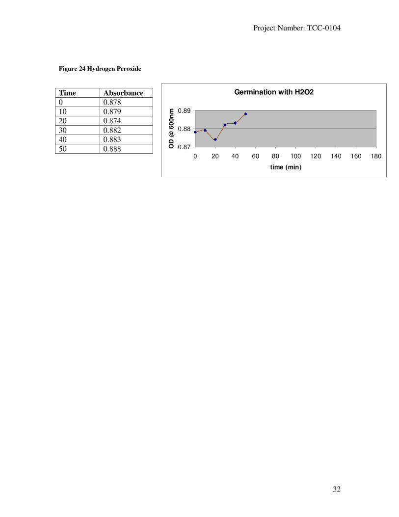
Figures 23-25 show the effect of hydrogen peroxide alone. Since spores were
mixed with hydrogen peroxide which is a known oxidant, there was activity occurring;
due to the change in absorbance recorded over the given time. Hydrogen peroxide is
known to have a CT time of about 5 logs in 10mins. This means virtually all of the spores
would be killed (not viable) after 10mins [33].
Figures 26-31 represents the data for the addition of hypochlorite, a strong
disinfectant known to destroy the spore viability. In these figures it is much more
apparent that something is occurring to the spores in the suspension. The recorded
absorbencies were at a much lower number compared to the control of spores and water.
The absorbance decreased over time, much more quickly then in the other experiments
indicating the spore is less refractile and can not scatter light as efficiently anymore. The
CT for hypochlorite was measured to be 4 logs in about 17 minutes[34]. This helps to
conclude that hypochlorite acts quickly on the germination process affecting the spore
and its enzymatic activities.
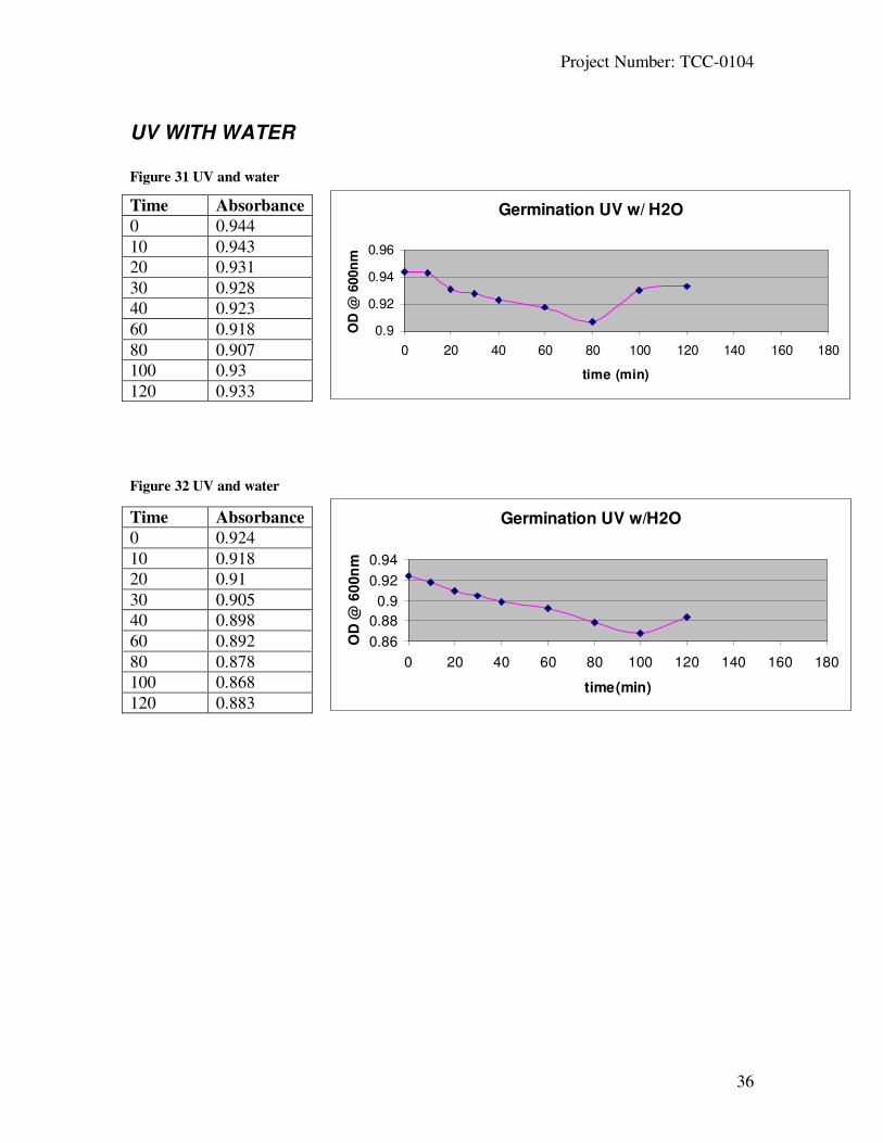
Figures 32-34 describes the experiment when the spores were exposed to UV in a
solution of water only, and they became much less spore-like over time. This is known
due the fact that UV irradiation for 30 minutes is known to kill all of the spores by DNA
damage and although it was not tested for in this MQP whether or not the spores were
dead, it is apparent from the graph that the UV light did have an effect on their
germination process.
Figures 35-37 show spores when exposed to UV light but this time with L-alanine
present. L-alanine is known as an initiator of the germination process. UV irradiation
results in the formation of spore photoproduct, (SP) 5-thyminyl-5,6-dihydrothymineis
which can effect the absorbance of the spore suspension [27] Since UV is acting to
destroy the spores’ DNA and L-alanine is initiating germination it is obvious there would
be some reaction occurring with in the spore, thus affecting its appearance and ability to
scatter/absorb light.

Project Number: TCC-0104
42
Over all conclusions
After viewing the results for each experiment, it is apparent that each reagent had
some affect on the spore’s form. L-alanine is known the have an affect on the
germination process by initiating certain genes and biochemical pathways that begin the
whole process. In the results it is apparent that there was activity occurring once it was
added into the spore suspension. There is continuous activity in the absorbance indicating
activity within the spore suspension.
Results from just spores and water are expected to have a steady absorbance rate,
since there was no initiator present to stimulate the germination process. From the graphs
this remained true, since in each one there was very little change in absorbance. This
means the spore kept its distinct form throughout the entire time of observation.
For the hydrogen peroxide with fixer experiments there should have been some
activity due to the fact that hydrogen peroxide is known to oxidize contaminants, which
would have an effect on the spore and its form, thus affecting the absorbance measured.
Once the fixer was added to help remove the H2O2 present in the spore suspension, it
appeared that some precipitate was forming due to the increased cloudiness and build up
in the cuvette. Therefore it is unclear of the effect the hydrogen/fixer combination had on
the spores’ germination process. In the control experiment (Figure 19), which had no
spores, a change in absorbance was observed. This shows some precipitate formed and
had an effect on the absorbance recorded.
Since the fixer had an effect of its own on the spore suspension, hydrogen
peroxide by itself was used. Since it is such a strong oxidizing agent and often times
used as a disinfectant it is known to have some effect on the spore and its germination
process. At high concentrations, H2O2 can cause a major break up of the spores coat
structure, cortex as well as the core all of which will have an effect on the spores
structure. If a change in the structure has occurred this can affect the absorbance, which
can be observed in the graphs. The absorbance, though not a major change, was
extremely unsteady, with high and low peaks being recorded.

Project Number: TCC-0104
43
Hypochlorite is often times used as a sporicidal due to its ability to kill bacteria.
Spore killing by hypochlorite appears to make the germination process defective, which
could be due to the severe damage to the spore's inner membrane. The graphs from the
experiments done using hypochlorite had the lowest absorbance and most decreased
absorbance measured. This indicates that hypochlorite had an effect on the spore, leaving
it non-viable since its typical spore form has been disrupted.
When the spores were irradiated on the UV block it is assumed that there will be a
change in absorbance due to the fact that UV light has an effect on the germination
process of spores. In the experiments done using only water and spores there was no
additional agent added as in the experiments done with L-alanine. These graphs show that
the UV had more of an affect on the spore and spore structure due to the change in
absorbance over time. When L-alanine was added into the solution after the spores had
been UV treated there was less of an obvious change. This could be due to the fact that
the L-alanine might have initiated germination process; allowing the spore to maintain
their distinct spore form and not affecting the absorbance as much.

Project Number: TCC-0104
44
Future Research
Spores of pathogens such as Bacillius anthracis, have been used in the past as
biological weapons. It is becoming more and more of a concern of future attacks and how
to detect them before they occur. The next task, after the exact pathways in which a spore
germinates have been discovered, is to detect spores in a rapid and effective manner.
One way to identify spores, before they have a chance to cause any harm, would be to
modify a procedure that can be used to detect many spores by use of fluorescence
microscopy.
The use of this technique would replace the currently used spore assays which
involve a time consuming protocol and allow it to be used to monitor for the presence of
B. anthracis spores in airborne releases.
Spores have a unique chemical that is specific to only spores, called dipicolinic
acid, commonly referred to as DPA. There is currently no one simple/quick test that will
allow someone to observe spores, but one would think that if there was a way to test for
DPA it would be extremely easy and quick to identify and then to quickly eradicate the
spores before they cause any harm.
It is known that terbium fluoresces at about 490 or 545nm when irradiated at
240nm while under a florescence microscope and is viewed in the green spectra around
540nm. It has also been shown that terbium and DPA will combine to form a [Tb(dpa)]
complex. The main idea behind this future MQP is to have the terbium cation bind to the
DPA anion derived from Bacillus spores. This MQP would allow to test and observe the
DPA-terbium complex fluorescing at 540nm which is sufficient to be seen under the
microscope and then use this as a way to help detect spores.
Project Number: TCC-0104
45
References
1- Nicholson, Wayne, L. DNA photochemistry, DNA repair, and bacterial spore structure
as determinants of spore resistance to solar UV radiation; Morningside College,
Sioux City, IA 51106;
http://www.photobiology.com/photobiology2000/nichol/
2- Brandl, Helmut. Patchiness of Bacillus spores in soils (with special reference to
Bacillus Anthracis); Cantonal Bio-safety Office, Zurich, Switzerland. March
2003;
http://www.research-projects.unizh.ch/math/unit72300/area81/p4663.htm
3- Wolters, D. A., M. P. Washburn, and J. R. Yates III. 2001. An automated
multidimensional protein identification technology for shotgun proteomics. Anal.
Chem. 73:5683-5690
4- Darling, David. The Encyclopedia of Astrobiology, Astronomy, and Spaceflight;
http://www.daviddarling.info/encyclopedia/B/bacspores.html
5- Aronson, A I & Fitz-James, P. Structure and morphogenesis of the bacterial spore
coat;
http://www.pubmedcentral.nih.gov/articlerender.fcgi?tool=pubmed&pubmedid=7
86255
6-Riesenman, Paul J and Nicholson, Wayne L. Role of the Spore Coat Layers in Bacillus
subtilis Spore Resistance to Hydrogen Peroxide, Artificial UV-C, UV-B, and
Solar UV Radiation. [13 September 1999]; Tucson, Arizona 85721;
http://aem.asm.org/cgi/content/full/66/2/620
7- Scully, M.O. FAST CARS Engineering a Laser Spectroscopic Technique for Rapid
Identification of Bacterial Spores. [August 2002]
8- Species specific bacterial spore detection using lateral flow immunoassay with DPA
triggered Tb luminescence. Pasadena, CA; NASA’s Jet Propulsion Laboratory. [
9- Todar, Kenneth. The Genus Bacillus. [2003] University of Wisconsin-Madison
Department of Bacteriology;
http://textbookofbacteriology.net/Bacillus.html
10- Setlow, B. Germanation of spores of B. subtilis with dodecylamine. Journal of
Applied Microbiology. Vol. 95, p. 637-348. [2003]

Project Number: TCC-0104
46
11- Smith, D A & Moir, A,. The Genetics of Bacterial Spore Germination. Annual Review of Microbiology; October 1990, Vol. 44: Pages 531-553
http://arjournals.annualreviews.org/doi/pdf/10.1146/annurev.mi.44.100190.0
02531?cookieSet=1
12. Vater, J. The decoding of the Bacillus subtilis genome .[May 1996]; Berlin, Germany.
Technical University of Berlin,
http://europa.eu.int/comm/research/success/en/pur/0291e.html]
13- Heritage, John. Medical Microbiology - A Brief Introduction. [October 2003]
University of Leeds;
http://www.bmb.leeds.ac.uk/mbiology/ug/ugteach/icu8/introduction/bacteria.
html
14- Jennings DH & Lysek G Spore Germination. [1996] Bios, pp125 - 129
http://bugs.bio.usyd.edu.au/Mycology/Reprodn_Dispersal/sporeGermination.shtm
l
15- Herman, Paul K. Yeast spore germination: a requirement for Ras protein activity
during re-entry into the cell cycle. [1997] Embo Journal; Vol.16, 6171–6181;
http://www.nature.com/cgi-
taf/DynaPage.taf?file=/emboj/journal/v16/n20/full/7590591a.html
16- Ireland, John A. W. Amino Acid- and Purine Ribonucleoside-Induced Germination of
Bacillus anthracis Sterne Endospores: gerS Mediates Responses to Aromatic Ring
Structures [March 2002] Journal of Bacteriology; Vol. 184, No. 5 1296-1303
http://jb.asm.org/cgi/content/abstract/184/5/1296
17. Shapiro, Michael P. Killing of Bacillus subtilis Spores by a Modified Fenton Reagent
Containing CuCl2 and Ascorbic Acid. Applied and Environmental Microbiology,
[April 2004] p. 2535-2539, Vol. 70, No. 4
http://aem.asm.org/cgi/content/full/70/4/2535
18 – Cross, J. B. Killing of Bacillus Spores by Aqueous Dissolved Oxygen, Ascorbic Acid,
and Copper Ions. Applied and Environmental Microbiology. [April 2003]; 69(4):
2245–2252.
http://www.ncbi.nlm.nih.gov/entrez/query.fcgi?cmd=Retrieve&db=PubMed&list
_uids=12676707&dopt=Abstract
19 – Miller, Jeff. Microsporidia (Protozoa): A Handbook of Biology and Research
Techniques. [August 1997]
http://pearl.agcomm.okstate.edu/scsb387/sporege.htm

Project Number: TCC-0104
47
20- Busck, Kristen. Cleaning and Disinfecting: The Effects of Germicides on
Microorganisms. Ecolab's Professional Products Division, Minn.
http://www.infectioncontroltoday.com/articles/191clean.html
21- Brundrett, Mark. Working with Mycorrhizas in Forestry and Agriculture. Mycorrhiza
Vol. 6 p. 509. [1996]
http://www.ffp.csiro.au/research/mycorrhiza/vam.html
22- Walling, Cheves Fenton’s Reagent Revisited, Accounts of Chem. Research, Vol. 8,
pp. 125-131 [1975].
http://www.h2o2.com/applications/industrialwastewater/fentonsreagent.html
23 Lenntech Water treatment. Disinfectants:Sodium hypochlorite; The Netherlands
[2003]
http://www.lenntech.com/water-disinfection/disinfectants-sodium-
hypochlorite.htm
24- [Online] Accessed April 2, 2005
http://www.sciencedirect.com/science?_ob=ArticleURL&_udi=B6T30-
41FTSCC-
8&_coverDate=11%2F30%2F2000&_alid=264098903&_rdoc=1&_fmt=&_orig=
search&_qd=1&_cdi=4932&_sort=d&view=c&_acct=C000005878&_version=1
&_urlVersion=0&_userid=74021&md5=9edba5f79edb317bfbf876ea216bf4f1
25 - USAMRIID's Medical Management of Biological Casualties Handbook. Fourth
Edition February 2001; pages 9-10.
http://www.millennium-ark.net/News_Files/NBC/Decontam3.html
26- Setlow P. Mechanisms of killing of Bacillus subtilis spores by hypochlorite and
chlorine dioxide. Journal of Applied Microbiology, July 2003, vol. 95, no. 1, pp.
54-67
http://www.ingentaconnect.com/content/bsc/jam/2003/00000095/00000001/art00
008
27- Riesenman, Paul J. Role of the Spore Coat Layers in Bacillus subtilis Spore
Resistance to Hydrogen Peroxide, Artificial UV-C, UV-B, and Solar UV
Radiation. AEM. [Nov.1999]
http://aem.asm.org/cgi/content/abstract/66/2/620
28- [Online] Accessed March 29, 2005
http://www.sciencedirect.com/science?_ob=ArticleURL&_udi=B6T30-
41FTSCC-
8&_coverDate=11%2F30%2F2000&_alid=264098903&_rdoc=1&_fmt=&_orig=

Project Number: TCC-0104
48
search&_qd=1&_cdi=4932&_sort=d&view=c&_acct=C000005878&_version=1
&_urlVersion=0&_userid=74021&md5=9edba5f79edb317bfbf876ea216bf4f1
29 – Foster, Simon. In vivo roles of germination-specific lytic enzymes of Bacillus
subtilis. Microbiology vol.147;, p.2925-2932 [2001]
30 Setlow P. Mechanisms for the prevention of damage to DNA in spores of Bacillus
species Annual. Review Microbiology p 49:29-54; [1995].
http://mcb.berkeley.edu/labs/kustu/mcb112/nov16.htm
31- Caipo, M.L. Bacillus Megaterium Spore Germination is influenced by inocculum
size. Journal of Applied Microbiology; vol. 92, p.879-884 [2002]
http://foodsci.rutgers.edu/schaffner/pdf%20files/Caipo%20JAM%202002.pdf
32 Cortezzo, B. Analysis of the action of compounds that inhibit germination of spores.
Journal of Applied Microbiology. Vol. 96 725-741. [2003]
33- Savino, A. Hydrogen Peroxide in the bacterial decontamination of Dental units.
http://www.anthos.com/HydrogenPeroxide.pdf
34 – Page, Martin. Strategies for Integrated Control of Biological Agents by Small
Utilities. [2004]
http://mtac.sws.uiuc.edu/mtacdocs/finalreports/2003_ThesisMainSporesFinalRep
ort.pdf