Glutamic Pyruvate Transaminase GPT2 Promotes Tumorigenesis ...

Cancer Cell
Article
SPOP Promotes Tumorigenesisby Acting as a Key Regulatory Hubin Kidney CancerGuoqiang Li,1,3,16 Weimin Ci,2,16 Subhradip Karmakar,4,5,16 Ke Chen,1 Ruby Dhar,4,5 Zhixiang Fan,9 Zhongqiang Guo,1,10
Jing Zhang,1 Yuwen Ke,1 Lu Wang,1,3 Min Zhuang,12 Shengdi Hu,13 Xuesong Li,10 Liqun Zhou,10 Xianghong Li,11
Matthew F. Calabrese,14 Edmond R. Watson,14 Sandip M. Prasad,7 Carrie Rinker-Schaeffer,7 Scott E. Eggener,7
Thomas Stricker,4,8 Yong Tian,13 Brenda A. Schulman,14,15 Jiang Liu,1,* and Kevin P. White4,5,6,*1Key Laboratory of Genome Sciences and Information2Laboratory of Disease Genomics and Individualized Medicine
Beijing Institute of Genomics, Chinese Academy of Sciences, Beijing 100101, China3University of Chinese Academy of Sciences, Beijing 100049, China4Institute for Genomics and Systems Biology, University of Chicago and Argonne National Laboratory, Chicago, IL 60637, USA5Department of Human Genetics6Section on Genetic Medicine, Department of Medicine7Section of Urology, Department of Surgery8Department of Pathology
University of Chicago, Chicago, IL 60637, USA9Kunming Medical University, Kunming, Yunnan 650500, China10Department of Urology, First Hospital of Peking University, Beijing 100034, China11Department of Pathology, Peking University Cancer Hospital, Beijing 100142, China12Department of Pharmaceutical Chemistry, University of California, San Francisco, San Francisco, CA 94158, USA13Laboratory of Animal Research Center, Institute of Biophysics, Chinese Academy of Sciences, Beijing 100101, China14Department of Structural Biology15Howard Hughes Medical Institute
St. Jude Children’s Research Hospital, Memphis, TN 38105, USA16These authors contributed equally to this work*Correspondence: [email protected] (J.L.), [email protected] (K.P.W.)
http://dx.doi.org/10.1016/j.ccr.2014.02.007
SUMMARY
Hypoxic stress and hypoxia-inducible factors (HIFs) play important roles in a wide range of tumors. Wedemonstrate that SPOP, which encodes an E3 ubiquitin ligase component, is a direct transcriptional targetof HIFs in clear cell renal cell carcinoma (ccRCC). Furthermore, hypoxia results in cytoplasmic accumulationof SPOP, which is sufficient to induce tumorigenesis. This tumorigenic activity occurs through the ubiquiti-nation and degradation of multiple regulators of cellular proliferation and apoptosis, including the tumorsuppressor PTEN, ERK phosphatases, the proapoptotic molecule Daxx, and the Hedgehog pathwaytranscription factor Gli2. Knockdown of SPOP specifically kills ccRCC cells, indicating that it may be apromising therapeutic target. Collectively, our results indicate that SPOP serves as a regulatory hub topromote ccRCC tumorigenesis.
Significance
Tumor cells can adapt to hypoxic microenvironments in several different ways to promote tumor growth, includingstimulating angiogenesis. Herein, we demonstrate that hypoxia can drive the cytoplasmic accumulation of SPOP andthat cytoplasmic accumulation of SPOP is sufficient to convey tumorigenic properties on to otherwise nontumorigenic cellsby targeting PTEN and several other tumor suppressor molecules for E3 ligase-mediated degradation. This tumor-promoting function of SPOP stands in contrast to its proapoptotic role in the cell nucleus. These results have elucidateda major mechanism that contributes to tumorigenesis in ccRCC, connecting hypoxia response and ubiquitin-mediateddegradation of tumor suppressors. The oncogenic role of cytoplasmic SPOPmakes it a promising candidate for therapeuticintervention.
Cancer Cell 25, 455–468, April 14, 2014 ª2014 Elsevier Inc. 455

Cancer Cell
SPOP Promotes Tumorigenesis in Kidney Cancer
INTRODUCTION
Renal cell carcinoma (RCC) is the eighth leading malignancy in
the United States, accounting for 4% of all cancers. More than
209,000 new cases and 102,000 deaths are estimated to occur
worldwide each year (Rini et al., 2009). Approximately 30% of
RCCpatients present withmetastatic disease at the time of diag-
nosis and nearly half of the remainder will subsequently develop
metastasis (McDermott et al., 2005; Negrier et al., 1998). As RCC
is highly resistant to chemotherapy, first-line treatment of meta-
static disease in the 1990s and 2000s relied on immunotherapies
such as interleukin-2 and interferon alpha, despite low response
rates (5% to 20%) (Fyfe et al., 1996; McDermott et al., 2005;
Wardle, 1991). However, hypoxic response mediated though
hypoxia-inducible factors (HIFs) is a key feature of most solid tu-
mors but is particularly important in kidney cancers (Chi et al.,
2006). Accordingly, recent targeted therapies that inhibit HIF-
regulated pathways, including angiogenesis inhibitors directed
against vascular endothelial growth factor (VEGF) and platelet-
derived growth factor (PDGF) signaling pathways, have been
developed. Although these therapies represent improvements
in patient care, the majority of patients with advanced disease
remain refractory to treatment, suggesting that certain critical
HIF targets remain unknown (Escudier et al., 2007; Motzer
et al., 2007, 2013).
It has been shown that, under normoxic conditions, the
oxygen-sensitive HIFa subunit is degraded by ubiquitination
via the von Hippel-Lindau (VHL) tumor suppressor gene (Kaelin,
2002). However, under hypoxic conditions, HIFa degradation is
suppressed, leading to enhanced nuclear localization of HIFa
and transcription of various target genes, including the
angiogenic gene VEGF (Kaelin, 2008). Inactivating mutations or
silencing of the VHL tumor suppressor gene can also suppress
the degradation of HIFa, and VHL mutation or silencing is found
in at least 80% of all clear cell RCCs (ccRCCs) (Kim and Kaelin,
2004; Motzer and Molina, 2009; Nickerson et al., 2008). ccRCCs
are the most common form of kidney cancer, accounting for
75% of all cases (Lopez-Beltran et al., 2006). Results from
ccRCC xenograft experiments indicate that HIF accumulation
is critical for VHL tumor suppressor function during ccRCC
oncogenesis (Kaelin, 2008). It has also been found that RCCs
have evolved an alternative hypoxia signaling pathway
compared with normal renal cells (Jiang et al., 2003) and that
the ccRCCs are in a high hypoxia response state compared to
the normal kidney samples and other subtypes of kidney cancer
(Chi et al., 2006).
Although hypoxia response plays a critical role in kidney
cancer, other cell regulatory pathways are also important for
tumor development and progression (Brugarolas, 2007). For
example, another promising target for RCC therapies is the
mammalian Target of Rapamycin (mTOR) pathway, which is
abnormally activated during the development of kidney cancer
(Robb et al., 2007). mTOR is negatively regulated by the tumor
suppressor phosphatase and tensin homolog (PTEN) (Hollander
et al., 2011), which is mutated or downregulated in many
cancers, including ccRCC (Vivanco and Sawyers, 2002). The
mTOR pathway positively regulates cell growth and proliferation
through enhanced mRNA translation by phosphorylated S6
kinase 1 and 4E binding protein-1 (Sarbassov et al., 2005). In
456 Cancer Cell 25, 455–468, April 14, 2014 ª2014 Elsevier Inc.
spite of its promise, the mTOR inhibitor temsirolimus produces
only low objective responses in patients with advanced RCC,
although it contributes to a modest improvement in overall
survival (Hudes et al., 2007; Kapoor and Figlin, 2009). Further
investigation of the mechanisms of PTEN downregulation in
ccRCC is critical for an understanding of RCC pathogenesis.
Our previous studies demonstrated that the SPOP protein is
overexpressed in 85% of kidney cancers and that nearly 100%
of primary and metastatic ccRCCs exhibit SPOP accumulation
(Liu et al., 2009). SPOP is a BTB/POZ domain protein, and
MEL-26, the C. elegans SPOP ortholog, was first identified as
an adaptor for the E3 ligase Cullin3 (Cul3). In C. elegans,
MEL-26 promotes the meiotic/mitotic transition through the
degradation of MEI-1/katanin (Mains et al., 1990; Pintard et al.,
2003; Xu et al., 2003). In Drosophila, D-SPOP (also known as
Roadkill) can promote the ubiquitination and degradation of
the Gli transcription factor ortholog Cubitus interruptus and the
JNK phosphatase Puckered to regulate the Hedgehog (Hh)
and tumor necrosis factor (TNF) pathways, respectively (Kent
et al., 2006; Zhang et al., 2006; Liu et al., 2009). In humans, the
roles for SPOP in regulating the Hh and TNF pathways have
been conserved (Zhang et al., 2006; Liu et al., 2009), and several
other SPOP substrates have been identified as well, including
the death domain-associated protein (Daxx) (Kwon et al.,
2006), the polycomb group protein BMI-1, and the Histone
variant MacroH2A (Hernandez-Munoz et al., 2005). Together,
these previous results indicate that SPOP plays important roles
during cell apoptosis, proliferation, and animal development,
and they suggest that overexpression of SPOP in ccRCCs may
lead to dysregulation of pathways involved in tumorigenesis.
However, it is unclear how SPOP becomes overexpressed or
whether it may function directly in kidney tumorigenesis. Adding
to this puzzle, recent deep sequencing studies of breast, pros-
tate, and endometrial cancers found that SPOP is frequently
mutated, and the SPOP locus is observed to undergo loss of
heterozygosity (LOH), indicating that SPOP may act as a tumor
suppressor (Berger et al., 2011; Li et al., 2011). However, no
SPOP mutations have been detected in kidney cancers thus
far (Liu et al., 2009; Cancer Genome Atlas Research Network,
2013). In this study, we aim to determine whether SPOP
promotes tumorigenesis in the kidney.
RESULTS
HIF Regulates SPOP ExpressionImmunohistochemistry results have suggested that SPOP is
overexpressed in virtually all ccRCCs (Liu et al., 2009). We
confirmed this finding by immunoblot assays in multiple pairs
of ccRCC primary tumor tissue and matched normal adjacent
tissue (Figure 1A). However, the mechanism underlying this
SPOP overexpression is unknown. Considering the frequent
accumulation of HIF in ccRCC patients, we were interested in
whether HIF can drive SPOP overexpression. We identified a
HIF-1a binding peak in the first intron of SPOP by chromatin
immunoprecitation sequencing (ChIP-seq) (Figure 1B) and
further validated it by ChIP-quantitative PCR (qPCR) (Figure S1A
available online). To determine whether this intronic DNA
sequence region functions as a HIF-responsive regulatory
element, we performed a luciferase reporter assay by inserting

Cancer Cell
SPOP Promotes Tumorigenesis in Kidney Cancer
the wild-type HIF binding sequence into a pGL3-promoter re-
porter vector. In human embryonic kidney HEK293 cells, the
wild-type sequence exhibits increased luciferase activity under
hypoxic exposure (1% O2/5% CO2/94% N2) compared to nor-
moxia, while HIF-1a knockdown impairs the luciferase activity
(Figure 1C). Additionally, we constructed several mutations or
deletions of the predicted hypoxia response elements (HREs;
50-RCGTG-30 and 50-[A/C]ACAG-30) (Miyazaki et al., 2002).
Although mutants of CACAG showed marginal decrease in lucif-
erase activity (Figure S1B), both point mutations and deletion of
the CGTG site showed significant decrease in luciferase activity
(Figure 1C), demonstrating that the HIF binding site is functional
in this hypoxia-responsive cis-regulatory element.
To further elucidate whether HIF can regulate SPOP, the VHL
wild-type ccRCC cell line Caki-2 was cultured under hypoxic
conditions. Hypoxic culture resulted in the accumulation of
HIF-1a and HIF-2a proteins. Both SPOP mRNA (Figure 1D) and
protein (Figure 1E) levels increased in ccRCC cells under hypoxic
conditions. Additionally, ectopic overexpression of either HIF-1a
or HIF-2a led to SPOP accumulation in HEK293 cells (Figure 1F),
indicating that both HIF-1a and HIF-2a can regulate SPOP
expression. Although knockdown of HIF-1a alone in Caki-2 cells,
which express both HIF-1a and HIF-2a (Kucejova et al., 2011),
can reduce SPOP protein abundance (Figure 1G), double knock-
down of HIF-1a and HIF-2a is more effective than either alone
(Figure S1C). Consistent with these results, knocking down
HIF-2a by siRNA in the ccRCC cell line A498 that predominantly
expresses HIF-2a (Shinojima et al., 2007) resulted in a reduction
in the mRNA (Figure 1H) and protein (Figure 1I) abundance of
SPOP, as well as the known HIF target VEGF. Because VHL
mutation is one of the most common causes of kidney cancer
and affects abundance of HIFs, we examined SPOP abundance
after restoring VHL in the VHL null cell lines 786-O and A498.
Accordingly, restoration of VHL led to decreased protein abun-
dance of SPOP (Figure 1J), and conversely, VHL knockdown in
HEK293 cells resulted in an increase in SPOP (Figure S1D).
Taken together, these results demonstrate that HIF can directly
regulate SPOP expression.
SPOP Accumulation in the Cytoplasm of ccRCC CellsSPOPwas first identified as a nuclear protein (Nagai et al., 1997),
andseveral studieshaveconfirmed its nuclear location inHEK293
andHeLacells (Bunceet al., 2008;Hernandez-Munozet al., 2005;
Kwon et al., 2006). To investigate SPOP localization in kidney
cancer, we used a monoclonal anti-SPOP antibody (Liu et al.,
2009) to stain kidney tissue biopsies. We found that SPOP was
predominately localized in the nucleus of normal kidney tissue.
However, we observed SPOP accumulation in the cytoplasm of
neoplastic ccRCC cells, with residual SPOP remaining in the
nucleus (Figure 2A). Immunocytochemistry staining also re-
vealed that SPOP predominately accumulated in the cytoplasm
of Caki-2 cells but was primarily localized within the nucleus
of HeLa (Figure 2B) and HEK293 cells (Figure S2A). Ectopically
expressed GFP-tagged SPOP in ccRCC cell lines (Caki-2 and
A498) and in non-ccRCC cell lines (HEK293 and HeLa cells)
yielded similar results as those for endogenous SPOP (Fig-
ure S2B). Furthermore, cell fractionation assays confirmed the
cytoplasmic localizationofSPOP inccRCCcell linesandpredom-
inantly nuclear localization in HEK293 cells (Figure S2C).
Hypoxia Drives SPOP Accumulation in the CytoplasmPrevious studies have shown that some proteins accumulate in
the cytoplasm under stress conditions such as hypoxia (Arimoto
et al., 2008). Given that ccRCC tumorigenesis is associated with
hypoxia signaling, we tested whether hypoxic conditions could
stimulate SPOP accumulation in the cytoplasm. Immunofluores-
cence staining showed that endogenous SPOP localizes in the
nucleus of HeLa cells under normoxia, while hypoxic treatment
(1% O2/5% CO2/94%N2) led to SPOP accumulation in the cyto-
plasm (Figure 2B). Additionally, HIF overexpression further
enhanced the cytoplasmic accumulation of SPOP (Figure 2B).
SPOP accumulation in the cytoplasm under hypoxic treatment
was further confirmed by cell fractionation assays (Figures 2C
and 2D). Although previous data showed that ectopically ex-
pressed GFP-SPOP localized in the nucleus of HeLa and
HEK293 cells under normoxia (Figure S2B), we found that it
also accumulated in the cytoplasm under hypoxia in these cells
(Figure S2D). Thus, hypoxia appears to be a sufficient condition
to stimulate the cytoplasmic accumulation of SPOP.
Cytoplasmic SPOP Promotes TumorigenesisNext, we wanted to test whether cytoplasmic SPOP is associ-
ated with tumorigenic phenotypes. Sequence analysis indicated
that SPOP contains a nuclear localization signal (NLS) at its C
terminus, amino acids 367–373 (PRKRLKQ). Crystallographic
analysis indicates that the deletion of the NLS peptide from the
SPOP C terminus is unlikely to affect the binding of SPOP with
the E3 ligase Cullin3 or SPOP substrates (Zhuang et al., 2009).
Additionally, the in vitro ubiquitination of SPOP substrates is
unaffected by deletion of the NLS from SPOP (Zhuang et al.,
2009). Therefore, we deleted the NLS peptide from SPOP to
test whether SPOP lacking the NLS would accumulate in the
cytoplasm when ectopically expressed in non-ccRCC cells. As
expected, ectopic expression of GFP-SPOP lacking the NLS
accumulates in the cytoplasm of HEK293 cells (Figure S2E).
We refer to SPOP lacking the NLS as SPOP-cyto.
We overexpressed SPOP-cyto and wild-type SPOP in non-
ccRCC cell lines to determine whether there are differences in
phenotypic effects. A bromodeoxyuridine (BrdU) incorporation
assay showed that wild-type SPOP inhibited cell proliferation
(Figure 3A), which is consistent with previous results that
SPOP can induce apoptosis in HEK293 (Liu et al., 2009) and
HeLa cells (Kwon et al., 2006) (Figure S3A). It is striking that
SPOP-cyto enhanced proliferation rather than inducing
apoptosis (Figure 3A). As a marker for apoptotic state of the
cells, we used the proapoptotic protein Bax, which has been
shown to be inactivated by phosphorylation at Ser184 (Gardai
et al., 2004). Overexpression of SPOP-cyto increased the level
of Ser184-phosphorylated Bax (p-Bax), indicating that SPOP-
cyto can inhibit Bax-mediated apoptosis (Figure 3B). Overex-
pression of SPOP-cyto also increased the protein abundance
of the cellular proliferation markers ser10-phosphorylated
histone H3 (p-Histone H3) (Bhatia et al., 2011) and proliferating
cell nuclear antigen (PCNA), consistent with the observation
that SPOP-cyto promotes cell proliferation (Figure 3B). Thus,
the accumulation of cytoplasmic SPOP appears to alter the
function of the protein in comparison to its nuclear form, chang-
ing SPOP function from proapoptotic to antiapoptotic and pro-
proliferative. Furthermore, siRNA knockdown of SPOP
Cancer Cell 25, 455–468, April 14, 2014 ª2014 Elsevier Inc. 457

(legend on next page)
Cancer Cell
SPOP Promotes Tumorigenesis in Kidney Cancer
458 Cancer Cell 25, 455–468, April 14, 2014 ª2014 Elsevier Inc.

Cancer Cell
SPOP Promotes Tumorigenesis in Kidney Cancer
(Figure S3B) induced apoptosis in ccRCC cells where SPOP is in
the cytoplasm, but not in HeLa or HEK293 cells (Figures 3C, S3C,
and S3D). Knockdown of SPOP in ccRCC cells also resulted in
decreased levels of p-Bax (Ser184), PCNA, and p-Histone H3
levels (Figure S3E). However, we wondered whether hypoxia-
induced cytoplasmic localization of SPOP in non-ccRCC cells
would also lead to an SPOP ‘‘addicted’’ state in which knock-
down of SPOP would lead to cell death. Indeed, RNAi knock-
down of SPOP in HeLa cells under hypoxia exposure led to
significantly increased apoptosis compared to cells treated
with only hypoxia stress (Figure S3F), similar to the results of
SPOP knockdown in ccRCC cell lines. Together, these results
indicate that inhibition of SPOP can specifically induce
apoptosis and inhibit proliferation in ccRCC cells but not in
non-ccRCC cells, suggesting that SPOP could serve as a
therapeutic target specific to cancer cells.
We further investigated whether cytoplasmic SPOP could
promote tumorigenesis in a nude mouse xenograft model.
HEK293 cells have previously been used to demonstrate the
tumorigenic potential of oncogenes (Hamid et al., 2005). We
generated stable polyclonal HEK293 cell lines transfected
with SPOP-cyto, SPOP, or empty vector (pcDNA3) (Figure S3G),
and the levels of exogenous SPOP-cyto and SPOP are com-
parable to the endogenous protein in primary ccRCC samples
(Figure 1A). Subcutaneous injection of the stably transfected
HEK293-SPOP-cyto cells into nude mice induced tumor for-
mation in approximately 80% of mice (15/19) within 6 weeks,
whereas wild-type SPOP and control empty vector produced
no visible tumor growth (0/19 and 0/19, respectively)
(Figure 3D). Furthermore, histopathologic analyses of the xeno-
graft tumors reveal typical cancerous lesions (Figure S3H).
Thus, cytoplasmic SPOP, but not nuclear SPOP, promotes
tumorigenesis.
SPOP Mediates the Ubiquitination and Degradationof Tumor Suppressor PTENWe next wanted to investigate the mechanism by which
cytoplasmic SPOP promotes tumorigenesis. SPOP was previ-
ously identified as a regulatory ‘‘hub’’ molecule in Drosophila
Figure 1. HIF Activates SPOP Expression
(A) Immunoblots of endogenous SPOP in four paired ccRCC tumor and adjacen
tissue (T) versus normal tissue (N) as evaluated using ImageJ software. SPOP wa
was used as a loading control.
(B) HIF-1a ChIP-seq peak within the first intron of the SPOP gene (arrow). ChIP sig
HIF-1a antibody ChIP.
(C) Luciferase reporter assays indicate that the HIF binding peak in the first intron
HRE (CGTG)mutated or deleted HIF binding sequence were cloned into a pGL3-p
the results are represented as values relative to the empty vector. Hypoxia was in
was performed with two independent siRNAs to HIF-1a.
(D) SPOP mRNA expression is activated under hypoxic conditions in the Caki-2
(E) SPOP protein abundance increases after hypoxia treatment in Caki-2 cells. T
(F) In either HIF-1a- or HIF-2a-transfected HEK293 cells, SPOP protein expressi
(G) Knockdown HIF-1a downregulated the expression of SPOP in Caki-2 cells. R
(H) SPOP mRNA expression is suppressed after two independent siRNAs’ knoc
control cells exposed to negative control siRNAs. VEGF was used as a positive
(I) SPOP protein abundance decreases after knockdown of HIF-2a in A498 cells
(J) SPOP protein abundance decreases when restoring of VHL in 786-O and A4
loading control.
Data in (C), (D), and (H) are presented as means ± SD of at least three independen
See also Figure S1.
(Liu et al., 2009) and can degrade substrates in multiple signaling
pathways via Cul3-mediated ubiquitination (Bunce et al., 2008;
Hernandez-Munoz et al., 2005; Kwon et al., 2006; Xu et al.,
2003; Zhuang et al., 2009). SPOP crystal structure studies
demonstrated that all known SPOP substrates share a
conserved SPOP-binding consensus (SBC) motif (F-p-S-S/
T-S/T; F is nonpolar, p is polar) (Zhuang et al., 2009). Thus, we
hypothesized that cytoplasmic SPOP might promote cancer
phenotypes by mediating the degradation of cytoplasmic
proteins that contain SBC motifs.
Previously, we found that SPOP can mediate the ubiquitina-
tion and degradation of Puc, a dual-specificity phosphatase
(DUSP) class molecule in Drosophila that regulates JNK
signaling (Liu et al., 2009). DUSPs have been implicated asmajor
modulators of critical signaling pathways that are dysregulated
in various diseases (Patterson et al., 2009). Therefore, we
computationally searched the human proteome for DUSP
domain proteins that contain the SBC motif. We found seven
DUSP domain containing proteins with at least one SBC motif
(Table S1), including the cytoplasmic tumor suppressor protein
PTEN with substrate specificity to phosphatidylinositol phos-
phates (Myers et al., 1998) and that contains the SBC motif
ASSST (residues 359–363).
Genetic alterations targeting the PTEN tumor suppressor are
among the most frequently noted somatic mutations in human
cancers (Sansal and Sellers, 2004). PTEN functions to antago-
nize phosphoinositide 3-kinase (PI3K)/Akt signaling through its
lipid phosphatase activity, thereby controlling cell growth,
survival, and metabolic processes (Wu et al., 1998). Based on
previous crystal structures, the SBCmotif in PTEN is in a flexible
region that is not folded into any three-dimensional domains and
is thus potentially accessible for SPOP binding (Lee et al., 1999).
To understand the molecular basis for potential SPOP MATH
domain interactions with the PTEN SBC, we determined the
crystal structure of the SPOPMATH complex with a peptide cor-
responding to PTEN residues 354–368, which constitutes the
PTEN SBC motif. The SPOP MATH domain forms an antiparallel
b sandwich, and the PTEN SBC motif adopts an extended
conformation to bind within the MATH central shallow groove
t nontumor tissue samples. Numbers indicate the relative density of the tumor
s detected by immunoblotting with an anti-SPOP antibody (clone 6C). Tubulin
nal intensity represents differential binding between input/mock IP control and
of SPOP (hg18, chr17:45,109,830–45,109,897) is functional. The wild-type and
romoter vector. Luciferase activities were normalized to Renella luciferase, and
duced using a hypoxia chamber (1% O2/5% CO2/94% N2). RNAi knockdown
ccRCC cell line. VEGF was used as a positive control.
ubulin was used as a loading control.
on was induced. GAPDH was used as a loading control.
NAi knockdown was performed as in (C).
kdown of HIF-2a in 786-O ccRCC cells. mRNA expression was normalized to
control.
. Tubulin was used as a loading control.
98 cells. Cells were harvested 72 hr after transfection. GAPDH was used as a
t experiments. *p < 0.05, **p < 0.01, and ***p < 0.001, based on Student’s t test.
Cancer Cell 25, 455–468, April 14, 2014 ª2014 Elsevier Inc. 459

Figure 2. Hypoxia Drives SPOP Accumulation in the Cytoplasm of ccRCC Cells
(A) Immunohistochemistry reveals that SPOP accumulates in the cytoplasm of ccRCC tumor cells but in the nuclei of adjacent nontumor cells. An SPOP-specific
monoclonal antibody (SPOP-5G) was used for staining (diaminobenzidine, brown staining). Scale bar, 20 mm.
(B) SPOP localizes in the nucleus in HeLa cells under normoxic conditions but accumulates in the cytoplasm under hypoxic conditions (1% O2), comparable to
that in Caki-2 cells; additional HIF expression enhances SPOP accumulation in the cytoplasm. Cells were stained with SPOP-5G antibody (green), and the nuclei
were counterstained with DAPI (40,6-diamidino-2-phenylindole; blue). Scale bar, 10 mm.
(C) Separation of the nuclear (N) and cytoplasmic (C) fractions confirms SPOP accumulation in the cytoplasm of HeLa cells under mormoxic conditions. Histone
H3 and tubulin served as nuclear and cytoplasmic markers, respectively.
(D) Separation of the nuclear (N) and cytoplasmic (C) fractions confirms SPOP accumulation in the cytoplasm of HeLa cells under hypoxic conditions (1% O2).
Histone H3 and tubulin served as nuclear and cytoplasmic markers, respectively.
See also Figure S2.
Cancer Cell
SPOP Promotes Tumorigenesis in Kidney Cancer
(Figures S4A and S4B). SPOP MATH-SBC interactions are
anchored by both hydrophobic and polar interactions with the
PTEN SBC residues ASSST (Figure 4A).
We further validated whether PTEN acts as a SPOP sub-
strate in cells. Coimmunoprecipitation (Co-IP) data showed
that both SPOP and SPOP-cyto can bind PTEN, whereas the
deletion of the PTEN SBC motif eliminated binding (Figure 4B).
Overexpressed SPOP-cyto also bound endogenous PTEN in
HeLa cells (Figure 4C). Furthermore, exogenous PTEN abun-
dance was reduced when PTEN was coexpressed with
SPOP-cyto in HeLa cells, whereas a PTEN SBC mutant was
resistant to SPOP-cyto-mediated degradation (Figure 4D). A
cycloheximide (CHX) chase assay also indicated that PTEN is
rapidly degraded by SPOP-cyto after cotransfection (Figure 4E).
Notably, SPOP can mediate the ubiquitination of PTEN, but
the SBC-mutated PTEN was not affected by SPOP, as
demonstrated by in vivo (Figure 4F) and in vitro ubiquitination
assays (Figure 4G; Figure S4C). Taken together, these results
indicate that SPOP promotes the degradation of tumor sup-
pressor PTEN.
460 Cancer Cell 25, 455–468, April 14, 2014 ª2014 Elsevier Inc.
SPOP Mediates the Degradation of ERK PhosphataseDUSP7Our computational scan for SBC motifs also indicated that
DUSP6 and DUSP7, ERK-specific cytoplasmic MAPK phospha-
tases, are candidate SPOP targets (Table S1). DUSP6 contains
the motif CSSSS (residues 155–159), and DUSP7 contains the
motif VDSSS (residues 191–195). The classical ERK pathway
has long been associated with the ability of cancer cells to
grow independently, and this pathway is dysregulated in approx-
imately 30% of human tumors (Dhillon et al., 2007; Keyse, 2008).
Reports from several groups have indicated that the level of ERK
phosphorylation is significantly elevated in ccRCCs (Campbell
et al., 2009; Lee et al., 2009). We found that the levels of ERK
phosphorylation increased after dual knockdown of DUSP6
and DUSP7 in Caki-2 cells (Figure S5A), indicating that DUSP6
and DUSP7 regulate the ERK pathway in ccRCC cells.
Based on their SBC domains, DUSP6 and DUSP7 have similar
predicted SPOP interaction properties; therefore, we focused
our biochemical analysis on DUSP7. Co-IP assays showed
that both SPOP and SPOP-cyto bind DUSP7, whereas a mutant

Figure 3. Cytoplasmic SPOP Promotes Tumorigenesis(A) SPOP-cyto promotes cell proliferation. HEK293 cells were transfected with
the indicated vectors for 48 hr, and cell proliferation was measured by
BrdU incorporation. Values are normalized to empty vector-transfected
control cells.
(B) SPOP-cyto overexpression upregulates the indicated antiapoptotic marker
p-Bax and proliferation markers, p-Histone H3 and PCNA. SPOP-5G antibody
was used to blot SPOP. HPRT served as a loading control.
(C) RNAi knockdown of SPOP induces apoptosis in A498 but not HeLa cells.
Apoptosis was evaluated by caspase 3/7 activity 48 hr after siRNA trans-
fection.
(D) SPOP-cyto promotes tumorigenesis in a xenograft model. HEK293-
pcDNA3, HEK293-SPOP, or HEK293-SPOP-cyto polyclonal stable cell lines
were injected subcutaneously into the nude mice. Six weeks later, the number
of mice that formed tumors in each group was counted.
Data in (A) and (C) are presented as the means ± SD of three independent
experiments. *p < 0.05 and **p < 0.01, based on Student’s t test.
See also Figure S3.
Cancer Cell
SPOP Promotes Tumorigenesis in Kidney Cancer
DUSP7 SBC motif eliminated binding with SPOP (Figure 5A).
Furthermore, overexpressed SPOP-cyto bound to endogenous
DUSP7 in HeLa cells (Figure 5B). DUSP7 is also downregulated
in SPOP- andSPOP-cyto-overexpressing cells (Figure 5C), while
neither SPOP nor SPOP-cyto can degrade the DUSP7-SBC
mutant (Figure 5D). A CHX chase assay indicated that DUSP7
is rapidly degraded by SPOP-cyto (Figure S5B). Notably,
SPOP mediated the ubiquitination of DUSP7 but not its SBC
mutant in both in vivo (Figure 5E) and in vitro (Figure 5F; Fig-
ure S5C) ubiquitination assays.
Cytoplasmic SPOP Acts as a Regulatory Hubby Modulating Multiple Pathways during KidneyTumorigenesisPI3K/Akt and ERK pathways are hyperactivated in different
types of tumors, including ccRCC (Campbell et al., 2009; Lee
et al., 2009). While it has been shown that these pathways are
sometimes genetically altered in ccRCC, such mutations
contribute to a limited percentage of ccRCC patients (Cancer
Genome Atlas Research Network, 2013; Sato et al., 2013).
Considering that SPOP is overexpressed in nearly 100% of
ccRCCs, wewonderedwhether cytoplasmic SPOPmight dereg-
ulate these pathways through mediating degradation of PTEN,
DUSP6, and DUSP7 in ccRCC. Thus, we examined the effects
of RNAi knockdown of SPOP on these signaling pathways in
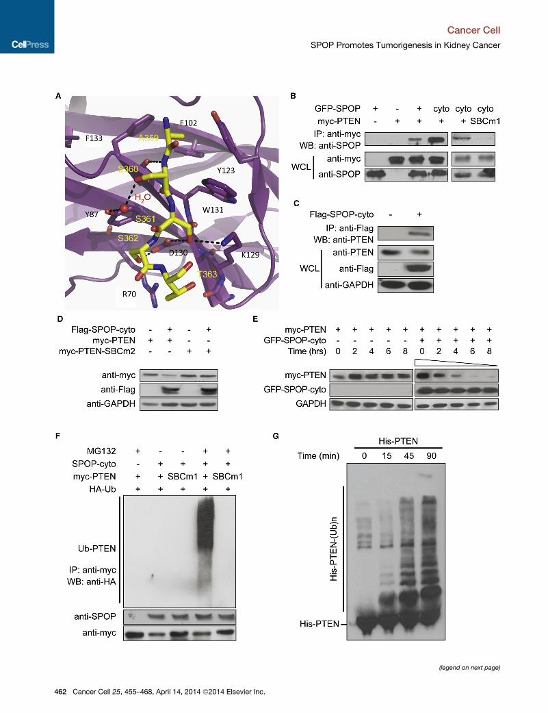
A498 ccRCC cells. As shown in Figure 6A, SPOP knockdown
resulted in increased levels of PTEN and DUSP7 and a decrease
in phosphorylated Akt levels and ERK, respectively (Figure 6A).
These results indicate that SPOP can deregulate these pathways
by degrading PTEN and DUSP7 in ccRCC.
SPOP has been found to also degrade many other substrates,
some of which are candidates for promoting tumorigenesis if
posttranslationally degraded, including Daxx (Kwon et al.,
2006) and Gli2 (Wang et al., 2010). Daxx protein is expressed
in both the nucleus and cytoplasm. In the cytoplasm, Daxx has
been reported to interact with ASK1 and other apical kinases
to induce cell death (Salomoni and Khelifi, 2006). Degradation
of Daxx would impair the ability of the cells to respond to
apoptotic cues. Gli2 acts as a mediator of Hh signaling and
has both transcriptional repression and activation domains
(Sasaki et al., 1999). Gli2 is primarily localized in cytoplasm
and can transfer to nucleus under Hh stimulation (Kim et al.,
2009). Both Daxx and Gli2 are upregulated when SPOP is
knocked down in Caki-2 ccRCC cells (Figure 6B). Additionally,
knockdown of Gli2 in ccRCC cells showed a further increase in
the proliferation marker p-Histone H3 (Figure S6A). To further
explore the possibility that cytoplasmic SPOP degrades these
key substrates in ccRCC, we examined their protein abundance
under hypoxic exposure in non-ccRCC cells. As expected, the
abundance of PTEN, DUSP7, and Daxx proteins were all
reduced in HeLa cells when SPOP was increased and accumu-
lated in the cytoplasm under hypoxia (Figure 6C).
To further validate the role of SPOP on these targets, we
examined the abundance of these SPOP targets in primary
ccRCC tissue samples. We found an inverse relationship
between PTEN levels and SPOP levels in 100% (14/14) of exam-
ined primary ccRCC tumor samples. These ccRCC tumor
tissues exhibited high levels of SPOP and low levels of PTEN,
consistent with previous reports that PTEN levels are reduced
in ccRCCs (Brenner et al., 2002). By contrast, normal adjacent
kidney tissues exhibit the opposite relationship (Figure 6D).
Similarly, we observed inverse relationships for all other exam-
ined substrates, DUSP7 (10/10), Daxx (14/14), and Gli2 (14/14).
Finally, to determine whether the decrease of SPOP targets
promotes ccRCC tumor cell survival, we restored PTEN and
DUSP7 by ectopic expression in A498 ccRCC cells and found
that cellular apoptosis was strongly induced (Figure 6E). Consis-
tent with this finding, knockdown of DUSP6 and DUSP7 together
led to an increase of cell proliferation in ccRCC cells (Figures S6B
and S6C).
DISCUSSION
Our results indicate that HIF can drive SPOP overexpression in
ccRCC and that overexpressed SPOP accumulates in the
cytoplasm of ccRCC. In turn, accumulation of cytoplasmic
Cancer Cell 25, 455–468, April 14, 2014 ª2014 Elsevier Inc. 461
(legend on next page)
Cancer Cell
SPOP Promotes Tumorigenesis in Kidney Cancer
462 Cancer Cell 25, 455–468, April 14, 2014 ª2014 Elsevier Inc.

Figure 5. SPOPMediates the Ubiquitination
and Degradation of DUSP7
(A) Ectopically expressed SPOP or SPOP-cyto
can interact with DUSP7 in an in vivo Co-IP
assay, whereas DUSP7-SBCm (replace SBCmotif
VDSSS with VDGGG) eliminates the interaction.
HeLa cells were transfected with the indicated
constructs and incubated with 10 mM MG132 for
4 hr before harvesting.
(B) Co-IP and immunoblots indicate that overex-
pressed SPOP-cyto can interact and degrade
endogenous DUSP7 in HeLa cells. GAPDH served
as a loading control.
(C) SPOP promotes DUSP7 degradation. HEK293
cells were transfected with the indicated con-
structs. GFP-SPOP and GFP-SPOP-cyto were
detected with monoclonal antibody SPOP-6C.
GAPDH was used as loading control.
(D) Immunoblots demonstrate that neither SPOP
nor SPOP-cyto can degrade DUSP7-SBCm.
(E) In vivo ubiquitination assay reveals that SPOP
promotes DUSP7 ubiquitination through the
DUSP7 SBC domain. Cell lysates were prepared
under a denaturing condition. Myc-DUSP7 was
immunoprecipitated, and HA-Ub was detected by
immunoblotting.
(F) In vitro ubiquitination assay demonstrates that
DUSP7 is a substrate of SPOP.
See also Figure S5.
Cancer Cell
SPOP Promotes Tumorigenesis in Kidney Cancer
SPOP in ccRCC cells appears to directly result in the degrada-
tion of PTEN, DUSP6, DUSP7, Daxx, and Gli2. We suggest
that the concerted loss of function of these proteins in ccRCCs
leads to tumorigenic phenotypes. Therefore, cytoplasmic
SPOP appears to drive tumorigenesis by acting as a key regula-
tory hub protein that orchestrates cancer phenotypes through
the modulation of several critical cellular pathways (Figure 6F).
Figure 4. SPOP Mediates the Ubiquitination and Degradation of PTEN
(A) Crystal structure of the SPOP MATH domain complex with a peptide corresponding to the PTEN SBC m
(B) Co-IP reveals that SPOP and SPOP-cyto bind PTEN, whereas a PTEN mutant lacking a functional SBC
359–363) is unable to bind SPOP. HeLa cells were transfected with the indicated plasmids and incubate
GFP-SPOP and GFP-SPOP-cyto were detected with monoclonal antibody SPOP-6C. WB, western blot; WC
(C) Co-IP and immunoblots indicate that overexpressed SPOP-cyto can interact with and degrade endogeno
control. WCL, whole cell lysates.
(D) SPOP promotes the degradation of PTEN but not a PTEN SBC mutant (SBC2: the SBC motif ASSST w
loading control.
(E)Measurement of PTEN protein abundance by CHX chase assay. HEK293 cells were transfectedwith the ind
the cells were treated with CHX (100 mg/ml) for 2–8 hr, and western blotting was performed. GFP-SPOP-cyto
GAPDH was used as a loading control.
(F) In vivo ubiquitination assay reveals that SPOP promotes PTEN ubiquitination through the PTEN SBC dom
conditions. myc-PTEN was immunoprecipitated, and hemagglutinin-ubiquitin (HA-Ub) was detected by imm
(G) In vitro ubiquitination assay demonstrates that PTEN is a substrate of SPOP.
See also Figure S4 and Table S1.
Cancer Cell 25, 455–4
The HIF signaling pathway and the
PTEN/mTOR pathway are currently the
major therapeutic targets in ccRCC treat-
ment. We suggest here that, as a regula-
tory hub driving tumorigenesis in ccRCC,
SPOP has the potential to be an efficient
drug target. Knocking down SPOP leads
to apoptosis in ccRCC cell lines but not
in HEK293 and HeLa cells, where SPOP is in the nucleus. In
addition to considering SPOP itself as a target, we also suggest
that combinatorial modulation of the signaling pathways
regulated by SPOP may have the potential to be more effective
than the current targeted therapies alone.
Several issues about SPOP function in ccRCC remain to
be explored. For example, given the promiscuity of SPOP in
otif.
motif (SBC1, deletion of the SBC peptide residues
d with 10 mM MG132 for 4 hr before harvesting.
L, whole cell lysates.
us PTEN in HeLa cells. GAPDH served as a loading
as replaced with GGSGG). GAPDH was used as a
icated plasmids. Thirty-six hours after transfection,
was detected with monoclonal antibody SPOP-6C.
ain. Cell lysates were prepared under denaturing
unoblot.
68, April 14, 2014 ª2014 Elsevier Inc. 463
(legend on next page)
Cancer Cell
SPOP Promotes Tumorigenesis in Kidney Cancer
464 Cancer Cell 25, 455–468, April 14, 2014 ª2014 Elsevier Inc.

Cancer Cell
SPOP Promotes Tumorigenesis in Kidney Cancer
targeting critical proliferative and apoptotic pathway compo-
nents, it is unlikely that the proteins studied here are the only crit-
ical cellular targets.While we focused our identification of targets
on proteins with DUSP domains, genomewide, there are dozens
of potential SPOP substrates. However, themodulation of PTEN,
ERK, and Hh (through Gli2) signaling clearly has major effects on
proliferation, and these results clearly demonstrate that SPOP is
acting as a critical hub in a network involving multiple cancer-
related pathways. In doing so, SPOP appears to be both neces-
sary and sufficient for tumorigenic phenotypes.
Another open question is what molecular mechanism
accounts for SPOP mislocalization to the cytoplasm. Although
we demonstrate that hypoxia can drive accumulation of SPOP
in cytoplasm, a number of possibilities exist for how it transfers
from the nucleus to the cytoplasm or vice versa. Since no
SPOP mutation has been detected in kidney cancers thus far
(Liu et al., 2009; Cancer Genome Atlas Research Network,
2013), posttranscriptional modifications on SPOP, such as
phosphorylation and ubiquitination on its nuclear localization
signal, may regulate its location. It has been extensively re-
viewed elsewhere that posttranscriptional modifications can
regulate nuclear import (Hung and Link, 2011; Nardozzi et al.,
2010). Alternatively, this translocation may also be regulated by
interaction with transfer proteins regulated by hypoxia, since
protein-protein interactions also serve as an important regulato-
ry mechanism of protein translocation (Hung and Link, 2011).
Localized and targeted degradation of SPOP is also an intriguing
mechanism, although attempts in our hands to determine
whether it could be a target of obvious candidates such as
VHL have been negative (S.K. and K.P.W., unpublished data).
Exploring the molecular mechanisms of cytoplasmic SPOP
accumulation is an important direction to pursue in the future.
Finally, our previous screening also observed that SPOP was
overexpressed in several other tumors, including some cases
of endometrial and germ cell tumors (Liu et al., 2009). The role
of SPOP in these tumor types is unexplored, as is the role of
mutant SPOP or its LOH in prostate and breast cancers (Berger
et al., 2011; Li et al., 2011). The identification of loss-of-function
SPOP in these other tissues warrants caution in development of
any therapeutic interventions that systemically inhibit SPOP
function. Indeed, SPOP may be acting as a tumor suppressor
in breast and/or prostate cancers, but our results indicate that
it acts as an oncoprotein in ccRCCs. Considering that hypoxia
stress plays important roles in many different tumors, future
efforts to develop tools that selectively modulate SPOP may
be beneficial for treating a wide range of tumors.
Figure 6. SPOP Regulates Multiple Targets in Kidney Cancer
(A) Immunoblots indicate that knockdown of SPOP induces PTEN and DUSP7 p
(Thr202/Tyr204) levels in A498 cells. GAPDH served as a loading control.
(B) Immunoblots demonstrate that knockdown of SPOP in Caki-2 cells induced
(C) Daxx, PTEN, and DUSP7 protein abundance decrease under hypoxia treatm
(D) Immunohistochemistry staining indicates a reduction in multiple SPOP targ
compared with their adjacent normal tissues (diaminobenzidine, brown staining)
(E) Restoring of PTEN and DUSP7 induces apoptosis in A498. Caspase 3/7 activ
and expressed as means ± SD of three independent experiments. *p < 0.05 bas
(F) Schematic overview of SPOP action as a regulatory hub in promoting tumorig
cancer cells it accumulates in the cytoplasm and promotes tumorigenesis by targ
ubiquitin-mediated degradation.
See also Figure S6.
EXPERIMENTAL PROCEDURES
ChIP
ChIP was performed as described elsewhere (Polo et al., 2008). Briefly, ChIPs
for HIF-1a were performed with 10 to 20 3 106 Caki-2 cells under normoxic
and hypoxic conditions using 5 mg HIF-1a antibody (Novus, NB100-134). An
immunoglobulin G antibody (Upstate Biotechnology) was used as a negative
control. Enriched DNA fragments were detected by qPCR and are presented
as enrichment relative to input.
Luciferase Reporter Assay
DNA fragments spanning the potential HIF binding peak in SPOP (chromo-
some 17 [chr17]: 45,109,830–45,109,897 and a series of mutations in the
potential hypoxia-response elements (HRE) were cloned into PGL4.23 [luc2/
minP] vector (Promega, E841A). Plasmids were then cotransfected in K293
cells with a transfection-controlling plasmid pGL4.23 [hRluc/TK] (Promega,
E692A). After 24 hr, cells were transfected with two independent siRNA oligos
to HIF-1a (Dharmacon, J-004018-07 and J-004018-10) and further cultured
under normoxic and hypoxic (1% O2/5% CO2/94% N2) conditions for another
20 hr. Luciferase activity wasmeasured using the Dual-glo Luciferase Assay kit
(Promega, E2920).
Immunohistochemistry
Primary human kidney cancer samples were obtained from the Department
of Urology, First Hospital of Peking University with patients’ consent and
approval of the institutional review board of Peking University. Immuno-
histochemistry was performed according to the method previously used
(Cen et al., 2007) with appropriate antibodies (Supplemental Experimental
Procedures).
Immunofluorescence Microscopy
Immunofluorescence was carried out as described elsewhere (Gottfried et al.,
2004). Mouse anti-SPOP mAb (clone 5G) was used as the primary antibody.
The slides were examined using a Leica Tcs Sp5 confocal laser scanning
microscope (Supplemental Experimental Procedures).
Proliferation Assay
HEK293 cells were transfected with SPOP, SPOP-cyto, or control empty
vector. Cell proliferation was measured by BrdU incorporation (Roche, Cell
Proliferation ELISA kit) according to the manufacturer’s instructions.
Apoptosis Assay
Caspase 3/7 activities were measured using the Apo-ONE Homogenous
Caspase Assay kit (Promega) according to the manufacturer’s instructions.
After incubation for 2 hr at room temperature, fluorescence was detected by
a Fluoroskan Ascent FL microplate reader (Thermo Scientific).
Tumor Xenograft Experiments
Stable HEK293 cell lines expressing SPOP, SPOP-cyto, or the control empty
vector were established as described elsewhere (Kass et al., 2007). Briefly, a
total of 5 3 106 tumor cells were injected subcutaneously into BALB/c nude
mice. Animals were sacrificed at 6 weeks after cell injection to investigate
the tumor formation. A total of 19 mice were used for each construct, with
rotein accumulation and decreased phosphor-Akt (Thr308) and phospho-ERK
Daxx and Gli2 protein accumulation. GAPDH was used as loading control.
ent in HeLa cells.
ets: PTEN, DUSP7, Daxx, and Gli2 are reduced in ccRCC patient samples
. One pair of representative samples is shown. Scale bar, 50 mm.
ity was analyzed to evaluate cell apoptosis. Values were normalized to control
ed on Student’s t test.
enesis in ccRCC. Although SPOP is localized to the nucleus in normal cells, in
eting tumor suppressor (PTEN, DUSP7, Gli2) and proapototic protein (Daxx) for
Cancer Cell 25, 455–468, April 14, 2014 ª2014 Elsevier Inc. 465

Cancer Cell
SPOP Promotes Tumorigenesis in Kidney Cancer
experiments replicated under similar conditions at the Chinese Academy of
Sciences (CAS) and The University of Chicago. All of the animal experiments
were approved by the Committee on the Use of Live Animals in Teaching
and Research at the Institute of Biophysics, CAS, as well as the Institutional
Care and Use Committee at the University of Chicago in accordance with
the National Institutes of Health (NIH) Guide for the Care and Use of Laboratory
Animals.
Immunoprecipitation
Cells were transiently transfected with the indicated plasmids. Twenty-four
hours after transfection, cells were harvested with radio immunoprecipitation
assay (RIPA) buffer and briefly sonicated at 4�C. Lysates were immunopre-
cipitated with anti-myc- or anti-flag-conjugated agarose beads (Sigma).
Precipitates were analyzed on SDS-polyacrylamide gels.
In Vivo Ubiquitination Assay
In vivo ubiquitination assays were based on the protocol described else-
where (Liu et al., 2009). Briefly, HeLa cells were transfected with the indi-
cated plasmids. Twenty-four hours after transfection, cells were treated
with 10 mM MG132 (Calbiochem) for 4 hr before harvesting. Cells were
then lysed in denaturing buffer (1% SDS/50 mM Tris [pH 7.5], 0.5 mM
EDTA/1 mM dithiothreitol). After incubation for 5 min at 100�C, the lysate
was sonicated and diluted 10 times with RIPA lysis buffer and subjected
to Co-IP with anti-c-myc-conjugated agarose beads (Sigma, rabbit antibody)
followed by immunoblotting analysis with anti-hemagglutinin antibody
(Sigma, H9658).
In Vitro Ubiquitination Assay
In vitro ubiquitination was performed as reported elsewhere (Zhuang et al.,
2009) (Supplemental Experimental Procedures).
ACCESSION NUMBERS
The Gene Expression Omnibus accession number for the ChIP-seq data
is GSE54327. The coordinates and structural data of the PTEN SBC motif
binding with SPOP have been deposited to RCSB with the accession
number 4O1V.
SUPPLEMENTAL INFORMATION
Supplemental Information includes Supplemental Experimental Procedures,
six figures, and one table and can be found with this article online at http://
dx.doi.org/10.1016/j.ccr.2014.02.007.
ACKNOWLEDGMENTS
We thank Erin E. Mowers for critical reading and editing of the manuscript.
W.C. was supported by grant 2011CB510101 from the Ministry of Science
and Technology (MOST) 973 Program. K.Y. was supported by the National
863 High-Tech Foundation (grant 2014AA020608). K.C. was supported by
National Science Foundation of China (NSFC) grant 81101940. W.C. was sup-
ported by NSFC grants 91231112 and 31171244. M.C. was supported by a
Howard Hughes Medical Institute (HHMI) postdoctoral fellowship from the
Damon Runyon Cancer Research Foundation (DRG 2021-9). T.S. was sup-
ported by a National Research Service Award from the NIH. B.S. was sup-
ported by ALSAC, the HHMI, NIH grant 5R01GM069530, and NIH grant
5P30CA021765. Y.T. was supported by CAS grant 09CF011001. J.L. was sup-
ported by the MOST 973 Program (2011CB510101), NSFC grant 81171902,
and Beijing Science Foundation grant 5102031. K.P.W. was supported by
the W.M. Keck Foundation, the United States National Institute of General
Medical Sciences grant P50GM081892 and by the Searle Funds at The
Chicago Community Trust from the Chicago Biomedical Consortium.
Received: March 25, 2013
Revised: October 26, 2013
Accepted: February 14, 2014
Published: March 20, 2014
466 Cancer Cell 25, 455–468, April 14, 2014 ª2014 Elsevier Inc.
REFERENCES
Arimoto, K., Fukuda, H., Imajoh-Ohmi, S., Saito, H., and Takekawa, M. (2008).
Formation of stress granules inhibits apoptosis by suppressing stress-respon-
sive MAPK pathways. Nat. Cell Biol. 10, 1324–1332.
Berger, M.F., Lawrence, M.S., Demichelis, F., Drier, Y., Cibulskis, K.,
Sivachenko, A.Y., Sboner, A., Esgueva, R., Pflueger, D., Sougnez, C., et al.
(2011). The genomic complexity of primary human prostate cancer. Nature
470, 214–220.
Bhatia, B., Hsieh, M., Kenney, A.M., and Nahle, Z. (2011). Mitogenic Sonic
hedgehog signaling drives E2F1-dependent lipogenesis in progenitor cells
and medulloblastoma. Oncogene 30, 410–422.
Brenner, W., Farber, G., Herget, T., Lehr, H.A., Hengstler, J.G., and Thuroff,
J.W. (2002). Loss of tumor suppressor protein PTEN during renal carcino-
genesis. Int. J. Cancer 99, 53–57.
Brugarolas, J. (2007). Renal-cell carcinoma—molecular pathways and thera-
pies. N. Engl. J. Med. 356, 185–187.
Bunce, M.W., Boronenkov, I.V., and Anderson, R.A. (2008). Coordinated
activation of the nuclear ubiquitin ligase Cul3-SPOP by the generation of phos-
phatidylinositol 5-phosphate. J. Biol. Chem. 283, 8678–8686.
Campbell, L., Nuttall, R., Griffiths, D., and Gumbleton, M. (2009). Activated
extracellular signal-regulated kinase is an independent prognostic factor in
clinically confined renal cell carcinoma. Cancer 115, 3457–3467.
Cancer Genome Atlas Research Network (2013). Comprehensive
molecular characterization of clear cell renal cell carcinoma. Nature 499,
43–49.
Cen, L., Arnoczky, K.J., Hsieh, F.-C., Lin, H.-J., Qualman, S.J., Yu, S., Xiang,
H., and Lin, J. (2007). Phosphorylation profiles of protein kinases in alveolar
and embryonal rhabdomyosarcoma. Mod. Pathol. 20, 936–946.
Chi, J.-T., Wang, Z., Nuyten, D.S.A., Rodriguez, E.H., Schaner, M.E., Salim, A.,
Wang, Y., Kristensen, G.B., Helland, A., Børresen-Dale, A.-L., et al. (2006).
Gene expression programs in response to hypoxia: cell type specificity and
prognostic significance in human cancers. PLoS Med. 3, e47.
Dhillon, A.S., Hagan, S., Rath, O., and Kolch, W. (2007). MAP kinase signalling
pathways in cancer. Oncogene 26, 3279–3290.
Escudier, B., Eisen, T., Stadler, W.M., Szczylik, C., Oudard, S., Siebels, M.,
Negrier, S., Chevreau, C., Solska, E., Desai, A.A., et al.; TARGET Study
Group (2007). Sorafenib in advanced clear-cell renal-cell carcinoma. N. Engl.
J. Med. 356, 125–134.
Fyfe, G.A., Fisher, R.I., Rosenberg, S.A., Sznol, M., Parkinson, D.R., and Louie,
A.C. (1996). Long-term response data for 255 patients with metastatic renal
cell carcinoma treated with high-dose recombinant interleukin-2 therapy.
J. Clin. Oncol. 14, 2410–2411.
Gardai, S.J., Whitlock, B.B., Xiao, Y.Q., Bratton, D.B., and Henson, P.M.
(2004). Oxidants inhibit ERK/MAPK and prevent its ability to delay neutrophil
apoptosis downstream of mitochondrial changes and at the level of XIAP.
J. Biol. Chem. 279, 44695–44703.
Gottfried, Y., Rotem, A., Lotan, R., Steller, H., and Larisch, S. (2004). The
mitochondrial ARTS protein promotes apoptosis through targeting XIAP.
EMBO J. 23, 1627–1635.
Hamid, T., Malik, M.T., and Kakar, S.S. (2005). Ectopic expression of PTTG1/
securin promotes tumorigenesis in human embryonic kidney cells. Mol.
Cancer 4, 3.
Hernandez-Munoz, I., Lund, A.H., van der Stoop, P., Boutsma, E., Muijrers, I.,
Verhoeven, E., Nusinow, D.A., Panning, B., Marahrens, Y., and van Lohuizen,
M. (2005). Stable X chromosome inactivation involves the PRC1 Polycomb
complex and requires histone MACROH2A1 and the CULLIN3/SPOP ubiquitin
E3 ligase. Proc. Natl. Acad. Sci. USA 102, 7635–7640.
Hollander, M.C., Blumenthal, G.M., and Dennis, P.A. (2011). PTEN loss in the
continuum of common cancers, rare syndromes and mousemodels. Nat. Rev.
Cancer 11, 289–301.
Hudes, G., Carducci, M., Tomczak, P., Dutcher, J., Figlin, R., Kapoor, A.,
Staroslawska, E., Sosman, J., McDermott, D., Bodrogi, I., et al.; Global
Cancer Cell
SPOP Promotes Tumorigenesis in Kidney Cancer
ARCC Trial (2007). Temsirolimus, interferon alfa, or both for advanced
renal-cell carcinoma. N. Engl. J. Med. 356, 2271–2281.
Hung, M.-C., and Link, W. (2011). Protein localization in disease and therapy.
J. Cell Sci. 124, 3381–3392.
Jiang, Y., Zhang, W., Kondo, K., Klco, J.M., St Martin, T.B., Dufault, M.R.,
Madden, S.L., Kaelin, W.G., Jr., and Nacht, M. (2003). Gene expression
profiling in a renal cell carcinoma cell line: dissecting VHL and hypoxia-
dependent pathways. Mol. Cancer Res. 1, 453–462.
Kaelin, W.G., Jr. (2002). Molecular basis of the VHL hereditary cancer
syndrome. Nat. Rev. Cancer 2, 673–682.
Kaelin, W.G., Jr. (2008). The von Hippel-Lindau tumour suppressor protein: O2
sensing and cancer. Nat. Rev. Cancer 8, 865–873.
Kapoor, A., and Figlin, R.A. (2009). Targeted inhibition of mammalian target of
rapamycin for the treatment of advanced renal cell carcinoma. Cancer 115,
3618–3630.
Kass, E.M., Ahn, J., Tanaka, T., Freed-Pastor, W.A., Keezer, S., and Prives, C.
(2007). Stability of checkpoint kinase 2 is regulated via phosphorylation at
serine 456. J. Biol. Chem. 282, 30311–30321.
Kent, D., Bush, E.W., and Hooper, J.E. (2006). Roadkill attenuates Hedgehog
responses through degradation of Cubitus interruptus. Development 133,
2001–2010.
Keyse, S.M. (2008). Dual-specificity MAP kinase phosphatases (MKPs) and
cancer. Cancer Metastasis Rev. 27, 253–261.
Kim, W.Y., and Kaelin, W.G. (2004). Role of VHL gene mutation in human
cancer. J. Clin. Oncol. 22, 4991–5004.
Kim, J., Kato, M., and Beachy, P.A. (2009). Gli2 trafficking links Hedgehog-
dependent activation of Smoothened in the primary cilium to transcriptional
activation in the nucleus. Proc. Natl. Acad. Sci. USA 106, 21666–21671.
Kucejova, B., Pena-Llopis, S., Yamasaki, T., Sivanand, S., Tran, T.A.T.,
Alexander, S., Wolff, N.C., Lotan, Y., Xie, X.-J., Kabbani, W., et al. (2011).
Interplay between pVHL and mTORC1 pathways in clear-cell renal cell
carcinoma. Mol. Cancer Res. 9, 1255–1265.
Kwon, J.E., La, M., Oh, K.H., Oh, Y.M., Kim, G.R., Seol, J.H., Baek, S.H.,
Chiba, T., Tanaka, K., Bang, O.S., et al. (2006). BTB domain-containing
speckle-type POZ protein (SPOP) serves as an adaptor of Daxx for ubiquitina-
tion by Cul3-based ubiquitin ligase. J. Biol. Chem. 281, 12664–12672.
Lee, J.O., Yang, H., Georgescu, M.M., Di Cristofano, A., Maehama, T., Shi, Y.,
Dixon, J.E., Pandolfi, P., and Pavletich, N.P. (1999). Crystal structure of the
PTEN tumor suppressor: implications for its phosphoinositide phosphatase
activity and membrane association. Cell 99, 323–334.
Lee, H.J., Kim, D.I., Kang, G.H., Kwak, C., Ku, J.H., and Moon, K.C. (2009).
Phosphorylation of ERK1/2 and prognosis of clear cell renal cell carcinoma.
Urology 73, 394–399.
Li, C., Ao, J., Fu, J., Lee, D.F., Xu, J., Lonard, D., and O’Malley, B.W. (2011).
Tumor-suppressor role for the SPOP ubiquitin ligase in signal-dependent
proteolysis of the oncogenic co-activator SRC-3/AIB1. Oncogene 30, 4350–
4364.
Liu, J., Ghanim, M., Xue, L., Brown, C.D., Iossifov, I., Angeletti, C., Hua, S.,
Negre, N., Ludwig, M., Stricker, T., et al. (2009). Analysis of Drosophila seg-
mentation network identifies a JNK pathway factor overexpressed in kidney
cancer. Science 323, 1218–1222.
Lopez-Beltran, A., Scarpelli, M., Montironi, R., and Kirkali, Z. (2006).
2004 WHO classification of the renal tumors of the adults. Eur. Urol. 49,
798–805.
Mains, P., Kemphues, K., Sprunger, S., Sulston, I., and Wood, W. (1990).
Mutations affecting the meiotic and mitotic divisions of the early
Caenorhabditis elegans embryo. Genetics 126, 593–605.
McDermott, D.F., Regan, M.M., Clark, J.I., Flaherty, L.E., Weiss, G.R., Logan,
T.F., Kirkwood, J.M., Gordon, M.S., Sosman, J.A., Ernstoff, M.S., et al. (2005).
Randomized phase III trial of high-dose interleukin-2 versus subcutaneous
interleukin-2 and interferon in patients with metastatic renal cell carcinoma.
J. Clin. Oncol. 23, 133–141.
Miyazaki, K., Kawamoto, T., Tanimoto, K., Nishiyama, M., Honda, H., and
Kato, Y. (2002). Identification of functional hypoxia response elements in the
promoter region of the DEC1 and DEC2 genes. J. Biol. Chem. 277, 47014–
47021.
Motzer, R.J., and Molina, A.M. (2009). Targeting renal cell carcinoma. J. Clin.
Oncol. 27, 3274–3276.
Motzer, R.J., Hutson, T.E., Tomczak, P., Michaelson, M.D., Bukowski, R.M.,
Rixe, O., Oudard, S., Negrier, S., Szczylik, C., Kim, S.T., et al. (2007).
Sunitinib versus interferon alfa in metastatic renal-cell carcinoma. N. Engl. J.
Med. 356, 115–124.
Motzer, R.J., Escudier, B., Tomczak, P., Hutson, T.E., Michaelson, M.D.,
Negrier, S., Oudard, S., Gore, M.E., Tarazi, J., Hariharan, S., et al. (2013).
Axitinib versus sorafenib as second-line treatment for advanced renal cell
carcinoma: overall survival analysis and updated results from a randomised
phase 3 trial. Lancet Oncol. 14, 552–562.
Myers, M.P., Pass, I., Batty, I.H., Van der Kaay, J., Stolarov, J.P., Hemmings,
B.A., Wigler, M.H., Downes, C.P., and Tonks, N.K. (1998). The lipid phospha-
tase activity of PTEN is critical for its tumor supressor function. Proc. Natl.
Acad. Sci. USA 95, 13513–13518.
Nagai, Y., Kojima, T., Muro, Y., Hachiya, T., Nishizawa, Y., Wakabayashi, T.,
and Hagiwara, M. (1997). Identification of a novel nuclear speckle-type protein,
SPOP. FEBS Lett. 418, 23–26.
Nardozzi, J.D., Lott, K., and Cingolani, G. (2010). Phosphorylation meets
nuclear import: a review. Cell Commun. Signal. 8, 32.
Negrier, S., Escudier, B., Lasset, C., Douillard, J.Y., Savary, J., Chevreau, C.,
Ravaud, A., Mercatello, A., Peny, J., Mousseau, M., et al. (1998). Recombinant
human interleukin-2, recombinant human interferon alfa-2a, or both in meta-
static renal-cell carcinoma. Groupe Francais d’Immunotherapie. N. Engl. J.
Med. 338, 1272–1278.
Nickerson, M.L., Jaeger, E., Shi, Y., Durocher, J.A., Mahurkar, S., Zaridze, D.,
Matveev, V., Janout, V., Kollarova, H., Bencko, V., et al. (2008). Improved
identification of von Hippel-Lindau gene alterations in clear cell renal tumors.
Clin. Cancer Res. 14, 4726–4734.
Patterson, K.I., Brummer, T., O’Brien, P.M., and Daly, R.J. (2009).
Dual-specificity phosphatases: critical regulators with diverse cellular targets.
Biochem. J. 418, 475–489.
Pintard, L., Willis, J.H., Willems, A., Johnson, J.-L.F., Srayko, M., Kurz, T.,
Glaser, S., Mains, P.E., Tyers, M., Bowerman, B., et al. (2003). The BTB protein
MEL-26 is a substrate-specific adaptor of the CUL-3 ubiquitin-ligase. Nature
425, 311–316.
Polo, J.M., Ci, W., Licht, J.D., and Melnick, A. (2008). Reversible disruption of
BCL6 repression complexes by CD40 signaling in normal and malignant B
cells. Blood 112, 644–651.
Rini, B.I., Campbell, S.C., and Escudier, B. (2009). Renal cell carcinoma.
Lancet 373, 1119–1132.
Robb, V.A., Karbowniczek, M., Klein-Szanto, A.J., and Henske, E.P. (2007).
Activation of the mTOR signaling pathway in renal clear cell carcinoma.
J. Urol. 177, 346–352.
Salomoni, P., and Khelifi, A.F. (2006). Daxx: death or survival protein? Trends
Cell Biol. 16, 97–104.
Sansal, I., and Sellers, W.R. (2004). The biology and clinical relevance of the
PTEN tumor suppressor pathway. J. Clin. Oncol. 22, 2954–2963.
Sarbassov, D.D., Ali, S.M., and Sabatini, D.M. (2005). Growing roles for the
mTOR pathway. Curr. Opin. Cell Biol. 17, 596–603.
Sasaki, H., Nishizaki, Y., Hui, C., Nakafuku, M., and Kondoh, H. (1999).
Regulation of Gli2 and Gli3 activities by an amino-terminal repression domain:
implication of Gli2 and Gli3 as primary mediators of Shh signaling.
Development 126, 3915–3924.
Sato, Y., Yoshizato, T., Shiraishi, Y., Maekawa, S., Okuno, Y., Kamura, T.,
Shimamura, T., Sato-Otsubo, A., Nagae, G., Suzuki, H., et al. (2013).
Integrated molecular analysis of clear-cell renal cell carcinoma. Nat. Genet.
45, 860–867.
Shinojima, T., Oya, M., Takayanagi, A., Mizuno, R., Shimizu, N., and Murai, M.
(2007). Renal cancer cells lacking hypoxia inducible factor (HIF)-1alpha
expression maintain vascular endothelial growth factor expression through
HIF-2alpha. Carcinogenesis 28, 529–536.
Cancer Cell 25, 455–468, April 14, 2014 ª2014 Elsevier Inc. 467

Cancer Cell
SPOP Promotes Tumorigenesis in Kidney Cancer
Vivanco, I., and Sawyers, C.L. (2002). The phosphatidylinositol 3-Kinase AKT
pathway in human cancer. Nat. Rev. Cancer 2, 489–501.
Wang, C., Pan, Y., and Wang, B. (2010). Suppressor of fused and Spop regu-
late the stability, processing and function of Gli2 and Gli3 full-length activators
but not their repressors. Development 137, 2001–2009.
Wardle, E.N. (1991). Cyclophosphamide pulse therapy in relapsing nephrotic
syndrome. Nephron 58, 377.
Wu, X., Senechal, K., Neshat, M.S., Whang, Y.E., and Sawyers, C.L. (1998).
The PTEN/MMAC1 tumor suppressor phosphatase functions as a negative
regulator of the phosphoinositide 3-kinase/Akt pathway. Proc. Natl. Acad.
Sci. USA 95, 15587–15591.
468 Cancer Cell 25, 455–468, April 14, 2014 ª2014 Elsevier Inc.
Xu, L., Wei, Y., Reboul, J., Vaglio, P., Shin, T.H., Vidal, M., Elledge, S.J., and
Harper, J.W. (2003). BTB proteins are substrate-specific adaptors
in an SCF-like modular ubiquitin ligase containing CUL-3. Nature 425,
316–321.
Zhang, Q., Zhang, L., Wang, B., Ou, C.Y., Chien, C.T., and Jiang, J. (2006). A
hedgehog-induced BTB protein modulates hedgehog signaling by degrading
Ci/Gli transcription factor. Dev. Cell 10, 719–729.
Zhuang, M., Calabrese, M.F., Liu, J., Waddell, M.B., Nourse, A., Hammel, M.,
Miller, D.J., Walden, H., Duda, D.M., Seyedin, S.N., et al. (2009). Structures of
SPOP-substrate complexes: insights intomolecular architectures of BTB-Cul3
ubiquitin ligases. Mol. Cell 36, 39–50.


![CircDLST promotes the tumorigenesis and metastasis of ...RNA translation [9], and facilitate the transcription of their parental genes [10, 11], of which circAGO2 promotes the tumor](https://static.fdocuments.us/doc/165x107/60bbe64b3540d83a1377085f/circdlst-promotes-the-tumorigenesis-and-metastasis-of-rna-translation-9-and.jpg)