SPONTANEOUS PERFORATION IN THE NEWBORN …adc.bmj.com/content/archdischild/35/182/378.full.pdf ·...
5
SPONTANEOUS PERFORATION OF THE COLON IN THE NEWBORN INFANT BY S. E. LEVIN and C. ISAACSON From the Department of Paediatrics, Baragwanath Hospital, and the South African Institute for Medical Research, Johannesburg (RECEIVED FOR PUBLICATION SEPTEMBER 15, 1959) If one excludes those cases of perforation which are secondary to volvulus, intestinal atresia, con- genital bands, intussusception, herniation of the bowel, meconium ileus or Hirschsprung's disease, there remains a group of newborn infants who develop perforation of the colon without apparent cause. Spontaneous perforation of the colon in the neonate was first described by Breslau in 1863. It is a rare event and up until 1939 only 26 case reports could be traced in the medical literature by Thelander. A number of factors have been considered in the aetiology of spontaneous perforation such as trauma during delivery (Zillner, 1884), congenital defects of the bowel with diverticulum formation (Fischer, 1928), localized vascular insufficiency, and intrauterine or post-partum infection (Thelander, 1939). In 1888 Paltauf reported five cases which he attributed to coprostasis. This aetiological concept has recently received support from the clinico-surgical and pathological studies by Zachary (1957) and Emery (1957) of a group of newborn infants in whom inspissated faecal plugs gave rise to stercoral ulceration with perforation. These authors suggest that the observed faecal impaction may be due to an imbalance between fluid absorption and of propulsion of material along the colon. More recently Haas (1958) has reported a newborn infant with perforation of the transverse colon in whom a haemangiomatous abnormality at the site of perforation was demonstrated. There still remains a group of newborn infants with one or more perforations of the colon and in whom no clear cut cause can be discovered. In this paper two further cases of spontaneous perforation of the colon are reported. In one of these, perforation occurred during or immediately after birth and this was associated with marked intraperitoneal haemorrhage. The cause of the perforation was undetermined. The other child was a premature infant who developed the per- foration nine days after delivery and histological examination showed fungal infection at the site of perforation. Case Reports Case 1. This female infant was delivered at term as a vertex presentation by a district midwife after an uneventful pregnancy. The child appeared to be vigorous after birth, but abdominal distension with oedema of the vulva was noted. One blood-stained stool was passed after birth and this led to the admission of the child to this hospital. On examination, six hours after delivery, the child appeared somewhat pale but not greatly distressed. The weight was 5 lb. 5 oz. and the rectal temperature was 950 F. The most striking feature was marked distension of the abdomen. There were prominent dilated veins on the anterior abdominal wall which was oedematous in its lower half. There was also oedema of the vulva. Percussion of the abdomen elicited shifting dullness. A radiograph of the abdomen showed air in the stomach and small bowel. There were no fluid levels indicating obstruction. Examination of venous blood revealed a haemoglobin level of 9-0 g./ 100 ml. An abdominal paracentesis at McBurney's point yielded blood-stained fluid with a haemoglobin level of 2-5 g./100 ml. A preoperative diagnosis of rupture of the liver was made and the child was transfused with 100 ml. of group 0 Rh positive compatible blood. At laparotomy blood-stained fluid escaped from the peritoneal cavity. Floating in this fluid were fragments of fresh meconium (later confirmed histologically). Careful inspection of small and large bowel failed to reveal a perforation. A Meckel's diverticulum was found, but it appeared to be intact. No rupture of the liver was found. The omentum was stained green by the meconium but there was no evidence of peritonitis. The abdomen was closed again and the child was maintained on intravenous fluid (0 2% saline in 5% invert sugar) while gastric suction was applied. There was no indication of postoperative bleeding, but 378 copyright. on 29 July 2018 by guest. Protected by http://adc.bmj.com/ Arch Dis Child: first published as 10.1136/adc.35.182.378 on 1 August 1960. Downloaded from
Transcript of SPONTANEOUS PERFORATION IN THE NEWBORN …adc.bmj.com/content/archdischild/35/182/378.full.pdf ·...

SPONTANEOUS PERFORATION OF THE COLONIN THE NEWBORN INFANT
BY
S. E. LEVIN and C. ISAACSONFrom the Department ofPaediatrics, Baragwanath Hospital, andthe South African Institute for Medical Research, Johannesburg
(RECEIVED FOR PUBLICATION SEPTEMBER 15, 1959)
If one excludes those cases of perforation whichare secondary to volvulus, intestinal atresia, con-genital bands, intussusception, herniation of thebowel, meconium ileus or Hirschsprung's disease,there remains a group of newborn infants whodevelop perforation of the colon without apparentcause.
Spontaneous perforation of the colon in theneonate was first described by Breslau in 1863.It is a rare event and up until 1939 only 26 casereports could be traced in the medical literature byThelander.A number of factors have been considered in the
aetiology of spontaneous perforation such astrauma during delivery (Zillner, 1884), congenitaldefects of the bowel with diverticulum formation(Fischer, 1928), localized vascular insufficiency, andintrauterine or post-partum infection (Thelander,1939). In 1888 Paltauf reported five cases whichhe attributed to coprostasis. This aetiologicalconcept has recently received support from theclinico-surgical and pathological studies by Zachary(1957) and Emery (1957) of a group of newborninfants in whom inspissated faecal plugs gave rise tostercoral ulceration with perforation. These authorssuggest that the observed faecal impaction may bedue to an imbalance between fluid absorption andof propulsion of material along the colon. Morerecently Haas (1958) has reported a newborn infantwith perforation of the transverse colon in whoma haemangiomatous abnormality at the site ofperforation was demonstrated.
There still remains a group of newborn infantswith one or more perforations of the colon and inwhom no clear cut cause can be discovered.
In this paper two further cases of spontaneousperforation of the colon are reported. In one ofthese, perforation occurred during or immediatelyafter birth and this was associated with markedintraperitoneal haemorrhage. The cause of the
perforation was undetermined. The other childwas a premature infant who developed the per-foration nine days after delivery and histologicalexamination showed fungal infection at the site ofperforation.
Case ReportsCase 1. This female infant was delivered at term
as a vertex presentation by a district midwife after anuneventful pregnancy. The child appeared to bevigorous after birth, but abdominal distension withoedema of the vulva was noted. One blood-stainedstool was passed after birth and this led to the admissionof the child to this hospital.On examination, six hours after delivery, the child
appeared somewhat pale but not greatly distressed.The weight was 5 lb. 5 oz. and the rectal temperaturewas 950 F. The most striking feature was markeddistension of the abdomen. There were prominentdilated veins on the anterior abdominal wall which wasoedematous in its lower half. There was also oedemaof the vulva. Percussion of the abdomen elicitedshifting dullness. A radiograph of the abdomen showedair in the stomach and small bowel. There were nofluid levels indicating obstruction. Examination ofvenous blood revealed a haemoglobin level of 9-0 g./100 ml.An abdominal paracentesis at McBurney's point
yielded blood-stained fluid with a haemoglobin level of2-5 g./100 ml. A preoperative diagnosis of ruptureof the liver was made and the child was transfused with100 ml. of group 0 Rh positive compatible blood.At laparotomy blood-stained fluid escaped from theperitoneal cavity. Floating in this fluid were fragmentsof fresh meconium (later confirmed histologically).Careful inspection of small and large bowel failed toreveal a perforation. A Meckel's diverticulum wasfound, but it appeared to be intact. No rupture of theliver was found. The omentum was stained green by themeconium but there was no evidence of peritonitis.The abdomen was closed again and the child was
maintained on intravenous fluid (0 2% saline in 5%invert sugar) while gastric suction was applied. Therewas no indication of postoperative bleeding, but
378
copyright. on 29 July 2018 by guest. P
rotected byhttp://adc.bm
j.com/
Arch D
is Child: first published as 10.1136/adc.35.182.378 on 1 A
ugust 1960. Dow
nloaded from
SPONTANEOUS PERFORATION OF THE COLON48 hours after the operation she suddenly collapsed anddied.At necropsy the peritoneal cavity contained a small
quantity of semi-fluid dark brown material. There wassome adherence of the bowel loops. A small perfora-tion 3-4 mm. in diameter was found in the region of thehepatic flexure of the ascending colon and a smallquantity of meconium was emerging from the perfora-tion. The anatomical position of the bowel at autopsyappeared normal and there was no evidence of volvulusor intussusception. There was thus no obvious causefor the perforation. Other organs showed no abnor-malities except for some patches of atelectasis of thelungs.
Histology of sections of the bowel at the site of per-foration revealed merely a break in the continuity of thewall with some necrosis of the wall adjacent to theperforation. Ganglion cells were present in normalnumbers. The pancreas showed no obvious lesion.The lungs were congested and oedematous.
Case 2. This male infant was born at this hospitalweighing 3 lb. The mother developed puerperal sepsisand the infant at 3 days old was transferred to thepremature infant unit, his weight then being 2 lb. 11 oz.Apart from mild icterus, no abnormalities were detected
FIG. 1.-Radiograph of the abdomen showing a large accumulationof air under the diaphragm and in the right scrotal sac.
on clinical examination. He was given feeds of un-diluted expressed breast milk from the third day. Onthe fifth day of life the jaundice had become moreintense. The serum bilirubin level was 12 mg./100 ml.Meconium was passed on the fourth and sixth daysof life but no stools were noted on the seventh, eighthand ninth days of life. On the ninth day, the infantvomited a feed which was not stained with bile. Someslight distension of the abdomen was noted. A glycerinesuppository was prescribed and oral feeding of expressedbreast milk was continued. Later in the day two brownwatery stools were passed and one of them was blood-stained.The following day (day 10) the infant continued to
vomit and developed gross gaseous distension of theabdomen. The clinical state of hydration was satis-factory. A radiograph of the abdomen revealed thepresence of a well-marked pneumoperitoneum (Fig. 1).Gas was present in the small bowel without evidenceof fluid levels, and for this reason the site of perforationwas thought to be in the colon. Feeds were stoppedand intravenous therapy was started with 0-2% salinein 5% invert sugar with the addition of 2-0 g. of potas-sium chloride to 1,000 ml. of solution. The child wasalso given 100,000 units of penicillin intramuscularlyevery eight hours and 50 mg. of streptomycin intra-muscularly every 12 hours. On the eleventh day thechild's abdomen was still markedly distended and therewas oedema of the scrotum. An ill-defined mass couldbe palpated in the left flank. The infant had alsodeveloped oral thrush which was treated with 50,000units of Nystatin ('mycostatin') every eight hours. Onthe tenth day the child passed two small scybala. Thechild's condition continued to deteriorate and deathtook place on the twelfth day of life.At necropsy there was marked meconium peri-
tonitis. The peritoneal cavity contained 150 ml. of
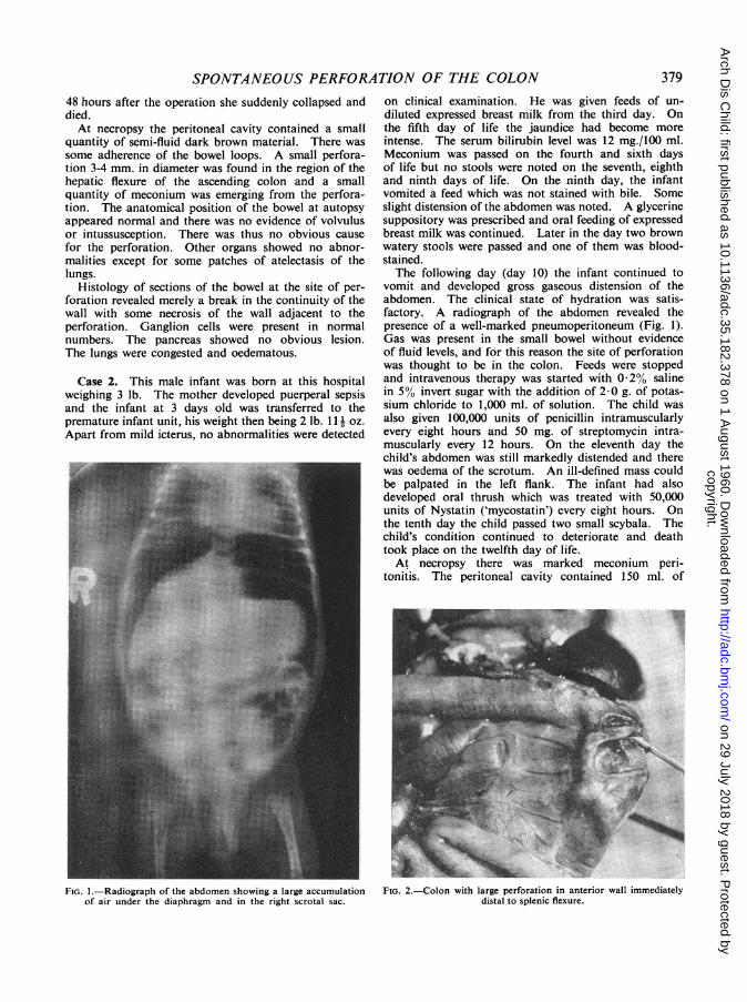
FIG. 2.-Colon with large perforation in anterior wall immediatelydistal to splenic flexure.
379
copyright. on 29 July 2018 by guest. P
rotected byhttp://adc.bm
j.com/
Arch D
is Child: first published as 10.1136/adc.35.182.378 on 1 A
ugust 1960. Dow
nloaded from
ARCHIVES OF DISEASE IN CHILDHOOD
W.'^ . ' MR
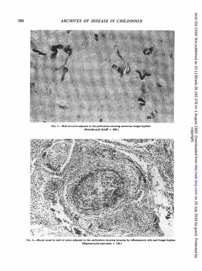
FIG. 3.-Wall of colon adjacent to the perforation showing numerous fungal hyphae.(Periodic-acid Schiff x 480.)
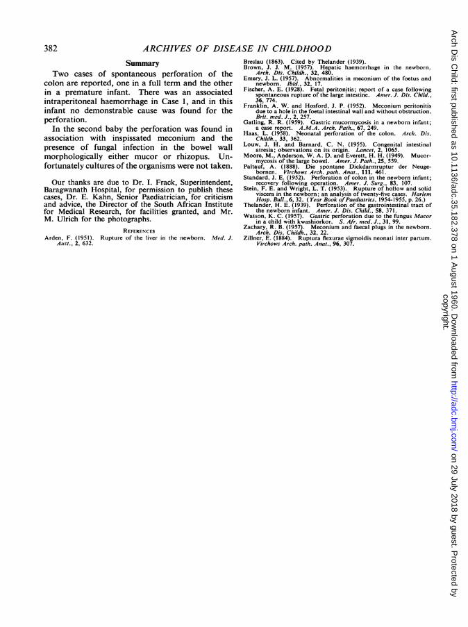
FIG. 4.-Blood vessel in wall of colon adjacent to the perforation showing invasion by inflammatory cells and fungal hyphae.(Haematoxylin and eosin x 150.)
380
copyright. on 29 July 2018 by guest. P
rotected byhttp://adc.bm
j.com/
Arch D
is Child: first published as 10.1136/adc.35.182.378 on 1 A
ugust 1960. Dow
nloaded from

SPONTANEOUS PERFORATION OF THE COLONgreen purulent fluid. Loops of bowel were denselyadherent to the lateral abdominal wall on the left.A large perforation 0-5 cm. in diameter (Fig. 2) waspresent about 2-5 cm. below the splenic flexure in theanterior wall of the descending colon.
There were still some yellow flecks of inspissatedmeconium around the perforation and floating in theperitoneal exudate. Anatomically the bowel was nor-mal, and there was no evidence of a volvulus or anintussusception.
Section of the bowel wall at the site of the perforationshowed the surrounding mucosa to be necrotic andhaemorrhagic. There was haemorrhage into the sub-mucosa and infiltration of the entire wall by poly-morphs. The serosa was acutely inflamed and coveredby necrotic debris. Fungal hyphae consistent in appear-ance with the fungus mucor, or the related rhizopus,were present in the lumen of the bowel, in the wall andin the serosa (Fig. 3). The fungal filaments were largeand showed absence of cross-wall formation with freeand irregular branching, features suggesting origin fromthe mucoraceae. In many areas the fungi were invadingthe walls of blood vessels and had come to lie withintheir lumens (Fig. 4). Unfortunately cultures of thefungus were not made before the tissues were fixed informalin.
Histology of the pancreas was normal.
Discussion
Our first case presented the clinical picture ofintraperitoneal haemorrhage. Hence a preopera-tive diagnosis was made of rupture of the liver,which is the commonest solid viscus to be damagedin the newborn infant (Stein and Wright, 1953;Arden, 1951; Brown, 1957).At operation the surprise finding of meconium
in the peritoneal cavity, in addition to the blood,indicated perforation of a hollow viscus. The siteof perforation was not detected at laparotomy.This difficulty of locating the site of perforationhas been encountered by others, and it happenedin four of the cases reported by Thelander (1939).Standard (1952) in reporting his case, which wassuccessfully treated surgically, admits that theperforation would not have been visible but forthe presence of leaking barium suspension whichhe had given to the infant. In a case reported byFranklin and Hosford (1952), the perforation wasonly found at the second operation.
Necropsy in our first case failed to reveal acause for the perforation, but it must be admittedthat a close study of the blood supply to the affectedsegment of bowel was not carried out. The clinicaland experimental studies of Louw and Barnard(1955) have indicated that vascular occlusion playsan important role in the production of intestinal
atresia in the newborn infant. Their work mayhave some application in the elucidation of theaetiology of spontaneous perforation of the colon.We suggest that, in future, a careful study be madeof the blood supply to the area of a spontaneousperforation of the colon. We have assumed that theintraperitoneal bleeding in our case arose from thesame site as the perforation, but this was not obviousat autopsy, the bleeding having ceased post-operatively.Case 2 is of interest in that this premature infant
was progressing satisfactorily until the ninth dayof life. The child was starting to regain its post-natal weight loss, when it failed to pass stools forthree successive days. As in Case 1 there was thepassage of one blood-stained stool and it is possiblethat this finding in the presence of abdominaldistension may be of help in the diagnosis of spon-taneous perforation of the bowel.Three possibilities suggest themselves as to the
cause of the perforation in Case 2. Firstly thisperforation could have followed on stercoral ulcera-tion caused by inspissated meconium. The passageof two small hard pieces of faeces and the finding ofresidual thickened meconium at necropsy are infavour of this suggestion. However, we were unableto analyse the water content of the meconium as wasdone by Emery (1957).
Secondly the perforation may be aetiologicallyconnected with the finding of mucor (or rhizopus)in the bowel wall at the site of perforation. Mucorhas been found in association with perforation inother parts of the gastro-intestinal tract. Moore,Anderson and Everett (1949) described a wide-spread ulcerative colitis in a non-diabetic femaleaged 37 years caused by mucor which resulted indeath after perforation and generalized peritonitis.Watson (1957) reported perforation of the stomachin association with mucormycosis in a child of26 months suffering from malnutrition. A similarperforation was described by Gatling (1959) in a4-day-old neonate. Thus this fungal infectionmay have been the primary cause of the perforationin Case 2. It is also possible that the fungusaggravated a perforation caused initially by hardenedmeconium.
Until recently the prognosis of spontaneousperforation was thought to be uniformly hopeless.However, in 1952, Standard, in America, andFranklin and Hosford, in Britain, reported survivalsfollowing surgical repair. Thus laparotomy isprobably advisable whenever the condition of thepatient permits. Preliminary decompression ofthe abdomen may be necessary to increase vitalcapacity (Standard, 1952).
381
copyright. on 29 July 2018 by guest. P
rotected byhttp://adc.bm
j.com/
Arch D
is Child: first published as 10.1136/adc.35.182.378 on 1 A
ugust 1960. Dow
nloaded from
382 ARCHIVES OF DISEASE IN CHILDHOODSummary
Two cases of spontaneous perforation of thecolon are reported, one in a full term and the otherin a premature infant. There was an associatedintraperitoneal haemorrhage in Case 1, and in thisinfant no demonstrable cause was found for theperforation.
In the second baby the perforation was found inassociation with inspissated meconium and thepresence of fungal infection in the bowel wallmorphologically either mucor or rhizopus. Un-fortunately cultures of the organisms were not taken.
Our thanks are due to Dr. I. Frack, Superintendent,Baragwanath Hospital, for permission to publish thesecases, Dr. E. Kahn, Senior Paediatrician, for criticismand advice, the Director of the South African Institutefor Medical Research, for facilities granted, and Mr.M. Ulrich for the photographs.
REFERENCESArden, F. (1951). Rupture of the liver in the newborn. Med. J.
Aust., 2, 632.
Breslau (1863). Cited by Thelander (1939).Brown, J. J. M. (1957). Hepatic haemorrhage in the newborn.
Arch. Dis. Childh., 32, 480.Emery, J. L. (1957). Abnormalities in meconium of the foetus and
newborn. Ibid., 32, 17.Fischer, A. E. (1928). Fetal peritonitis; report of a case following
spontaneous rupture of the large intestine. Amer. J. Dis. Child.,36, 774.
Franklin, A. W. and Hosford, J. P. (1952). Meconium peritonitisdue to a hole in the foetal intestinal wall and without obstruction.Brit. med. J., 2, 257.
Gatling, R. R. (1959). Gastric mucormycosis in a newborn infant;a case report. A.M.A. Arch. Path., 67, 249.
Haas, L. (1958). Neonatal perforation of the colon. Arch. Dis.Childh., 33, 362.
Louw, J. H. and Barnard, C. N. (1955). Congenital intestinalatresia; observations on its origin. Lancet, 2, 1065.
Moore, M., Anderson, W. A. D. and Everett, H. H. (1949). Mucor-mycosis of the large bowel. Amer. J. Path., 25, 559.
Paltauf, A. (1888). Die spontane Dickdarmruptur der Neuge-bornen. Virchows Arch. path. Anat., 111, 461.
Standard, J. E. (1952). Perforation of colon in the newborn infant;recovery following operation. Amer. J. Surg., 83, 107.
Stein, F. E. and Wright, L. T. (1953). Rupture of hollow and solidviscera in the newborn; an analysis of twenty-five cases. HarlemHosp. Bull., 6, 32. (Year Book ofPaediatrics, 1954-1955, p. 26.)
Thelander, H. E. (1939). Perforation of the gastrointestinal tract ofthe newborn infant. Amer. J. Dis. Child., 58, 371.
Watson, K. C. (1957). Gastric perforation due to the fungus Mucorin a child with kwashiorkor. S. Afr. med. J., 31, 99.
Zachary, R. B. (1957). Meconium and faecal plugs in the newborn.Arch. Dis. Childh., 32, 22.
Zillner, E. (1884). Ruptura flexurae sigmoidis neonati inter partum.Virchows Arch. path. Anat., 96, 307.
copyright. on 29 July 2018 by guest. P
rotected byhttp://adc.bm
j.com/
Arch D
is Child: first published as 10.1136/adc.35.182.378 on 1 A
ugust 1960. Dow
nloaded from