Spontaneous Loss of Virulence in Natural Populations...
13
Spontaneous Loss of Virulence in Natural Populations of Listeria monocytogenes Mylène M. Maury, a,b Viviane Chenal-Francisque, a,b * Hélène Bracq-Dieye, a,b Lei Han, c Alexandre Leclercq, a,b Guillaume Vales, a,b Alexandra Moura, b,d Edith Gouin, f Mariela Scortti, c,e Olivier Disson, b,d José A. Vázquez-Boland, c,e Marc Lecuit a,b,d,g Institut Pasteur, National Reference Centre and WHO Collaborating Centre for Listeria, Paris, France a ; Institut Pasteur, Biology of Infection Unit, Paris, France b ; Microbial Pathogenesis Unit, Medical School (Biomedical Sciences), University of Edinburgh, Edinburgh, United Kingdom c ; Inserm U1117, Paris, France d ; Division of Infection and Immunity, The Roslin Institute, University of Edinburgh, Edinburgh, United Kingdom e ; Institut Pasteur, Bacteria-Cell Interactions Unit, Paris, France f ; Paris Descartes University, Sorbonne Paris Cité, Institut Imagine, Necker-Enfants Malades University Hospital, Division of Infectious Diseases and Tropical Medicine, APHP, Paris, France g ABSTRACT The pathogenesis of Listeria monocytogenes depends on the ability of this bacterium to escape from the phagosome of the host cells via the action of the pore-forming toxin listeriolysin O (LLO). Expression of the LLO-encoding gene (hly) requires the transcriptional activator PrfA, and both hly and prfA genes are essential for L. monocytogenes virulence. Here, we used the hemolytic activity of LLO as a phenotypic marker to screen for spontaneous virulence-attenuating mutations in L. monocytogenes. Sixty nonhemolytic isolates were identified among a collection of 57,820 confirmed L. monocytogenes strains isolated from a variety of sources (0.1%). In most cases (56/60; 93.3%), the nonhemolytic phenotype resulted from nonsense, missense, or frameshift mutations in prfA. Five strains carried hly mutations leading to a single amino acid substitution (G299V) or a premature stop codon causing strong virulence attenuation in mice. In one strain, both hly and gshF (encoding a glutathione synthase required for full PrfA activity) were missing due to genomic re- arrangements likely caused by a transposable element. The PrfA/LLO loss-of-function (PrfA /LLO ) mutants belonged to phylogenetically diverse clades of L. monocyto- genes, and most were identified among nonclinical strains (57/60). Consistent with the rare occurrence of loss-of-virulence mutations, we show that prfA and hly are under purifying selection. Although occurring at a low frequency, PrfA /LLO muta- tional events in L. monocytogenes lead to niche restriction and open an evolutionary path for obligate saprophytism in this facultative intracellular pathogen. KEYWORDS Listeria monocytogenes, virulence, hemolysis, genomics, spontaneous mutations L isteria monocytogenes is a foodborne pathogen that can cause a severe invasive disease, called listeriosis, in people and animals. As a facultative intracellular bac- terium, L. monocytogenes has evolved a range of virulence determinants allowing intracellular survival (1, 2). One key virulence factor is listeriolysin O (LLO), a pore- forming toxin responsible for the characteristic -hemolytic phenotype of L. monocy- togenes that allows the bacterium to escape from the phagosome of host cells and replicate intracellularly (3, 4). LLO is encoded by hly, located in Listeria pathogenicity island 1 (LIPI-1) (5). Expression of the genes within this central pathogenicity locus, including hly, is under the control of the transcriptional activator PrfA, the master regulator of L. monocytogenes virulence genes (6, 7). The hemolytic activity conferred Received 28 July 2017 Accepted 2 August 2017 Accepted manuscript posted online 21 August 2017 Citation Maury MM, Chenal-Francisque V, Bracq-Dieye H, Han L, Leclercq A, Vales G, Moura A, Gouin E, Scortti M, Disson O, Vázquez- Boland JA, Lecuit M. 2017. Spontaneous loss of virulence in natural populations of Listeria monocytogenes. Infect Immun 85:e00541-17. https://doi.org/10.1128/IAI.00541-17. Editor Nancy E. Freitag, University of Illinois at Chicago Copyright © 2017 Maury et al. This is an open- access article distributed under the terms of the Creative Commons Attribution 4.0 International license. Address correspondence to Marc Lecuit, [email protected], and Jose ´ A. Va ´zquez-Boland, [email protected]. * Present address: Viviane Chenal-Francisque, Biology of Intracellular Bacteria, Institut Pasteur, Paris, France. J.A.V.-B. and M.L. share senior authorship of this article. M.M.M., V.C.-F., H.B.-D., and L.H. contributed equally to this article. MOLECULAR GENOMICS crossm November 2017 Volume 85 Issue 11 e00541-17 iai.asm.org 1 Infection and Immunity on October 13, 2018 by guest http://iai.asm.org/ Downloaded from
-
Upload
vuongthien -
Category
Documents
-
view
214 -
download
0
Transcript of Spontaneous Loss of Virulence in Natural Populations...

Spontaneous Loss of Virulence inNatural Populations of Listeriamonocytogenes
Mylène M. Maury,a,b Viviane Chenal-Francisque,a,b* Hélène Bracq-Dieye,a,b
Lei Han,c Alexandre Leclercq,a,b Guillaume Vales,a,b Alexandra Moura,b,d
Edith Gouin,f Mariela Scortti,c,e Olivier Disson,b,d José A. Vázquez-Boland,c,e
Marc Lecuita,b,d,g
Institut Pasteur, National Reference Centre and WHO Collaborating Centre for Listeria, Paris, Francea; InstitutPasteur, Biology of Infection Unit, Paris, Franceb; Microbial Pathogenesis Unit, Medical School (BiomedicalSciences), University of Edinburgh, Edinburgh, United Kingdomc; Inserm U1117, Paris, Franced; Division ofInfection and Immunity, The Roslin Institute, University of Edinburgh, Edinburgh, United Kingdome; InstitutPasteur, Bacteria-Cell Interactions Unit, Paris, Francef; Paris Descartes University, Sorbonne Paris Cité, InstitutImagine, Necker-Enfants Malades University Hospital, Division of Infectious Diseases and Tropical Medicine,APHP, Paris, Franceg
ABSTRACT The pathogenesis of Listeria monocytogenes depends on the ability ofthis bacterium to escape from the phagosome of the host cells via the action of thepore-forming toxin listeriolysin O (LLO). Expression of the LLO-encoding gene (hly)requires the transcriptional activator PrfA, and both hly and prfA genes are essentialfor L. monocytogenes virulence. Here, we used the hemolytic activity of LLO as aphenotypic marker to screen for spontaneous virulence-attenuating mutations in L.monocytogenes. Sixty nonhemolytic isolates were identified among a collection of57,820 confirmed L. monocytogenes strains isolated from a variety of sources (0.1%).In most cases (56/60; 93.3%), the nonhemolytic phenotype resulted from nonsense,missense, or frameshift mutations in prfA. Five strains carried hly mutations leadingto a single amino acid substitution (G299V) or a premature stop codon causingstrong virulence attenuation in mice. In one strain, both hly and gshF (encoding aglutathione synthase required for full PrfA activity) were missing due to genomic re-arrangements likely caused by a transposable element. The PrfA/LLO loss-of-function(PrfA�/LLO�) mutants belonged to phylogenetically diverse clades of L. monocyto-genes, and most were identified among nonclinical strains (57/60). Consistent withthe rare occurrence of loss-of-virulence mutations, we show that prfA and hly areunder purifying selection. Although occurring at a low frequency, PrfA�/LLO� muta-tional events in L. monocytogenes lead to niche restriction and open an evolutionarypath for obligate saprophytism in this facultative intracellular pathogen.
KEYWORDS Listeria monocytogenes, virulence, hemolysis, genomics, spontaneousmutations
Listeria monocytogenes is a foodborne pathogen that can cause a severe invasivedisease, called listeriosis, in people and animals. As a facultative intracellular bac-
terium, L. monocytogenes has evolved a range of virulence determinants allowingintracellular survival (1, 2). One key virulence factor is listeriolysin O (LLO), a pore-forming toxin responsible for the characteristic �-hemolytic phenotype of L. monocy-togenes that allows the bacterium to escape from the phagosome of host cells andreplicate intracellularly (3, 4). LLO is encoded by hly, located in Listeria pathogenicityisland 1 (LIPI-1) (5). Expression of the genes within this central pathogenicity locus,including hly, is under the control of the transcriptional activator PrfA, the masterregulator of L. monocytogenes virulence genes (6, 7). The hemolytic activity conferred
Received 28 July 2017 Accepted 2 August2017
Accepted manuscript posted online 21August 2017
Citation Maury MM, Chenal-Francisque V,Bracq-Dieye H, Han L, Leclercq A, Vales G,Moura A, Gouin E, Scortti M, Disson O, Vázquez-Boland JA, Lecuit M. 2017. Spontaneous loss ofvirulence in natural populations of Listeriamonocytogenes. Infect Immun 85:e00541-17.https://doi.org/10.1128/IAI.00541-17.
Editor Nancy E. Freitag, University of Illinois atChicago
Copyright © 2017 Maury et al. This is an open-access article distributed under the terms ofthe Creative Commons Attribution 4.0International license.
Address correspondence to Marc Lecuit,[email protected], and Jose A. Vazquez-Boland,[email protected].
* Present address: Viviane Chenal-Francisque,Biology of Intracellular Bacteria, Institut Pasteur,Paris, France.
J.A.V.-B. and M.L. share senior authorship of thisarticle. M.M.M., V.C.-F., H.B.-D., and L.H.contributed equally to this article.
MOLECULAR GENOMICS
crossm
November 2017 Volume 85 Issue 11 e00541-17 iai.asm.org 1Infection and Immunity
on October 13, 2018 by guest
http://iai.asm.org/
Dow
nloaded from

by LLO is considered a cardinal marker for L. monocytogenes detection and/or identi-fication in clinical and food microbiology. L. monocytogenes is divided into four phy-logenetic lineages (8–10), 13 serotypes (11) that can be approximated by PCR sero-grouping (12), and more than 100 clonal complexes (CCs, as defined by multilocussequence typing [MLST]) (13), which are unevenly virulent (14). Weakly or nonhemolyticL. monocytogenes strains have been reported (15–19), but the frequency and phyloge-netic diversity of the strains displaying an altered hemolysis phenotype are unknown,as well as their underlying genetic and microbiological features.
This study aimed at (i) estimating the frequency of naturally occurring nonhemolyticL. monocytogenes isolates and their distribution among L. monocytogenes lineages andMLST clonal complexes, (ii) understanding the molecular bases of the nonhemolyticphenotype, and (iii) assessing its impact on virulence. By using phenotypic andgenomic approaches, mutagenesis, and in vivo assays, we show that mutations leadingto loss of hemolytic activity in L. monocytogenes, although rare, affect a wide range ofclonal complexes of the major lineages I and II and lead to a decreased virulence.
RESULTSIdentification and characterization of nonhemolytic L. monocytogenes strains.
We examined the prevalence of nonhemolytic L. monocytogenes strains among the57,820 L. monocytogenes isolates collected between 1987 and 2008 at the FrenchNational Reference Centre for Listeria (NRCL) and the WHO Collaborating Centre forListeria (WHOCCL). Sixty L. monocytogenes isolates (0.1%) were identified as nonhemo-lytic on horse blood agar plates. These were isolated from food (n � 33), foodproduction environments (n � 2), nonhuman unknown sources (n � 22), and humanclinical cases (n � 3). Phenotypic characterization using the API Listeria system con-firmed all 60 nonhemolytic isolates as L. monocytogenes. These belonged to lineages I(n � 23, 38.3%) and II (n � 37, 61.7%) and were grouped within serogroups IIa (n � 36),IVb (n � 13), IIb (n � 10), and IIc (n � 1) (see Table S1 in the supplemental material).MLST showed that the 60 nonhemolytic isolates belonged to 15 different clonalcomplexes, including the hypovirulent CC9 (n � 1), CC121 (n � 3), CC31 (n � 20), andsequence type 13 (ST13) (n � 3) (14, 20) as well as the hypervirulent CC1 (n � 3), CC2(n � 7), CC4 (n � 1), and CC6 (n � 1) (14) (Fig. 1 and Table S1). Core genome MLST(cgMLST) typing identified 39 different cgMLST types (CTs) (21). Nine CTs comprisedmore than one strain, suggesting a possible epidemiological link between them (21)(Table S1). In particular, among the 20 nonhemolytic CC31 strains, 10 belonged toCT878, and 2 belonged to CT2659, suggesting that the overrepresentation of CC31could be in part due to multiple sampling of the same source in the context of anepidemiological investigation. These results show that nonhemolytic strains are phy-logenetically very diverse and that the loss of hemolytic activity is caused by indepen-dent events across the L. monocytogenes population.
To investigate the impact of the loss of hemolytic activity on L. monocytogenesfitness, we analyzed the growth of all nonhemolytic strains in brain heart infusion (BHI)broth at 22°C and 37°C, using strain EGDe as control (Fig. S1). At 22°C, in a largemajority of cases, the growth of nonhemolytic strains was within the same range as thatof EGDe, as revealed by the areas under the growth curves (AUCs). In contrast, at 37°C,the temperature at which prfA is known to be maximally expressed (22), most of thenonhemolytic strains showed lower growth (lower AUCs) than EGDe. Some of thenonhemolytic strains showed particularly decreased fitness at one or both tempera-tures: CLIP 2000/86467 (PrfAT170*, at 22°C, where the asterisk indicates a truncation atresidue T170 of PrfA), CLIP 1998/75799 (PrfAI51*-LLON261*, at 37°C), and, at both tem-peratures, strains CLIP 1998/76801 (Δhly-ΔgshF), CLIP 1996/70991 (PrfAQ21*), CLIP 1994/58618 (PrfAA129P), and CLIP 1996/71614 (PrfAY207*) (Fig. S1).
Molecular basis of the nonhemolytic phenotype: PrfA variants and activity. Thecentral regulator of Listeria virulence, PrfA, is required for the expression of a set of keyvirulence determinants, known as the PrfA regulon, including the hly gene (6, 7, 23).Consequently, mutations altering the function of either PrfA or LLO could lead to a
Maury et al. Infection and Immunity
November 2017 Volume 85 Issue 11 e00541-17 iai.asm.org 2
on October 13, 2018 by guest
http://iai.asm.org/
Dow
nloaded from
FIG 1 Phylogenetic tree summarizing all the genetic features causing the loss of hemolytic activity among the 60 nonhemolytic L. monocytogenesstrains. Single-linkage-based clustering was obtained based on the cgMLST allelic profiles, as described previously (21). The scale bar indicates the
(Continued on next page)
Spontaneous Loss of Listeria monocytogenes Virulence Infection and Immunity
November 2017 Volume 85 Issue 11 e00541-17 iai.asm.org 3
on October 13, 2018 by guest
http://iai.asm.org/
Dow
nloaded from

nonhemolytic phenotype. Sequence analyses identified frameshifts and missense andnonsense mutations in prfA in 56 nonhemolytic strains, leading to amino acid substi-tutions or protein truncations in PrfA (Fig. 1; Table S1). Phenotypic analysis underPrfA-activating and -nonactivating conditions using the PrfA-dependent virulence fac-tors PlcB (phospholipase C) and Hpt as reporters (see Materials and Methods) (24)confirmed the complete loss of function of the central virulence gene regulator in allof these strains (Fig. 1; Fig. S2).
Forty-three out of the 56 PrfA� strains, distributed in lineages I and II, expressed atruncated PrfA at 14 distinct positions distributed along the entire PrfA protein (TableS1). All analyzed strains of CC59 and CC31 exhibited a truncation at positions 59 and185, respectively, suggesting a common ancestor for each of these groups of strains.Seven PrfA� strains presented a single amino acid substitution in PrfA compared to thesequence of the reference strain EGDe (GenBank accession number: NC_003210).Among them, one occurred in the �-roll region of PrfA (G72D in strain CLIP 1997/75561,CC9). Mutations located in this region are known to affect PrfA activation or the abilityof PrfA to form a stable complex with the RNA polymerase and initiate transcription ofthe target virulence genes (25–27). One PrfA� mutation occurred in the DNA-bindinghelix-turn-helix (HTH) domain of PrfA (G175C in strain CLIP 2006/01642, CC6), and twoothers occurred in its C-terminal part (K220T in strains CLIP 1994/60344, CLIP 2000/80770, and CLIP 2001/87255, all ST13; and L221F in strain CLIP 1994/56373, CC1). Theseregions are known to be important for the binding of PrfA to PrfA-binding sites oftarget DNAs (25, 26). In addition, the A129P substitution, located between the �-rolland the hinge �D regions, occurred in a CC224 strain (CLIP 1994/58618). Finally, six ofthe PrfA� strains, all belonging to CC155, showed a reversion of the prfA stop codondue to the insertion of 5 nucleotides at position 712 in the prfA sequence, leading toa longer PrfA protein (238 amino acids in EGDe versus 293 amino acids in the CC155strains of this study).
One of the four nonhemolytic mutants (CC1 strain CLIP 1998/76801) exhibited awild-type (WT) PrfA sequence compared to that of EGDe but showed a PrfA� pheno-type. This observation suggested that a mechanism interfering upstream of PrfAfunction was affected. Glutathione, synthetized by L. monocytogenes through theglutathione synthase encoded by gshF (lmo2770), is critical for PrfA activation (28).Interestingly, although it is part of the L. monocytogenes core genome (14, 21), gshF wasabsent in the genome of the CLIP 1998/76801 strain (Fig. 1) (see below), which couldexplain the absence of PrfA activity in this strain.
Analysis of spontaneous LLO mutants. Analysis of hly sequences in the 60nonhemolytic strains identified multiple mutations leading to amino acid substitutionsin LLO (Table S1). Several substitutions (N31H, S35L, V438I, and K523S) were identifiedin at least 48 hemolytic L. monocytogenes strains of our database (�4,100 genomes),suggesting that they do not cause LLO loss of function. However, an S250N substitutionwas found only in three nonhemolytic strains of this study (CLIP 2008/01432, 2008/01433, and 2008/01435, all CC77) and could therefore result in LLO loss of function.Since these strains also expressed a truncated and nonactive PrfA, which is sufficient toexplain the nonhemolytic phenotype of these strains, we did not pursue this further.
Two out of the three nonhemolytic strains showing a WT PrfA sequence and a PrfA�
phenotype (CC121 strains CLIP 2007/01406 and CLIP 2007/01014) exhibited a singleamino acid substitution in LLO (the G299V substitution encoded by hly [hlyG299V], orLLOG299V), which was not present in any of the other strains. The third strain (CC2, CLIP
FIG 1 Legend (Continued)percentage of cgMLST similarity. Strain names have been simplified to avoid redundancy and should be preceded by CLIP (Collection of the InstitutPasteur). PrfA activities and mutations (first and second columns, respectively), gshF presence/absence profile (third column), and LLO mutations andpresence/absence profile (fourth column) are mapped on the phylogeny. The position and the nature of amino acid substitutions are indicated ingray zones. Positions of premature stop codons are indicated next to black asterisks in light pink zones. The absence of gshF and hly in the CLIP1998/76801 strain is indicated in black. MLST clonal complexes are shown on the right. The black star highlights the CLIP 1998/76801 strain thatcontains multiple copies of a transposable element that induced huge genomic rearrangements. ND, not determined (unknown and nonhumanorigin).
Maury et al. Infection and Immunity
November 2017 Volume 85 Issue 11 e00541-17 iai.asm.org 4
on October 13, 2018 by guest
http://iai.asm.org/
Dow
nloaded from

1989/13656) harbored a premature stop codon at position 484 in LLO (hlyC484*, orLLOC484*). The absence of any other specific feature in these three strains that could belinked to the loss of hemolytic activity suggested that the G299V mutation and thetruncation at position 484 in LLO could be the cause of the loss of hemolytic activity inthese strains. In addition, two CC7 strains expressing a truncated PrfA (CLIP 1998/75799and CLIP 1989/14490) also showed a premature stop codon in LLO at position 261(hlyN261*) due to the insertion of one nucleotide.
In the CLIP 1998/76801 strain mentioned above, hly could not be detected by PCR,and the hly region could not be assembled from Illumina reads. In order to resolve thisregion, we sequenced this strain using single-molecule, real-time (SMRT) sequencingtechnology (Pacific Biosciences, CA, USA). The CLIP 1998/76801 complete genome (CC1;2.84 Mb) was compared to the closely related F2365 complete genome (CC1; NCBIaccession number NC_002973) as a reference. This showed that the LIPI-1 region hadundergone an inversion of more than 40 kb (Fig. 2A). This large rearrangement splitLIPI-1 into two parts with a concomitant loss of hly and partial truncation of the 5=region of the adjacent mpl gene. Six open reading frames (ORFs) were insertedupstream of mpl in CLIP 1998/76801 compared to the sequence of F2365 and com-prised genes encoding a transposition protein (tnsB) and a DNA invertase (hin), whichare likely the cause of the rearrangement, as well as cadmium resistance genes (cadAand cadC) (Fig. 2A).
FIG 2 Comparison of the CLIP 1998/76801 and F2365 genomes. (A) Gene content of the LIPI-1 region in F2365 (GenBank accession numberNC_002973) in comparison to the corresponding region in the nonhemolytic CLIP 1998/76801 strain, as indicated. LIPI-1 genes are highlightedin red. mpl is composed of 1,532 bp in F2365 but 1,133 bp in CLIP 1998/76801. (B) Gene content of the gshF region in F2365 in comparison tothe corresponding region in CLIP 1998/76801. In panels A and B, genes that are present in CLIP 1998/76801 but absent in F2365 are indicatedin orange. Genes encoding the transposition protein (tnsB), the DNA invertase (hin), and cadmium resistance (cadA and cadC) are indicated. (C)Global comparison of the F2365 and the CLIP 1998/76801 genomes. Positions of the eight copies of the transposable element are indicated indark blue. Identity percentages (indicated by gray zones of variable intensities) between sequences were determined by nucleotide BLAST (55).Genome comparisons were performed using Easyfig, version 2.1 (56).
Spontaneous Loss of Listeria monocytogenes Virulence Infection and Immunity
November 2017 Volume 85 Issue 11 e00541-17 iai.asm.org 5
on October 13, 2018 by guest
http://iai.asm.org/
Dow
nloaded from

We confirmed that gshF is absent in CLIP 1998/76801, together with 12 otherupstream and downstream genes related to sugar metabolism (Fig. 2B). These geneswere replaced by 11 ORFs encoding a transposition protein (tnsB), a DNA invertase (hin),and cadmium resistance (cadA and cadC), similar to the genes inserted in the LIPI-1region. In total, eight similar copies of this transposable element were found in theCLIP 1998/76801 genome, as well as many other large rearrangements and deletions(Fig. 2C). Similar transposable elements were detected in one Listeria ivanovii strain inthe NCBI database (GenBank accession number KR780025.1; 99% nucleotide identity)and in 128 L. monocytogenes strains (�99.87% nucleotide similarity) of the 4,091genome sequences available at the NRCL at the time of the study. These strainscomprised 14.1% of all the CC1 strains (90/638, representing two distinct monophyleticgroups within the phylogeny of CC1 [data not shown]) and all the CC59 strains (n � 38).No significant link of this element with food or clinical origins was found within CC1.
Assessment of hly and prfA transcription. In order to test the effect of the
identified mutations on hly and prfA transcription, quantitative reverse transcription-PCRs (qRT-PCRs) were performed for a representative set of nonhemolytic strains (onestrain per type of loss-of-hemolysis mutation) (Table S1). All nonhemolytic strainsshowed prfA transcription levels equivalent to or higher than those of EGDe, except forstrains CLIP 1998/75799 (PrfAI51*-LLON261* mutations) and CLIP 1998/77604 (PrfAT76*
mutation), which showed no amplification, likely due to poor primer annealing (eightmismatches with the prfA-R primer) (Fig. S3). As expected, strains with an altered PrfA(amino acid substitution or truncation) showed no or extremely reduced hly transcrip-tion levels. These results show that for these strains the loss of hemolytic activity is dueto prfA posttranscriptional events leading to the absence of PrfA activity. In the strainCLIP 2007/01406 (LLOG299V), hly was transcribed at a level similar to that in EGDe,whereas in CLIP 1989/13656 (LLOC484*), hly transcription was weaker.
In vitro characterization of the hlyG299V and hlyC484* mutations. In order to
characterize the functional impact of the G299V substitution (CLIP 2007/01406 and CLIP2007/01014) and of the truncation at position 484 in LLO (CLIP 1989/13656), weintroduced a plasmid containing either a wild-type hly gene (hlyWT) or a mutatedversion of this gene (hlyG299V or hlyC484*, encoding LLOG299V and LLOC484*, respectively)in an EGDΔhly strain. While EGDΔhly::pPL2-hlyWT was hemolytic, EGDΔhly::pPL2-hlyG299V
or EGDΔhly::pPL2-hlyC484* remained nonhemolytic, as assessed on Columbia horseblood agar plates. These results demonstrate that the hlyG299V and hlyC484* mutationsare responsible for the absence of hemolytic activity in the strains CLIP 2007/01406,CLIP 2007/01014, and CLIP 1989/13656.
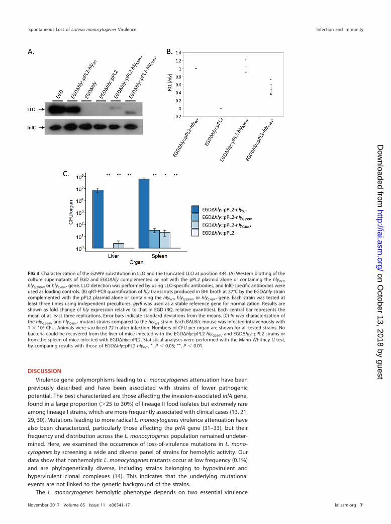
Western blot analyses of culture supernatants detected smaller amounts of LLOproduced by EGDΔhly::pPL2-hlyG299V and EGDΔhly::pPL2-hlyC484* bacteria than by theWT EGD and EGDΔhly::pPL2-hlyWT strains (Fig. 3A). qRT-PCR analyses showed that thehly transcription levels in both the EGDΔhly::pPL2-hlyG299V and EGDΔhly::pPL2-hlyC484*
strains are comparable to the level observed in EGDΔhly::pPL2-hlyWT although the levelis slightly weaker in EGDΔhly::pPL2-hlyC484* (Fig. 3B). Furthermore, the EGDΔhly::pPL2-hlyC484* mutant produced a shorter LLO protein than strains harboring the hlyWT,confirming that the premature stop codon identified in hly in the CLIP 1989/13656strain leads to the production of a truncated LLO. The hlyN261* mutation (Fig. 1; TableS1) was not tested in vitro as this premature stop codon is upstream of the hlyC484*
mutation, leading to an even shorter LLO.Virulence of hlyG299V and hlyC484* mutants. We finally assessed the virulence of
the EGDΔhly::pPL2-hlyG299V and EGDΔhly::pPL2-hlyC484* complemented strains relativeto that of the EGDΔhly::pPL2-hlyWT and EGDΔhly::pPL2 strains upon intravenous injec-tion in mice. The EGDΔhly::pPL2-hlyG299V and EGDΔhly::pPL2-hlyC484* strains were fourorders of magnitude less abundant than the EGDΔhly::pPL2-hlyWT strain in the liver andthe spleen (Fig. 3C). This demonstrates that the virulence of L. monocytogenes express-ing either LLOG299V or LLOC484* is strongly attenuated in vivo.
Maury et al. Infection and Immunity
November 2017 Volume 85 Issue 11 e00541-17 iai.asm.org 6
on October 13, 2018 by guest
http://iai.asm.org/
Dow
nloaded from
DISCUSSION
Virulence gene polymorphisms leading to L. monocytogenes attenuation have beenpreviously described and have been associated with strains of lower pathogenicpotential. The best characterized are those affecting the invasion-associated inlA gene,found in a large proportion (�25 to 30%) of lineage II food isolates but extremely rareamong lineage I strains, which are more frequently associated with clinical cases (13, 21,29, 30). Mutations leading to more radical L. monocytogenes virulence attenuation havealso been characterized, particularly those affecting the prfA gene (31–33), but theirfrequency and distribution across the L. monocytogenes population remained undeter-mined. Here, we examined the occurrence of loss-of-virulence mutations in L. mono-cytogenes by screening a wide and diverse panel of strains for hemolytic activity. Ourdata show that nonhemolytic L. monocytogenes mutants occur at low frequency (0.1%)and are phylogenetically diverse, including strains belonging to hypovirulent andhypervirulent clonal complexes (14). This indicates that the underlying mutationalevents are not linked to the genetic background of the strains.
The L. monocytogenes hemolytic phenotype depends on two essential virulence
FIG 3 Characterization of the G299V substitution in LLO and the truncated LLO at position 484. (A) Western blotting of theculture supernatants of EGD and EGDΔhly complemented or not with the pPL2 plasmid alone or containing the hlyWT,hlyG299V, or hlyC484* gene. LLO detection was performed by using LLO-specific antibodies, and InlC-specific antibodies wereused as loading controls. (B) qRT-PCR quantification of hly transcripts produced in BHI broth at 37°C by the EGDΔhly straincomplemented with the pPL2 plasmid alone or containing the hlyWT, hlyG299V, or hlyC484* gene. Each strain was tested atleast three times using independent precultures. gyrB was used as a stable reference gene for normalization. Results areshown as fold change of hly expression relative to that in EGD (RQ, relative quantities). Each central bar represents themean of at least three replications. Error bars indicate standard deviations from the means. (C) In vivo characterization ofthe hlyG299V and hlyC484* mutant strains compared to the hlyWT strain. Each BALB/c mouse was infected intravenously with1 � 104 CFU. Animals were sacrificed 72 h after infection. Numbers of CFU per organ are shown for all tested strains. Nobacteria could be recovered from the liver of mice infected with the EGDΔhly::pPL2-hlyG299V and EGDΔhly::pPL2 strains orfrom the spleen of mice infected with EGDΔhly::pPL2. Statistical analyses were performed with the Mann-Whitney U test,by comparing results with those of EGDΔhly::pPL2-hlyWT. *, P � 0.05; **, P � 0.01.
Spontaneous Loss of Listeria monocytogenes Virulence Infection and Immunity
November 2017 Volume 85 Issue 11 e00541-17 iai.asm.org 7
on October 13, 2018 by guest
http://iai.asm.org/
Dow
nloaded from

determinants, the central virulence regulator PrfA and LLO, encoded by prfA and hly,respectively. Indeed, all nonhemolytic strains identified in this study carried mutationsin at least one of these genes. The large majority of nonhemolytic strains (56/60; 93.3%)carried prfA mutations (frameshifts, missense or nonsense nucleotide changes, orreversion of the stop codon into a glutamine codon). Although no PrfA activity couldbe detected and hly was not transcribed in these strains, prfA was transcribed at levelssimilar to the level in strain EGDe. This suggests that the loss of PrfA activity in thesestrains likely results from PrfA misfolding, instability, and/or inactivating amino acidsubstitution. Some inactivating amino acid substitutions in PrfA occurred in the �-roll,HTH motif, or C-terminal domain, in line with the critical role of these regions in PrfAactivity (25–27, 31). As PrfA is the major transcriptional regulator of the virulence genesof L. monocytogenes and is essential for its pathogenicity (23, 34), the virulence of PrfA�
strains is expected to be highly attenuated, as previously described (31–33). The first L.monocytogenes strain naturally producing a C-terminally extended PrfA polypeptide (55residues longer) was identified in this study and showed no PrfA activity and no hlytranscription.
Nonhemolytic hly mutants with affected LLO activity were less frequent (5/60; 8.3%)in our study than strains with loss-of-hemolysis mutations in prfA. Our analysis identi-fied for the first time a spontaneous amino acid substitution in LLO (hlyG299V) andpremature stop codons in hly (hlyN261* and hlyC484*) leading to the loss of LLO activity.Lower quantities of LLO were detected in the culture supernatants of the EGDΔhly::pPL2-hlyG299V and EGDΔhly::pPL2-hlyC484* constructs than in the EGD and EGDΔhly::pPL2-hlyWT strains. The quantities of hly transcripts were similar in the EGDΔhly::pPL2-hlyG299V and the EGDΔhly::pPL2-hlyWT control strain, indicating that LLOG299V is likelyless stable than WT LLO. In contrast, EGDΔhly::pPL2-hlyC484* showed a lower hlytranscription level than that of WT hly, suggesting an impaired stability of the hlyC484*
transcript. In vivo experiments confirmed that the nonhemolytic strains harboring thehlyG299V or hlyC484* mutation have strongly attenuated virulence in mice. In line withthese results, only three nonhemolytic strains were isolated from human samples.Although we did not have access to the detailed clinical data of these patients (datingback to the 1980s and 1990s), one possibility would be that they were heavilyimmunocompromised, mirroring previous reports on isolation of the nonpathogenic L.monocytogenes relative Listeria innocua from immunosuppressed individuals (35).
One of the LLO-negative (LLO�) strains (CLIP 1998/76801) underwent huge genomicrearrangements that likely caused the loss of hly and gshF, encoding a glutathionesynthase reported as being required for PrfA activity (28). CLIP 1998/76801 is the onlystrain in our entire genome database (�4,100 entries) that lacks gshF. Interestingly,each copy of the transposable element that likely caused the genomic rearrangementsobserved in this strain carried putative cadmium resistance determinants that could beadvantageous in environments in which virulence determinants are not needed. Similartransposable elements were detected in monophyletic groups of CC1 and CC59 strains,suggesting that they have been horizontally transmitted in the L. monocytogenespopulation.
The predominance of PrfA� mutants among the nonhemolytic strains could reflectthe fact that prfA is a pleiotropic regulatory gene that controls the expression of anumber of virulence determinants, the expression of which is known to entail asignificant fitness cost under nonhost conditions (24). Our results show that, at 22°C,the majority of PrfA� strains have a fitness level similar to that of EGDe, suggesting thatthe absence of PrfA activity does not impact L. monocytogenes fitness under nonpatho-genic conditions. Nevertheless, reduced fitness was observed at 37°C (mammalian hosttemperature) compared to that of EGDeΔprfA. This result suggests that nonhemolyticstrains are more adapted to a nonpathogenic lifestyle, independently of PrfA. Consis-tent with this, most of the nonhemolytic L. monocytogenes isolates were from nonclini-cal origins. The ratio of nonsynonymous to synonymous substitutions (dN/dS) esti-mated for prfA (dN/dS � 0.08892) and hly (dN/dS � 0.03674) using a data set of 100genomes representative of L. monocytogenes phylogenetic diversity (14) confirmed
Maury et al. Infection and Immunity
November 2017 Volume 85 Issue 11 e00541-17 iai.asm.org 8
on October 13, 2018 by guest
http://iai.asm.org/
Dow
nloaded from

that, similar to L. monocytogenes core genes (dN/dS � 0.05353, on average [21]), thesegenes are under purifying selection. Thus, any deleterious mutations affecting thesegenes tend to be eliminated from the L. monocytogenes population. The relatively lowfrequency of deleterious mutations in prfA and hly indicates that there might be astrong selection for L. monocytogenes to retain its virulence capacity (36). Our resultsalso suggest that once strains lose their virulence capacity (e.g., due to a prfA mutation),other virulence genes may become irrelevant and prone to accumulate mutations, asobserved in our PrfA�/LLO� and PrfA�/GshF� strains. Previous studies have alreadyidentified strains with multiple mutations occurring in several major virulence genes(20). Strains with virulence-attenuating mutations are therefore prone to enter into anevolutionary path toward obligate saprophytism. The L. monocytogenes phylogenomicclade comprises another pathogenic species, Listeria ivanovii, which contains a set ofPrfA-regulated genes, as well as nonpathogenic species, some of which contain rem-nants thereof (e.g., Listeria seeligeri or L. innocua) (37, 38). While infrequent, spontane-ous virulence-disabling mutations such as those described here could have been keyinitial events in the emergence and evolution of the L. monocytogenes-related non-pathogenic Listeria species.
MATERIALS AND METHODSBacterial strains and growth media. The 60 nonhemolytic L. monocytogenes isolates included in
this study were identified among a collection of 57,820 L. monocytogenes strains collected between 1987and 2008 by the French National Reference Centre for Listeria (NRCL) and World Health OrganizationCollaborating Centre for Listeria (WHOCCL) in the context of the epidemiological surveillance of listeri-osis. This global collection included isolates of food (n � 36,630), clinical (n � 5,980), environmental (n �3,647), veterinary (n � 1,713), and unknown (n � 9,850) origins. Isolates were revived by plating themonto Columbia agar, and single colonies were grown on Columbia agar slants. L. monocytogenes strainswere routinely grown in BHI broth at 37°C, and Escherichia coli strains were grown at 37°C in LB brothor agar plates.
Phenotypic characterization of Listeria isolates. Miniaturized enzymatic and sugar fermentationtests (API-Listeria identification microgallery; bioMérieux, France), in combination with the hemolyticactivity assessment of strains, were used for phenotypic identification of Listeria species (39). Hemolyticactivity was tested on Columbia horse blood agar plates (bioMérieux, France). L. monocytogenes CLIP74910 and Listeria innocua CLIP 74915 were used as positive and negative controls of hemolysis,respectively.
Genome sequencing and analyses. Genomic DNA was extracted using a DNeasy Blood and TissueExtraction kit (Qiagen, Denmark) and used for whole-genome sequencing on an Illumina NextSeq 500(2 � 150 bp) platform (Illumina, CA, USA). Reads were trimmed with AlienTrimmer (40) to eliminateadapter sequences and discard reads with Phred scores of �20. De novo assembly of Illumina reads wasperformed using SPAdes Genome Assembler, version 3.1 (41). The complete genome of the CLIP1998/76801 strain was obtained by PacBio RS II sequencing (Pacific Biosciences, CA, USA) using DNApurified with a Wizard genomic DNA purification kit (Promega, WI, USA). Genome annotation wasperformed using Prokka, version 1.11 (42).
PCR serogroups (12, 43), MLST profiles (13), and cgMLST profiles (21) were deduced from genomeassemblies using the BIGSdb-L. monocytogenes platform (http://bigsdb.pasteur.fr/listeria) (21). Genomeassemblies were made publicly available in the BIGSdb-L. monocytogenes platform (see Table S1 in thesupplemental material).
Assessment of prfA and hly evolutionary trends. prfA and hly sequences were extracted from 100genomes that were selected to represent the species diversity based on MLST and pulsed-field gelelectrophoresis (PFGE) typing (14) and aligned using Muscle, version 3.8 (44). This data set includedgenomes from 13 food isolates, 45 human clinical isolates, 19 animal isolates, 1 environmental isolate,and 22 isolates of unknown origin. They comprised 41 genomes of lineage I, 53 of lineage II, 5 of lineageIII, and 1 of lineage IV and represented 5 singletons and 34 clonal complexes based on MLST. Nononhemolytic strain was included in this analysis. Alignments were used to estimate the ratios ofnonsynonymous to synonymous substitutions (dN/dS) in prfA and hly using the codeml programincluded in the PAML package, version 4.4 (45).
Assessment of PrfA activity. PrfA activity was assessed by measuring the activity of PrfA-regulatedplcB and hpt gene products as previously described (46, 47). For PlcB, lecithinase tests were performedin egg yolk BHI agar, and for Hpt, glucose-1-phosphate acidification tests were carried out in phenol redbroth, in both cases with and without 0.5% (wt/vol) activated charcoal (Merck, NJ, USA). Mediumsupplementation with charcoal leads to the partial activation of PrfA, presumably due to sequestrationof repressor substances from the culture medium. Three L. monocytogenes genotypes from strain P14were used as controls: (i) a prfAWT strain characterized by an activatable PrfA phenotype (lack of PlcB andHpt activity in normal medium and strong activity in charcoal-supplemented medium), (ii) a ΔprfA strainwhich remains negative for PlcB and Hpt activity in the presence of charcoal, and (iii) a constitutivelyactivated prfA (prfA*) strain with strong PlcB and Hpt activity independent of charcoal supplementation(24, 46, 48).
Spontaneous Loss of Listeria monocytogenes Virulence Infection and Immunity
November 2017 Volume 85 Issue 11 e00541-17 iai.asm.org 9
on October 13, 2018 by guest
http://iai.asm.org/
Dow
nloaded from
RNA extractions. Nonhemolytic strains and EGDΔhly::pPL2-hlyWT, EGDΔhly::pPL2, EGDΔhly::pPL2-hlyG299V, and EGDΔhly::pPL2-hlyC484* constructs were cultured overnight on BHI agar at 37°C. One colonywas used to inoculate 5 ml of BHI broth. After overnight growth at 37°C, 500 �l of culture was added to10 ml of BHI broth, and the whole exponential-phase culture (at 37°C) was centrifuged at 5,000 � g for5 min. The pellet was suspended with 400 �l of resuspension buffer (10% glucose, 12.5 mM Tris, 10 mMEDTA in nuclease-free water) and transferred to a lysing tube (containing 0.1 mm of ceramic breads, 500�l of acid phenol, and 60 �l of 0.5 M EDTA). A Precellys 24 homogenizer (Bertin Instruments, France)was used at 6,500 rpm for 23 s two times (10-s break), and the resulting mixture was centrifuged at14,000 � g at 4°C for 10 min. The upper aqueous phase was transferred into a tube containing 1 ml ofTRIzol and 100 �l of chloroform, mixed by inversions, and centrifuged. The upper aqueous phase wastransferred into a tube containing 200 �l of chloroform, mixed by inversions, and centrifuged. The upperaqueous phase was transferred into a storage tube (containing 650 �l of isopropanol and 65 �l of 3 Msodium acetate), mixed by inversions, precipitated for 20 min at �20°C, and centrifuged for 20 min. Thesupernatant was rinsed twice with 75% ethanol. The air-dried pellet was dissolved in 300 �l ofnuclease-free water. RNA concentrations were measured with a DeNovix DS-11 spectrophotometer(DeNovix, DE, USA) and diluted to obtain 500 ng of RNA in 12.5 �l of nuclease-free water.
Quantification of hly and prfA transcripts by qRT-PCR. For the qRT-PCRs, cDNAs were generatedprior to quantitative PCRs (qPCRs). DNase treatment was performed with RNase-free DNase I (NewEngland BioLabs, MA, USA) according to the instructions of the manufacturer. Briefly, 0.5 �l of RNAseOUT,0.5 �l of DNase I, and 1.5 �l of 10� buffer were added to the 12.5 �l of diluted RNAs, followed by a finaladdition of 1.5 �l of 0.05 M EDTA. cDNAs were generated by reverse transcription using Moloney murineleukemia virus (MMLV) reverse transcriptase (Invitrogen, CA, USA) and random hexamers for primingaccording to the instructions of the manufacturer. Briefly, 2 �l of 10 mM deoxynucleoside triphosphates(dNTPs), 2 �l of 2.5 �M random primers, and 3.5 �l of nuclease-free water were added to the 16.5 �l ofthe previously DNase-treated sample, and then 8 �l of 5� first-strand buffer, 4 �l of 0.1 M dithiothreitol(DTT), and 2 �l of nuclease-free water were added, followed by 0.5 �l of MMLV reverse transcriptase.
All quantitative PCRs were prepared using SYBR green real-time PCR master mixes and a StepOnePlusreal-time PCR system (Applied Biosystems, CA, USA). Each primer pair was used for separate reactionsusing PCR mixtures containing 1 �l of a 9 �M concentration of each primer (Table S2), 5 �l of SYBR mix,1 �l of cDNA diluted at 1:5, and 3 �l of nuclease-free water. Real-time PCRs were carried out in MicroAmpFast Optical 96-well reaction plates (Applied Biosystems, CA, USA) using the following protocol: initialdenaturation at 95°C for 10 min, followed by 40 cycles of denaturation at 95°C for 15 s and primerannealing/elongation at 60°C for 1 min. Each strain was tested at least three times using independentprecultures. gyrB was used as a stable reference gene for normalization. Results are shown as fold changeof the target gene expression level relative to that of EGDe or EGD (relative quantities [RQ]), which wasdeduced from the cycle threshold (CT) values using the 2�ΔΔCT methodology.
Fitness studies. The microbial growth of nonhemolytic strains, EGDe, EGD, and EGDeΔprfA wasmonitored over time in BHI broth at 22°C and 37°C using absorbance measurements (optical density at600 nm [OD600]) through a Bioscreen C system (Oy Growth Curves Ab Ltd., Helsinki, Finland). Bacteriawere first cultured overnight on BHI agar at 22°C or 37°C, and one colony was used to inoculate 5 ml ofBHI broth. After overnight growth, the stationary-phase cultures were diluted to reach an OD600 of 0.1and transferred into Bioscreen C 96-well plates. The OD600 values of noninoculated wells (blanks) weresubtracted from those of inoculated ones to delete the background noise. Each strain was tested threetimes. Mean OD600 values per strain were used to calculate the areas under the curves over time. For this,data were fitted to parametric models (Gompertz, modified Gompertz, logistic, and Richards laws) usingthe gcFit function of the grofit R package, version 1.1.1-1 (49). The model that best fitted the data wasselected by means of an Akaike information criterion (AIC) (50) and used to derive areas under thegrowth curves.
DNA manipulations and cloning. We used a two-step cloning strategy to introduce the wild-typehly (hlyWT), hlyG299V, or hlyC484* genes in the L. monocytogenes strain EGDΔhly. First, we cloned separatelythe hlyWT, hlyG299V, and hlyC484* gene sequences into the Listeria integrative vector pPL2 (51). Primers usedare listed in Table S2. To deliver plasmids into L. monocytogenes, Escherichia coli S17.1 (colistin andnalidixic acid sensitive) was transformed with the plasmids, followed by conjugation with L. monocyto-genes EGDΔhly (colistin and nalidixic acid resistant). L. monocytogenes EGDΔhly bacteria were selected on7 �g/ml chloramphenicol (bacteria containing the pPL2 derivatives), 10 �g/ml colicin, and 50 �g/mlnalidixic acid (selection of resistant L. monocytogenes versus sensitive E. coli bacteria). Since all ourconstructs were made on a similar EGD background, the PrfA* phenotype of EGD was not expected tohave any impact on our results and conclusions.
Western blotting. Protein extracts were obtained from EGD, EGDΔhly, EGDΔhly::pPL2, EGDΔhly::pPL2-hlyWT, EGDΔhly::pPL2-hlyG299V, and EGDΔhly::pPL2-hlyC484* as follows. Bacteria were grown over-night in BHI broth at 37°C. After centrifugation of bacterial cultures (30 min at 2,151 � g), all proteins ofthe supernatant were precipitated by using trichloroacetic acid (20%) and washed using acetone.Proteins were then separated by SDS-PAGE (8% acrylamide gel and 3.9% stacking gel) and transferred toa polyvinylidene difluoride transfer membrane (Bio-Rad, CA, USA). The membrane was incubatedovernight at 4°C with a blocking buffer containing dried milk (5%), phosphate-buffered saline (PBS; 1%),and Tween (0.1%) and washed with PBS (1%) and Tween (0.1%). The membrane was then incubated firstwith a polyclonal anti-LLO (52, 53) or anti-InlC antibody (54) (1/20,000; 1 h at room temperature) andsecond with an anti-rabbit antibody (1/3,000; 1 h at room temperature). The membrane was washed withPBS (1%) and Tween (0.1%) between each incubation step with antibodies. Antibody-antigen interactions
Maury et al. Infection and Immunity
November 2017 Volume 85 Issue 11 e00541-17 iai.asm.org 10
on October 13, 2018 by guest
http://iai.asm.org/
Dow
nloaded from

were revealed using a SuperSignal West Pico chemiluminescent substrate (Thermo Fischer Scientific, MA,USA).
Animal studies. The virulence of L. monocytogenes strains EGDΔhly::pPL2-hlyWT, EGDΔhly::pPL2-hlyG299V, EGDΔhly::pPL2-hlyC484*, and EGDΔhly::pPL2 was assessed in vivo. BALB/c mice were infected viathe intravenous route with 1 � 104 CFU per animal. At 72 h postinfection, mice were sacrificed for spleenand liver dissection. CFU were enumerated by plating dilutions of the whole homogenized organs ontoBHI plates. Statistical analyses were performed with the Mann-Whitney U test, by comparing the resultswith those of EGDΔhly::pPL2-hlyWT. All procedures were in agreement with the guidelines of theEuropean Commission for the handling of laboratory animals, directive 86/609/EEC, and were approvedby the Animal Care and Use Committee of the Institut Pasteur, as well as by the ethical committee of ParisCentre et Sud under the number 2010-0020.
SUPPLEMENTAL MATERIAL
Supplemental material for this article may be found at https://doi.org/10.1128/IAI.00541-17.
SUPPLEMENTAL FILE 1, XLSX file, 0.1 MB.SUPPLEMENTAL FILE 2, XLSX file, 0.1 MB.SUPPLEMENTAL FILE 3, PDF file, 0.1 MB.SUPPLEMENTAL FILE 4, PDF file, 0.2 MB.SUPPLEMENTAL FILE 5, PDF file, 0.2 MB.SUPPLEMENTAL FILE 6, PDF file, 0.1 MB.
ACKNOWLEDGMENTSWe acknowledge Anne Morvan, Thomas Cantinelli, Nathalie Tessaud-Rita and Laeti-
tia Bellon for their help in strain collection and isolation at the National ReferenceCentre for Listeria. We thank Pascale Cossart for providing antibodies against LLO andthe Institut Pasteur P2M platform for genome sequencing.
Work in M.L.’s laboratory was supported by Institut Pasteur, Inserm, LabEx IBEID, theEuropean Research Council, and Santé Publique France. Work in J.A.V.-B.’s laboratorywas supported by the Wellcome Trust (program grant WT074020MA) and partially bycore Roslin Institute funding from BBSRC (BB/J004227/1).
The funders had no role in study design, data collection and interpretation, or thedecision to submit the work for publication.
REFERENCES1. Freitag NE, Port GC, Miner MD. 2009. Listeria monocytogenes—from
saprophyte to intracellular pathogen. Nat Rev Microbiol 7:623– 628.https://doi.org/10.1038/nrmicro2171.
2. Cossart P. 2011. Illuminating the landscape of host-pathogen interac-tions with the bacterium Listeria monocytogenes. Proc Natl Acad Sci U S A108:19484–19491. https://doi.org/10.1073/pnas.1112371108.
3. Portnoy DA, Jacks PS, Hinrichs DJ. 1988. Role of hemolysin for theintracellular growth of Listeria monocytogenes. J Exp Med 167:1459 –1471. https://doi.org/10.1084/jem.167.4.1459.
4. Hamon MA, Ribet D, Stavru F, Cossart P. 2012. Listeriolysin O: the Swissarmy knife of Listeria. Trends Microbiol 20:360 –368. https://doi.org/10.1016/j.tim.2012.04.006.
5. Vazquez-Boland JA, Dominguez-Bernal G, Gonzalez-Zorn B, Kreft J, Goe-bel W. 2001. Pathogenicity islands and virulence evolution in Listeria.Microbes Infect 3:571–584. https://doi.org/10.1016/S1286-4579(01)01413-7.
6. Leimeister-Wachter M, Haffner C, Domann E, Goebel W, Chakraborty T.1990. Identification of a gene that positively regulates expression of list-eriolysin, the major virulence factor of Listeria monocytogenes. Proc NatlAcad Sci U S A 87:8336–8340. https://doi.org/10.1073/pnas.87.21.8336.
7. Chakraborty T, Leimeister-Wachter M, Domann E, Hartl M, Goebel W,Nichterlein T, Notermans S. 1992. Coordinate regulation of virulencegenes in Listeria monocytogenes requires the product of the prfA gene.J Bacteriol 174:568 –574. https://doi.org/10.1128/jb.174.2.568-574.1992.
8. Piffaretti JC, Kressebuch H, Aeschbacher M, Bille J, Bannerman E, Musser JM,Selander RK, Rocourt J. 1989. Genetic characterization of clones of thebacterium Listeria monocytogenes causing epidemic disease. Proc Natl AcadSci U S A 86:3818–3822. https://doi.org/10.1073/pnas.86.10.3818.
9. Wiedmann M, Bruce JL, Keating C, Johnson AE, McDonough PL, Batt CA.1997. Ribotypes and virulence gene polymorphisms suggest three dis-
tinct Listeria monocytogenes lineages with differences in pathogenicpotential. Infect Immun 65:2707–2716.
10. Orsi RH, den Bakker HC, Wiedmann M. 2011. Listeria monocytogeneslineages: Genomics, evolution, ecology, and phenotypic characteristics.Int J Med Microbiol 301:79 –96. https://doi.org/10.1016/j.ijmm.2010.05.002.
11. Seeliger HPR, Jones D. 1986. Genus Listeria, p 1235–1245. In Sneath PHA,Mair NS, Sharpe ME, Holt JG (ed), Bergey’s manual of systematic bacte-riology, vol 2. Williams and Wilkins, Baltimore, MD.
12. Doumith M, Buchrieser C, Glaser P, Jacquet C, Martin P. 2004. Differen-tiation of the major Listeria monocytogenes serovars by multiplex PCR. JClin Microbiol 42:3819 –3822. https://doi.org/10.1128/JCM.42.8.3819-3822.2004.
13. Ragon M, Wirth T, Hollandt F, Lavenir R, Lecuit M, Le Monnier A, BrisseS. 2008. A new perspective on Listeria monocytogenes evolution. PLoSPathog 4:e1000146. https://doi.org/10.1371/journal.ppat.1000146.
14. Maury MM, Tsai YH, Charlier C, Touchon M, Chenal-Francisque V,Leclercq A, Criscuolo A, Gaultier C, Roussel S, Brisabois A, Disson O,Rocha EP, Brisse S, Lecuit M. 2016. Uncovering Listeria monocytogeneshypervirulence by harnessing its biodiversity. Nat Genet 48:308 –313.https://doi.org/10.1038/ng.3501.
15. Tabouret M, De Rycke J, Audurier A, Poutrel B. 1991. Pathogenicity ofListeria monocytogenes isolates in immunocompromised mice in relationto listeriolysin production. J Med Microbiol 34:13–18. https://doi.org/10.1099/00222615-34-1-13.
16. Allerberger F, Dierich M, Petranyi G, Lalic M, Bubert A. 1997. Nonhemo-lytic strains of Listeria monocytogenes detected in milk products usingVIDAS immunoassay kit. Zentralbl Hyg Umweltmed 200:189 –195.
17. Moreno LZ, Paixao R, de Gobbi DD, Raimundo DC, Porfida Ferreira TS,Micke Moreno A, Hofer E, dos Reis CM, Matte GR, Matte MH. 2014.
Spontaneous Loss of Listeria monocytogenes Virulence Infection and Immunity
November 2017 Volume 85 Issue 11 e00541-17 iai.asm.org 11
on October 13, 2018 by guest
http://iai.asm.org/
Dow
nloaded from

Phenotypic and genotypic characterization of atypical Listeria monocy-togenes and Listeria innocua isolated from swine slaughterhouses andmeat markets. Biomed Res Int 2014:742032. https://doi.org/10.1155/2014/742032.
18. Palerme JS, Pan PC, Parsons CT, Kathariou S, Ward TJ, Jacob ME. 2016.Isolation and characterization of atypical Listeria monocytogenes associ-ated with a canine urinary tract infection. J Vet Diagn Invest 28:604 – 607.https://doi.org/10.1177/1040638716661381.
19. Burall LS, Grim C, Gopinath G, Laksanalamai P, Datta AR. 2014. Whole-genome sequencing identifies an atypical Listeria monocytogenes strainisolated from pet foods. Genome Announc 2:e01243-14. https://doi.org/10.1128/genomeA.01243-14.
20. Roche SM, Grepinet O, Kerouanton A, Ragon M, Leclercq A, Temoin S,Schaeffer B, Skorski G, Mereghetti L, Le Monnier A, Velge P. 2012.Polyphasic characterization and genetic relatedness of low-virulenceand virulent Listeria monocytogenes isolates. BMC Microbiol 12:304.https://doi.org/10.1186/1471-2180-12-304.
21. Moura A, Criscuolo A, Pouseele H, Maury MM, Leclercq A, Tarr C, Bjork-man JT, Dallman T, Reimer A, Enouf V, Larsonneur E, Carleton H, Bracq-Dieye H, Katz LS, Jones L, Touchon M, Tourdjman M, Walker M, StroikaS, Cantinelli T, Chenal-Francisque V, Kucerova Z, Rocha EP, Nadon C,Grant K, Nielsen EM, Pot B, Gerner-Smidt P, Lecuit M, Brisse S. 2016.Whole genome-based population biology and epidemiological surveil-lance of Listeria monocytogenes. Nat Microbiol 2:16185. https://doi.org/10.1038/nmicrobiol.2016.185.
22. Johansson J, Mandin P, Renzoni A, Chiaruttini C, Springer M, Cossart P.2002. An RNA thermosensor controls expression of virulence genes inListeria monocytogenes. Cell 110:551–561. https://doi.org/10.1016/S0092-8674(02)00905-4.
23. Scortti M, Monzo HJ, Lacharme-Lora L, Lewis DA, Vazquez-Boland JA.2007. The PrfA virulence regulon. Microbes Infect 9:1196 –1207. https://doi.org/10.1016/j.micinf.2007.05.007.
24. Vasanthakrishnan RB, de Las Heras A, Scortti M, Deshayes C, ColegraveN, Vazquez-Boland JA. 2015. PrfA regulation offsets the cost of Listeriavirulence outside the host. Environ Microbiol 17:4566 – 4579. https://doi.org/10.1111/1462-2920.12980.
25. Vega Y, Rauch M, Banfield MJ, Ermolaeva S, Scortti M, Goebel W,Vazquez-Boland JA. 2004. New Listeria monocytogenes prfA* mutants,transcriptional properties of PrfA* proteins and structure-function of thevirulence regulator PrfA. Mol Microbiol 52:1553–1565. https://doi.org/10.1111/j.1365-2958.2004.04052.x.
26. Herler M, Bubert A, Goetz M, Vega Y, Vazquez-Boland JA, Goebel W.2001. Positive selection of mutations leading to loss or reduction oftranscriptional activity of PrfA, the central regulator of Listeria monocy-togenes virulence. J Bacteriol 183:5562–5570. https://doi.org/10.1128/JB.183.19.5562-5570.2001.
27. Deshayes C, Bielecka MK, Cain RJ, Scortti M, de las Heras A, Pietras Z,Luisi BF, Nunez Miguel R, Vazquez-Boland JA. 2012. Allosteric mutantsshow that PrfA activation is dispensable for vacuole escape but requiredfor efficient spread and Listeria survival in vivo. Mol Microbiol 85:461– 477. https://doi.org/10.1111/j.1365-2958.2012.08121.x.
28. Reniere ML, Whiteley AT, Hamilton KL, John SM, Lauer P, Brennan RG,Portnoy DA. 2015. Glutathione activates virulence gene expression of anintracellular pathogen. Nature 517:170 –173. https://doi.org/10.1038/nature14029.
29. Jacquet C, Doumith M, Gordon JI, Martin PM, Cossart P, Lecuit M. 2004.A molecular marker for evaluating the pathogenic potential of food-borne Listeria monocytogenes. J Infect Dis 189:2094 –2100. https://doi.org/10.1086/420853.
30. Nightingale KK, Windham K, Martin KE, Yeung M, Wiedmann M. 2005.Select Listeria monocytogenes subtypes commonly found in foods carrydistinct nonsense mutations in inlA, leading to expression of truncatedand secreted internalin A, and are associated with a reduced invasionphenotype for human intestinal epithelial cells. Appl Environ Microbiol71:8764 – 8772. https://doi.org/10.1128/AEM.71.12.8764-8772.2005.
31. Velge P, Herler M, Johansson J, Roche SM, Temoin S, Fedorov AA,Gracieux P, Almo SC, Goebel W, Cossart P. 2007. A naturally occurringmutation K220T in the pleiotropic activator PrfA of Listeria monocyto-genes results in a loss of virulence due to decreasing DNA-bindingaffinity. Microbiology 153:995–1005. https://doi.org/10.1099/mic.0.2006/002238-0.
32. Miner MD, Port GC, Bouwer HG, Chang JC, Freitag NE. 2008. A novel prfAmutation that promotes Listeria monocytogenes cytosol entry but re-
duces bacterial spread and cytotoxicity. Microb Pathog 45:273–281.https://doi.org/10.1016/j.micpath.2008.06.006.
33. Rupp S, Aguilar-Bultet L, Jagannathan V, Guldimann C, Drogemuller C,Pfarrer C, Vidondo B, Seuberlich T, Frey J, Oevermann A. 2015. A natu-rally occurring prfA truncation in a Listeria monocytogenes field straincontributes to reduced replication and cell-to-cell spread. Vet Microbiol179:91–101. https://doi.org/10.1016/j.vetmic.2015.03.002.
34. de las Heras A, Cain RJ, Bielecka MK, Vazquez-Boland JA. 2011. Regula-tion of Listeria virulence: PrfA master and commander. Curr Opin Micro-biol 14:118 –127. https://doi.org/10.1016/j.mib.2011.01.005.
35. Perrin M, Bemer M, Delamare C. 2003. Fatal case of Listeria innocuabacteremia. J Clin Microbiol 41:5308 –5309. https://doi.org/10.1128/JCM.41.11.5308-5309.2003.
36. Bruno JC, Jr, Freitag NE. 2011. Listeria monocytogenes adapts to long-term stationary phase survival without compromising bacterial viru-lence. FEMS Microbiol Lett 323:171–179. https://doi.org/10.1111/j.1574-6968.2011.02373.x.
37. den Bakker HC, Cummings CA, Ferreira V, Vatta P, Orsi RH, Degoricija L,Barker M, Petrauskene O, Furtado MR, Wiedmann M. 2010. Comparativegenomics of the bacterial genus Listeria: genome evolution is charac-terized by limited gene acquisition and limited gene loss. BMC Genomics11:688. https://doi.org/10.1186/1471-2164-11-688.
38. Orsi RH, Wiedmann M. 2016. Characteristics and distribution of Listeriaspp., including Listeria species newly described since 2009. Appl Micro-biol Biotechnol 100:5273–5287. https://doi.org/10.1007/s00253-016-7552-2.
39. Fujisawa T, Mori M. 1994. Evaluation of media for determining hemolyticactivity and that of API Listeria system for identifying strains of Listeriamonocytogenes. J Clin Microbiol 32:1127–1129.
40. Criscuolo A, Brisse S. 2013. AlienTrimmer: a tool to quickly and accuratelytrim off multiple short contaminant sequences from high-throughputsequencing reads. Genomics 102:500 –506. https://doi.org/10.1016/j.ygeno.2013.07.011.
41. Bankevich A, Nurk S, Antipov D, Gurevich AA, Dvorkin M, Kulikov AS,Lesin VM, Nikolenko SI, Pham S, Prjibelski AD, Pyshkin AV, Sirotkin AV,Vyahhi N, Tesler G, Alekseyev MA, Pevzner PA. 2012. SPAdes: a newgenome assembly algorithm and its applications to single-cell sequenc-ing. J Comput Biol 19:455– 477. https://doi.org/10.1089/cmb.2012.0021.
42. Seemann T. 2014. Prokka: rapid prokaryotic genome annotation. Bioin-formatics 30:2068 –2069. https://doi.org/10.1093/bioinformatics/btu153.
43. Leclercq A, Chenal-Francisque V, Dieye H, Cantinelli T, Drali R, Brisse S,Lecuit M. 2011. Characterization of the novel Listeria monocytogenes PCRserogrouping profile IVb-v1. Int J Food Microbiol 147:74 –77. https://doi.org/10.1016/j.ijfoodmicro.2011.03.010.
44. Edgar RC. 2004. MUSCLE: multiple sequence alignment with high accu-racy and high throughput. Nucleic Acids Res 32:1792–1797. https://doi.org/10.1093/nar/gkh340.
45. Yang Z. 2007. PAML 4: phylogenetic analysis by maximum likelihood.Mol Biol Evol 24:1586 –1591. https://doi.org/10.1093/molbev/msm088.
46. Ermolaeva S, Karpova T, Novella S, Wagner M, Scortti M, Tartakovskii I,Vazquez-Boland JA. 2003. A simple method for the differentiation ofListeria monocytogenes based on induction of lecithinase activity bycharcoal. Int J Food Microbiol 82:87–94. https://doi.org/10.1016/S0168-1605(02)00399-9.
47. Chico-Calero I, Suarez M, Gonzalez-Zorn B, Scortti M, Slaghuis J, GoebelW, European Listeria Genome Consortium, Vazquez-Boland JA. 2002.Hpt, a bacterial homolog of the microsomal glucose-6-phosphate trans-locase, mediates rapid intracellular proliferation in Listeria. Proc NatlAcad Sci U S A 99:431– 436. https://doi.org/10.1073/pnas.012363899.
48. Ermolaeva S, Novella S, Vega Y, Ripio MT, Scortti M, Vazquez-Boland JA.2004. Negative control of Listeria monocytogenes virulence genes by adiffusible autorepressor. Mol Microbiol 52:601– 611. https://doi.org/10.1111/j.1365-2958.2004.04003.x.
49. Kahm M, Hasenbrink G, Lichtenberg-Frate H, Ludwig J, Kschischo M.2010. grofit: fitting biological growth curves with R. J Stat Softw 33:1–21.https://doi.org/10.18637/jss.v033.i07.
50. Akaike H. 1974. A new look at the statistical model identification. IEEETrans Automat Contr 19:716 –723. https://doi.org/10.1109/TAC.1974.1100705.
51. Lauer P, Chow MY, Loessner MJ, Portnoy DA, Calendar R. 2002. Con-struction, characterization, and use of two Listeria monocytogenes site-specific phage integration vectors. J Bacteriol 184:4177– 4186. https://doi.org/10.1128/JB.184.15.4177-4186.2002.
52. Gouin E, Dehoux P, Mengaud J, Kocks C, Cossart P. 1995. iactA of Listeria
Maury et al. Infection and Immunity
November 2017 Volume 85 Issue 11 e00541-17 iai.asm.org 12
on October 13, 2018 by guest
http://iai.asm.org/
Dow
nloaded from

ivanovii, although distantly related to Listeria monocytogenes actA, re-stores actin tail formation in an L. monocytogenes actA mutant. InfectImmun 63:2729 –2737.
53. Ribet D, Hamon M, Gouin E, Nahori MA, Impens F, Neyret-Kahn H,Gevaert K, Vandekerckhove J, Dejean A, Cossart P. 2010. Listeria mono-cytogenes impairs SUMOylation for efficient infection. Nature 464:1192–1195. https://doi.org/10.1038/nature08963.
54. Archambaud C, Gouin E, Pizarro-Cerda J, Cossart P, Dussurget O. 2005.Translation elongation factor EF-Tu is a target for Stp, a serine-threonine
phosphatase involved in virulence of Listeria monocytogenes. Mol Micro-biol 56:383–396. https://doi.org/10.1111/j.1365-2958.2005.04551.x.
55. Altschul SF, Madden TL, Schaffer AA, Zhang J, Zhang Z, Miller W, LipmanDJ. 1997. Gapped BLAST and PSI-BLAST: a new generation of proteindatabase search programs. Nucleic Acids Res 25:3389 –3402. https://doi.org/10.1093/nar/25.17.3389.
56. Sullivan MJ, Petty NK, Beatson SA. 2011. Easyfig: a genome comparisonvisualizer. Bioinformatics 27:1009 –1010. https://doi.org/10.1093/bioinformatics/btr039.
Spontaneous Loss of Listeria monocytogenes Virulence Infection and Immunity
November 2017 Volume 85 Issue 11 e00541-17 iai.asm.org 13
on October 13, 2018 by guest
http://iai.asm.org/
Dow
nloaded from