Spontaneous adrenal pheochromocytoma rupture complicated by
7
CASE REPORT Open Access Spontaneous adrenal pheochromocytoma rupture complicated by intraperitoneal hemorrhage and shock Joseph S Hanna 1 , Philip J Spencer 2 , Cornelia Savopoulou 4 , Edward Kwasnik 3 and Reza Askari 1* Abstract MEN2A is a hereditary syndrome characterized by medullary thyroid carcinoma, hyperparathyroidism, and pheochromocytoma. Classically patients with a pheochromocytoma initially present with the triad of paroxysmal headaches, palpitations, and diaphoresis accompanied by marked hypertension. However, although reported as a rare presentation, spontaneous hemorrhage within a pheochromocytoma can present as an abdominal catastrophe. Unrecognized, this transformation can rapidly result in death. We report the only documented case of a thirty eight year old gentleman with MEN2A who presented to a community hospital with hemorrhagic shock and peritonitis secondary to an unrecognized hemorrhagic pheochromocytoma. The clinical course is notable for an inability to localize the source of hemorrhage during an initial damage control laparotomy that stabilized the patient sufficiently to allow emergent transfer to our facility, re-exploration for continued hemorrhage and abdominal compartment syndrome, and ultimately angiographic embolization of the left adrenal artery for control of the bleeding. Following recovery from his critical illness and appropriate medical management for pheochromocytoma, he returned for interval bilateral adrenal gland resection, from which his recovery was unremarkable. Our review of the literature highlights the high mortality associated with the undertaking of an operative intervention in the face of an unrecognized functional pheochromocytoma. This reinforces the need for maintaining a high index of suspicion for pheochromocytoma in similar cases. Our case also demonstrates the need for a mutimodal treatment approach that will often be required in these cases. Background Multiple endocrine neoplasia 2A (MEN2A) is a rare autosomal dominant syndrome caused by missense mutations in the RET proto-oncogene associated with medullary thyroid cancer, pheochromocytoma and hyperparathyroidism. Pheochromocytoma is a rare cate- cholamine-secreting tumor of the adrenal glands most often presenting with the characteristic symptoms of paroxysmal hypertension, palpitations, diaphoresis, and headache. Acute onset abdominal pain and nausea may be the only presenting symptoms of spontaneous intra- abdominal hemorrhage, a rare and highly lethal compli- cation. We present a case of spontaneous intra-abdom- inal hemorrhage secondary to a ruptured pheochromocytoma, subsequent management, and a review of the literature. Case Presentation M.J., a 38-year-old man developed sudden severe abdominal pain, nausea, and vomiting after shoveling snow. Prior to this event, he denies having had any epi- sodes of hypertension, tachycardia or diaphoresis, although several months prior he was diagnosed with essential hypertension and was started on lisinopril. In addition, he denied any recent abdominal or flank trauma. Of note, his past medical history is significant for a diagnosis of MEN2A which was made at the age of 18 months, and a prophylactic total thyroidectomy at age 10 secondary to elevated serum calcitonin levels. Since that time he has had no further follow-up, although of his two children, his daughter has been diagnosed with MEN2A and undergone a prophylactic total thyroidectomy 2 years prior to this event. On * Correspondence: [email protected] 1 Department of Trauma, Burns, Surgical Critical Care, Brigham and Women’s Hospital, 75 Francis St., Boston, 02115, USA Full list of author information is available at the end of the article Hanna et al. World Journal of Emergency Surgery 2011, 6:27 http://www.wjes.org/content/6/1/27 WORLD JOURNAL OF EMERGENCY SURGERY © 2011 Hanna et al; licensee BioMed Central Ltd. This is an Open Access article distributed under the terms of the Creative Commons Attribution License (http://creativecommons.org/licenses/by/2.0), which permits unrestricted use, distribution, and reproduction in any medium, provided the original work is properly cited.
Transcript of Spontaneous adrenal pheochromocytoma rupture complicated by
CASE REPORT Open Access
Spontaneous adrenal pheochromocytoma rupturecomplicated by intraperitoneal hemorrhage andshockJoseph S Hanna1, Philip J Spencer2, Cornelia Savopoulou4, Edward Kwasnik3 and Reza Askari1*
Abstract
MEN2A is a hereditary syndrome characterized by medullary thyroid carcinoma, hyperparathyroidism, andpheochromocytoma. Classically patients with a pheochromocytoma initially present with the triad of paroxysmalheadaches, palpitations, and diaphoresis accompanied by marked hypertension. However, although reported as arare presentation, spontaneous hemorrhage within a pheochromocytoma can present as an abdominalcatastrophe. Unrecognized, this transformation can rapidly result in death. We report the only documented case ofa thirty eight year old gentleman with MEN2A who presented to a community hospital with hemorrhagic shockand peritonitis secondary to an unrecognized hemorrhagic pheochromocytoma. The clinical course is notable foran inability to localize the source of hemorrhage during an initial damage control laparotomy that stabilized thepatient sufficiently to allow emergent transfer to our facility, re-exploration for continued hemorrhage andabdominal compartment syndrome, and ultimately angiographic embolization of the left adrenal artery for controlof the bleeding. Following recovery from his critical illness and appropriate medical management forpheochromocytoma, he returned for interval bilateral adrenal gland resection, from which his recovery wasunremarkable. Our review of the literature highlights the high mortality associated with the undertaking of anoperative intervention in the face of an unrecognized functional pheochromocytoma. This reinforces the need formaintaining a high index of suspicion for pheochromocytoma in similar cases. Our case also demonstrates theneed for a mutimodal treatment approach that will often be required in these cases.
BackgroundMultiple endocrine neoplasia 2A (MEN2A) is a rareautosomal dominant syndrome caused by missensemutations in the RET proto-oncogene associated withmedullary thyroid cancer, pheochromocytoma andhyperparathyroidism. Pheochromocytoma is a rare cate-cholamine-secreting tumor of the adrenal glands mostoften presenting with the characteristic symptoms ofparoxysmal hypertension, palpitations, diaphoresis, andheadache. Acute onset abdominal pain and nausea maybe the only presenting symptoms of spontaneous intra-abdominal hemorrhage, a rare and highly lethal compli-cation. We present a case of spontaneous intra-abdom-inal hemorrhage secondary to a ruptured
pheochromocytoma, subsequent management, and areview of the literature.
Case PresentationM.J., a 38-year-old man developed sudden severeabdominal pain, nausea, and vomiting after shovelingsnow. Prior to this event, he denies having had any epi-sodes of hypertension, tachycardia or diaphoresis,although several months prior he was diagnosed withessential hypertension and was started on lisinopril. Inaddition, he denied any recent abdominal or flanktrauma. Of note, his past medical history is significantfor a diagnosis of MEN2A which was made at the age of18 months, and a prophylactic total thyroidectomy atage 10 secondary to elevated serum calcitonin levels.Since that time he has had no further follow-up,although of his two children, his daughter has beendiagnosed with MEN2A and undergone a prophylactictotal thyroidectomy 2 years prior to this event. On
* Correspondence: [email protected] of Trauma, Burns, Surgical Critical Care, Brigham and Women’sHospital, 75 Francis St., Boston, 02115, USAFull list of author information is available at the end of the article
Hanna et al. World Journal of Emergency Surgery 2011, 6:27http://www.wjes.org/content/6/1/27 WORLD JOURNAL OF
EMERGENCY SURGERY
© 2011 Hanna et al; licensee BioMed Central Ltd. This is an Open Access article distributed under the terms of the Creative CommonsAttribution License (http://creativecommons.org/licenses/by/2.0), which permits unrestricted use, distribution, and reproduction inany medium, provided the original work is properly cited.
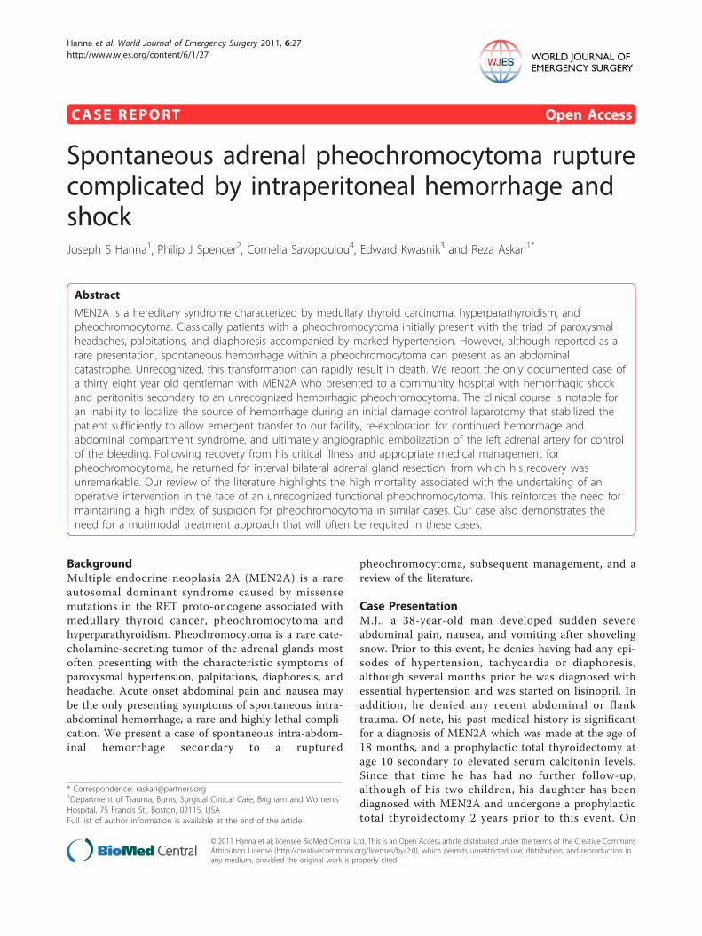
arrival, paramedics found him near syncopal and dia-phoretic with a heart rate of 180 bpm and systolic bloodpressure of 64 mmHg. Fluid resuscitation was initiatedand the patient was taken to an outside hospital. Initialevaluation at the local level II trauma center was notablefor a heart rate of 150 bpm, systolic blood pressure of70 mmHg, diffuse peritoneal signs, a hematocrit of 34%,INR of 1.0 and PTT of 30.4. Following resuscitationwith additional crystalloid and 2 units of packed redblood cells (pRBC), his hematocrit was 34%, INR 2.4and PTT 66.2. A non-contrast abdominal computedtomogram revealed bilateral adrenal masses and a largeamount of intra and retroperitoneal hemorrhage (Figure1). In light of his hemodynamic instability and CT find-ings, emergent laparotomy was undertaken. Upon enter-ing the abdomen, a large amount of blood wasencountered and immediate control of the abdominalaorta was obtained to manage the ongoing hemorrhageand facilitate resuscitation which ultimately required 12units of pRBCs, 4 units of fresh frozen plasma (FFP)and 6 units of platelets. A bleeding source was identifiedin the left upper quadrant (LUQ) in the retroperitonealfat which was oversewn. The abdomen was packed withlaparotomy pads and closed; the blood loss was esti-mated to be 8000 cc.The patient was subsequently transferred to our facil-
ity for further care. On arrival he was intubated andsedated with a blood pressure of 90/35 mmHg, heartrate 129 bpm, Hct 36.3%, INR 2.7 and fibrinogen 117mg/dL. On initial examination his abdomen was tenseand distended, and his extremities were cold. Ongoinghemorrhage was suspected given the coagulopathy and
persistent hypotension, therefore aggressive resuscitationwith blood products was resumed. An initial bladderpressure of 33 mmHg along with poor urine output,hypotension and a tense abdominal examination raisedsuspicion for an evolving abdominal compartment syn-drome; therefore a second emergent exploration wasundertaken. On entry into the abdominal cavity, theright colon was found to be frankly ischemic and persis-tent hemorrhage from the LUQ was again noted. As thesource of bleeding could not be readily identified, anemergent splenectomy was performed, and laparotomypads were again packed into the LUQ. Once adequatecontrol of the bleeding was obtained with packing,attention was turned to performing a right hemicolect-omy. A Bogota bag with a wound V.A.C (KCI, TX) wasthen fashioned for temporary abdominal closure. Fol-lowing closure of the abdomen, the patient suffered car-diac arrest with pulseless electrical activity. Advancedcardiac life support measures were initiated and a per-fusing rhythm was obtained shortly thereafter. Given thehistory of MEN2A and bilateral adrenal masses, thediagnosis of occult pheochromocytoma was entertained.The blood pressure swings were controlled with phento-lamine and a sodium nitroprusside infusion with goodeffect. The patient was returned to the surgical intensivecare unit for further management.In the intensive care unit, the patient continued to
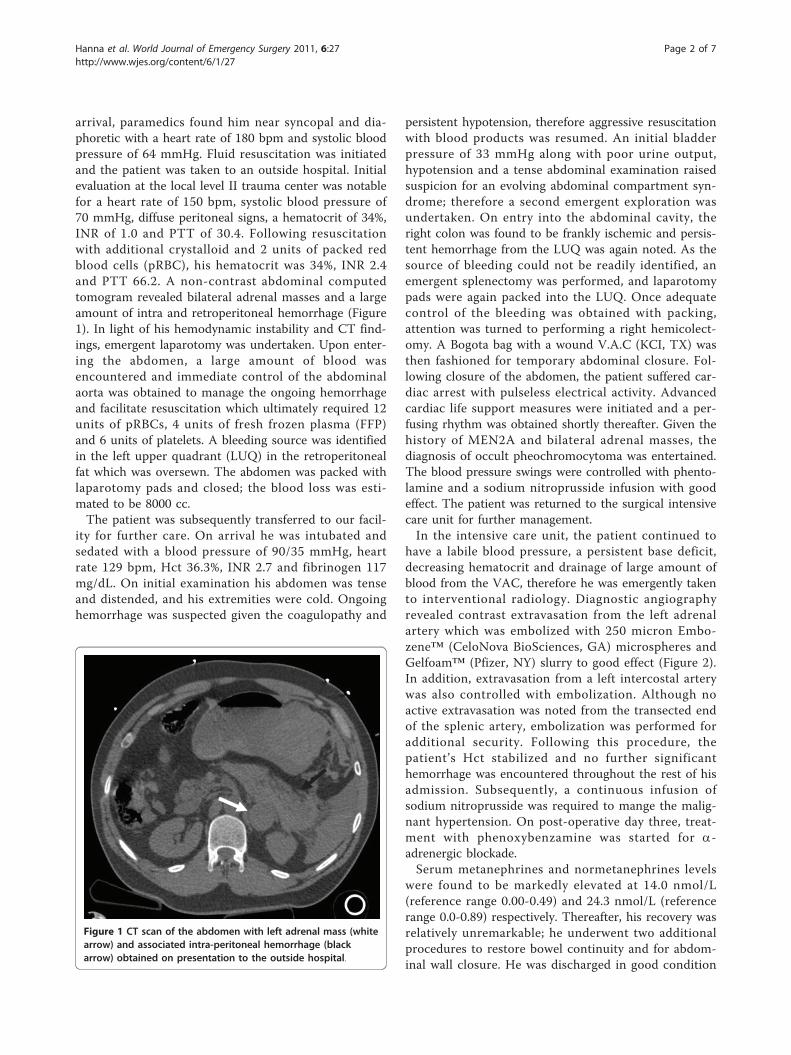
have a labile blood pressure, a persistent base deficit,decreasing hematocrit and drainage of large amount ofblood from the VAC, therefore he was emergently takento interventional radiology. Diagnostic angiographyrevealed contrast extravasation from the left adrenalartery which was embolized with 250 micron Embo-zene™ (CeloNova BioSciences, GA) microspheres andGelfoam™ (Pfizer, NY) slurry to good effect (Figure 2).In addition, extravasation from a left intercostal arterywas also controlled with embolization. Although noactive extravasation was noted from the transected endof the splenic artery, embolization was performed foradditional security. Following this procedure, thepatient’s Hct stabilized and no further significanthemorrhage was encountered throughout the rest of hisadmission. Subsequently, a continuous infusion ofsodium nitroprusside was required to mange the malig-nant hypertension. On post-operative day three, treat-ment with phenoxybenzamine was started for a-adrenergic blockade.Serum metanephrines and normetanephrines levels
were found to be markedly elevated at 14.0 nmol/L(reference range 0.00-0.49) and 24.3 nmol/L (referencerange 0.0-0.89) respectively. Thereafter, his recovery wasrelatively unremarkable; he underwent two additionalprocedures to restore bowel continuity and for abdom-inal wall closure. He was discharged in good condition
Figure 1 CT scan of the abdomen with left adrenal mass (whitearrow) and associated intra-peritoneal hemorrhage (blackarrow) obtained on presentation to the outside hospital.
Hanna et al. World Journal of Emergency Surgery 2011, 6:27http://www.wjes.org/content/6/1/27
Page 2 of 7
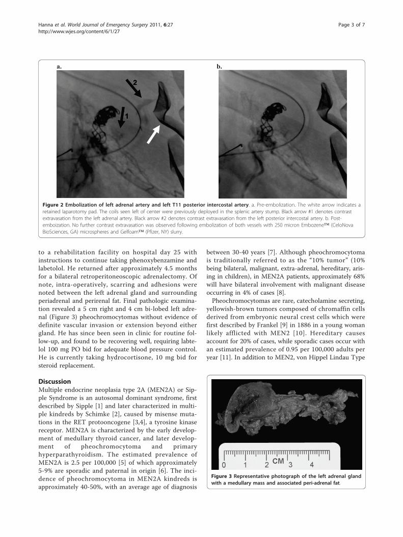
to a rehabilitation facility on hospital day 25 withinstructions to continue taking phenoxybenzamine andlabetolol. He returned after approximately 4.5 monthsfor a bilateral retroperitoneoscopic adrenalectomy. Ofnote, intra-operatively, scarring and adhesions werenoted between the left adrenal gland and surroundingperiadrenal and perirenal fat. Final pathologic examina-tion revealed a 5 cm right and 4 cm bi-lobed left adre-nal (Figure 3) pheochromocytomas without evidence ofdefinite vascular invasion or extension beyond eithergland. He has since been seen in clinic for routine fol-low-up, and found to be recovering well, requiring labte-lol 100 mg PO bid for adequate blood pressure control.He is currently taking hydrocortisone, 10 mg bid forsteroid replacement.
DiscussionMultiple endocrine neoplasia type 2A (MEN2A) or Sip-ple Syndrome is an autosomal dominant syndrome, firstdescribed by Sipple [1] and later characterized in multi-ple kindreds by Schimke [2], caused by misense muta-tions in the RET protooncogene [3,4], a tyrosine kinasereceptor. MEN2A is characterized by the early develop-ment of medullary thyroid cancer, and later develop-ment of pheochromocytoma and primaryhyperparathyroidism. The estimated prevalence ofMEN2A is 2.5 per 100,000 [5] of which approximately5-9% are sporadic and paternal in origin [6]. The inci-dence of pheochromocytoma in MEN2A kindreds isapproximately 40-50%, with an average age of diagnosis
between 30-40 years [7]. Although pheochromocytomais traditionally referred to as the “10% tumor” (10%being bilateral, malignant, extra-adrenal, hereditary, aris-ing in children), in MEN2A patients, approximately 68%will have bilateral involvement with malignant diseaseoccurring in 4% of cases [8].Pheochromocytomas are rare, catecholamine secreting,
yellowish-brown tumors composed of chromaffin cellsderived from embryonic neural crest cells which werefirst described by Frankel [9] in 1886 in a young womanlikely afflicted with MEN2 [10]. Hereditary causesaccount for 20% of cases, while sporadic cases occur withan estimated prevalence of 0.95 per 100,000 adults peryear [11]. In addition to MEN2, von Hippel Lindau Type
a. b.
Figure 2 Embolization of left adrenal artery and left T11 posterior intercostal artery. a. Pre-embolization. The white arrow indicates aretained laparotomy pad. The coils seen left of center were previously deployed in the splenic artery stump. Black arrow #1 denotes contrastextravasation from the left adrenal artery. Black arrow #2 denotes contrast extravasation from the left posterior intercostal artery. b. Post-emboization. No further contrast extravasation was observed following embolization of both vessels with 250 micron Embozene™ (CeloNovaBioSciences, GA) microspheres and Gelfoam™ (Pfizer, NY) slurry.
Figure 3 Representative photograph of the left adrenal glandwith a medullary mass and associated peri-adrenal fat.
Hanna et al. World Journal of Emergency Surgery 2011, 6:27http://www.wjes.org/content/6/1/27
Page 3 of 7
2, von Recklinghausen’s neurofibromatosis type 1, andfamilial paragangliomas are associated with the develop-ment of pheochromocytomas. Eighty percent of all pheo-chromocytomas arise within the adrenal medulla, whileextra-adrenal lesions are most commonly found in thesympathetic ganglia as well as the organs of Zuckerkandl.Of note, it is estimated that 5% of adrenal incidentalomasare likely pheochromocytomas [12]. In addition to secret-ing the catecholes dopamine, epinephrine and norepi-nephrine, numerous other hormones have been isolatedfrom pheochromocytomas including adrenocorticotropin,vasoactive intestinal peptide, neuropeptide Y, IL-6, calci-tonin, and chromogranin A.Classically patients initially present with the triad of
paroxysmal headaches, palpitations, and diaphoresisaccompanied by marked hypertension. Of interest, it isestimated that pheochromocytomas are present in 0.1-0.6% of patients with hypertension [13]. In addition tothese symptoms, pallor, nausea, flushing, anxiety or asense of doom, palpitations and abdominal pain can bepart of the constellation of presenting symptoms. Moreominously, patients may present in fulminant cardio-genic shock [14], multiorgan failure, or with acutehemorrhage. Several biochemical assays are available tofacilitate diagnosis, however, plasma free metanephrineshad the highest sensitivity and urinary VMA had thehighest specificity in a recent multicenter cohort trial[15] in the detection of pheochromocytomas. Once bio-chemical evidence of pheochromocytoma is obtained,imaging for localization should be undertaken to guidesurgical resection. Computed tomography and magneticresonance imaging provides high sensitivity for lesiondetection, though poor specificity. Alternative imagingmodalities such as I123 or I131 MIBG scintigraphy orPET may be utilized when CT or MRI fail to reveal thelesion or if malignancy is suspected.Although both Roux (Switzerland) and Mayo (US) are
credited with concomitantly performing the first suc-cessful resections of pheochromocytomas in 1926,neither described any peri-operative hemodynamicinstability, and both patients survived [16]. However, inthe ensuing years, further attempts at resection weremet with a mortality rate of approximately 25%. It wasnot until 1956 when Priestley recorded a case series of51 patients who underwent resection without anydeaths. His success is attributable to the use of phento-lamine and norepinephrine to manage the hemodynamicinstability that is typically encountered [16]. Lessonslearned during the early years of surgical managementhave led to the recognition of the importance of initialperi-operative a-blockade and volume expansion fol-lowed by b-blockade for management of tachycardiaand hypertension in anticipation of elective surgicalresection.
Implementation of these management principles in theemergent setting can often be challenging as patientpresentation can be widely variable, ranging from minorretroperitoneal hemorrhage with hypertension orabdominal pain to shock and impending cardiovascularcollapse. In the setting of a contained retroperitonealhemorrhage, every effort should be made to avoid emer-gent or urgent surgical intervention. Not surprisingly,review of the literature reveals a mortality of ~25% asso-ciated with emergent surgical intervention for containedhemorrhage; in contrast, adequate medical preparationas described above results in a mortality rate similar tothat observed for elective adrenalectomy in the absenceof hemorrhage. Medical optimization should includeadequate blood resuscitation, correction of any coagulo-pathy to limit continued hemorrhage, hemodynamicsupport as needed, and ultimately a-blockade followedby volume expansion and b-blockade in an in-patientsetting. This simplistic algorithm must be tempered bythe recognition that providing supportive care in thesetting of cardiovascular collapse mediated by adrenalcompression from an evolving retroperitoneal hematomaand the resulting catecholamine excess may tax even themost advanced intensive care unit. Emergent surgicalintervention may be considered in cases refractory tomaximal medical management as recently described byMay and colleagues [17] with recognition of the atten-dant high morbidity and mortality.Spontaneous hemorrhage within a pheochromocytoma
resulting in capsular rupture and retroperitoneal orintra-peritoneal hemorrhage has long been recognizedas a rare, but catastrophic and highly lethal event. Inaddition, trauma [17] and medications [18,19] have alsobeen implicated in hemorrhagic complications. In areview of the literature, we have identified 49 documen-ted cases between 1944 and 2010 [14,17-52] of which,including this report, 12 involved spontaneous intra-peritoneal hemorrhage [19,53-61] (Table 1). Review ofthese twelve cases revealed that emergent laparotomyresulted in a mortality of 29%, consistent with the mor-tality observed prior to the routine use of pre-operativea-adrenergic blockade [16]. More telling is the 100%mortality associated with misdiagnosis and failure tocontrol the hemorrhage in a timely fashion. Though thecase number is small, these data suggest that althoughundertaking an emergent exploration for this indicationis fraught with danger, it offers the patient the bestopportunity for survival. In the absence of adequate a-adrenergic blockade in these extreme cases, the intra-operative and post-operative care must be tailored tothe clinical picture as it evolves. Thus, the anaesthesiaand surgical teams must be prepared to manage suddencardiovascular collapse, fulminant heart failure, massivepulmonary edema, and ongoing hemorrhage. Immediate
Hanna et al. World Journal of Emergency Surgery 2011, 6:27http://www.wjes.org/content/6/1/27
Page 4 of 7
availability of a perfusionist and cell-saver, an intra-aor-tic counter-pulsation pump, a percutaneous right ventri-cular assist device, a ventilator capable of maintaininghigh positive end-expiratory pressures with advancedventilation modes (ex. APRV, BiLevel), an establishedmassive transfusion protocol, and interventional radiolo-gists are vital in the successful management of thesechallenging cases. If the tumor is completely removed,post-operative a-blockade is not typically necessary;however, if transcatheter arterial embolization (TAE) isused as a temporizing measure, continued a-blockadebecomes essential as discussed below.In the present case, we were faced with a unique set of
circumstances which dictated an unconventional courseof management. Although the patient’s medical historynotable for total thyroidectomy as a child and the pre-sence of the bilateral adrenal masses raised suspicion forMEN2A and possible pheochromocytoma, given hisinitial presentation in extremis with hemoperitoneum thedecision to undertake an emergent exploratory laparot-omy was warranted. Although only partial control of thehemorrhage was obtained at that time, it should bestressed that inaction may have resulted in this patient’sdemise given the historically observed 100% mortality inthe face of conservatively managed intra-peritonealhemorrhage from pheochromocytoma. Upon arrival toour facility, we were faced with an evolving abdominalcompartment syndrome in addition to acute hemorrhageof unclear etiology. In the course of the second laparot-omy, hemodynamic instability, the need to address thesequelae of abdominal hypertension, and worsening coa-gulopathy precluded further exploration of the LUQ forthe continued source of hemorrhage. Moreover, giventhe presence of bilateral adrenal masses in the setting ofa history of MEN2A, further exploration of the adrenalswithout proper a-blockade presented addition significant
risk of morbidity and mortality. Therefore the decisionwas made to proceed with angiographic embolization inthe setting of continued bleeding.TAE as a therapeutic option for pheochromocytoma
was first described in 1978 by Bunuan [62] and colle-gues. Their effort to use gel foam TAE was met withsignificant hemodynamic instability resulting in emer-gent laparotomy for excision of the necrotic tumor.Since this initial experience, TAE has been reported inthe literature as a palliative option in the managementof malignant pheochromocytoma when surgical extirpa-tion is not feasible [63,64]. More germane to the presentcase, the use of TAE for management of acute sponta-neous intraperitoneal hemorrhage from a pheochromo-cytoma has not been previously reported, although itsuse in retroperitoneal hemorrhage as been described bytwo separate groups [17,50]. In the present case anyfurther effort to explore the LUQ for the source ofhemorrhage may very well have resulted in the patient’sdemise. We therefore elected to salvage the situation byemploying damage control techniques to quickly get thepatient out of the operating room to facilitate TAE ofthe suspected hemorrhaging pheochromocytoma. Inter-estingly, in addition to embolization of a left adrenalartery in this case, a bleeding left intercostal artery wasalso identified. In an effort to better define the anatomyof the suprarenal arteries, Toni and colleagues reviewedaortography performed on patients without knownsuprarenal disease [65]. They identified the origin of theleft suprarenal artery as a left intercostal branch in 3%of the patients in their study. As described in all ofthese reports, post-TAE hypertension can present a for-midable challenge. In this case, malignant hypertensionwas successfully managed with infusion of sodium nitro-prusside in the acute setting, followed by administrationof phenoxybenzamine.
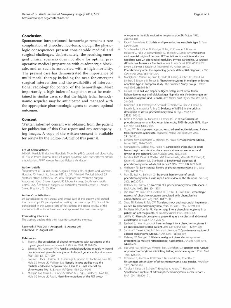
Table 1 Features of previously reported pheochromocytomas complicated by intra-peritoneal hemorrhage
Pt Symptoms DxKnown
Intervention Outcome
Hanna 2010 38M Shock, abdominal pain No Emergent exploration alive
Li 2009 50M HTN, abdominal pain, palpable mass No Delayed exploration alive
Chan 2003 35F abdominal pain No Emergent exploration dead
Lee 1987 31M abdominal pain, orthostasis No Emergent exploration alive
Greatorex 1984 46M HTN, CP, palpitation, HA, emesis, tachychardia No Emergent exploration alive
Wenisch 1982 62F abdominal pain, nausea, palpable mass No Emergent exploration alive
Bednarski 1981 69M abdominal pain, dyspnea No None dead
van Royen 1978 53M HTN, abdominal pain, palpable mass, bronchospasm No None dead
Van Way 1976 76F HTN, abdominal pain Yes Emergent exploration alive
Gielchinsky 1972 36M abdominal pain, peritonitis Yes Delayed exploration alive
Cahill 1944 53F abdominal pain No Emergent exploration dead
61 shock, sudden death No None dead
A summary of the 11 previously described cases of ruptured pheochromocytoma with free intraperitoneal hemorrhage including the present case. The relevantsymptoms on presentation, timing of operative intervention and outcome are summarized.
Hanna et al. World Journal of Emergency Surgery 2011, 6:27http://www.wjes.org/content/6/1/27
Page 5 of 7
ConclusionSpontaneous intraperitoneal hemorrhage remains a rarecomplication of pheochromocytoma, though the physio-logic consequences present considerable medical andsurgical challenges. Unfortunately, the resulting emer-gent clinical scenario does not allow for optimal pre-operative medical preparation with a-adrenergic block-ade, and as such is associated with a high mortality.The present case has demonstrated the importance ofmulti-modal therapy including the need for emergentsurgical intervention and the availability of interven-tional radiology for control of the hemorrhage. Mostimportantly, a high index of suspicion must be main-tained in similar cases so that the highly lethal hemody-namic sequelae may be anticipated and managed withthe appropriate pharmacologic agents to ensure optimaloutcomes.
ConsentWritten informed consent was obtained from the patientfor publication of this Case report and any accompany-ing images. A copy of the written consent is availablefor review by the Editor-in-Chief of this journal.
List of AbbreviationsMEN2A: Multiple Endocrine Neoplasia Type 2A; pRBC: packed red blood cells;FFP: fresh frozen plasma; LUQ: left upper quadrant; TAE: transcatheter arterialembolization; APRV: Airway Pressure Release Ventilation
Author details1Department of Trauma, Burns, Surgical Critical Care, Brigham and Women’sHospital, 75 Francis St., Boston, 02115, USA. 2Harvard Medical School, 25Shattuck Street, Boston, 02115, USA. 3Brigham and Women’s SurgicalAssociates, South Shore Hospital, 90 Libbey Parkway, South Weymouth,02190, USA. 4Division of Surgery, St. Elizabeth’s Medical Center, 11 NevinsStreet, Brighton, 02135, USA.
Authors’ contributionsJH participated in the surgical and critical care of this patient and draftedthe manuscript. PS participated in drafting the manuscript. CS, EK and RAparticipated in the surgical care of this patient and critical review of themanuscript. All authors have read and approved the final manuscript.
Competing interestsThe authors declare that they have no competing interests.
Received: 5 May 2011 Accepted: 15 August 2011Published: 15 August 2011
References1. Sipple J: The association of pheochromocytoma with carcinoma of the
thyroid gland. American Journal of Medicine 1961, 31:163-166.2. Schimke RN, Hartmann WH: Familial amyloid-producing medullary thyroid
carcinoma and pheochromocytoma. A distinct genetic entity. Ann InternMed 1965, 63:1027-1039.
3. Gardner E, Papi L, Easton DF, Cummings T, Jackson CE, Kaplan M, Love DR,Mole SE, Moore JK, Mulligan LM: Genetic linkage studies map themultiple endocrine neoplasia type 2 loci to a small interval onchromosome 10q11. 2. Hum Mol Genet 1993, 2:241-246.
4. Mulligan LM, Kwok JB, Healey CS, Elsdon MJ, Eng C, Gardner E, Love DR,Mole SE, Moore JK, Papi L: Germ-line mutations of the RET proto-
oncogene in multiple endocrine neoplasia type 2A. Nature 1993,363:458-460.
5. Raue F, Frank-Raue K: Update multiple endocrine neoplasia type 2. FamCancer 2010.
6. Schuffenecker I, Ginet N, Goldgar D, Eng C, Chambe B, Boneu A,Houdent C, Pallo D, Schlumberger M, Thivolet C, Lenoir GM: Prevalenceand parental origin of de novo RET mutations in multiple endocrineneoplasia type 2A and familial medullary thyroid carcinoma. Le Grouped’Etude des Tumeurs a Calcitonine. Am J Hum Genet 1997, 60:233-237.
7. Bryant J, Farmer J, Kessler LJ, Townsend RR, Nathanson KL:Pheochromocytoma: the expanding genetic differential diagnosis. J NatlCancer Inst 2003, 95:1196-1204.
8. Modigliani E, Vasen HM, Raue K, Dralle H, Frilling A, Gheri RG, Brandi ML,Limbert E, Niederle B, Forgas L: Pheochromocytoma in multiple endocrineneoplasia type 2: European study. The Euromen Study Group. J InternMed 1995, 238:363-367.
9. Frankel F: Ein Fall von doppelseitigem, völlig latent verlaufenenNebennierentumor und gleichzeitiger Nephritis mit Veränderungen amCirculationsapparat und Retinitis. Arch Pathol Anat Physiol Klin Med 1886,244-263.
10. Neumann HPH, Vortmeyer A, Schmidt D, Werner M, Erlic Z, Cascon A,Bausch B, Januszewicz A, Eng C: Evidence of MEN-2 in the originaldescription of classic pheochromocytoma. N Engl J Med 2007,357:1311-1315.
11. Beard CM, Sheps SG, Kurland LT, Carney JA, Lie JT: Occurrence ofpheochromocytoma in Rochester, Minnesota, 1950 through 1979. MayoClin Proc 1983, 58:802-804.
12. Young WF: Management approaches to adrenal incidentalomas. A viewfrom Rochester, Minnesota. Endocrinol Metab Clin North Am 2000,29:159-185, x.
13. Lenders JWM, Eisenhofer G, Mannelli M, Pacak K: Phaeochromocytoma.Lancet 2005, 366:665-675.
14. Mohamed HA, Aldakar MO, Habib N: Cardiogenic shock due to acutehemorrhagic necrosis of a pheochromocytoma: a case report andreview of the literature. Can J Cardiol 2003, 19:573-576.
15. Lenders JWM, Pacak K, Walther MM, Linehan WM, Mannelli M, Friberg P,Keiser HR, Goldstein DS, Eisenhofer G: Biochemical diagnosis ofpheochromocytoma: which test is best? JAMA 2002, 287:1427-1434.
16. Welbourn RB: Early surgical history of phaeochromocytoma. Br J Surg1987, 74:594-596.
17. May EE, Beal AL, Beilman GJ: Traumatic hemorrhage of occultpheochromocytoma: a case report and review of the literature. Am Surg2000, 66:720-724.
18. Delaney JP, Paritzky AZ: Necrosis of a pheochromocytoma with shock. NEngl J Med 1969, 280:1394-1395.
19. Van Way CW, Faraci RP, Cleveland HC, Foster JF, Scott HW: Hemorrhagicnecrosis of pheochromocytoma associated with phentolamineadministration. Ann Surg 1976, 184:26-30.
20. Shaw TR, Rafferty P, Tait GW: Transient shock and myocardial impairmentcaused by phaeochromocytoma crisis. Br Heart J 1987, 57:194-198.
21. McAlister WH, Koehler PR: Hemorrhage into a pheochromocytoma in apatient on anticoagulants. J Can Assoc Radiol 1967, 18:404-406.
22. Jelliffe RS: Phaeochromocytoma presenting as a cardiac and abdominalcatastrophe. Br Med J 1952, 2:76-77.
23. Ejerblad S, Hemmingsson A: Haemorrhage into a pheochromocytoma inan anticoagulant-treated patient. Acta Chir Scand 1981, 147:497-500.
24. Sumino Y, Tasaki Y, Satoh F, Mimata H, Nomura Y: Spontaneous rupture ofadrenal pheochromocytoma. J Urol 2002, 168:188-189.
25. Delaney PV, Mungall IP: Bilateral malignant phaeochromocytomaspresenting as massive retroperitoneal haemorrage. J Ir Med Assoc 1971,64:428-429.
26. Sue-Ling HM, Foster ME, Wheeler MH, McMahon MJ: Spontaneous ruptureof phaeochromocytoma mimicking leaking aortic aneurysm. J R Soc Med1989, 82:53-54.
27. Grossman E, Knecht A, Holtzman E, Nussinovich N, Rosenthal T:Uncommon presentation of pheochromocytoma: case studies. Angiology1985, 36:759-765.
28. Tanaka K, Noguchi S, Shuin T, Kinoshita Y, Kubota Y, Hosaka M:Spontaneous rupture of adrenal pheochromocytoma: a case report. JUrol 1994, 151:120-121.
Hanna et al. World Journal of Emergency Surgery 2011, 6:27http://www.wjes.org/content/6/1/27
Page 6 of 7
29. Suga K, Motoyama K, Hara A, Kume N, Ariga M, Matsunaga N: Tc-99 mMIBG imaging in a huge clinically silent pheochromocytoma with cysticdegeneration and massive hemorrhage. Clin Nucl Med 2000, 25:796-800.
30. Lehman DJ, Rosof J: Massive hemorrhage into an adrenalpheochromocytoma. N Engl J Med 1956, 254:474-476.
31. Forty J, Dale RF: Ruptured phaeochromocytoma: a case report. J R CollSurg Edinb 1989, 34:109-110.
32. Belden CJ, Powers C, Ros PR: MR demonstration of a cysticpheochromocytoma. J Magn Reson Imaging 1995, 5:778-780.
33. Hatada T, Nakai T, Aoki I, Gondo N, Katou N, Yoshinaga K, Nakasaku O,Utsunomiya J: Acute abdominal symptoms caused by hemorrhagicnecrosis of a pheochromocytoma: report of a case. Surg Today 1994,24:363-367.
34. Nicholls K: Massive adrenal haemorrhage complicating adrenalneoplasm. Med J Aust 1979, 2:560-562.
35. Jones DJ, Durning P: Phaeochromocytoma presenting as an acuteabdomen: report of two cases. Br Med J (Clin Res Ed) 1985, 291:1267-1268.
36. Gilliland IC, Daniel O: Phaeochromocytoma presenting as an abdominalemergency. Br Med J 1951, 2:275-277.
37. Saltz NJ, Luttwak EM, Schwartz A, Goldberg GM: Danger of aortography inthe localization of pheochromocytoma. Ann Surg 1956, 144:118-123.
38. Brody IA: Shock after administration of prochlorperazine in patient withpheochromocytoma; report of a case with spontaneous tumordestruction. J Am Med Assoc 1959, 169:1749-1752.
39. Jacobs LM, Williams LF, Hinrichs HR: Hemorrhage into apheochromocytoma. JAMA 1978, 239:1156.
40. Nyman D, Wahlberg P: Necrotic phaeochromocytoma with gastrichaemorrhage, shock, and uncommonly high catecholamine excretion.Acta Med Scand 1970, 187:381-383.
41. Terachi T, Terai A, Yoshida S, Yokota K, Fukunaga M: Spontaneous ruptureof adrenal pheochromocytoma: a case report. Urol Int 1989, 44:235-237.
42. O’Hickey S, Hilton AM, Whittaker JS: Phaeochromocytoma associated withadult respiratory distress syndrome. Thorax 1987, 42:157-158.
43. Scott I, Parkes R, Cameron DP: Phaeochromocytoma and cardiomyopathy.Med J Aust 1988, 148:94-96.
44. Andersen PT, Baadsgaard SE, Larsen BP: Repetitive bleeding from apheochromocytoma presenting as an abdominal emergency. Casereport. Acta Chir Scand 1986, 152:69-70.
45. Huston JR, Stewart WR: Hemorrhagic Pheochromocytoma with Shock andAbdominal Pain. Am J Med 1965, 39:502-504.
46. Primhak RA, Spicer RD, Variend S: Sudden death after minor abdominaltrauma: an unusual presentation of phaeochromocytoma. Br Med J (ClinRes Ed) 1986, 292:95-96.
47. Scully R, Mark E, McNeely B: Case records of the Massachusetts GeneralHospital. Weekly clinicopathological exercises. Case 15-1988. A 26-year-old woman with cardiomyopathy, multiple strokes, and an adrenalmass. N Engl J Med 1988, 318:970-981.
48. Ong KL, Tan TH: Ruptured phaeochromocytoma–a rare differentialdiagnosis of acute abdomen. Singapore Med J 1996, 37:113-114.
49. McFarland GE, Bliss WR: Hemorrhage From Spontaneous Rupture of aPheochromocytoma of the Right Adrenal Gland: A Case Report. Ann Surg1951, 133:404-407.
50. Pua U, Wong DE: Transarterial embolisation of spontaneous adrenalpheochromocytoma rupture using polyvinyl alcohol particles. SingaporeMed J 2008, 49:e126-130.
51. Lago Montero A, Silva Abuin J, Gómez Zancajo VR, Montero Gómez J:Massive retroperitoneal hemorrhage as the 1st manifestation of apheochromocytoma. Arch Esp Urol 1986, 39:269-273.
52. Chlebus M, Lapiński M, Torbicki A, Chlebus H, Szostek M, Wocial B,Staszkiewicz W, Januszewicz W: [Pheochromocytoma with hemorrhagicnecrosis and rupture with symptoms of acute abdomen and shock]. PolArch Med Wewn 1996, 96:58-61.
53. Li C, Xu Y-min: Spontaneous intraperitoneal bleeding caused by adrenalpheochromocytoma. Chin Med J 2009, 122:2193-2195.
54. Lee PH, Blute R, Malhotra R: A clinically “silent” pheochromocytoma withspontaneous hemorrhage. J Urol 1987, 138:1429-1432.
55. Greatorex RA, Raftery AT: Intraperitoneal rupture of aphaeochromocytoma. J R Soc Med 1984, 77:513-514.
56. Gielchinsky I, Petty C, Dierdorff S: Treatment of hemorrhagic necrosiswithin a pheochromocytoma with symptoms of acute abdomen. AmSurg 1972, 38:380-384.
57. Cahill G: The Hormonal Tumors of the Adrenal Gland. PennsylvaniaMedical Journal 1944, 47:655-667.
58. Chan MKY, Tse HW, Mok FPT: Ruptured phaeochromocytoma–a lesson inacute abdomen. Hong Kong Med J 2003, 9:221-223.
59. Wenisch HJ, Klempa I: Rupture of a pheochromocytoma into the freeabdominal cavity. Case report. Chirurg 1982, 53:154-156.
60. Bednarski Z: Pheochromocytoma as a cause of fatal abdominalhemorrhage. Pol Tyg Lek 1981, 36:531-532.
61. van Royen EA, Alberts C, de Vos R, Becker AE: Pheochromocytoma as acause of “acute abdomen”. Ned Tijdschr Geneeskd 1978, 122:573-577.
62. Bunuan HD, Alltree M, Merendino KA: Gel foam embolization of afunctioning pheochromocytoma. Am J Surg 1978, 136:395-398.
63. Takahashi K, Ashizawa N, Minami T, Suzuki S, Sakamoto I, Hayashi K,Tomiyasu S, Sumikawa K, Kitamura K, Eto T, Yano K: Malignantpheochromocytoma with multiple hepatic metastases treated bychemotherapy and transcatheter arterial embolization. Intern Med 1999,38:349-354.
64. Baguet JP, Hammer L, Tremel F, Mangin L, Mallion JM: Metastaticphaeochromocytoma: risks of diagnostic needle puncture and treatmentby arterial embolisation. J Hum Hypertens 2001, 15:209-211.
65. Toni R, Mosca S, Favero L, Ricci S, Roversi R, Toni G, Vezzadini P: Clinicalanatomy of the suprarenal arteries: a quantitative approach byaortography. Surg Radiol Anat 1988, 10:297-302.
doi:10.1186/1749-7922-6-27Cite this article as: Hanna et al.: Spontaneous adrenalpheochromocytoma rupture complicated by intraperitonealhemorrhage and shock. World Journal of Emergency Surgery 2011 6:27.
Submit your next manuscript to BioMed Centraland take full advantage of:
• Convenient online submission
• Thorough peer review
• No space constraints or color figure charges
• Immediate publication on acceptance
• Inclusion in PubMed, CAS, Scopus and Google Scholar
• Research which is freely available for redistribution
Submit your manuscript at www.biomedcentral.com/submit
Hanna et al. World Journal of Emergency Surgery 2011, 6:27http://www.wjes.org/content/6/1/27
Page 7 of 7


![Myelolipoma: A Rare Adrenal Incidentaloma · insufficiency, and pheochromocytoma [23-28]. The extensive use of abdominal CT-scan and magnetic resonance imaging has led to a dramatic](https://static.fdocuments.us/doc/165x107/5f0e41897e708231d43e5c5c/myelolipoma-a-rare-adrenal-incidentaloma-insufficiency-and-pheochromocytoma-23-28.jpg)


![Review Article Adrenal Tumors with Unexpected Outcome: A ...adrenal mass, even in absence of clinical signs of Cush-ing s syndrome or pheochromocytoma [ , , , ]. Also, clinicians should](https://static.fdocuments.us/doc/165x107/60f8675ba943c364ae34ff3c/review-article-adrenal-tumors-with-unexpected-outcome-a-adrenal-mass-even.jpg)