SpoIIB Localizes to Active Sites of Septal Biogenesis and...
13
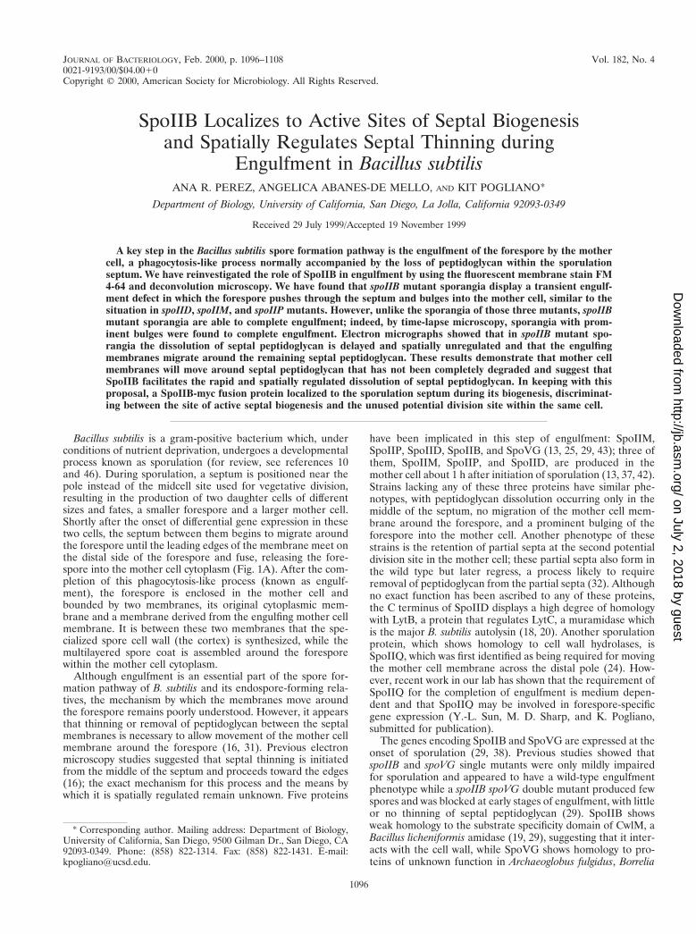
JOURNAL OF BACTERIOLOGY, 0021-9193/00/$04.0010 Feb. 2000, p. 1096–1108 Vol. 182, No. 4 Copyright © 2000, American Society for Microbiology. All Rights Reserved. SpoIIB Localizes to Active Sites of Septal Biogenesis and Spatially Regulates Septal Thinning during Engulfment in Bacillus subtilis ANA R. PEREZ, ANGELICA ABANES-DE MELLO, AND KIT POGLIANO* Department of Biology, University of California, San Diego, La Jolla, California 92093-0349 Received 29 July 1999/Accepted 19 November 1999 A key step in the Bacillus subtilis spore formation pathway is the engulfment of the forespore by the mother cell, a phagocytosis-like process normally accompanied by the loss of peptidoglycan within the sporulation septum. We have reinvestigated the role of SpoIIB in engulfment by using the fluorescent membrane stain FM 4-64 and deconvolution microscopy. We have found that spoIIB mutant sporangia display a transient engulf- ment defect in which the forespore pushes through the septum and bulges into the mother cell, similar to the situation in spoIID, spoIIM, and spoIIP mutants. However, unlike the sporangia of those three mutants, spoIIB mutant sporangia are able to complete engulfment; indeed, by time-lapse microscopy, sporangia with prom- inent bulges were found to complete engulfment. Electron micrographs showed that in spoIIB mutant spo- rangia the dissolution of septal peptidoglycan is delayed and spatially unregulated and that the engulfing membranes migrate around the remaining septal peptidoglycan. These results demonstrate that mother cell membranes will move around septal peptidoglycan that has not been completely degraded and suggest that SpoIIB facilitates the rapid and spatially regulated dissolution of septal peptidoglycan. In keeping with this proposal, a SpoIIB-myc fusion protein localized to the sporulation septum during its biogenesis, discriminat- ing between the site of active septal biogenesis and the unused potential division site within the same cell. Bacillus subtilis is a gram-positive bacterium which, under conditions of nutrient deprivation, undergoes a developmental process known as sporulation (for review, see references 10 and 46). During sporulation, a septum is positioned near the pole instead of the midcell site used for vegetative division, resulting in the production of two daughter cells of different sizes and fates, a smaller forespore and a larger mother cell. Shortly after the onset of differential gene expression in these two cells, the septum between them begins to migrate around the forespore until the leading edges of the membrane meet on the distal side of the forespore and fuse, releasing the fore- spore into the mother cell cytoplasm (Fig. 1A). After the com- pletion of this phagocytosis-like process (known as engulf- ment), the forespore is enclosed in the mother cell and bounded by two membranes, its original cytoplasmic mem- brane and a membrane derived from the engulfing mother cell membrane. It is between these two membranes that the spe- cialized spore cell wall (the cortex) is synthesized, while the multilayered spore coat is assembled around the forespore within the mother cell cytoplasm. Although engulfment is an essential part of the spore for- mation pathway of B. subtilis and its endospore-forming rela- tives, the mechanism by which the membranes move around the forespore remains poorly understood. However, it appears that thinning or removal of peptidoglycan between the septal membranes is necessary to allow movement of the mother cell membrane around the forespore (16, 31). Previous electron microscopy studies suggested that septal thinning is initiated from the middle of the septum and proceeds toward the edges (16); the exact mechanism for this process and the means by which it is spatially regulated remain unknown. Five proteins have been implicated in this step of engulfment: SpoIIM, SpoIIP, SpoIID, SpoIIB, and SpoVG (13, 25, 29, 43); three of them, SpoIIM, SpoIIP, and SpoIID, are produced in the mother cell about 1 h after initiation of sporulation (13, 37, 42). Strains lacking any of these three proteins have similar phe- notypes, with peptidoglycan dissolution occurring only in the middle of the septum, no migration of the mother cell mem- brane around the forespore, and a prominent bulging of the forespore into the mother cell. Another phenotype of these strains is the retention of partial septa at the second potential division site in the mother cell; these partial septa also form in the wild type but later regress, a process likely to require removal of peptidoglycan from the partial septa (32). Although no exact function has been ascribed to any of these proteins, the C terminus of SpoIID displays a high degree of homology with LytB, a protein that regulates LytC, a muramidase which is the major B. subtilis autolysin (18, 20). Another sporulation protein, which shows homology to cell wall hydrolases, is SpoIIQ, which was first identified as being required for moving the mother cell membrane across the distal pole (24). How- ever, recent work in our lab has shown that the requirement of SpoIIQ for the completion of engulfment is medium depen- dent and that SpoIIQ may be involved in forespore-specific gene expression (Y.-L. Sun, M. D. Sharp, and K. Pogliano, submitted for publication). The genes encoding SpoIIB and SpoVG are expressed at the onset of sporulation (29, 38). Previous studies showed that spoIIB and spoVG single mutants were only mildly impaired for sporulation and appeared to have a wild-type engulfment phenotype while a spoIIB spoVG double mutant produced few spores and was blocked at early stages of engulfment, with little or no thinning of septal peptidoglycan (29). SpoIIB shows weak homology to the substrate specificity domain of CwlM, a Bacillus licheniformis amidase (19, 29), suggesting that it inter- acts with the cell wall, while SpoVG shows homology to pro- teins of unknown function in Archaeoglobus fulgidus, Borrelia * Corresponding author. Mailing address: Department of Biology, University of California, San Diego, 9500 Gilman Dr., San Diego, CA 92093-0349. Phone: (858) 822-1314. Fax: (858) 822-1431. E-mail: [email protected]. 1096 on July 2, 2018 by guest http://jb.asm.org/ Downloaded from
Transcript of SpoIIB Localizes to Active Sites of Septal Biogenesis and...
JOURNAL OF BACTERIOLOGY,0021-9193/00/$04.0010
Feb. 2000, p. 1096–1108 Vol. 182, No. 4
Copyright © 2000, American Society for Microbiology. All Rights Reserved.
SpoIIB Localizes to Active Sites of Septal Biogenesisand Spatially Regulates Septal Thinning during
Engulfment in Bacillus subtilisANA R. PEREZ, ANGELICA ABANES-DE MELLO, AND KIT POGLIANO*
Department of Biology, University of California, San Diego, La Jolla, California 92093-0349
Received 29 July 1999/Accepted 19 November 1999
A key step in the Bacillus subtilis spore formation pathway is the engulfment of the forespore by the mothercell, a phagocytosis-like process normally accompanied by the loss of peptidoglycan within the sporulationseptum. We have reinvestigated the role of SpoIIB in engulfment by using the fluorescent membrane stain FM4-64 and deconvolution microscopy. We have found that spoIIB mutant sporangia display a transient engulf-ment defect in which the forespore pushes through the septum and bulges into the mother cell, similar to thesituation in spoIID, spoIIM, and spoIIP mutants. However, unlike the sporangia of those three mutants, spoIIBmutant sporangia are able to complete engulfment; indeed, by time-lapse microscopy, sporangia with prom-inent bulges were found to complete engulfment. Electron micrographs showed that in spoIIB mutant spo-rangia the dissolution of septal peptidoglycan is delayed and spatially unregulated and that the engulfingmembranes migrate around the remaining septal peptidoglycan. These results demonstrate that mother cellmembranes will move around septal peptidoglycan that has not been completely degraded and suggest thatSpoIIB facilitates the rapid and spatially regulated dissolution of septal peptidoglycan. In keeping with thisproposal, a SpoIIB-myc fusion protein localized to the sporulation septum during its biogenesis, discriminat-ing between the site of active septal biogenesis and the unused potential division site within the same cell.
Bacillus subtilis is a gram-positive bacterium which, underconditions of nutrient deprivation, undergoes a developmentalprocess known as sporulation (for review, see references 10and 46). During sporulation, a septum is positioned near thepole instead of the midcell site used for vegetative division,resulting in the production of two daughter cells of differentsizes and fates, a smaller forespore and a larger mother cell.Shortly after the onset of differential gene expression in thesetwo cells, the septum between them begins to migrate aroundthe forespore until the leading edges of the membrane meet onthe distal side of the forespore and fuse, releasing the fore-spore into the mother cell cytoplasm (Fig. 1A). After the com-pletion of this phagocytosis-like process (known as engulf-ment), the forespore is enclosed in the mother cell andbounded by two membranes, its original cytoplasmic mem-brane and a membrane derived from the engulfing mother cellmembrane. It is between these two membranes that the spe-cialized spore cell wall (the cortex) is synthesized, while themultilayered spore coat is assembled around the foresporewithin the mother cell cytoplasm.
Although engulfment is an essential part of the spore for-mation pathway of B. subtilis and its endospore-forming rela-tives, the mechanism by which the membranes move aroundthe forespore remains poorly understood. However, it appearsthat thinning or removal of peptidoglycan between the septalmembranes is necessary to allow movement of the mother cellmembrane around the forespore (16, 31). Previous electronmicroscopy studies suggested that septal thinning is initiatedfrom the middle of the septum and proceeds toward the edges(16); the exact mechanism for this process and the means bywhich it is spatially regulated remain unknown. Five proteins
have been implicated in this step of engulfment: SpoIIM,SpoIIP, SpoIID, SpoIIB, and SpoVG (13, 25, 29, 43); three ofthem, SpoIIM, SpoIIP, and SpoIID, are produced in themother cell about 1 h after initiation of sporulation (13, 37, 42).Strains lacking any of these three proteins have similar phe-notypes, with peptidoglycan dissolution occurring only in themiddle of the septum, no migration of the mother cell mem-brane around the forespore, and a prominent bulging of theforespore into the mother cell. Another phenotype of thesestrains is the retention of partial septa at the second potentialdivision site in the mother cell; these partial septa also form inthe wild type but later regress, a process likely to requireremoval of peptidoglycan from the partial septa (32). Althoughno exact function has been ascribed to any of these proteins,the C terminus of SpoIID displays a high degree of homologywith LytB, a protein that regulates LytC, a muramidase whichis the major B. subtilis autolysin (18, 20). Another sporulationprotein, which shows homology to cell wall hydrolases, isSpoIIQ, which was first identified as being required for movingthe mother cell membrane across the distal pole (24). How-ever, recent work in our lab has shown that the requirement ofSpoIIQ for the completion of engulfment is medium depen-dent and that SpoIIQ may be involved in forespore-specificgene expression (Y.-L. Sun, M. D. Sharp, and K. Pogliano,submitted for publication).
The genes encoding SpoIIB and SpoVG are expressed at theonset of sporulation (29, 38). Previous studies showed thatspoIIB and spoVG single mutants were only mildly impairedfor sporulation and appeared to have a wild-type engulfmentphenotype while a spoIIB spoVG double mutant produced fewspores and was blocked at early stages of engulfment, with littleor no thinning of septal peptidoglycan (29). SpoIIB showsweak homology to the substrate specificity domain of CwlM, aBacillus licheniformis amidase (19, 29), suggesting that it inter-acts with the cell wall, while SpoVG shows homology to pro-teins of unknown function in Archaeoglobus fulgidus, Borrelia
* Corresponding author. Mailing address: Department of Biology,University of California, San Diego, 9500 Gilman Dr., San Diego, CA92093-0349. Phone: (858) 822-1314. Fax: (858) 822-1431. E-mail:[email protected].
1096
on July 2, 2018 by guesthttp://jb.asm
.org/D
ownloaded from

burgdorferi, Bacillus megaterium, and Clostridium acetobutyli-cum (1, 30). It was recently reported that spoVG mutant spo-rangia initiate polar septation more rapidly than wild-type spo-rangia, suggesting that SpoVG serves directly or indirectly as arepressor of polar septation (30). The reason for the synergybetween spoIIB and spoVG mutations remains unclear.
Here we report the further characterization of the roles ofSpoIIB and SpoVG in engulfment. Using a sensitive assay forengulfment, we observed that a spoIIB null mutant displays atransient engulfment phenotype not described previously butsimilar to that of other engulfment mutants which fail to de-grade septal peptidoglycan. Ultimately, however, the spoIIBmutant is able to complete engulfment, suggesting that thedefect affects only the speed of engulfment. In contrast, en-gulfment proceeds normally in the spoVG mutant, suggestingthat this gene’s product is not directly involved in engulfment.We have also demonstrated that SpoIIB localizes to active sitesof septal biogenesis and that its localization is dependent on anas-yet-unidentified structure within the polar septum. Ourstudies suggest that SpoIIB is necessary for efficient dissolutionof septal peptidoglycan.
MATERIALS AND METHODS
Bacterial strains and strain construction. B. subtilis strains used in this workare listed in Table 1. Strains were constructed from the prototroph B. subtilisPY79 by conventional genetic techniques (8). KP548 was constructed by trans-forming KP10 with pTn917VstrR, which is able to replace the existing macrolide-lincosamide-streptogramin B resistance marker with a spectinomycin resistancemarker following a double-recombination event (44). Construction of strainKP545 is described in the next section. Escherichia coli KJ622 (TGI pcnB24-1)was constructed by P1 transduction from donor strain MJC97 (pcnB24-1:Tn10VstrR,tetS) to recipient TG1, selecting for the Tn10 (Strr) transposon linkedto pcnB (26), and screening for isolates with low plasmid copy numbers.
Construction of myc-tagged SpoIIB. Using primers 59-CTTAGAATTCGGGTTAAACATATCGGG39 and 59AATTTCTCGAGTTTTACGACGGCTAACAG39, a 612-bp fragment corresponding to the 39 end of the spoIIB coding regionwas isolated from strain PY79. EcoRI (New England Biolabs) and AvaI (Boehr-inger Mannheim) restriction sites (underlined) were introduced to facilitatecloning. Taq polymerase (Qiagen) was used under the following conditions toobtain the fragment: an initial denaturation at 94°C for 5 min; 30 cycles ofdenaturation at 94°C for 1 min, annealing at 47°C for 1 min, and extension at72°C for 1 min; and a final extension at 72°C for 10 min. The PCR product waspurified by using a QIAquick PCR Purification Kit (Qiagen) and digested withEcoRI and AvaI. The vector plasmid pKL94 (a gift from K. Lemon), encoding ac-myc tag, was linearized with EcoRI and AvaI. Fragments were purified by usingGeneclean II (Bio101); the fragment and vector were then ligated and trans-formed into strain KJ622 (TGI pcnB, described above). PKL94 was isolated byusing a Plasmid Midi Kit (Qiagen) and transformed into PY79. A single homol-ogous recombination event integrated the spoIIB-myc-containing plasmid at thechromosomal spoIIB gene, resulting in strain KP545. Competent KP545 was thentransformed with KP444, KP174, and KP548 chromosomal DNA to obtainKP547, KP549, and KP550, respectively.
Resuspension sporulation. Sporulation was induced by the method of Sterliniand Mandelstam (45). The membrane stain FM 4-64 (Molecular Probes) wasincluded in the resuspension medium as described by Pogliano et al. (32). TheDNA stain 49,6-diamidino-2-phenylindole (DAPI) was added to the cells imme-diately prior to viewing, as described previously (32).
Since KP547 contains FtsZ under the isopropyl-b-D-thiogalactopyranoside(IPTG)-inducible Pspac promoter, all samples were grown in the presence of 1mM IPTG to provide the FtsZ required for cell division. To deplete FtsZ duringresuspension sporulation, cells were grown in the presence of 1 mM IPTG untilreaching an optical density at 600 nm of 0.2, centrifuged, washed twice in CHmedium, and divided in half. One portion was resuspended in CH mediumcontaining 1 mM IPTG, while the other portion was resuspended in CH mediumlacking IPTG. The cultures were allowed to grow until the optical density at 600nm was between 0.5 and 0.6, at which time they were resuspended in sporulationsalts with or without (for the depletion) 1 mM IPTG.
Deconvolution and time-lapse microscopy. Deconvolution microscopy andimage processing was performed essentially as described by Pogliano et al. (32).However, for time-lapse experiments, cells were grown in the presence ofFM4-64 at 1 mg ml21 and images were collected every 20 min. Of the 29 spoIIBmutant sporangia that could be monitored throughout the time course, 18 com-pleted engulfment following bulge formation, 1 completed engulfment with nodetectable bulging, 4 failed to initiate engulfment, 2 initiated but did not com-plete engulfment, 3 lysed before the completion of engulfment, and one formeda disporic sporangium. Fields of cells from these time-lapse experiments can beseen on the Pogliano lab website (http://www.biology.ucsd.edu/labs/pogliano).
Immunofluorescence microscopy. Immunofluorescence microscopy was per-formed as described by Arigoni et al. (2) with the following modifications. A0.5-ml portion of the sporulating culture was fixed in a solution containing 20mM sodium phosphate (pH 7.2), 3% (wt/vol) paraformaldehyde, and 0.007%(wt/vol) glutaraldehyde (the last two were obtained from EM Sciences). Sampleswere fixed for 20 min at room temperature, washed with phosphate-bufferedsaline three times, and placed on ice for a maximum of 4 h. Sporangia werepermeabilized with lysozyme (0.8 mg ml21) for 4 min and washed with phos-phate-buffered saline. Prior to addition of the primary antibody, sporangia wereblocked with 2% bovine serum albumin at room temperature for 15 min. Sam-ples were incubated overnight at 4°C with the primary antibodies, an anti-c-myc
FIG. 1. Model for engulfment in the wild type and in spoIID, spoIIM, spoIIP,and spoIIB mutants. (A) Engulfment in the wild type. After polar septation,septal peptidoglycan is degraded, beginning in the middle of the septal disc(arrow) and proceeding toward the edges. The mother cell membranes move upand around the growing forespore (center sporangium), which ultimately be-comes fully enclosed within the mother cell (far-right sporangium). (B) Engulf-ment in spoIID, spoIIM, and spoIIP mutants. As in the wild type, there is initialdissolution of the septal peptidoglycan in the center of the septal disc (leftsporangium, arrow); however, degradation is not complete. When the foresporegrows, it breaks through this weakened region of the septum, resulting in thebulging of the forespore into the mother cell (right sporangium). (C) Engulfmentin spoIIB mutants. In spoIIB mutants, the septal peptidoglycan is incompletelydegraded throughout the septum (left-most sporangium, arrows). When theforespore grows, it breaks this weakened septal peptidoglycan, resulting in broadbulges of the forespore into the mother cell, with peptidoglycan being displacedinto the mother cell. Despite this residual peptidoglycan, engulfment is com-pleted.
TABLE 1. B. subtilis strains used in this studya
Strain Genotype Reference orsource
PY79 Prototrophic 51KP10 spoVG::VHU265 39KP52 spoIIBD::erm spoVG::Tn917VHU265 29KP69 spoIIE::Tn917VHU7 39KP174 D(spoIIAA-spoIIAC)Vspc Gift from
P. StragierKP343 spoIIBD::erm 29KP444 Pspac-ftsZ::phleo 4KP519 spoIIM::Tn917 39KP548 spoVG::Tn917::spc This studyKP545b spoIIB-mycVcm This studyKP547b Pspac-ftsZ::phleo spoIIB-mycVcm This studyKP549b D(spoIIAA-spoIIAC)Vspc spoIIB-
mycVcmThis study
KP550b spoVG::spec spoIIB-mycVcm This study
a All strains are congenic with PY79.b Wild-type spoIIB gene replaced with spoIIB-myc fusion.
VOL. 182, 2000 B. SUBTILIS SpoIIB 1097
on July 2, 2018 by guesthttp://jb.asm
.org/D
ownloaded from

mouse antibody (Boehringer Mannheim) at 1.1 mg ml21 and an affinity-purifiedanti-FtsZ rabbit antibody (a gift from P. Levin, purified by N. Osborne) at 1:500.Slides were washed with phosphate-buffered saline and then incubated with thesecondary antibodies, an affinity-purified fluorescein isothiocyanate (FITC)-la-beled donkey anti-mouse antibody (at 5 mg ml21) and an affinity-purified Cy5-labeled donkey anti-rabbit antibody (at 5 mg ml21) (both from Jackson Immu-nolabs), at room temperature for 3 h. DAPI (0.2 mg ml21; Molecular Probes)and FM 4-64 (10 mg ml21; Molecular Probes) were added with the equilibrationbuffer. We noted that FM 4-64 bleached rapidly in fixed cells exposed to UVirradiation. Therefore, before collecting images of fluorescein fluorescence, webleached the FM 4-64, using a DAPI filter set to ensure that there was no FM4-64 fluorescence remaining (which otherwise might be visualized together withfluorescein). To ensure that FM 4-64 did not affect protein localization, controlexperiments were performed without the stain; identical results were obtained. Awild-type strain, PY79, without the c-myc fusion was used as a negative control;it showed faint, punctate staining that did not localize to the septa (data notshown).
Electron microscopy. Sporulating cultures of PY79 and KP343 were preparedfor electron microscopy by four methods, three of which are slight modificationsof a protocol frequently used for preparation of B. subtilis (32). The fourthmethod was devised to allow better visualization of the engulfing membranes,which were often obscured by the darkly stained cell wall in the first threemethods. Method I exactly followed a previously described protocol (32) whichemployed a fixation with 4% (wt/vol) glutaraldehyde in 0.1 M sodium phosphate(pH 7.0) followed by a secondary fixation with 1% (wt/vol) osmium tetroxide in0.1 M sodium phosphate (pH 7.0) and then a wash in 0.5 M ammonium chloride(see Fig. 4A, C, I, and M to O). Method II introduced the 0.5 M ammoniumchloride wash before (rather than after) the osmium tetroxide fixation (see Fig.4B, E to H, L, and R), whereas method III lacked the ammonium chloride wash(see Fig. 4D). Following agarose enrobing, Spurr’s resin embedding, and ultra-thin sectioning, the samples were poststained with 1% (wt/vol) uranyl acetate in30% (vol/vol) ethanol and Reynold’s lead as described previously (32). MethodIV was a slight modification of a protocol used to enhance membrane staining inother organisms (14). Sporangia were fixed for 8 to 14 h at 4°C in 0.1 M sodiumcacodylate buffer (pH 7.4) containing 4% (wt/vol) glutaraldehyde and 2 mMcalcium chloride. Cells were postfixed for between 8 and 14 h at 4°C in 0.1 Msodium cacodylate buffer (pH 7.4) containing 1% (wt/vol) osmium tetroxide and3.0% (wt/vol) potassium ferricyanide. Samples were enrobed in agarose, diced,and stained with 1% (wt/vol) uranyl acetate in 30% (vol/vol) ethanol for 8 to 14 hat 25°C. They were embedded in Spurr’s resin, which was allowed to polymerizeat 65°C for at least 48 h. Ultrathin sections were applied to a grid and poststainedwith 0.25% (wt/vol) potassium permanganate and Reynold’s lead as describedelsewhere (14). This protocol was utilized because we were not interested indefining cytoplasmic contents (such as ribosomes) that stain darkly with uranylacetate and Reynold’s lead, obscuring the membranes. We found that this lastmethod readily allowed visualization of the engulfing membranes (see Fig. 4J, K,P, and Q).
Western blot analysis. Western blotting was performed as described by Pogli-ano et al. (34), with the exception that trichloroacetic acid was not added to thecell suspension. After addition of the 23 sodium dodecyl sulfate loading bufferto the lysozyme-treated cells, samples were heated for 10 min at 80°C and theneither used immediately or placed at 270°C for storage. A 30-ml aliquot ofsample was electrophoresed in a 12.5% polyacrylamide gel. Following electro-phoresis, the gel was equilibrated in Towbin’s buffer (25 mM Tris, 192 mMglycine, 20% [vol/vol] methanol, pH 8.3) and transferred onto an Immobilon Ptransfer membrane (Millipore) by using a Trans-Blot SD semidry transfer cell(Bio-Rad) at 15 V for 45 min. After being blocked, the membrane was incubatedovernight at 4°C with a 40-mg ml21 solution of mouse anti-c-myc antibodies(Boehringer Mannheim) or a 1:500 dilution of affinity-purified rabbit anti-FtsZantibodies. Horseradish peroxidase-labeled anti-mouse or anti-rabbit secondaryantibodies (Amersham) were used at a 1:1,500 dilution and incubated with themembrane for 1 h at room temperature. Enhanced chemiluminescence (ECL;Amersham) was used for Western blot analysis.
Measurements of sporangia during engulfment. Wild-type (PY79) sporangiawere analyzed from three progressions (reference 32 and this work [time-lapsemicroscopy of wild-type controls] [see the Pogliano lab website {http://www.biology.ucsd.edu/labs/pogliano} for fields of cells used in these experiments]) byusing NIH Image 1.61.1 software. TIFF-formatted images (see sections on mi-croscopy and image analysis in reference 32) were imported into NIH Image; theimage shading was inverted (to black membranes on a white background), andthe images were processed to display membrane edges. This process representssolid membranes as bilayers, and therefore careful measurements were taken inbetween the bilayers perpendicular to, and in the middle of the polar septum asfollows: to determine sporangial length, measurements were taken across thesporangium, from one pole to the opposite pole; to determine forespore growth,measurements were taken from the middle of the polar septum to the proximatepole; and to represent mother cell size, measurements were taken from themiddle of the polar septum to the distal pole. Only sporangia which were flat, asindicated by the clear visualization of both ends of the sporangium withoutblurring, were scored. Twenty-three sporangia from the first and last time pointswere measured. While the forespore grew by an average 6 standard deviation of21.9% 6 11.9%, the combined length of both cells decreased slightly, by an
average of 1.5% 6 3.1%. One sporangium was excluded from the data analysisbecause it had an unusual phenotype at the 1-h time point.
RESULTS
The spoIIB mutant shows a transient engulfment phenotype.The SpoIIB protein was predicted to be involved in the earlystages of engulfment (29) based on the phenotype of a spoIIBspoVG double mutant, although neither mutation alone re-sulted in an obvious engulfment defect. To determine whethereither spoIIB or spoVG mutations alone caused a subtle, notpreviously identified engulfment defect, we used the vitalmembrane stain FM 4-64 to investigate engulfment at earlytimes in the spore formation process. When used with decon-volution microscopy, FM 4-64 clearly reveals the sporulationseptum before, during, and after engulfment, without notablyaffecting either growth or sporulation (32). Cultures weregrown in the presence of FM 4-64 and induced to sporulate byresuspension, and samples were taken 2, 3, and 4 h after theonset of sporulation (t2.0, t3.0, and t4.0, respectively). The bac-teria were stained with DAPI and prepared for deconvolutionmicroscopy as described previously (32) (see Materials andMethods). In wild-type sporangia, the sporulation septum ap-peared flat shortly after its biogenesis (32) (data not shown).At the onset of engulfment, the sporulation septum began tocurve around the smaller forespore (Fig. 2A, arrow 1) movingup the sides of the forespore (Fig. 2A, arrow 2) until theforespore was completely surrounded by the engulfing mem-branes (Fig. 2A, arrow 3). During, and shortly after engulf-ment, the forespore chromosome stained brightly with DAPIand appeared to be more fully condensed than the mother cellchromosome (40) (Fig. 2B, arrow). By t2.0, approximately 50%of wild-type sporangia had completed engulfment (Table 2),while 1 h later (at t3.0), 72% had completed engulfment (Fig.2C and D; Table 2).
Engulfment was completed much more slowly in spoIIB mu-tant sporangia than in those of the wild type; by t2.0, only 4%of spoIIB mutant sporangia had completed engulfment (Fig.1E and F; Table 2). Also, 25% of spoIIB mutant sporangiashowed bulging of the forespore into the mother cell, as evi-denced by the displacement of the sporulation septum into themother cell, resulting in the formation of sags or bubbles ofseptal membranes (Fig. 2E, arrow). In wild-type sporangia,bulging of the forespore into the mother cell is rarely observedby FM 4-64 staining (zero bulges in .1,500 sporangia at t1.5,1.8% at t2.0); when bulges are seen, they are less prominentthan those in spoIIB mutant sporangia. However, a similarbulging phenotype is observed in other engulfment mutants,such as spoIIM (Fig. 2S, arrow) (43), spoIID (25), and spoIIP(13). We noted three differences between bulges in these mu-tants and those in spoIIB mutants. First, the bulges are some-what more centrally located in spoIIM (Fig. 2S), spoIID, andspoIIP mutants than in spoIIB mutants (Fig. 2E). Second, theforespore bulges in spoIIM (Fig. 2S) (43), spoIID (25, 32), andspoIIP (13) mutants contain a striking constriction at the sep-tum that is less prominent in spoIIB bulges (Fig. 2E). Finally,in many spoIIB mutant sporangia with bulging forespores, theengulfing membranes appeared to have partially migratedaround the forespore (Fig. 2E, arrow). Migration of the septalmembranes around the forespore does not occur in spoIIM,spoIID, or spoIIP mutant sporangia (13, 25, 43) (Fig. 2S), whichinstead begin lysing approximately 3 to 4 h after the onset ofsporulation (31, 43) (confirmed by our observations). We alsonoted that the chromosomes in the bulging forespores ofspoIIB, spoIIM, spoIID, and spoIIP mutants often appearedunusual in that they were sometimes partitioned between the
1098 PEREZ ET AL. J. BACTERIOL.
on July 2, 2018 by guesthttp://jb.asm
.org/D
ownloaded from
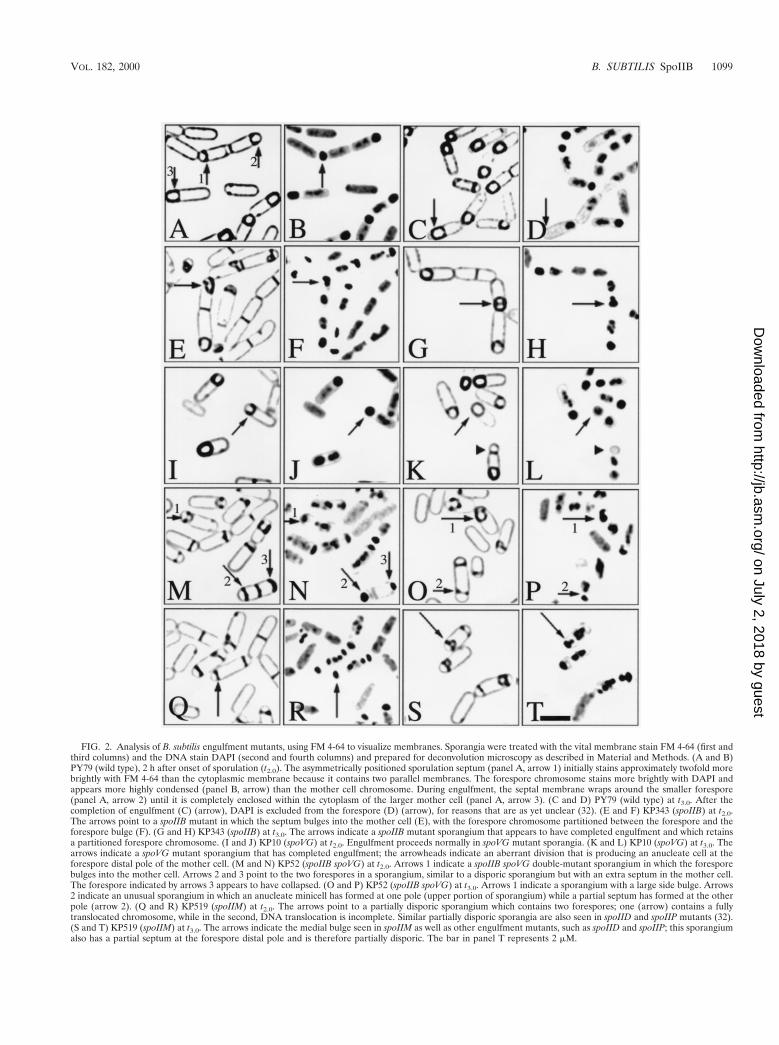
FIG. 2. Analysis of B. subtilis engulfment mutants, using FM 4-64 to visualize membranes. Sporangia were treated with the vital membrane stain FM 4-64 (first andthird columns) and the DNA stain DAPI (second and fourth columns) and prepared for deconvolution microscopy as described in Material and Methods. (A and B)PY79 (wild type), 2 h after onset of sporulation (t2.0). The asymmetrically positioned sporulation septum (panel A, arrow 1) initially stains approximately twofold morebrightly with FM 4-64 than the cytoplasmic membrane because it contains two parallel membranes. The forespore chromosome stains more brightly with DAPI andappears more highly condensed (panel B, arrow) than the mother cell chromosome. During engulfment, the septal membrane wraps around the smaller forespore(panel A, arrow 2) until it is completely enclosed within the cytoplasm of the larger mother cell (panel A, arrow 3). (C and D) PY79 (wild type) at t3.0. After thecompletion of engulfment (C) (arrow), DAPI is excluded from the forespore (D) (arrow), for reasons that are as yet unclear (32). (E and F) KP343 (spoIIB) at t2.0.The arrows point to a spoIIB mutant in which the septum bulges into the mother cell (E), with the forespore chromosome partitioned between the forespore and theforespore bulge (F). (G and H) KP343 (spoIIB) at t3.0. The arrows indicate a spoIIB mutant sporangium that appears to have completed engulfment and which retainsa partitioned forespore chromosome. (I and J) KP10 (spoVG) at t2.0. Engulfment proceeds normally in spoVG mutant sporangia. (K and L) KP10 (spoVG) at t3.0. Thearrows indicate a spoVG mutant sporangium that has completed engulfment; the arrowheads indicate an aberrant division that is producing an anucleate cell at theforespore distal pole of the mother cell. (M and N) KP52 (spoIIB spoVG) at t2.0. Arrows 1 indicate a spoIIB spoVG double-mutant sporangium in which the foresporebulges into the mother cell. Arrows 2 and 3 point to the two forespores in a sporangium, similar to a disporic sporangium but with an extra septum in the mother cell.The forespore indicated by arrows 3 appears to have collapsed. (O and P) KP52 (spoIIB spoVG) at t3.0. Arrows 1 indicate a sporangium with a large side bulge. Arrows2 indicate an unusual sporangium in which an anucleate minicell has formed at one pole (upper portion of sporangium) while a partial septum has formed at the otherpole (arrow 2). (Q and R) KP519 (spoIIM) at t2.0. The arrows point to a partially disporic sporangium which contains two forespores; one (arrow) contains a fullytranslocated chromosome, while in the second, DNA translocation is incomplete. Similar partially disporic sporangia are also seen in spoIID and spoIIP mutants (32).(S and T) KP519 (spoIIM) at t3.0. The arrows indicate the medial bulge seen in spoIIM as well as other engulfment mutants, such as spoIID and spoIIP; this sporangiumalso has a partial septum at the forespore distal pole and is therefore partially disporic. The bar in panel T represents 2 mM.
VOL. 182, 2000 B. SUBTILIS SpoIIB 1099
on July 2, 2018 by guesthttp://jb.asm
.org/D
ownloaded from

forespore bulge and the forespore (Fig. 2F and T, arrows). Byt3.0 (Fig. 2G), approximately 26% of spoIIB mutant sporangiahad completed engulfment (Fig. 2G and H). Some of theengulfed sporangia contained a slight medial constriction (Fig.2G, arrow) and retained the partitioned forespore chromo-some seen in the sporangia containing bulges at t2.0 (Fig. 2H,arrow), suggesting that these sporangia had completed engulf-ment after the formation of a forespore bulge.
Time-lapse microscopy of engulfment in a spoIIB mutant.The results described above suggested that engulfment inspoIIB mutant sporangia is preceded by the bulging of theforespore into the mother cell. To demonstrate that sporangiawith bulges are capable of completing engulfment, we per-formed time-lapse deconvolution microscopy of FM 4-64-stained spoIIB mutant sporangia, using a method previouslydescribed (32) (Fig. 3). Sporangia were applied to a coverslip90 min after the onset of sporulation, and images were col-lected every 20 min for 1 h. At the first time point, manysporangia had completed polar septation (Fig. 3A and B) andsome showed bulging of the forespore into the mother cell(Fig. 3C). At the second time point, the two sporangia thatinitially had flat polar septa showed a bulging of the foresporeinto the mother cell (Fig. 3A and B, 20-min time point), whileengulfment was under way in the sporangium that had startedwith a bulge (Fig. 3C, 40-min time point) (see the Pogliano labwebsite [http://www.biology.ucsd.edu/lab/pogliano] for fields ofsporangia from these time-lapse experiments). By the end ofthe time course, 19 of 29 sporangia had completed engulfment;in 18 of these 19, a forespore bulge was evident at one or moretime points. Thus, although prominent bulging of the fore-spore into the mother cell is not normally part of the engulf-ment pathway in B. subtilis, this event does not inhibit thesuccessful completion of engulfment in spoIIB mutant spo-rangia.
FIG. 3. Time-lapse deconvolution microscopy of engulfment in spoIIB mu-tant sporangia. A culture of KP343 (spoIIB) was grown in the presence of FM4-64, and sporulation was induced by resuspension. At 1.5 h after the initiationof sporulation, the culture was affixed to a coverslip (see Materials and Meth-ods). Images were collected every 20 min over a period of 1 h, as indicated by thetime line above the sporangia. The panels of this figure display a progression ofthree different sporangia, from polar septation (A and B) or bulge formation (C)through recovery of the bulge and, finally, completion of engulfment. The fieldsfrom which these examples were taken are presented in the Pogliano lab website(http://www.biology.ucsd.edu/labs/pogliano). The bar in panel C represents 2 mM.
TA
BL
E2.
Eng
ulfm
ent
phen
otyp
esof
vari
ous
B.s
ubtil
isst
rain
sas
asse
ssed
byF
M4-
64m
embr
ane
stai
ning
and
fluor
esce
nce
mic
rosc
opy
atth
ein
dica
ted
times
ofth
esp
orul
atio
npr
oces
s
Eng
ulfm
ent
phen
otyp
e
%of
spor
angi
aex
hibi
ting
phen
otyp
eat
indi
cate
dtim
efo
rge
noty
pea:
wt
spoI
IBsp
oVG
spoI
IBsp
oVG
spoI
IM
t 2t 3
t 2t 3
t 2t 3
t 2t 3
t 2t 3
Stra
ight
and
curv
edb
40.2
(111
)24
.9(6
6)49
(438
)30
.3(1
08)
51.5
(83)
26.0
(51)
50.2
(312
)35
.8(1
41)
56.4
(269
)36
.3(1
74)
Eng
ulfe
dc51
.8(1
43)
71.7
(190
)3.
9(3
5)26
.1(9
3)36
.6(5
9)62
.1(1
13)
1.1
(7)
3.7
(11)
0(0
)0
(0)
Bul
ged
1.1
(3)
0.4
(1)
20.4
(182
)26
.7(9
5)0.
0(0
)0.
0(0
)15
.0(9
3)18
.5(7
3)7.
3(3
5)26
.5(1
27)
Dis
pore
ew
ithbu
lgef
0.7
(2)
0.0
(0)
4.7
(42)
2.8
(10)
0.0
(0)
0.5
(1)
4.0
(25)
5.3
(21)
7.5
(36)
15.4
(74)
Dis
pori
ce4.
7(1
3)0.
8(2
)15
.8(1
41)
6.2
(23)
6.8
(11)
5.0
(9)
22.7
(141
)19
.8(7
8)27
.0(1
29)
20.5
(98)
Abn
orm
aldi
visi
ong
0.7
(2)
0.8
(2)
3.0
(27)
2.2
(8)
3.7
(6)
3.3
(6)
4.3
(27)
6.6
(26)
0(0
)0
(0)
Col
laps
eh0.
4(1
)1.
1(3
)2.
3(2
1)5.
1(1
8)1.
2(2
)1.
6(3
)2.
7(1
7)11
.2(4
4)1
(5)
0.6
(3)
Oth
er0.
4(1
)0.
4(1
)0.
3(3
)0.
3(1
)0.
0(0
)0.
0(0
)0.
0(0
)0.
0(0
)0.
6(3
)0.
6(3
)
Tot
alno
.of
cells
scor
ed56
644
51,
670
627
391
381
1,07
264
394
375
6
aT
henu
mbe
rof
spor
angi
ain
each
phen
otyp
iccl
ass
was
divi
ded
byth
eto
taln
umbe
rof
spor
angi
a(t
hose
cont
aini
ngfo
resp
ores
atan
yst
age
ofen
gulfm
ent,
visu
aliz
edby
FM
4-64
stai
ning
).A
tt 2
.0an
dt 3
.0,a
vera
ges
of50
.4an
d57
.8%
,res
pect
ivel
y,of
the
bact
eria
inth
ecu
lture
wer
esp
oran
gia.
The
tota
lnum
bers
ofsp
oran
gia
scor
edfo
rea
chcl
ass
are
give
nin
pare
nthe
sis.
wt,
wild
type
.b
Fla
tan
dcu
rved
pola
rse
pta,
incl
udin
gth
ose
inw
hich
the
sept
aha
dm
igra
ted
slig
htly
tow
ard
the
pole
s.c
Tho
sein
whi
chth
een
gulfi
ngm
embr
anes
appe
arto
fully
,or
alm
ost
fully
,enc
ircl
eth
efo
resp
ore.
dT
hose
show
ing
abu
lgin
gof
the
fore
spor
ein
toth
em
othe
rce
ll.e
Dis
pori
csp
oran
gia
incl
ude
thos
ew
ithtw
oco
mpl
ete
sept
aan
dtw
ofu
llytr
ansl
ocat
edfo
resp
ore
chro
mos
omes
(whi
chw
ere
noto
bser
ved
inth
ew
ildty
pe)
and
thos
ew
ithon
eco
mpl
ete
sept
um(w
itha
fully
tran
sloc
ated
fore
spor
ech
rom
osom
e)an
don
epa
rtia
lsep
tum
with
anin
com
plet
ely
tran
sloc
ated
fore
spor
ech
rom
osom
e(f
oran
exam
ple,
see
Fig
.1Q
and
R,a
rrow
).fD
ispo
ric
spor
angi
ain
whi
chon
epo
lar
sept
umbu
lges
into
the
mot
her
cell
(for
anex
ampl
e,se
eF
ig.1
San
dT
,arr
ow).
gSp
oran
gia
cont
aini
ngan
extr
ase
ptum
atm
idce
ll,re
sulti
ngin
anuc
leat
em
othe
rce
lls(f
oran
exam
ple,
see
Fig
.2M
,arr
ows
2an
d3)
.h
For
espo
res
inw
hich
the
mem
bran
esan
dD
NA
appe
arco
mpr
esse
dag
ains
tth
epo
leof
the
spor
angi
um(f
oran
exam
ple,
see
Fig
.2M
and
N,a
rrow
3).
1100 PEREZ ET AL. J. BACTERIOL.
on July 2, 2018 by guesthttp://jb.asm
.org/D
ownloaded from

Transmission electron microscopy analysis. To determinemore precisely the septal structure of spoIIB mutants duringthe process of engulfment, we performed transmission electronmicroscopy on cultures harvested at t2.0, a time at which bulgesare prevalent. After polar septation, a layer of peptidoglycanlies between the septal membranes (16) (Fig. 4A). In the wildtype, the septal peptidoglycan is thinned, apparently beginningin the middle of the septum (Fig. 4B, arrowhead) and movingtoward the edges of the septum (16, 31) (Fig. 4C, arrowhead).Once the peptidoglycan is completely removed (Fig. 4D), themother cell membranes move up and around the forespore(Fig. 4E and F) until the forespore is completely enclosedwithin the mother cell cytoplasm (Fig. 4G). In spoIIB mutantsporangia, the thickness of the peptidoglycan between themother cell and forespore membranes often appeared unevenacross the septum (Fig. 4I, J, L, and M, arrowheads), althoughoccasionally septal thinning appeared more similar to that seenin the wild type (Fig. 4K). These observations suggested that inspoIIB mutants, thinning of the septal peptidoglycan occurredrandomly throughout the septum. Bulges of the forespore intothe mother cell occurred both at the middle (Fig. 4N) and theedges (Fig. 4O and P) of the septum. Often the septal pepti-doglycan in these sporangia was displaced from the originalplane of the septum into the mother cell (Fig. 4N to P, arrow-heads), appearing to have been pushed aside as the foresporebulged into the mother cell. Occasionally, fragments of septalpeptidoglycan were observed on the forespore bulge (Fig. 4N,double arrowhead). This was in contrast to the situation in thewild type, in which neither large forespore bulges nor folds ofcell wall material were observed. In spoIIB mutant sporangia,engulfment occurred around residual septal peptidoglycan,which often extended almost 200 nm into the middle of the cell(Fig. 4P and Q). A large amount of septal peptidoglycan re-mained after engulfment was complete (Fig. 4R), perhaps ex-plaining the medial constriction of the engulfed foresporeshown in Fig. 2G and H (arrow). Thus, septal thinning ap-peared to be spatially unregulated in spoIIB mutant sporangiaand was not complete prior to the onset or completion ofengulfment.
The spoVG and spoIIB spoVG mutant phenotypes. Since thespoVG mutation enhances the sporulation deficiency of spoIIBmutations (29), we next examined the spoVG single-mutantand the spoIIB spoVG double-mutant phenotypes by FM 4-64membrane staining. The spoVG single mutant has been shownto display an early onset of polar septation (30), mild impair-ment of cortex synthesis (38), and a slight decrease in sporu-lation efficiency (29, 38). We found that at early times in thespore formation process, the ability of the spoVG single mutantto initiate and complete engulfment appeared similar to that ofthe wild type (Fig. 2I to L). We did not observe any sporangiawith the bulging phenotype characteristic of an early engulf-ment defect, and engulfment was completed at about the samerate as in the wild type (Table 2). We also observed that about10% of spoVG mutant sporangia display aberrant divisions,such as the minicell forming in the sporangium in Fig. 2K, aswell as other division phenotypes, which will be described be-low.
As previously reported (29), the spoIIB spoVG double mu-tant displays a strong impairment of engulfment (Fig. 2M toP). However, in addition to the flat polar septa previouslydescribed, we observed bulges of the forespore into the mothercell (Fig. 2M, arrow 1), some of which appeared to have initi-ated but not completed engulfment (Fig. 2O, arrow 1). Indeed,2 h after the onset of sporulation, 50.2% of sporangia had a flatpolar septum while sporangia that had the bulging phenotypeaccounted for 19.0% of the population. One hour later (at t3),
the population of sporangia was similar to that observed at t2(Table 2) and the bulging sporangia appeared to be lysing. Thismay explain why the bulging phenotype was not observed pre-viously, since samples for electron microscopy were taken 4 hafter induction of sporulation (29).
We also noted that 26% of spoIIB spoVG double-mutantsporangia (and, at a lower frequency, spoVG single-mutantsporangia) contained additional septa, either at polar or me-dial sites. Additional polar division events have been describedin sporangia lacking the activity of the mother cell transcrip-tion factor sE (16, 31, 40). In such sporangia, division occurs atboth potential sites of polar septation, resulting in the produc-tion of disporic sporangia with two forespore compartments,each with a chromosome, and a centrally located and anucleatemother cell (23, 32, 40). A second example is provided by theengulfment mutants spoIID, spoIIM, and spoIIP, which pro-duce partially disporic sporangia (32). Such sporangia containone forespore with a chromosome, while at the second poten-tial site of polar septation a septum (often incomplete) forms,but the chromosome is incompletely segregated into this sec-ond forespore (Fig. 2Q and R, arrow).
The spoIIB spoVG double-mutant division phenotype wasdistinct from these two previous examples, since the extra septawere positioned at midcell as well as at polar division sites.Thus, anucleate mother cells, forespores, and minicells wereproduced. For example, the sporangium indicated by arrows 2and 3 in Fig. 2M and N appears to have two forespores and twocentrally located, anucleate mother cells between the fore-spores. Such a sporangium could be produced if, followingformation of one polar septum, division occurred again atmidcell and at the remaining polar division site, with the chro-mosome ultimately being translocated into the second fore-spore. The disporic sporangia produced by the spoIIB spoVGdouble mutant fell into several classes differing in the position-ing and number of chromosomes. First, as in the classic dis-pores produced by sE-defective sporangia, some of the spoIIBspoVG dispores contained two forespores, each with a singlechromosome and an anucleate mother cell (data not shown).Others appeared similar to the partial dispores of engulfmentmutants, in which the second forespore septum was often in-complete and contained part of a chromosome. Finally, somedispores had an anucleate forespore (these perhaps shouldmore precisely be called minicells) and one or two chromo-somes with various degrees of segregation into the secondforespore (Fig. 2O and P, arrow 2).
Another significant phenotype (exhibited by 11% of the spo-rangia at t3.0) in the double mutant was strong membranestaining near one pole of the sporangium and a highly con-densed, and often crescent-shaped chromosome at the samesite (Fig. 2M and N, arrow 3). We suggest that these representcollapsed forespores and may be identical to the pygmy spo-rangia previously described for the spoVM ftsH double mutant(7).
Localization of SpoIIB-myc to the sporulation septum. Be-cause it appeared that SpoIIB is necessary for the efficientdegradation of septal peptidoglycan, we next investigated itssubcellular distribution. To do so, we constructed a fusion geneencoding an epitope-tagged SpoIIB-myc protein and used an-tibodies directed against the c-myc epitope to localize the fu-sion protein by indirect immunofluorescence microscopy. ThespoIIB-myc fusion gene replaced the wild-type spoIIB gene andwas able to support wild-type levels of spore formation (datanot shown), with no aberrant sporangia being produced atearly or late times of sporulation (data not shown).
The SpoIIB-myc fusion protein was visualized by usingmouse monoclonal antibodies directed against the c-myc
VOL. 182, 2000 B. SUBTILIS SpoIIB 1101
on July 2, 2018 by guesthttp://jb.asm
.org/D
ownloaded from

1102 PEREZ ET AL. J. BACTERIOL.
on July 2, 2018 by guesthttp://jb.asm
.org/D
ownloaded from

epitope and FITC-labeled secondary antibodies (green), whileFM 4-64 (red) and DAPI (blue) were used to visualize mem-branes and DNA, respectively. Although membrane structureis not as well-defined in immunofluorescence experiments as itis in living cells, the polar septum could be readily visualizedprior to the onset of engulfment (Fig. 5C, arrow) and after thecompletion of engulfment (Fig. 5G, arrow 3). The DAPI-stained chromosomes shown in Fig. 5 appear more punctatethan in many previous publications, a difference we attribute toour use of an optical sectioning deconvolution microscope aswell as to differences in handling of the micrographs in imageprocessing programs such as Adobe Photoshop (as illustratedin the Pogliano lab website [http://www.biology.ucsd.edu/labs/po-gliano]). Ninety minutes after the onset of sporulation, SpoIIB-myc localized to the sporulation septum, appearing evenly dis-tributed throughout the septum prior to the onset ofengulfment (Fig. 5A to D, arrow). For example, in the sporan-gium indicated by the arrow in Fig. 5B, SpoIIB-myc (green)localized between the forespore and mother cell chromosomes(Fig. 5A); when SpoIIB-myc staining was overlaid on the FM4-64 membrane staining in Fig. 5D, SpoIIB-myc was seen tolocalize to the sporulation septum (yellow). The random punc-tate immunostaining within the sporangia was also observed ina strain lacking the SpoIIB-myc fusion, although septal stainingwas absent. We therefore attribute the punctate signal to non-specific binding of either the primary antibodies or the fluo-rescently labeled secondary antibodies. The majority of spo-rangia with flat polar septa showed SpoIIB-myc localization tothe septum, while those with curved septa and those in whichengulfment was complete failed to show SpoIIB-myc localiza-tion (Fig. 5E to H, arrows 2 and 3; scored in Fig. 6). Theseresults suggest that SpoIIB, which is synthesized prior to polarseptation, rapidly localizes to newly formed septa and disap-pears as engulfment commences.
Because genetic evidence suggested that SpoVG and SpoIIBinteract (29), we investigated the localization of SpoIIB-myc inspoVG mutant sporangia. We found that SpoIIB-myc localizedand delocalized similarly to the wild type, with the exceptionthat SpoIIB-myc localized to both poles of spoVG dispores(data not shown).
SpoIIB localization is inhibited in FtsZ-depleted cells. Wenext investigated whether localization of SpoIIB depends onthe cell division protein FtsZ. FtsZ plays a central role inbacterial cell division, directing other cell division proteins tothe division site (5, 6, 9, 11, 27, 28, 47, 48) and forming a ringthat constricts as the septum is synthesized (5). To test whetherlocalization of SpoIIB depends on FtsZ, we examined SpoIIB-myc localization in a strain in which the ftsZ gene is under thecontrol of the inducible spac promoter (4). In such a strain,FtsZ can be depleted by growth in the absence of IPTG,resulting in the inhibition of cell division and the production of
filamentous cells. When FtsZ was depleted during sporulation,SpoIIB-myc failed to localize (Fig. 5U to X), although, as willbe discussed later, the protein was present in such sporangia.In contrast, normal SpoIIB-myc localization was observed fol-lowing induction of FtsZ by IPTG (data not shown). Thus,FtsZ assembly is required for SpoIIB localization.
SpoIIB colocalizes with FtsZ at sites of septal biogenesis.The rapid, FtsZ-dependent localization of SpoIIB-myc to thesporulation septum raised the possibility that SpoIIB-myc as-sembled into the sporulation septum during its biogenesis. Totest this possibility, we colocalized SpoIIB-myc and the celldivision protein FtsZ by immunofluorescence microscopy. Inthe early sporangium, FtsZ localizes in rings to the two poten-tial sites of polar septation (21) but becomes active at only onesite, where it constricts during septal biogenesis (5). If SpoIIBlocalized only to complete septa, SpoIIB-myc and FtsZ shouldnot colocalize; however, if SpoIIB localized to partial septa,then colocalization of SpoIIB-myc and FtsZ would be ob-served, although SpoIIB-myc would show unipolar localizationwhile FtsZ would show bipolar localization.
Sporulation was induced by resuspension, and samples wereprepared for immunofluorescence microscopy 1.5 and 2.5 hafter the onset of sporulation. SpoIIB-myc was detected byusing mouse monoclonal antibodies directed against the c-mycepitope and FITC-labeled secondary antibodies (green fluo-rescence, Fig. 5J, L, N, P, R, and T). FtsZ was detected byusing affinity-purified rabbit antibodies directed against FtsZand Cy5-labeled secondary antibodies (far-red fluorescence—false-colored red in Fig. 5K, L, O, P, S, and T). To correlateSpoIIB-myc and FtsZ localization with septal morphology andforespore chromosome segregation, we simultaneously visual-ized membranes with FM 4-64 (data not shown) and DNA withDAPI (blue fluorescence in Fig. 5I to T). We observed colo-calization of SpoIIB-myc and FtsZ in 39% of the sporangiathat had incompletely translocated chromosomes and flat polarsepta (Fig. 5I to L; scored in Fig. 6); we infer that in thesesporangia the sporulation septum was incomplete. In most ofthese sporangia, FtsZ showed bipolar staining (Fig. 5K, arrowand arrowhead) while SpoIIB-myc showed unipolar staining atthe sporulation septum (Fig. 5J, arrow). Most of the remainingsporangia with flat polar septa showed localization of SpoIIB-myc but not FtsZ to the septum (data not shown; scored in Fig.6); we infer that biogenesis of the sporulation septum wascomplete in these sporangia. None of the sporangia withcurved septa showed localization of FtsZ to the septum, while42% showed localization of SpoIIB-myc to the septum and theremaining 58% lacked both FtsZ and SpoIIB staining (Fig. 6).Thus, SpoIIB colocalizes with FtsZ at sites of polar septationbut does not localize to the inactive FtsZ complex within thesame cell. This suggests that SpoIIB localizes to the site of
FIG. 4. Examination of spoIIB sporangia by transmission electron microscopy. Strains PY79 (wild type) and KP343 (spoIIB) were induced to sporulate byresuspension, and samples taken either 2 h (A to H, J to M, and O to Q) or 3 h (I, N, and R) after the onset of sporulation. Samples were prepared for electronmicroscopy by protocols described in Materials and Methods. (A to G) Various stages of engulfment in the wild type. The sporulation septum initially contained darklystaining cell wall material (A), which became thinner (as indicated by arrowheads), apparently starting in the middle of the septal disc and proceeding toward the edges(B and C). When septal thinning was complete (D), the forespore and mother cell septal membranes were very close to one another. Engulfment commenced with aslight bowing of the septum (E), and the mother cell membrane migrated around the forespore (F), which ultimately was fully enclosed within the mother cell cytoplasm(G). (H to R) Various stages of engulfment in spoIIB mutants. In spoIIB mutant sporangia, the septum also initially contained a layer of darkly staining cell wall material(H), and septal thinning (indicated by arrowheads) occasionally started in the middle of the septum (K). However, more frequently, the septum was thinned in severaldifferent locations (I and J), and occasionally large regions of the septum appeared incompletely thinned (L and M) (area between arrowheads). In sporangia thatcontained bulges of the forespore into the mother cell, portions of the darkly staining septal cell wall material were often displaced from the original plane of the septuminto the mother cell (N, O, and P) (arrowheads), and sometimes fragments of cell wall material were observed (N) (double arrowhead). This was not observed inwild-type sporangia, in which the septal peptidoglycan was completely thinned before the onset of engulfment. The engulfing membranes moved around residual cellwall material that extended well into middle of the cell (Q), and after engulfment was complete (R), a substantial amount of septal peptidoglycan remained, which wefailed to observe in our wild-type strain. The bar in panel R represents 200 nm.
VOL. 182, 2000 B. SUBTILIS SpoIIB 1103
on July 2, 2018 by guesthttp://jb.asm
.org/D
ownloaded from

1104 PEREZ ET AL. J. BACTERIOL.
on July 2, 2018 by guesthttp://jb.asm
.org/D
ownloaded from

polar septation either shortly before or after the onset of septalbiogenesis.
Neither sF- nor sE-directed gene expression is required forlocalization or delocalization of SpoIIB. Shortly after polarseptation, sF becomes active in the forespore, directing theactivation of sE in the mother cell, thereby allowing the pro-duction of proteins essential for hydrolysis of septal pepti-doglycan and for engulfment. We were interested in knowingwhether either the assembly or disassembly of SpoIIB at theseptum depends on proteins under the control of these tran-scription factors. We therefore examined localization ofSpoIIB-myc in two strains, the first containing an insertionmutation in spoIIGB (encoding sE) and the second containinga deletion of spoIIAA-AC (encoding sF). Both strains lack sE
activity and thus divide sequentially at the two polar sites ofFtsZ assembly (15, 23, 32), with rapid initiation of division atthe second FtsZ ring following completion of the first septum(32). They also sequentially translocate a chromosome intoeach forespore following division (15, 23, 32). Identical resultswere obtained for the two strains; here we present only thoseof the mutant lacking the sF protein. After synthesis of the firstsporulation septum, SpoIIB-myc (green) localized to this sep-tum (Fig. 5N, arrowhead), as in the wild type. However,SpoIIB-myc also colocalized with the FtsZ ring (red) at thedistal pole (Fig. 5N to P, arrow), where biogenesis of thesecond sporulation septum would likely have commenced.Thus, in the absence of sF-directed gene expression, SpoIIB-myc sequentially localized to each sporulation septum. Shortlybefore the completion of DNA translocation into each fore-spore, SpoIIB-myc was lost from the septum (Fig. 5Q to T,arrowheads). Thus, the assembly of SpoIIB in the septum andits subsequent loss are independent of both sF- and sE-direct-ed gene expression and the onset of engulfment.
SpoIIB degradation is facilitated by its localization to divi-sion sites. To determine if the loss of localized SpoIIB-myc atthe onset of engulfment correlated with its degradation or wassolely due to exclusion from the septum, we performed a West-ern blot analysis to monitor SpoIIB-myc levels during sporu-lation (Fig. 7A). SpoIIB was first detected 1 h after the onsetof sporulation, when levels of SpoIIB-myc were maximal.
Ninety minutes after the onset of sporulation, a SpoIIB-mycbreakdown product was seen (Fig. 7A, arrow 2) that was absentin strains lacking the SpoIIB-myc fusion, and levels of theprotein gradually decreased until it was completely absent att3.0. Therefore, SpoIIB appears to be degraded early in engulf-ment, in correlation with its loss from polar septa. We nextwanted to determine the stability of SpoIIB-myc under condi-tions in which it fails to localize, such as after depletion ofFtsZ. In the absence of FtsZ, appearance of the putativeSpoIIB-myc breakdown product was delayed by 2 h (until t3.5)and SpoIIB-myc levels remained constant until at least t3.5(Fig. 7B). Thus, the instability of SpoIIB-myc depends on itslocalization to division sites.
DISCUSSION
spoIIB mutants display a transient engulfment defect. Wehave used FM 4-64 staining and deconvolution microscopy todemonstrate that spoIIB mutants display a transient engulf-ment phenotype not previously described. This phenotype,consisting of a bulging of the forespore into the mother cell, issimilar to that previously seen in spoIID, spoIIM, and spoIIPmutants, although spoIIB mutants are able to complete engulf-ment following bulge formation whereas spoIID, spoIIM, andspoIIP mutants are completely engulfment defective (Fig. 1).Electron microscopy showed that spoIIB mutants display anuneven dissolution of septal peptidoglycan and that engulf-ment occurs around residual peptidoglycan that often stretchesmost of the way across the mother cell. These results suggestthat SpoIIB serves to regulate the dissolution of septal pepti-doglycan, and they demonstrate that large amounts of residualseptal peptidoglycan do not always inhibit membrane migra-tion during engulfment, as has been observed in spoIID,spoIIM, and spoIIP mutants (13). Indeed a similar situation hasbeen noted in wild-type Bacillus sphaericus, in which engulf-ment proceeds around residual septal peptidoglycan (15). Per-haps SpoIIB was a late addition to the engulfment machineryof B. subtilis, serving to increase the speed and efficiency ofengulfment.
FIG. 5. Localization of SpoIIB-myc to the sporulation septum. Sporulation was induced by resuspension, and samples were prepared for immunofluorescencemicroscopy (see Materials and Methods). Chromosomes were visualized with DAPI (blue) (A, B, E, F, I to T, U, and V), and membranes were visualized by stainingwith FM 4-64 (red) (C, D, G, W, and X). The membranes appear less distinct in immunofluorescence experiments (which require chemical fixation and lysozymetreatment) than in living cells. However, the sporulation septum remains visible as a brightly staining region that is either flat (C [arrow] and G [arrow 1]), curved (G[arrow 2]), or, following the completion of engulfment, circular (G [arrow 3]). FITC-labeled secondary antibodies were used to visualize SpoIIB-myc (green) (B, D,F, H, J, L, N, P, R, T, V, and X), while Cy5-labeled secondary antibodies were used to visualize FtsZ (false-colored red) (K, L, O, P, S, and T). Overlays of SpoIIB-myc(green) and either DAPI (blue) (B, F, J, L, N, P, R, T, and V), FM 4-64 (red) (D, H, and X), FtsZ (red) (L, P, and T), or both DAPI (blue) and FtsZ (red) (L, M,and P) are shown. (A to D) Localization of SpoIIB-myc at t1.5 in the wild type. The arrows point to localized SpoIIB-myc (B and D) in a sporangium with a partiallytranslocated forespore chromosome (A) and a flat sporulation septum (C). The nonseptal, punctate staining is present in strains lacking the SpoIIB-myc fusion (notshown) and therefore represents nonspecific staining. The relatively high level of this staining is probably due to the low-level expression of the spoIIB gene (29), whichdecreases the specific signal. (E to H) Localization of SpoIIB-myc at t2.5 in the wild type. Arrows 1 point to a sporangium that has not completely translocated itschromosome (E) and which still displays SpoIIB-myc localization (F and H). Arrows 2 point to a sporangium in which engulfment is almost complete (G), hascompletely translocated its chromosome (E), and may show only faint SpoIIB-myc localization (F and H). Arrows 3 indicate a sporangium that has completedengulfment (G) and lacks SpoIIB-myc (F and H). (I to L) Colocalization of SpoIIB-myc and FtsZ to partial sporulation septa in the wild type at t1.5. The arrows pointto one end of a sporangium that shows SpoIIB-myc (green) (J and L) colocalizing with a partially constricted FtsZ ring (red) (O and P). The arrowheads point to theFtsZ ring (K and L) at the second potential division site in the same sporangium; SpoIIB-myc is absent from this site. (M to P) Colocalization of SpoIIB-myc and FtsZin sporangia lacking sF (DspoIIAA-AC) at t1.5. The arrows and arrowheads indicate the two sites of polar septation in a single sporangium. The arrowheads point toa sporulation septum that is likely to be complete, since DNA translocation is well advanced (M) and FtsZ immunostaining is absent (O) but SpoIIB-myc localizes (M).The arrows indicate the second potential site of polar septation, which shows colocalization of SpoIIB-myc (N and P) and FtsZ (O and P) and little chromosometranslocation (M). (Q to T) Colocalization of SpoIIB-myc and FtsZ in sporangia lacking sF (DspoIIAA-AC) at t2.5. The arrows indicate a forespore with a partiallytranslocated chromosome (Q) that has SpoIIB-myc localized throughout the septum (R and T). The arrowheads directly below the arrows indicate a forespore thathas most of its chromosome translocated (Q) and lacks SpoIIB immunostaining (R and T). The arrowhead pairs point to a sporangium that has completely translocatedboth its chromosomes (Q) and no longer shows localization of SpoIIB-myc (R and T). These sporangia no longer display FtsZ localization (S and T). Panel Y showsa sketch of SpoIIB immunostaining (green) versus that of membranes (black) and chromosomes (blue). (U to X) SpoIIB-myc localization at t1.5 after depletion of FtsZfrom the PspacftsZ strain KP547. (W) FtsZ was depleted (see Materials and Methods), inhibiting cell division and causing the production of filaments. Only punctateSpoIIB-myc staining (V and X), similar to the background, is observed. (Y) Sketch of SpoIIB immunostaining in panels Q to T. SpoIIB immunostaining in the twosporangia indicated by arrows and arrowheads in panels Q to T is shown in green. Membranes are shown in black, while the chromosomes are shown in blue. The scalebar in panel X represents 2 mM.
VOL. 182, 2000 B. SUBTILIS SpoIIB 1105
on July 2, 2018 by guesthttp://jb.asm
.org/D
ownloaded from

SpoIIB localizes to the sporulation septum during its bio-genesis. To further investigate the role of SpoIIB, we deter-mined its subcellular distribution during sporulation. Wefound that SpoIIB localizes to the sporulation septum duringseptal biogenesis, as evidenced by the colocalization of SpoIIBwith the cell division protein FtsZ only at the growing septumand not at the inactive FtsZ ring present at the opposite poleof the same cell. It will be interesting to determine if SpoIIBrecognizes some physical feature of the nascent sporulationseptum or if it recognizes a cell division protein that is itselfrecruited to the division site either after or shortly before theonset of septal biogenesis.
Two sporulation-specific proteins, SpoIIE (2, 3, 16, 17, 22)and SpoIIGA (12), have previously been shown to localize topotential division sites, and several proteins have been shownto localize to the sporulation septum after its synthesis is com-pleted (24, 33, 35, 36), but SpoIIB is the first shown to dis-criminate between the active site of septal biogenesis and theinactive division site in the same cell. Coincidentally, it hasrecently been observed that SpoIIIE, a protein required for thetranslocation of the chromosome into the forespore (49),shows a similar pattern of localization, initially colocalizingwith FtsZ during septum biogenesis (M. D. Sharp, personalcommunication) and then localizing to the septal midpoint (41,50). These observations raise the possibility that proteins re-quired late in septation or after septation is complete can belocalized to the septum after the onset of cell division. SinceSpoIIB is incorporated into the forming septum, it is wellpositioned to facilitate the prompt and spatially regulated dis-solution of septal peptidoglycan (as discussed further below).
SpoIIB is lost from the sporulation septum at the onset ofengulfment. We noted that SpoIIB-myc localization was tran-sient, since the protein no longer localized in sporangia thathad commenced engulfment. The loss of SpoIIB from theseptum did not require the dissolution of septal peptidoglycan,the onset of engulfment, or expression of any sF- or sE-de-pendent gene, because it occurred in mutants lacking sF. Thus,SpoIIB delocalization is not correlated with a particular mor-phological event but rather appears to occur at a set time after
septal biogenesis is completed, even in the absence of furthermorphogenesis. SpoIIB was degraded during sporulation butcould be stabilized if localization was inhibited (for example,by depletion of the cell division protein FtsZ). This suggeststhat either septation itself or localization to the septum isrequired for degradation of SpoIIB, similar to the situationobserved for SpoIIE (22).
The role of SpoIIB in dissolution of septal peptidoglycan. Inthe absence of SpoIIB, septal thinning appears to occurthroughout the septum and is often incomplete prior to theonset of engulfment. This suggests that SpoIIB contributes tothe spatial regulation of septal peptidoglycan dissolution, en-suring that septal thinning commences in the middle of theseptum and proceeds toward the edges. It also suggests a rolefor SpoIIB in the temporal regulation of septal peptidoglycandissolution, ensuring that septal thinning is complete prior tothe onset of engulfment. There are several possible mecha-nisms by which SpoIIB might contribute to the spatial andtemporal regulation of septal thinning. First, SpoIIB might bean enzyme that degrades septal peptidoglycan, or it mightregulate the activity of such an enzyme, ensuring that septalthinning commences in the middle of the septum and proceedstoward the edge. Alternatively, SpoIIB might recruit cell wallhydrolases to the polar septum. Because these hydrolases arelikely to be produced under the control of sE (16), they wouldbe synthesized after polar septation, while SpoIIB, which islocalized to the polar septum while it is being synthesized,would be ideally positioned to recruit these proteins to theseptum. Finally, it is also possible that SpoIIB is part of the celldivision machinery and serves to modify the structure of theseptal peptidoglycan to allow more efficient septal thinning.Further experiments are necessary to test these hypotheses.
Another look at the synergistic roles of SpoIIB and SpoVGin engulfment. In our study, we also investigated the interac-tion between SpoIIB and SpoVG (29). In agreement withprevious studies, we found that a spoVG null mutant has awild-type engulfment phenotype and that the spoIIB spoVG
FIG. 6. Scoring of the localization patterns of SpoIIB-myc and FtsZ at dif-ferent stages of engulfment and different times of the sporulation process. Sam-ples were harvested 1.5 or 2.5 h after the onset of sporulation and processed forimmunofluorescence microscopy. Sporangia were scored for localization ofSpoIIB-myc and FtsZ and for the morphology of the polar septum, as revealedby FM 4-64 staining. Class I sporangia (flat polar septa) include both sporangiain which biogenesis of the sporulation septum is incomplete and those in whichit is complete. The FtsZ ring distal to the active division site was sometimeslacking, for reasons that are unclear. Class II sporangia had curved sporulationsepta, indicating that engulfment had commenced, while class III sporangia hadcompleted engulfment. A total of 200 sporangia were scored from samplesobtained from cultures at t1.5 and t2.5.
FIG. 7. Western blot analysis of SpoIIB-myc during engulfment. (A) Levelsof SpoIIB-myc in the wild type. Samples from PY79, which lacks the spoIIB-mycfusion (indicated by minuses), and from KP545, which contains the spoIIB-mycfusion (indicated by pluses), were harvested and processed for Western blotanalysis at the indicated times of the sporulation process. In KP545 (1), SpoIIB-myc is first detected at t1.0 (arrow 1) but is rapidly degraded, showing a shorter-length product by t1.5 (arrow 2) and completely disappearing by t3.0. No signal isdetected in the strain lacking the spoIIB-myc fusion (2). (B) Levels of SpoIIB-myc in the presence or absence of FtsZ. Strain KP547 carries ftsZ under thecontrol of the IPTG-inducible Pspac promoter and the spoIIB-myc fusion. Cellswhich synthesize FtsZ are marked 1IPTG, while those that have been depletedof FtsZ are marked 2IPTG. When FtsZ is induced (1IPTG), SpoIIB is synthe-sized and degraded as in the wild type. However, in the absence of FtsZ(2IPTG), SpoIIB is stable until the last time point (t3.5). Arrow 1 indicatesfull-length SpoIIB-myc, and arrow 2 indicates a shorter product that is likely tobe a SpoIIB breakdown product.
1106 PEREZ ET AL. J. BACTERIOL.
on July 2, 2018 by guesthttp://jb.asm
.org/D
ownloaded from

double mutant has a more severe engulfment defect than thespoIIB single mutant. However, we also noted that spoIIBspoVG double-mutant cultures showed a variety of engulfment-related phenotypes, including sporangia in which the foresporehad bulged into the mother cell or in which the engulfingmembranes appeared to have initiated their migration aroundthe forespore (Fig. 2M to P; Table 2). Recent studies bySonenshein and colleague have suggested that SpoVG inhibitspolar septation, since spoVG mutants initiate polar septationearlier than normal (30). We also found evidence to supportthe hypothesis that SpoVG inhibits division, since both thespoVG null mutant and the spoIIB spoVG double mutant pro-duce sporangia with extra septa, either at the forespore distalpole (producing disporic sporangia) or at both midcell andpolar sites (producing disporic sporangia with an extra septumat midcell [Fig. 2M and N]). Thus, SpoVG appears to inhibitcell division events subsequent to the formation of the sporu-lation septum, in addition to controlling the onset of polarseptation.
Why do spoVG and spoIIB mutations act synergistically?First, it is possible that spoVG mutations cause alterations insporangial physiology or structure that make engulfment moredifficult in spoIIB mutants, perhaps as a direct consequence ofthe premature synthesis of the sporulation septum (30). Thetime prior to polar septation could be crucial for engulfment,either allowing structural alterations in the septal peptidogly-can or allowing synthesis of proteins whose functions are re-dundant to those of SpoIIB. Second, it is possible that theadditional active and potential division sites in spoVG mutantsporangia cause the inappropriate localization of proteins es-sential for engulfment to these sites, rather than to the sporu-lation septum. In either case, because engulfment is alreadyslow in spoIIB mutants, and because sporangia that fail tocomplete engulfment lyse (commencing around t3.5 [our un-published observations and references 31 and 43), the spoVGmutation need only slightly affect the speed or efficiency ofengulfment in spoIIB mutants to dramatically decrease sporeproduction.
A model for bulge formation. It has been proposed that ahigher osmolarity in the forespore than in the mother cell mayallow the forespore to grow larger during engulfment, possiblypushing the membranes around the forespore (10). Usingtime-lapse deconvolution microscopy (32), we found that, con-sistent with this model, the length of the forespore increasedduring engulfment by an average (6 standard deviation) of21.9% (6 11.9%) while the combined length of the two cellsremained essentially constant, showing a slight decrease of1.5% (6 3.1%) (see Materials and Methods and also the Po-gliano lab website [http://www.biology.ucsd.edu/labs/pogliano]).While it is not yet clear that the growth of the foresporecontributes to engulfment, it does provide a ready explanationfor the formation of bulging forespores in mutants defective inthe dissolution of septal peptidoglycan. In such mutants, fore-spore growth would exert pressure on a septum in which pep-tidoglycan hydrolysis was incomplete. For example, in spoIID,spoIIM, and spoIIP mutants, cell wall dissolution appears tooccur only in the center of the septum (Fig. 1B). As the fore-spore grows, it will break through this weakened area to forma centrally localized and constricted bulge. In contrast, inspoIIB mutants, cell wall degradation appears to occurthroughout the septum (Fig. 1C), and this will weaken theentire structure. Thus, as the forespore grows, the septumbreaks and is breached by the forespore, resulting in both sideand medial bulges. Significantly, while there is no engulfmentin spoIID, spoIIM, and spoIIP mutants, spoIIB mutants are able
to complete engulfment following bulge formation, despite theretention of peptidoglycan near the edges of the polar septum.
What implication do these findings have for the mechanismof engulfment? First, because the engulfing membranes canmigrate around a large amount of residual peptidoglycan (Fig.4B and C), peptidoglycan does not necessarily block migrationof the engulfing membranes, as has been proposed to explainthe engulfment defect of spoIIP mutants (13). Second, al-though membrane migration normally appears to commenceat the edges of the septum following thinning of the septalpeptidoglycan, migration must also be capable of starting inthe middle of the septum, moving between the forespore mem-brane and the residual septal peptidoglycan in spoIIB mutantsporangia (Fig. 4Q). Finally, while some of our findings sup-port the hypothesis that engulfment is facilitated by the growthof the forespore into the mother cell, the apparent uncouplingof these events in spoIIB mutants suggests the existence ofadditional mechanisms by which the membranes are drivenaround the forespore during engulfment.
ACKNOWLEDGMENTS
We are grateful to Petra Levin and Richard Losick for providing theFtsZ antibodies, to Nick Osborne for affinity purifying these antibod-ies, and to Terry G. Frey and Christian W. Renken for help in devisingthe alternative protocol for increasing membrane contrast during elec-tron microscopy. We thank C. Lance Washington for technical assis-tance; Adam Driks for helpful advice; Kiyoteru Tokuyasu for discus-sions; Richard Losick, Katherine Lemon, Petra Levin, Alan Grossman,and Michael Carson for providing strains and plasmids; and Joe Po-gliano for providing comments on the manuscript.
This work was supported by NIH grant GM-57045 to K.P., as well asby awards from the Arnold and Mabel Beckman Foundation and theSearle Scholars Program/The Chicago Community Trust. A.R.P. issupported by a MARC predoctoral fellowship from the NIH(GM19570-01).
REFERENCES
1. Altschul, S. F., W. Gish, W. Miller, E. W. Myers, and D. J. Lipman. 1990.Basic local alignment search tool. J. Mol. Biol. 215:403–410.
2. Arigoni, F., K. Pogliano, C. D. Webb, P. Stragier, and R. Losick. 1995.Localization of a protein implicated in establishment of cell type to sites ofasymmetric division. Science 270:637–640.
3. Barak, I., J. Behari, G. Olmedo, P. Guzman, D. P. Brown, E. Castro, D.Walker, J. Westpheling, and P. Youngman. 1996. Structure and function ofthe Bacillus subtilis SpoIIE protein and its localization to sites of sporulationseptum assembly. Mol. Microbiol. 19:1047–1060.
4. Beall, B., and J. Lutkenhaus. 1991. FtsZ in Bacillus subtilis is required forvegetative septation and for asymmetric septation during sporulation. GenesDev. 5:447–455.
5. Bi, E. F., and J. Lutkenhaus. 1991. FtsZ ring structure associated withdivision in Escherichia coli. Nature 354:161–164.
6. Bouche, J. P., and S. Pichoff. 1998. On the birth and fate of bacterial divisionsites. Mol. Microbiol. 29:19–26.
7. Cutting, S., M. Anderson, E. Lysenko, A. Page, T. Tomoyasu, K. Tatematsu,T. Tatsuta, L. Kroos, and T. Ogura. 1997. SpoVM, a small protein essentialto development in Bacillus subtilis, interacts with the ATP-dependent pro-tease FtsH. J. Bacteriol. 179:5534–5542.
8. Dubnau, D., and R. Davidoff-Abelson. 1971. Fate of transforming DNAfollowing uptake by competent Bacillus subtilis. J. Mol. Biol. 56:209–221.
9. Erickson, H. P. 1997. FtsZ, a tubulin homologue in prokaryotic cell division.Trends Cell Biol. 7:362–367.
10. Errington, J. 1993. Bacillus subtilis sporulation: regulation of gene expressionand control of morphogenesis. Microbiol. Rev. 57:1–33.
11. Faguy, D. M., and W. F. Doolittle. 1998. Cytoskeletal proteins: the evolutionof cell division. Curr. Biol. 8:R338–R341.
12. Fawcett, P., A. Melnikov, and P. Youngman. 1998. The Bacillus SpoIIGAprotein is targeted to sites of spore septum formation in a SpoIIE-indepen-dent manner. Mol. Microbiol. 28:931–943.
13. Frandsen, N., and P. Stragier. 1995. Identification and characterization ofthe Bacillus subtilis spoIIP locus. J. Bacteriol. 177:716–722.
14. Hayat, M. A. 1986. Basic techniques for transmission electron microscopy, p.182. Academic Press, Orlando, Fla.
15. Holt, S. C., J. J. Gauthier, and D. J. Tipper. 1975. Ultrastructural studies ofsporulation in Bacillus sphaericus. J. Bacteriol. 122:1322–1338.
VOL. 182, 2000 B. SUBTILIS SpoIIB 1107
on July 2, 2018 by guesthttp://jb.asm
.org/D
ownloaded from

16. Illing, N., and J. Errington. 1991. Genetic regulation of morphogenesis inBacillus subtilis: roles of sE and sF in prespore engulfment. J. Bacteriol.173:3159–3169.
17. King, N., O. Dreesen, P. Stragier, K. Pogliano, and R. Losick. 1999. Septa-tion, dephosphorylation, and the activation of sigma F during sporulation inBacillus subtilis. Genes Dev. 13:1156–1167.
18. Kuroda, A., M. H. Rashid, and J. Sekiguchi. 1992. Molecular cloning andsequencing of the upstream region of the major Bacillus subtilis autolysingene: a modifier protein exhibiting sequence homology to the major auto-lysin and the spoIID product. J. Gen. Microbiol. 138:1067–1076.
19. Kuroda, A., Y. Sugimoto, T. Funahashi, and J. Sekiguchi. 1992. Geneticstructure, isolation, and characterization of a Bacillus licheniformis cell wallhydrolase. Mol. Gen. Genet. 234:129–137.
20. Lazarevic, V., P. Margot, B. Soldo, and D. Karamata. 1992. Sequencing andanalysis of the Bacillus subtilis lytRABC divergon: a regulatory unit encom-passing the structural genes of the N-acetylmuramoyl-L-alanine amidase andits modifier. J. Gen. Microbiol. 138:1949–1961.
21. Levin, P., and R. Losick. 1996. Transcription factor SpoOA switches thelocalization of the cell division protein FtsZ from a medial to a bipolarpattern in Bacillus subtilis. Genes Dev. 10:478–488.
22. Levin, P. A., R. Losick, P. Stragier, and F. Arigoni. 1997. Localization of thesporulation protein SpoIIE in Bacillus subtilis is dependent upon the celldivision protein FtsZ. Mol. Microbiol. 25:839–846.
23. Lewis, P. J., S. R. Partridge, and J. Errington. 1994. s factors, asymmetry,and the determination of cell fate in Bacillus subtilis. Proc. Natl. Acad. Sci.USA 91:3849–3853.
24. Londono-Vallejo, J.-A., C. Frehel, and P. Stragier. 1997. spoIIQ, a forespore-expressed gene required for engulfment in Bacillus subtilis. Mol. Microbiol.24:29–39.
25. Lopez-Diaz, I., S. Clarke, and J. Mandelstam. 1986. spoIID operon of Ba-cillus subtilis: cloning and sequence. J. Gen. Microbiol. 132:341–354.
26. Lopilato, J., S. Bortner, and J. Beckwith. 1986. Mutations in a new chromo-somal gene of Escherichia coli K-12, pcnB, reduce plasmid copy number ofpBR322 and its derivatives. Mol. Gen. Genet. 205:285–290.
27. Lutkenhaus, J. 1993. FtsZ ring in bacterial cytokinesis. Mol. Microbiol.9:403–409.
28. Lutkenhaus, J., and S. G. Addinall. 1997. Bacterial cell division and the Zring. Annu. Rev. Biochem. 66:93–116.
29. Margolis, P. S., A. Driks, and R. Losick. 1993. Sporulation gene spoIIB fromBacillus subtilis. J. Bacteriol. 175:528–540.
30. Matsuno, K., and A. L. Sonenshein. 1999. Role of SpoVG in asymmetricseptation in Bacillus subtilis. J. Bacteriol. 181:3392–3401.
31. Piggot, P. J., and J. G. Coote. 1976. Genetic aspects of bacterial endosporeformation. Bacteriol. Rev. 40:908–962.
32. Pogliano, J., N. Osborne, M. D. Sharp, A. Abanes-De Mello, A. Perez, Y.-L.Sun, and K. Pogliano. 1999. A vital stain for studying membrane dynamics inbacteria: a novel mechanism controlling septation during Bacillus subtilissporulation. Mol. Microbiol. 31:1149–1159.
33. Pogliano, K., E. Harry, and R. Losick. 1995. Visualization of the subcellularlocation of sporulation proteins in Bacillus subtilis using immunofluorescencemicroscopy. Mol. Microbiol. 18:459–470.
34. Pogliano, K., A. E. M. Hofmeister, and R. Losick. 1997. Disappearance of the
sE transcription factor from the forespore and the SpoIIE phosphatase fromthe mother cell contributes to establishment of cell-specific gene expressionduring sporulation in Bacillus subtilis. J. Bacteriol. 179:3331–3341.
35. Price, K. D., and R. Losick. 1999. A four-dimensional view of assembly of amorphogenetic protein during sporulation in Bacillus subtilis. J. Bacteriol.181:781–790.
36. Resnekov, O., S. Alper, and R. Losick. 1996. Subcellular localization ofproteins governing the proteolytic activation of a developmental transcrip-tion factor in Bacillus subtilis. Genes Cells 1:529–542.
37. Rong, S., M. S. Rosenkrantz, and A. L. Sonenshein. 1986. Transcriptionalcontrol of the Bacillus subtilis spoIID gene. J. Bacteriol. 165:771–779.
38. Rosenbluh, A., C. D. B. Banner, R. Losick, and P. C. Fitz-James. 1981.Identification of a new developmental locus in Bacillus subtilis by construc-tion of a deletion mutation in a cloned gene under sporulation control. J.Bacteriol. 148:341–351.
39. Sandman, K., R. Losick, and P. Youngman. 1987. Genetic analysis of Bacil-lus subtilis spo mutations generated by Tn917-mediated insertional mutagen-esis. Genetics 117:603–617.
40. Setlow, B., N. Magill, P. Febbroriello, L. Nakhimovsky, D. E. Koppel, and P.Setlow. 1991. Condensation of the forespore nucleoid early in sporulation ofBacillus species. J. Bacteriol. 173:6270–6278.
41. Sharp, M. D., and K. Pogliano. 1999. An in vivo membrane fusion assayimplicates SpoIIIE in the final stages of engulfment during Bacillus subtilissporulation. Proc. Natl. Acad. Sci. USA 96:14553–14558.
42. Smith, K., and P. Youngman. 1993. Evidence that the spoIIM gene of Ba-cillus subtilis is transcribed by RNA polymerase associated with sE. J. Bac-teriol. 175:3618–3627.
43. Smith, K., M. E. Bayer, and P. Youngman. 1993. Physical and functionalcharacterization of the Bacillus subtilis spoIIM gene. J. Bacteriol. 175:3607–3617.
44. Steinmetz, M., and R. Richter. 1994. Plasmids designed to alter the antibioticresistance expressed by insertion mutations in Bacillus subtilis, through invivo recombination. Gene 142:79–83.
45. Sterlini, J. M., and J. Mandelstam. 1969. Commitment to sporulation inBacillus subtilis and its relationship to development of actinomycin resis-tance. Biochem. J. 113:29–37.
46. Stragier, P., and R. Losick. 1996. Molecular genetics of sporulation in Ba-cillus subtilis. Annu. Rev. Genet. 30:297–341.
47. Wang, X., J. Huang, A. Mukherjee, C. Cao, and J. Lutkenhaus. 1997. Anal-ysis of the interaction of FtsZ with itself, GTP, and FtsA. J. Bacteriol.179:5551–5559.
48. Weiss, D. S., J. C. Chen, J.-M. Ghigo, D. Boyd, and J. Beckwith. 1999.Localization of FtsI (PBP3) to the septal ring requires its membrane anchor,the Z ring, FtsA, FtsQ, and FtsL. J. Bacteriol. 181:508–520.
49. Wu, L. J., and J. Errington. 1994. Bacillus subtilis SpoIIIE protein requiredfor DNA segregation during asymmetric cell division. Science 264:572–575.
50. Wu, L. J., and J. Errington. 1997. Septal localization of the SpoIIIE chro-mosome partitioning protein in Bacillus subtilis. EMBO J. 16:2161–2169.
51. Youngman, P., J. B. Perkins, and R. Losick. 1984. A novel method for therapid cloning in Escherichia coli of Bacillus subtilis chromosomal DNA ad-jacent to Tn917 insertions. Mol. Gen. Genet. 195:424–433.
1108 PEREZ ET AL. J. BACTERIOL.
on July 2, 2018 by guesthttp://jb.asm
.org/D
ownloaded from