SPINAL YNDROMES GENERAL - …. Spinal Disorders/Spin1. GENERAL...Spinal Syndromes (GENERAL) ... -...
9
SPINAL SYNDROMES (GENERAL) Spin1 (1) Spinal Syndromes (GENERAL) Last updated: September 5, 2017 SPINAL STRUCTURES AFFECTED IN VARIOUS DISORDERS ........................................................................ 1 ANATOMIC SPINAL SYNDROMES ............................................................................................................. 1 CERVICAL CORD .................................................................................................................................... 2 THORACIC CORD.................................................................................................................................... 2 LUMBOSACRAL CORD ............................................................................................................................ 2 SPINAL CORD TRANSECTION, SPINAL SHOCK ........................................................................................ 2 SPINAL CORD HEMISECTION .................................................................................................................. 3 Brown-Sequard syndrome ................................................................................................................ 3 Dorsal (Posterior) Hemisection ........................................................................................................ 3 Ventral (Anterior) Hemisection ....................................................................................................... 3 ANTERIOR 2/3 TRANSECTION ................................................................................................................. 3 EXTRAMEDULLARY CORD COMPRESSION .............................................................................................. 4 CENTRAL CORD SYNDROME .................................................................................................................. 5 CAUDA EQUINA VS. CONUS MEDULLARIS SYNDROME ........................................................................... 5 SPINAL COMPLICATIONS ......................................................................................................................... 6 Paresthesias ...................................................................................................................................... 6 Decubitus Ulcers .............................................................................................................................. 6 Bladder dysfunction ......................................................................................................................... 6 GI complications .............................................................................................................................. 6 Sexual dysfunction ........................................................................................................................... 7 Malnutrition...................................................................................................................................... 7 Respiratory Failure ........................................................................................................................... 7 Venous Thrombosis & Pulmonary Embolism ................................................................................. 7 Spasticity .......................................................................................................................................... 7 Autonomic Dysfunction ................................................................................................................... 7 Psychiatric Dysfunction ................................................................................................................... 8 SPINAL PROGNOSIS .................................................................................................................................. 8 SPINAL REHABILITATION ........................................................................................................................ 8 Anatomic localization of MOTOR SYMPTOMS, UMN & LMN lesions → see p. Mov3 >> Anatomic localization of SENSORY SYMPTOMS → see p. S22 >> RADICULOPATHY → see p. PN1 >> Spinal IMAGING → see p. D70 >> INFECTIONS of spinal cord and vertebrae → see p. Inf3 >>, p. Inf5 >>, p. Inf7 >> TUMORS of spinal cord and vertebrae → see p. Onc50 >>, p. Onc54 >> How vertebra corresponds to spinal segment: C1: C1 C2-7: +1 T1-6: +2 T7-9: +3 T10: L1-2 T11: L3-4 T12-L1: other SPINAL STRUCTURES AFFECTED IN VARIOUS DISORDERS Dorsal funiculi (fasc. gracilis & cuneatus) Lateral funiculi (lateral pyramidal tract, UMN) Anterior horn (LMN) Anterolateral system Intermediolateral column (central autonomic motoneuron) Subacute Combined Degeneration (vit.B12 def.) + + ALS + + Primary Lateral Sclerosis + Familial Spastic Paraplegia ± + Spinal Muscular Atrophy (SMA), Progressive Bulbar Palsy + Syringomyelia ± ± + + (decussating fibers) ± Tabes dorsalis + Multiple sclerosis + + + Poliomyelitis + Shy-Drager syndrome ± ± + Tropical spastic paraparesis (HTLV) ± + HIV vacuolar myelopathy + + Anatomic Spinal Syndromes spinal cord contains, in small cross-sectional area, almost entire motor output and sensory input of trunk and limbs - spinal cord disorders are frequently devastating. LEVEL (below which sensory / motor / autonomic function is disturbed) – hallmark of spinal cord damage! (reflects spinal cord's segmental functional organization) Sensory level use painful (sharp pinprick) / temperature (dry tuning fork after immersion in cold water) stimulus applied to low back and sequentially moved up toward neck on each side. such sensory level (damage to spinothalamic tract) is located 1-2 segments* below actual level of unilateral spinal cord lesion (but it may be at level of lesion when bilateral). *sensory fibers after synapse in dorsal horn, ascend ipsilaterally for several segments before crossing just anterior to central canal to join opposite spinothalamic tract. Sweating level – determined by drawing spoon up torso. see p. D1 >>
-
Upload
hoanghuong -
Category
Documents
-
view
222 -
download
0
Transcript of SPINAL YNDROMES GENERAL - …. Spinal Disorders/Spin1. GENERAL...Spinal Syndromes (GENERAL) ... -...

SPINAL SYNDROMES (GENERAL) Spin1 (1)
Spinal Syndromes (GENERAL) Last updated: September 5, 2017
SPINAL STRUCTURES AFFECTED IN VARIOUS DISORDERS ........................................................................ 1
ANATOMIC SPINAL SYNDROMES ............................................................................................................. 1 CERVICAL CORD .................................................................................................................................... 2
THORACIC CORD .................................................................................................................................... 2
LUMBOSACRAL CORD ............................................................................................................................ 2
SPINAL CORD TRANSECTION, SPINAL SHOCK ........................................................................................ 2
SPINAL CORD HEMISECTION .................................................................................................................. 3
Brown-Sequard syndrome ................................................................................................................ 3
Dorsal (Posterior) Hemisection ........................................................................................................ 3
Ventral (Anterior) Hemisection ....................................................................................................... 3
ANTERIOR 2/3 TRANSECTION ................................................................................................................. 3
EXTRAMEDULLARY CORD COMPRESSION .............................................................................................. 4
CENTRAL CORD SYNDROME .................................................................................................................. 5
CAUDA EQUINA VS. CONUS MEDULLARIS SYNDROME ........................................................................... 5
SPINAL COMPLICATIONS ......................................................................................................................... 6 Paresthesias ...................................................................................................................................... 6
Decubitus Ulcers .............................................................................................................................. 6
Bladder dysfunction ......................................................................................................................... 6
GI complications .............................................................................................................................. 6
Sexual dysfunction ........................................................................................................................... 7
Malnutrition ...................................................................................................................................... 7
Respiratory Failure ........................................................................................................................... 7
Venous Thrombosis & Pulmonary Embolism ................................................................................. 7
Spasticity .......................................................................................................................................... 7
Autonomic Dysfunction ................................................................................................................... 7
Psychiatric Dysfunction ................................................................................................................... 8
SPINAL PROGNOSIS .................................................................................................................................. 8
SPINAL REHABILITATION ........................................................................................................................ 8
Anatomic localization of MOTOR SYMPTOMS, UMN & LMN lesions → see p. Mov3 >>
Anatomic localization of SENSORY SYMPTOMS → see p. S22 >>
RADICULOPATHY → see p. PN1 >>
Spinal IMAGING → see p. D70 >>
INFECTIONS of spinal cord and vertebrae → see p. Inf3 >>, p. Inf5 >>, p. Inf7 >>
TUMORS of spinal cord and vertebrae → see p. Onc50 >>, p. Onc54 >>
How vertebra corresponds to spinal segment:
C1: C1
C2-7: +1
T1-6: +2
T7-9: +3
T10: L1-2
T11: L3-4
T12-L1: other
SPINAL STRUCTURES AFFECTED IN VARIOUS DISORDERS
Dorsal
funiculi
(fasc.
gracilis &
cuneatus)
Lateral funiculi
(lateral
pyramidal tract,
UMN)
Anterior
horn
(LMN)
Anterolateral
system
Intermediolateral
column (central
autonomic
motoneuron)
Subacute
Combined
Degeneration
(vit.B12 def.)
+ +
ALS + +
Primary Lateral
Sclerosis +
Familial Spastic
Paraplegia ± +
Spinal Muscular
Atrophy (SMA),
Progressive
Bulbar Palsy
+
Syringomyelia ± ± +
+ (decussating
fibers) ±
Tabes dorsalis +
Multiple
sclerosis + + +
Poliomyelitis +
Shy-Drager
syndrome ± ± +
Tropical spastic
paraparesis
(HTLV)
± +
HIV vacuolar
myelopathy + +
Anatomic Spinal Syndromes
spinal cord contains, in small cross-sectional area, almost entire motor output and sensory input
of trunk and limbs - spinal cord disorders are frequently devastating.
LEVEL
(below which sensory / motor / autonomic function is disturbed) – hallmark of spinal cord damage!
(reflects spinal cord's segmental functional organization)
Sensory level
use painful (sharp pinprick) / temperature (dry tuning fork after immersion in cold water)
stimulus applied to low back and sequentially moved up toward neck on each side.
such sensory level (damage to spinothalamic tract) is located 1-2 segments* below actual
level of unilateral spinal cord lesion (but it may be at level of lesion when bilateral).
*sensory fibers after synapse in dorsal horn, ascend ipsilaterally for several segments
before crossing just anterior to central canal to join opposite spinothalamic tract.
Sweating level – determined by drawing spoon up torso. see p. D1 >>
SPINAL SYNDROMES (GENERAL) Spin1 (2)
SEGMENTAL SIGNS
- indicate upper and lower levels of spinal cord lesion:
1) band of altered sensation (hyperalgesia, hyperpathia).
2) flaccid paralysis, fasciculations, atrophy in muscles innervated by damaged segments.
3) absent deep tendon reflex.
N.B. with acute transverse lesions, SPINAL SHOCK may be mistaken for extensive damage to many cord
segments or polyneuropathy (e.g. Guillain-Barré).
CERVICAL CORD
Best localized by WEAKNESS pattern (sensory deficits have less localizing value):
1) cervicomedullary junction:
– extensive lesions involve adjacent medullary centers → vasomotor and respiratory collapse
→ neurogenic hypotension, apnea → unresponsiveness (difficult diagnosis) → death (in
absence of ventilatory support).
– partial lesions interrupt decussating pyramidal tract fibers destined for legs (cross below
those of arms) → "crural paresis" of lower limbs.
– compressive lesions produce weakness of ipsilateral shoulder & arm → ipsilateral leg →
contralateral leg → contralateral arm.
2) high cervical cord lesions - life-threatening (quadriplegia and respiratory paralysis*).
*breathing possible only by accessory muscles of respiration.
3) C4-5 - quadriplegia with preserved respiratory function (functional diaphragm)
4) C5-6 - sparing shoulder muscles (loss of biceps and brachioradialis reflexes).
5) C7 - sparing biceps (loss of triceps reflex).
6) C8 - sparing triceps (paralyzed fingers and wrist flexion); effort to close hand → extension of wrist
and slight flexion of fingers (“preacher's hand”).
ipsilateral HORNER'S SYNDROME may occur at any cervical level lesion.
damage to spinal tract of trigeminal nerve in high cervical region → characteristic ONION-SKIN
PATTERN FACE ANESTHESIA.
THORACIC CORD
Best localized by SENSORY LEVEL on trunk - nipples (T4), umbilicus (T10), etc. see p. D1 >>
observe abdominal wall musculature and umbilicus by asking patient to interlock fingers behind
head in supine position and attempt to sit up:
– lesions below T9 paralyze lower abdominal muscles → upward movement of umbilicus
(BEEVOR sign) + loss of lower superficial abdominal reflexes.
– unilateral lesions → movement of umbilicus to normal side; absent superficial abdominal
reflexes on involved side.
midline back pain is useful localizing sign.
LUMBOSACRAL CORD
lumbar-sacral segments progressively decrease in size - focal lesions are less easily localized.
L1-2 - cremasteric reflex.
L2-4 - thigh flexion and adduction, knee extension / patellar reflex.
L5-S1 - thigh extension, knee flexion, foot and ankle movements / ankle jerk.
S2-4 – anal sphincter tone / anal wink reflex.
SPINAL CORD TRANSECTION, SPINAL SHOCK
In all vertebrates, acute spinal cord concussion or complete cord transection is followed by SPINAL
SHOCK - transient profound depression of all SPINAL REFLEXES below level of injury (in addition to
complete PARALYSIS and ANESTHESIA below level):
1. Flaccid paralysis
2. Complete loss of sensations
3. Absence of all reflexes – skin (abdominal & cremasteric), tendon stretch, Babinski.
4. Hypotonic paralysis of bowel & bladder (ileus, gastroparesis, urinary and bowel retention) ±
priapism.
5. Hypotension* (not present if lesion is below lower thoracic level) with anhydrosis and
flushed warm peripheral skin (→ poikilothermy).
*without compensatory tachycardia (if high cervical lesion), i.e.
NEUROGENIC SHOCK (interrupted sympathetic outflow →
vasodilation & bradycardia)
N.B. it is possible to diagnose only UPPER LEVEL OF INJURY – sensory loss & flaccid paralysis level.
ascending myelitis - ascending spinal cord edema may rise upper level – may reach dangerous
levels (C4 and above); descending edema is asymptomatic.
CAUSE of spinal shock is uncertain (cessation of tonic bombardment of spinal neurons by excitatory
impulses in descending pathways undoubtedly plays role).
resting membrane potential of spinal motoneurons is 2-6 mV greater than normal.
spinal shock DURATION is proportionate to degree of encephalization of motor function in various
species:
in frogs & rats it lasts for minutes;
in dogs & cats it lasts for 1-2 hours;
in monkeys it lasts for days;
in humans it lasts for minimum of 2 weeks (if complications* are present - it is much longer!)
*e.g. infection, malnutrition, anemia, bedsores
spinal shock may superficially resemble Guillain-Barré syndrome.
RECOVERY FROM SPINAL SHOCK
- SPINAL REFLEXES below level return and become hyperactive (chronic stage of UMN lesion - flaccid
paralysis changes to spastic paralysis).
when reflex activity below level returns (i.e. spinal shock is over), check again for sensation /
voluntary motor control below level – if any is returned, cord transection is incomplete!
at lesion level, segmental LMN signs persist (injury to anterior horns or ventral roots); level where
peripheral (LMN) and central (UMN) paralysis abut is reliable indicator of lower level of spinal
cord injury!
Now it becomes possible to delineate UPPER & LOWER LEVELS OF INJURY.
recovery of reflex excitability may be due to:
1) denervation hypersensitivity to mediators released by remaining spinal excitatory
endings.
2) sprouting of collaterals from existing neurons → additional excitatory endings on
interneurons and motoneurons.
SPINAL SYNDROMES (GENERAL) Spin1 (3)
First reflexes to reappear:
a) sacral reflexes (bulbocavernosus, anal wink)!!! – may return within 24 hours of injury!
b) slight contraction of leg flexors and adductors in response to noxious stimulus.
c) knee jerks.
Once spinal reflexes begin to reappear, their threshold steadily drops.
various different stimuli may evoke REFLEX SPASMS (flexor or extensor) that involve many or all
of paralyzed muscles;
if cord section is incomplete, spasms can be associated with particularly bothersome
pain bursts (H: BACLOFEN).
repeated flexor spasms may occur for prolonged periods → contractures of flexor
muscles.
afferent stimuli irradiate from one spinal reflex center to another:
1) threshold of withdrawal reflex is especially low (minor noxious stimuli → prolonged
extremity withdrawal + marked flexion-extension patterns in other three limbs).
2) withdrawal reflex generalization may cause mass reflex (bladder and rectum evacuation,
sweating, piloerection, pallor, BP swings).
mass reflex can be used to give paraplegic patients degree of bladder and bowel
control (initiate urination and defecation by stroking or pinching thighs - intentional
mass reflex).
hyperactive stretch reflexes can cause magnet reaction (positive supporting reaction) (at least in
spinal animals). see p. A61 >>
in incomplete spinal cord transections, spinal locomotion generators can be turned on by tonic
discharge of discrete area in midbrain (mesencephalic locomotor region) → spinal patient can be
made to stand, and even to produce walking movements (e.g. on treadmill). see p. A61 >>
genital manipulation in spinal male produces erection and even ejaculation; in spinal female dogs,
vaginal stimulation causes tail deviation and movement of pelvis into copulatory position.
bladder becomes automatic spastic; about bladder and bowel dysfunction – see below (SPINAL
COMPLICATIONS).
See p. TrS5 >> for American Spinal Injury Association (ASIA) system for examination
and classification of spinal cord injury
TREATMENT (huge doses of glucocorticoids, etc) → see p. TrS5 >>
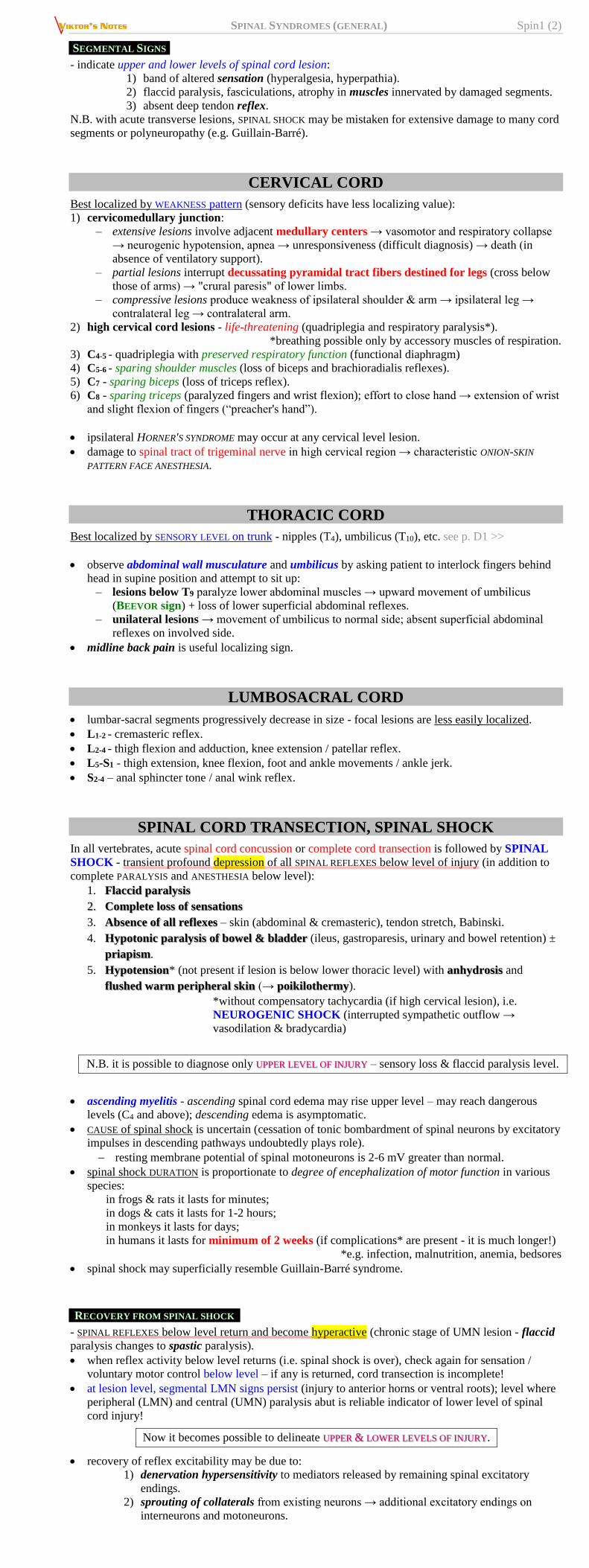
SPINAL CORD HEMISECTION
BROWN-SEQUARD SYNDROME
ETIOLOGY
1) traumatic hemisections (e.g. stab wound, lateral mass fracture in cervical spine)
2) extramedullary tumors
3) extramedullary abscesses
4) vasculitis (as in SLE).
CLINICAL FEATURES
- caudal to hemisection:
I. Contralateral effects – loss of pain-temperature sensation (tr. spinothalamicus).
N.B. sensory level is located 1-2 segments below level of lesion!!!
II. Ipsilateral effects:
1) UMN paralysis (tr. corticospinalis lat.);
if high cervical – hemidiaphragm paralysis.
2) loss of discriminative touch-proprioception (dorsal funiculus);
simple touch sensation may be unimpaired - anterolateral system carries touch sensation
from contralateral side.
ataxia cannot be seen clinically due to paralysis.
3) loss of sweating (descending autonomic fibers in ventral funiculus)
if high cervical – Horner syndrome.
4) SEGMENTAL* – anesthesia / radicular pain (dorsal root), LMN paralysis (ventral horn)
*i.e. hemisection segment
N.B. bowel and bladder control is usually intact!
DORSAL (POSTERIOR) HEMISECTION
1) dorsal funiculus – loss of vibration and position sense.
2) tr. corticospinalis lat. – paralysis.
VENTRAL (ANTERIOR) HEMISECTION
1) tr. spinothalamicus – loss of pain & temperature sense, loss of urge to urinate + preserved
dorsal funiculus function
2) tr. reticulospinalis – anhidrosis, vasodilation-hypotension, loss of voluntary* bladder-
bowel control; if rostral to C3 – paralysis of automatic breathing.
*reflex emptying intact
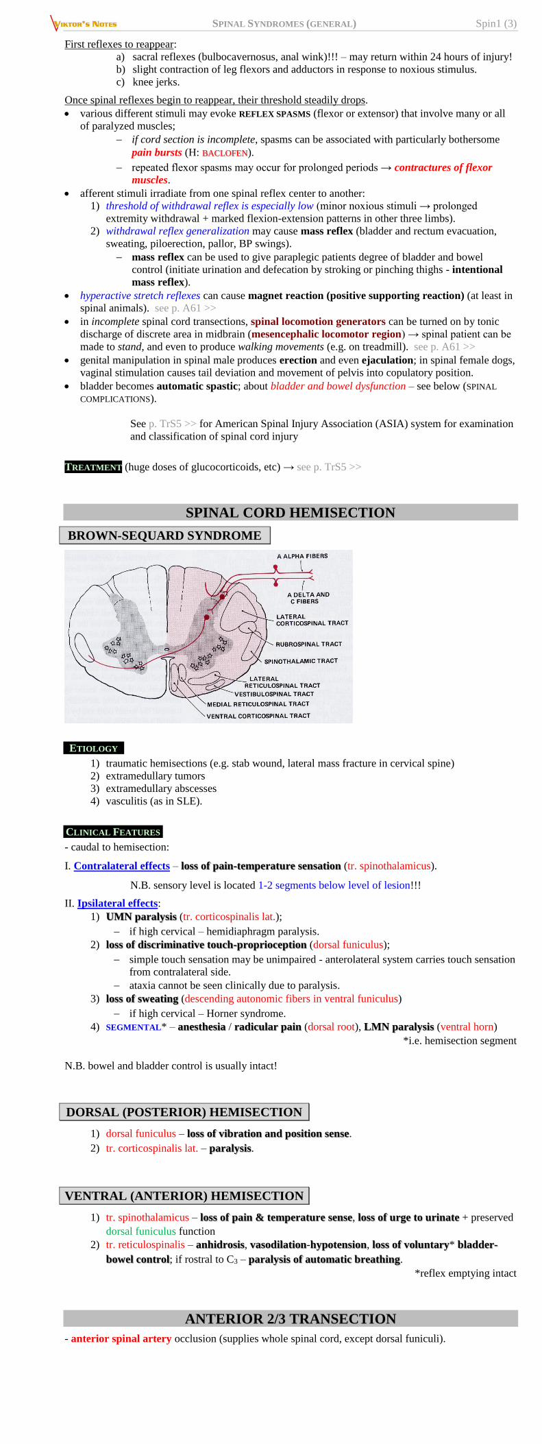
ANTERIOR 2/3 TRANSECTION
- anterior spinal artery occlusion (supplies whole spinal cord, except dorsal funiculi).
SPINAL SYNDROMES (GENERAL) Spin1 (4)
Clinically – VENTRAL HEMISECTION + spastic paralysis.
EXTRAMEDULLARY CORD COMPRESSION
ETIOLOGY
1. Spinal or epidural abscess / hematoma
2. Tumor (85% - vertebral metastases) - may present acutely even though tumor has been present
for weeks or longer.
3. Epidural granuloma (e.g. neurocysticercus).
4. Cervical or thoracic herniated intervertebral disk (central herniation may cause acute
compression without local pain).
5. Trauma
6. Atlantoaxial subluxation.
CLINICAL FEATURES
SEGMENTAL features - most reliable indication of lesion level (longitudinal location)!
1) LMN paralysis (ventral horn)
2) anesthesia / prominent radicular pain (dorsal root)
vs. intramedullary lesions - tend to produce poorly
localized burning pain rather than radicular pain
other strongly localizing symptoms – local back pain, tenderness over spine (N.B. some
lesions are painless!).
radicular pain may be exacerbated by Valsalva maneuvers, straight-leg raising test.
site of compression in transverse plane may determine clinical symptoms (e.g. laterally located
lesion → Brown-Sequard syndrome).
N.B. because most lesions twist cord and also interfere with vascular supply to sites
beyond compression, neurological signs may not demarcate exact transverse site!
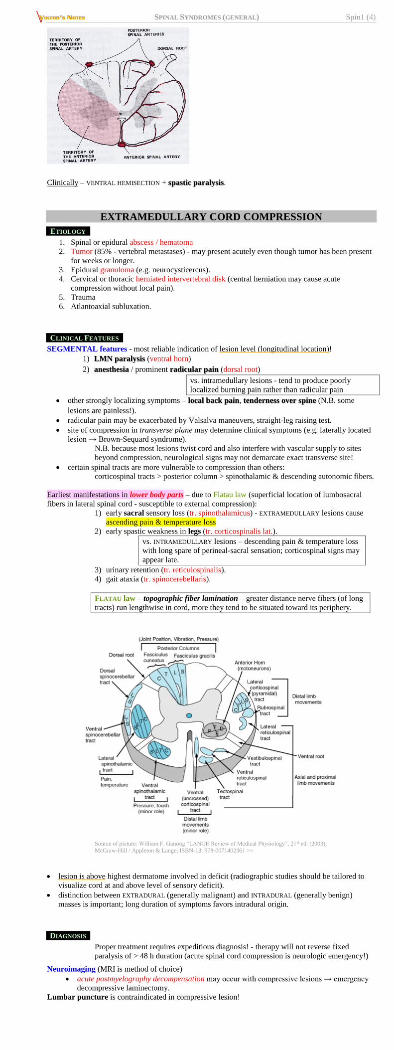
certain spinal tracts are more vulnerable to compression than others:
corticospinal tracts > posterior column > spinothalamic & descending autonomic fibers.
Earliest manifestations in lower body parts – due to Flatau law (superficial location of lumbosacral
fibers in lateral spinal cord - susceptible to external compression):
1) early sacral sensory loss (tr. spinothalamicus) - EXTRAMEDULLARY lesions cause
ascending pain & temperature loss
2) early spastic weakness in legs (tr. corticospinalis lat.).
vs. INTRAMEDULLARY lesions – descending pain & temperature loss
with long spare of perineal-sacral sensation; corticospinal signs may
appear late.
3) urinary retention (tr. reticulospinalis).
4) gait ataxia (tr. spinocerebellaris).
FLATAU law – topographic fiber lamination – greater distance nerve fibers (of long
tracts) run lengthwise in cord, more they tend to be situated toward its periphery.
Source of picture: William F. Ganong “LANGE Review of Medical Physiology”, 21st ed. (2003);
McGraw-Hill / Appleton & Lange; ISBN-13: 978-0071402361 >>
lesion is above highest dermatome involved in deficit (radiographic studies should be tailored to
visualize cord at and above level of sensory deficit).
distinction between EXTRADURAL (generally malignant) and INTRADURAL (generally benign)
masses is important; long duration of symptoms favors intradural origin.
DIAGNOSIS
Proper treatment requires expeditious diagnosis! - therapy will not reverse fixed
paralysis of > 48 h duration (acute spinal cord compression is neurologic emergency!)
Neuroimaging (MRI is method of choice)
acute postmyelography decompensation may occur with compressive lesions → emergency
decompressive laminectomy.
Lumbar puncture is contraindicated in compressive lesion!

SPINAL SYNDROMES (GENERAL) Spin1 (5)
TREATMENT
Spinal cord compression is emergency! see p. Onc56 >>
1) steroids – give immediately!
2) antibiotics (if indicated).
3) immediate radiotherapy (for cord compression due to malignancy).
4) surgical decompression (where radiotherapy is not effective)
CENTRAL CORD SYNDROME
- pathological process starts centrally and proceeds centrifugally → characteristically evolving motor
and sensory signs.
ETIOLOGY
1. Syringomyelia
2. Intramedullary cord tumors (esp. central canal ependymoma)
3. AVM 4. Anterior spinal artery ischemia.
5. Spinal cord trauma: see p. TrS5 >>
a) neck hyperextension in presence of narrow spinal canal → cord compression between
bony bars anteriorly and thickened ligamentum flavum posteriorly → cord
hypoperfusion in central watershed distribution.
b) hematomyelia (usually confined to central gray matter)
CLINICAL FEATURES
Characteristic initial presentation - combination of SEGMENTAL (at level of lesion) features:
1. Loss of pain and temperature sensation – due to lesion to central cord portion where
spinothalamic fibers decussate.
because only decussating spinothalamic tract fibers are affected, loss of pain and temperature
is bilateral but affects only those segments of spinal cord involved in pathological process
(suspended sensory loss with normal sensation above and below lesion).
may produce poorly localized burning pain.
vs. extramedullary cord compression – radicular pain
posterior column sensation is preserved (disassociated sensory loss).
2. LMN signs (SYRINGOMYELIA or TUMOR usually invade anterior horns early;);
in SYRINGOMYELIA (expands centrifugally), LMN damage follws after pain-temperature
involvement.
in SYRINGOMYELIA, segmental pattern characteristically begins in upper cervical segments
(distal arms suffer first!).
in CERVICAL TRAUMA, initial quadriplegia is replaced over minutes by leg recovery.
If lesion expands centrifugally, it may compromise other spinal structures:
1) lateral corticospinal tracts - late involvement!
vs. extramedullary cord compression – early, with legs affected first
2) ascending (vs. decussating) spinothalamic tract fibers
N.B. because spinothalamic tracts are topographically laminated (FLATAU law -
sacral fibers in most ventral-lateral position), sacral dermatomes are long preserved
(sacral sparing) – INTRAMEDULLARY lesions cause descending loss of pain and
temperature sensation.
vs. EXTRAMEDULLARY cord compression - ascending loss of pain
and temperature sensation with early sacral involvement
3) posterior columns
4) intermediolateral columns → autonomic manifestations (Horner's syndrome, sudomotor and
vasomotor dysfunction, trophic changes [esp. hands]).
CAUDA EQUINA vs. CONUS MEDULLARIS syndrome
CONUS MEDULLARIS – tapered caudal termination of spinal cord (lower sacral & coccygeal segments).
CAUDA EQUINA – collection of intradural elongated roots of lumbar & sacral spinal nerves.
Feature
Cauda Equina Conus Medullaris Pain Severe radicular pain (sciatica) &
low back pain
Back pain (less severe than
radicular pain)
Sensory loss Asymmetric saddle anesthesia* – all
modalities (radicular sensory loss)
Bilateral saddle anesthesia* (usually
restricted to perianal region) – all
modalities or touch preservation.
Motor deficits Asymmetrical areflexic para- / mono-
plegia
Absent!!! (or mild distal leg paresis)
Evacuation
disorder
Late and mild – hypotonic bladder
(urinary retention)**
Early - atonic bladder (urinary
retention with overflow
incontinence), atonic anal sphincter
(constipation with incontinence)
Impotence ± +
Bulbocavernosus
(S2-4) & anal wink
(S4-5) reflexes
+ ABSENT
*saddle anesthesia - sensory loss confined to S3-5 dermatomes.
**may be limited to asymptomatic bladder retention noted only on postvoid catheterization (>
100 mL)
Patient may not feel urge to urinate! (ask every patient with back pain about
difficulty with urination and defecation)
Nerve roots in cauda equina:
SPINAL SYNDROMES (GENERAL) Spin1 (6)
poorly developed epineurium - particularly susceptible to injury (in peripheral nerves well
developed epineurium protects against compressive and tensile stresses).
relative hypovascularity in proximal third of root (nutritional supply is supplemented with
increased vascular permeability* and diffusion from surrounding CSF).
*may result in edema compounding initial and sometimes seemingly slight injury.
Causes of CAUDA EQUINA syndrome:
1) tumor
2) abscess
3) lumbar spinal stenosis
4) lumbar disk disease
5) arachnoiditis
6) spinal anesthesia
7) trauma.
MRI is criterion standard for initial evaluation.
TREATMENT - directed at underlying cause.
in acute or traumatic syndrome, some suggest METHYLPREDNISOLONE (similar to traumatic
spinal cord injury); steroids have not shown significant benefit in penetrating trauma.
surgical decompression, e.g. lumbar laminectomy (timing is controversial - immediate,
early, and late surgery shows varying results; usual recommendation – within 24-48 hours). cauda equina injuries (involving peripheral nerves rather than spinal cord) are surgically
remediable for longer periods than conus medullaris injuries
SPINAL COMPLICATIONS
higher anatomical level of injury, greater risk of complications.
usual symptoms associated with medical illnesses may be lacking (because of destruction of
afferent pain pathways).
unexplained fever, spasticity worsening, neurologic function deterioration should prompt search
for underlying cause (infection, thrombophlebitis, intraabdominal pathology).
N.B. loss of normal thermoregulation can produce recurrent fever (quadriplegic fever)!
VCU protocol Recommendations for common SCI sequelae DVT Prophylaxis: Patient is considered high risk. Recommend Lovenox 40mg SQ BID plus SCD's or thigh-high
Ted Hose. Neurogenic Bladder: Continue Foley and monitor I&O’s. Consider transition to intermittent catheterization (IC
Q6h) if fluid output less than or around 2L daily. Keep IC volumes < 500cc. If voiding, check PVR/bladder scan and cath if >150mL PVR. If needing any form of IV fluids, can keep foley in place.
Neurogenic Bowel: Docusate BID, Senna QHS, Bisacodyl supp daily with rectal digital stimulation. Can add MiraLax as necessary.
Respiratory Insufficiency: Incentive spirometry, Mechanical in/exsufflation titrating to 40:40 or greater, chest PT. Trach. Spasticity prevention: ROM and positioning. Medications such as Baclofen, Tizanidine, Valium, or Dantrolene
as necessary. Pressure Ulcer Risk: Turn q2h, inspect skin daily or per unit protocol. If hypotension: Abd binder, TED hose. If meds required, consider midodrine (first-line) or Fluorinef. If signs of depression: Psychology consult or provide chaplain services as needed. Fevers: Evaluate for common sources (UTI, PNA, wounds, DVT, HO). Autonomic dysreflexia risk (Level T6 and above): if acute HTN occurs with diaphoresis and headache,
explore underlying noxious sources below the level of the injury, such as bladder distention, non-draining Foley, UTI, fecal impaction, pressure sore, tight clothes/splints, etc. and evaluate/treat. If none of those, consider CT abdomen/pelvis to evaluate for intraabdominal pathology.
Heterotopic ossification (HO) risk: monitor for decreased ROM (e.g., of the hip, knee, shoulder, elbow) or for increased serum alk phos.
Sexual: neurogenic sexual dysfunction. Psychosocial: support as needed.
PARESTHESIAS
- burning / shooting pains below level of spinal cord lesion.
causes:
a) selective deafferentation of spinothalamic pathway with preservation of function of dorsal
columns.
b) abnormal discharge of thalamic neurons.
c) another occult lesion in conus or cauda equina.
treat as neuropathic pain (e.g. GABAPENTIN, PREGABALIN); avoid narcotics (bowel and bladder
adverse effects).
DECUBITUS ULCERS see p. 2217 >>
- predisposed by immobility and lack of sensation!
BLADDER DYSFUNCTION see p. 2590a >>
SPINAL SHOCK – atonic bladder; H: cont Foley to prevent urinary retention (→ permanent bladder
atony). Consider intermittent catheterization (IC Q6h) if fluid output < 2L/d. Keep IC volumes < 500
mL. If voiding check PVR / bladder scan.
CHRONIC STAGE – bladder dysfunction depends on level of lesion:
a) lesions above sacral parasympathetic nucleus – within several days of injury,
automatic spastic bladder with detrusor-sphincter dyssynergia develops (bladder
re-education should begin promptly!);
b) lesions of conus medullaris or cauda equina – atonic bladder.
bone matrix protein breakdown + immobilization → osteoporosis, large Ca2+ release →
hypercalcemia and hypercalciuria → urinary calcium stones (H: urine acidification).
stones + dysfunctional bladder → urinary stasis → UTI (most common complication of spinal
cord injury!!!), hydronephrosis, autonomic dysreflexia.
GU tract is primary source of infection after cord trauma!
– prophylactic antibiotics are not indicated.
GI COMPLICATIONS
paralytic ileus almost universally occurs after cord trauma; H: nasogastric suctioning.
– in few days, small bowel function returns to normal, but large bowel and rectal function may be
lost permanently.
consider prophylaxis for GI stress ulcers.
SPINAL SYNDROMES (GENERAL) Spin1 (7)
for several weeks after acute spinal injury (anal sphincter is atonic) laxatives and digital
disimpaction are necessary in most patients to ensure at least biweekly evacuation;
– scheduled stool softeners (e.g. DOCUSATE BID), SENNA QHS, stool bulking agents (e.g.
PSYLLIUM), MIRALAX ac lunch.
– PRN BISACODYL supp; GLYCERIN suppositories are also useful (insert ≈ 20 min before desired
time of evacuation); PRN MILK OF MAGNESIA, MAGNESIUM CITRATE
– avoid anus stretching!
– flatus tube may be helpful.
later, start training for REGULAR DEFECATION - GLYCERIN suppositories on alternate days.
Both bowel and bladder sphincter reflexes can be trained to provide reflex emptying if lesions
spare lower motor neurons.
SEXUAL DYSFUNCTION
1. Mechanical and pharmacologic interventions
2. Psychosocial counseling
Men:
in men, priapism is seen early (esp. after high cord lesions) → reflex but no psychogenic erection.
semen quality and motility is reduced because of repeated UTIs.
Women:
paraplegia and tetraplegia result in menstrual cycle interruption for months, but this returns with
time - conception and pregnancy are possible.
women may experience life-threatening autonomic hyperreflexia during delivery.
MALNUTRITION
anorexia → early loss of weight occurs in many spinal patients.
patients (like all immobilized patients) catabolize large amounts of body protein → develop
negative nitrogen balance.
protein may be lost through bedsores.
prophylaxis / treatment - diet high in protein, calories, and vitamins (incl. parenteral
hyperalimentation).
calcium & vitamin D supplementation - to avoid osteoporosis.
RESPIRATORY FAILURE
respiratory failure is caused by:
a) neurological compromise
b) pain
c) retropharyngeal hematoma (from cervical trauma)
acute pulmonary edema has occurred after cervical spine injuries unassociated with significant
head injury.
respiratory failure is exacerbated by CNS depressants, immobilization in recumbency, abdominal
distention (from paralytic ileus).
atelectasis → pneumonia.
check at regular intervals - vital capacity, arterial blood gases / pulse oximetry.
for cervical cord lesions:
1) artificial ventilation (tracheal intubation → tracheostomy)
2) phrenic nerve pacemakers - for lesions at C5 or above. NeuRx DPS RA/4 Respiratory Stimulation System (FDA approved) - implantable
electronic device that stimulates diaphragm - allows to breathe for at least 4 hours a day
without a mechanical ventilator.
3) chest physical therapy
4) negative-pressure cuirass (to alleviate atelectasis, particularly if lesion is below C4).
N.B. in lesions above T10, there is no effective coughing!
H: regular nasotracheal suctioning, chest physiotherapy, use of rotating beds or frames
VENOUS THROMBOSIS & PULMONARY EMBOLISM
- high risk in acute cord injury.
1) calf-compression devices (for first two weeks)
2) anticoagulation: ENOXAPARIN (30 mg SC every 12 h) → WARFARIN (INR 2-3) for 3 months
in persistent paralysis.
SPASTICITY
- major late complication of spinal cord disease (weeks ÷ months after initial insult).
most severe spasticity - incomplete traumatic injury, multiple sclerosis.
if lesion involves upper cervical cord, spasms may involve all four extremities, trunk, and
bladder.
spasms of extremities are usually flexor (but may also be extensor).
severe spasticity may lead contractures.
treatment (if spasms are painful, interfere with rehabilitation, or delay healing of bedsores)
→ see p. Mov3 >>
AUTONOMIC DYSFUNCTION
descending pathways from brain normally coordinate sympathetic activity and modulate segmental
autonomic reflexes; spinal cord transection may be attended by autonomic hyperreflexia
(affecting bowel, bladder, sexual, temperature-regulation, and cardiovascular functions).
BLOOD PRESSURE is generally normal at rest, but precise feedback regulation normally supplied by
baroreceptor reflexes is absent:
wide swings in BP are common (quadriparetic patients exhibit both orthostatic
hypotension and supine hypertension after upward tilting).
vasopressin & renin-angiotensin-aldosterone system have enhanced role in maintenance
of orthostatic arterial pressure.
patients are at risk of bradycardia & cardiac arrest during tracheal suction (or other
maneuvers that activate vagovagal reflexes).
inability to sense heat or cold exposure below level of injury → dangerous increases / decreases
in body temperature.
PAROXYSMAL AUTONOMIC HYPERREFLEXIA (s. AUTONOMIC DYSREFLEXIA)
- in lesions above major splanchnic sympathetic outflow (i.e. lesions above T5-6; e.g. affects 85%
patients with lesion above C6).
SPINAL SYNDROMES (GENERAL) Spin1 (8)
trigger - noxious stimulus below level of cord lesion (e.g. fecal impaction, bladder distention,
catheter insertion, UTI, decubitus ulcer).
sensory inputs activate sympathetic neurons of intermediolateral nuclei in thoracic spinal cord →
massive reflex activation of sympathetic outflow below lesion → vasoconstriction (below level of
lesion), tachycardia, systemic hypertension (up to 300 mmHg!!!*)
*may lead to life-threatening hypertensive encephalopathy, stroke, retinal hemorrhage!
reflex pathways (via carotid and aortic baroreceptors) then inhibit sympathetic activity above cord
lesion → vasodilation (flushing, nasopharyngeal congestion, headache), diaphoresis above level
of lesion, bradycardia.
N.B. descending pathways are blocked - sympathetic hyperactivity below lesion continues.
prophylaxis-treatment:
1) removal of offending stimuli.
2) BP can often be lowered by tilting head upward.
3) ganglionic blockers (MECAMYLAMINE, 2.5-5 mg)
4) short-acting antihypertensives (e.g. CLONIDINE prophylactically to reduce hypertension
resulting from bladder stimulation; NIFEDIPINE).
AUTONOMIC HYPERREFLEXIA in addition to SOMATIC HYPERREFLEXIA (SPASTICITY) may lead to
accumulation of contractures, bladder, bowel, and skin disorders, which eventually cause severe
wasting and death!
PSYCHIATRIC DYSFUNCTION
depression (following initial period of denial) occurs in almost all patients and may be masked by
jocularity.
suicide rate is 5 times higher than in general population (lower for men; 2 times higher in
marginally disabled persons compared to more severely affected individuals).
narcotic addiction is also occasionally problem.
SPINAL PROGNOSIS
No effective means to promote repair of injured spinal cord tissue!
if total loss of motor power & sensation distal to level (feature of complete transection) persist for
> 24 hours* - 99% will not have functional recovery.
*ensure that spinal shock is not present and sacral sparing is carefully excluded
after acute spinal cord lesion, prospects for significant recovery fade after ≈ 4 months (recovery
plateaus between 6 and 12 months);
– many patients even after complete spinal cord injuries, regain 1-2 levels (or some key
muscles) after > 1 year – esp. important in high cervical lesions!
prognosis in TRANSECTED SPINAL CORDS used to be very poor (LIFE EXPECTANCY is greatly
decreased);
in past, renal failure was leading cause of death after spinal cord trauma.
currently, pulmonary problems (pneumonia, pulmonary emboli, sepsis) are single
most common cause of morbidity and mortality after spinal cord trauma.
– antibiotics and meticulous attention to nutrition, fluid balance, skin care, bladder function,
and general nursing care have reduced mortality to 6%.
SPORT aspects:
– any injury that necessitates internal surgical spinal stabilization obviates return to contact
sports.
– minor injuries that heal correctly with bracing may not limit athletic involvement.
Disability and survival associated with spinal cord damage are determined by:
1) level of lesion
2) completeness of transection
3) age (prognosis is better with younger age).
Expected Neurologic Function Following Complete Cord Lesions:
Level Self-Care Transfers Maximum Mobility
High quadriplegia
(C1-4)
Dependent on others;
requires respiratory support
(e.g. implantation of
diaphragmatic stimulators)
Dependent on
others
Motorized wheelchair
Low quadriplegia
(C5-8)
Partially independent with
adaptive equipment
May be
dependent or
independent
May use manual
wheelchair, drive
automobile with adaptive
equipment
Paraplegia
(below T1)
Independent Independent Ambulates short distances
with aids
Even complete high cervical cord lesions may be compatible with productive life!
SPINAL REHABILITATION
Functional recovery is continuous process in the first year after SCI
best carried out in experienced spinal centers.
best if single physician organizes long-term approach.
start early (once spine stabilization has been achieved).
– early range of motion prevents contractures, diminishes risk of venous thrombosis,
protects skin, and boosts morale.
– bed should be fitted with footboards to keep ankles and toes in neutral position.
– soft braces to fix lower extremities in neutral position.
– exercises to strengthen unaffected muscles.
– gradual progression toward vertical position (simultaneous monitoring of systemic BP -
horizontal position for prolonged period results in sympathetic tone loss)
major focus of rehabilitation:
1) bowel management
2) bladder management
3) transfer techniques
ultimate aim - AMBULATION & ECONOMIC INDEPENDENCE.
transient hypoxia (through measured breathing treatments), along with overground
walking training, improves walking speed and endurance after incomplete SCI - Class I
evidence Daily intermittent hypoxia enhances walking after chronic spinal cord injury. A randomized
trial. Heather B. Hayes, PhD*, Arun Jayaraman, PT, PhD*, Megan Herrmann, DPT, Gordon S.
Mitchell, PhD, William Z. Rymer, MD, PhD and Randy D. Trumbower, PT, PhD
Neurology January 14, 2014 vol. 82 no. 2 104-113
psychological support throughout disease course is necessary (severe depression can occur after
losing control of body).
SPINAL SYNDROMES (GENERAL) Spin1 (9)
special adaptive devices may allow patients to drive.
recently, role of CENTRAL PATTERN GENERATORS and possibility of activating standing and stepping
circuits after SCI even in chronic injury phase has been addressed; in 1914, Graham Brown demonstrated existence of central pattern generators for
walking in animals (neuronal networks capable of creating rhythmic motor activity in
absence of phasic sensory input).
theoretically, similar system exists in humans and can be activated by repeated
exercise or stimulation of walking pathways; exercise programs have been developed
(incl. suspended body weight support system over treadmill to facilitate walking and
bicycles designed for SCI).
using submotor threshold epidural spinal cord stimulation below injury level +
intensive rehab (step-and-stand) → motor recovery in chronically paralyzed individuals Johnson S1, Friedlander RM, Monaco EA 3rd “Complete spinal cord injury: an indication for
spinal cord stimulation?” Neurosurgery. 2014 Oct
Spinal Cord Independence Measure (SCIM)
recommended by “Clinical Assessment Following Acute Cervical Spinal Cord Injury” guidelines
(Level 1 evidence)
comprehensive FUNCTIONAL OUTCOME rating scale designed specifically for patients with spinal
cord injury - consists of 3 subscales:
1) self-care (6 items; score range, 0-20)
2) respiration and sphincter management (4 items; score range, 0-40)
3) mobility (9 items; score range, 0-40).
total score ranges from 0 to 100.
development of SCIM started in 1994.
third international version (SCIM III) to overcome intercultural differences was formulated in
2002.
also officially used in UK.
BIBLIOGRAPHY for ch. “Spinal Disorders” → follow this LINK >>
Viktor’s Notes℠ for the Neurosurgery Resident
Please visit website at www.NeurosurgeryResident.net