Spinal Surgery 1
4
1 Spinal Surgery 1 Mr Mushtaque A. Ishaque BSc(Hons) BChir(Cantab) DM FRCS FRCS(Ed) FRCS(Orth) Consultant Orthopaedic Spinal Surgeon and Senior Clinical Academy Teacher Hunterian Professor at The Royal College of Surgeons of England Teaching Aims Common Spinal Conditions Important Spinal Conditions Safe Common Spinal Pathologies Degenerative disc disease Lumbar and cervical disc herniation Spinal canal stenosis Spondylolysis Spondylolisthesis Important Spinal Pathologies Trauma Tumour Infection Deformity Inflammatory Cauda Equina Core Knowledge and Skills Anatomy, Physiology and Pathology History and Examination Draw up a Differential Diagnosis Anatomy of the Spine Central axis of the skeleton Supports the Skull Attachment to the thoracic cage and hence the pectoral girdle and upper limbs Linked via the pelvis to the lower limbs
Transcript of Spinal Surgery 1

1
Spinal Surgery 1
Mr Mushtaque A. Ishaque BSc(Hons) BChir(Cantab) DM FRCS FRCS(Ed) FRCS(Orth)
Consultant Orthopaedic Spinal Surgeon and Senior Clinical Academy Teacher Hunterian Professor at The Royal College of Surgeons of England
Teaching Aims
Common Spinal Conditions
Important Spinal Conditions
Safe
Common Spinal Pathologies
Degenerative disc disease Lumbar and cervical disc herniation
Spinal canal stenosis Spondylolysis
Spondylolisthesis
Important Spinal Pathologies
Trauma Tumour Infection
Deformity Inflammatory Cauda Equina
Core Knowledge and Skills
Anatomy, Physiology and Pathology
History and Examination
Draw up a Differential Diagnosis
Anatomy of the Spine
Central axis of the skeleton
Supports the Skull
Attachment to the thoracic cage and hence the pectoral girdle and upper limbs
Linked via the pelvis to the lower limbs
2
Anatomy
Strength of the column is from the size and architecture of the bony elements and the strength of the ligaments and muscles
Protects the neural elements
Anatomy
Cervical Spine Atlas and axis
5 Typical vertebrae
Coccyx
Sacrum 5 segments
Lumbar Spine 5 typical vertebrae
Thoracic Spine 12 Typical vertebrae
Anatomy Anatomy
Anatomy
Annulus fibrosis Nucleus pulposus
Intervertebral Disc
Anatomy
Cord C1 to T12
Conus medullaris T12 to L1
Cauda Equina L2 to sacrum
Long fibre tracts UMN
Nerve roots LMN
3
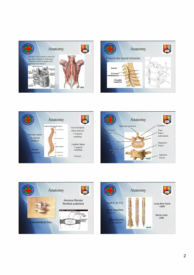
Anatomy Spinal Cord, Primary Rami & Sympathetic Chain
Dorsal Root Ganglion
Medial
Lateral
Posterior 1º Rami
Anatomy
Corticospinal Motor
Syndromes Anterior Central Mixed
Brown-Séquard Dorsal
Spinal Cord Columns and Syndromes
Anatomy
Dermatomal Map
Anatomy
Lower Limb Muscle Groups
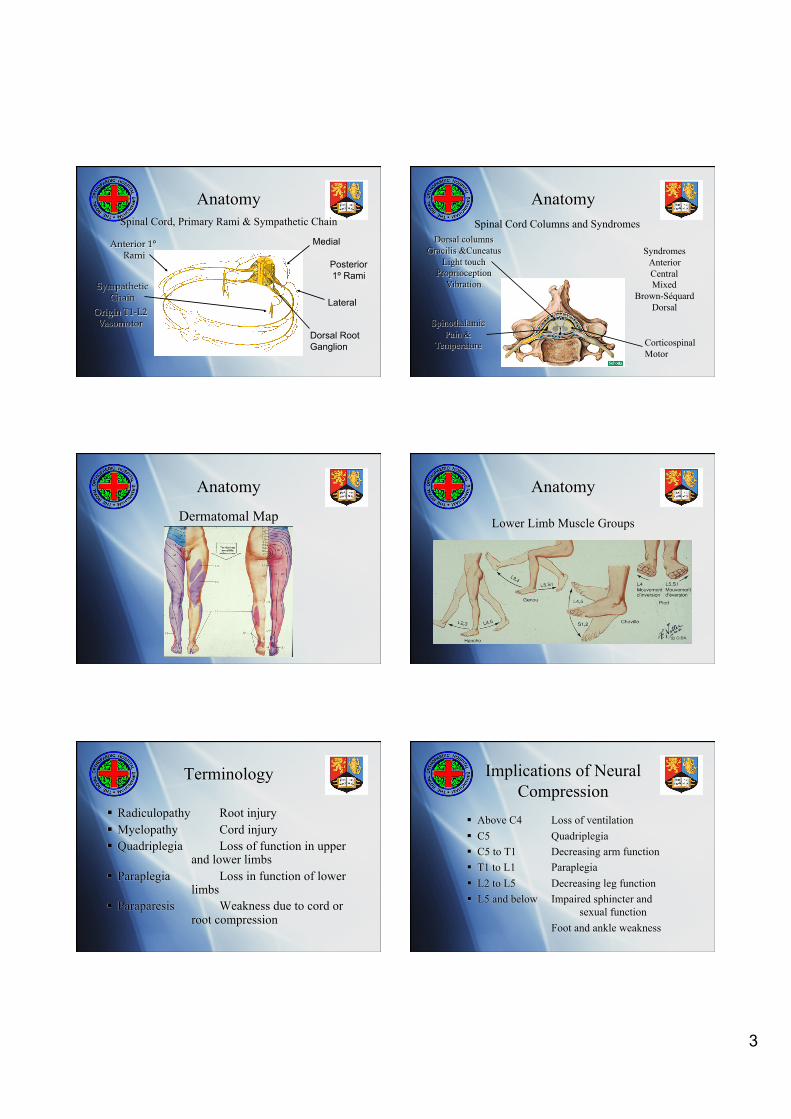
Terminology
Radiculopathy Root injury Myelopathy Cord injury Quadriplegia Loss of function in upper
and lower limbs Paraplegia Loss in function of lower
limbs Paraparesis Weakness due to cord or
root compression
Implications of Neural Compression
Above C4 Loss of ventilation C5 Quadriplegia C5 to T1 Decreasing arm function T1 to L1 Paraplegia L2 to L5 Decreasing leg function L5 and below Impaired sphincter and
sexual function Foot and ankle weakness

4
Cervical & Thoracic Cord Compression
Spastic paresis/paralysis
Increased tone and clonus
Brisk reflexes
Extensor plantar response
Retention, overflow and automatic bladder
Cauda Equina & Compression Below L1
Radicular weakness
Muscle wasting & fasciculation
Decreased tone & loss of reflexes
Autonomous dribbling bladder
Impotence
Myelopathy
Weakness of Upper limbs > Lower limbs Loss of fine motor skills Broad based shuffling gait Spasticity May have Upper limb radiculopathy Brisk reflexes and extensor plantars Rarely get urinary symptoms
Radiculopathy
Radiculitis - Root pain
Paraesthesia or numbness
Motor weakness
Sphincter disturbance
Ready to go !
Think about putting it all together
History and examination
Investigations
Correlate you findings