Spinal Cord, Spinal Nerves - De Anza College cord...Spinal cord ! Gray matter on the inside ! White...
21
Spinal Cord, Spinal Nerves A&P lab Dr. Kandula
Transcript of Spinal Cord, Spinal Nerves - De Anza College cord...Spinal cord ! Gray matter on the inside ! White...
Spinal Cord, Spinal Nerves
A&P lab Dr. Kandula
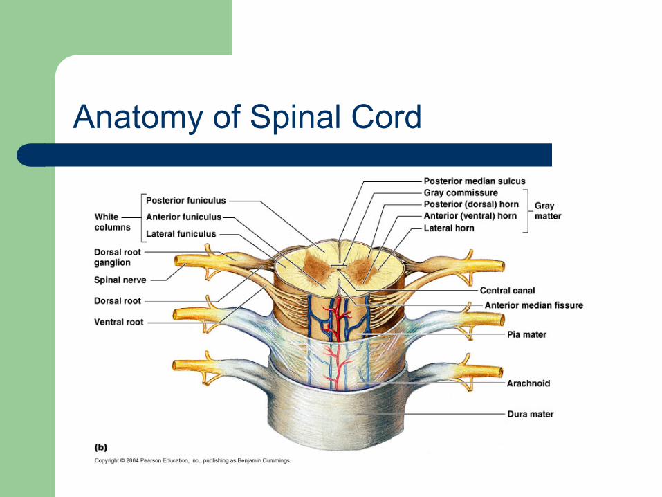
Anatomy of Spinal Cord
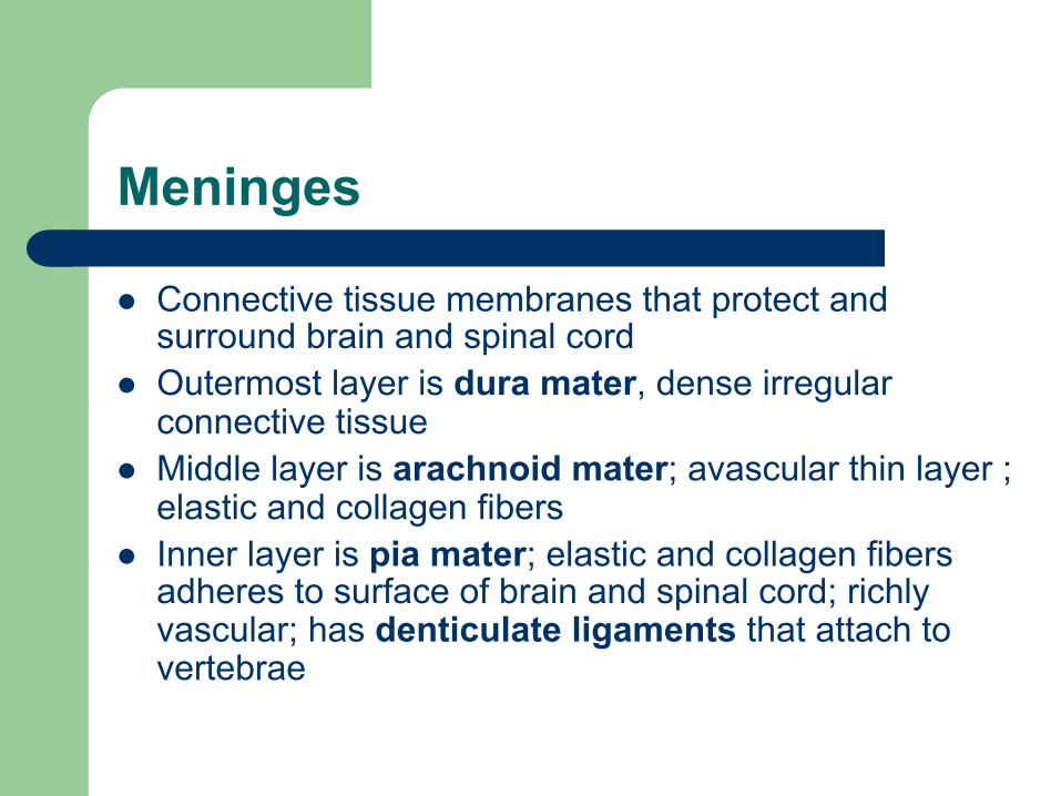
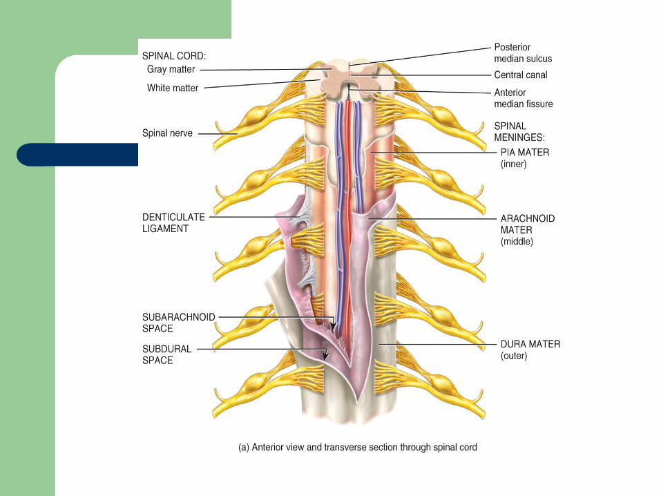
Meninges
l Connective tissue membranes that protect and surround brain and spinal cord
l Outermost layer is dura mater, dense irregular connective tissue
l Middle layer is arachnoid mater; avascular thin layer ; elastic and collagen fibers
l Inner layer is pia mater; elastic and collagen fibers adheres to surface of brain and spinal cord; richly vascular; has denticulate ligaments that attach to vertebrae
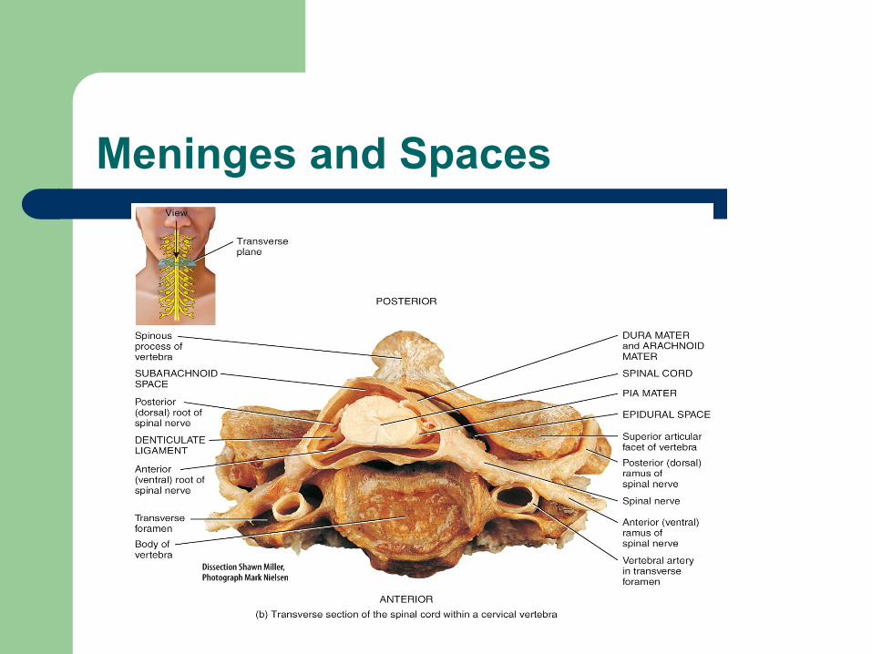
Meninges and Spaces
Spaces
l Epidural space - This is a space between the dura mater and the vertebral bone; it is filled with adipose tissue and connective tissue
l Subdural space – found between dura mater and arachnoid; contains interstitial fluid
l Subarachnoid space – between pia mater and arachnoid; contains cerebrospinal fluid
Denticulate ligaments
l Extensions of pia mater connect the spinal cord to the dura mater and the vertebral bodies
l Help to hold the spinal cord in position
Functions of the Spinal Cord
1. Process reflexes 2. Integrates EPSPs* and IPSPs** 3. Conducts sensory impulses to the brain and
motor impulses to effectors * Excitatory postsynaptic potentials ** Inhibitory postsynaptic potentials
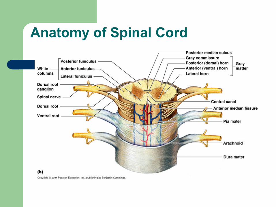
Anatomy of Spinal Cord
Spinal cord
l Begins at foramen magnum l 16 – 18 inches long l Ends at level of lumbar vertebra L1 or L2 in a
conical end called conus medullaris l Below this in the vertebral column – cauda
equina l Filum terminale – pia mater extension
Spinal cord
l 2 enlargements in transverse section cervical lumbar l Anterior surface – anterior median fissure l Posterior surface – posterior median sulcus l Central hollow tube – central canal filled with
CSF
Spinal cord
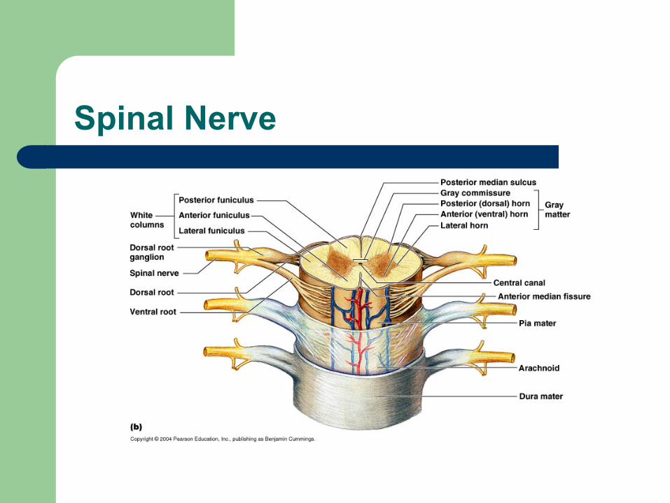
l Gray matter on the inside l White matter on the outside l Gray matter is composed of neuron bodies
– Found in center of spinal cord and looks like a butterfly
– Dorsal, ventral and lateral horns – Gray commissure
Spinal cord
l White matter is composed of axons covered in myelin
l 3 columns of white matter are called funiculi – Posterior, anterior and lateral – Each contains several tracts of axons.
Spinal Nerves
l Spinal nerves connect the CNS to sensory receptors, muscles, and glands and are part of the peripheral nervous system
l 31 pairs of spinal nerves l Anterior and posterior roots attach a spinal
nerve to a segment of the spinal cord
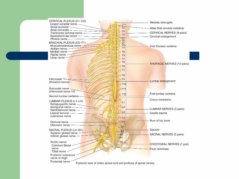
Spinal nerves
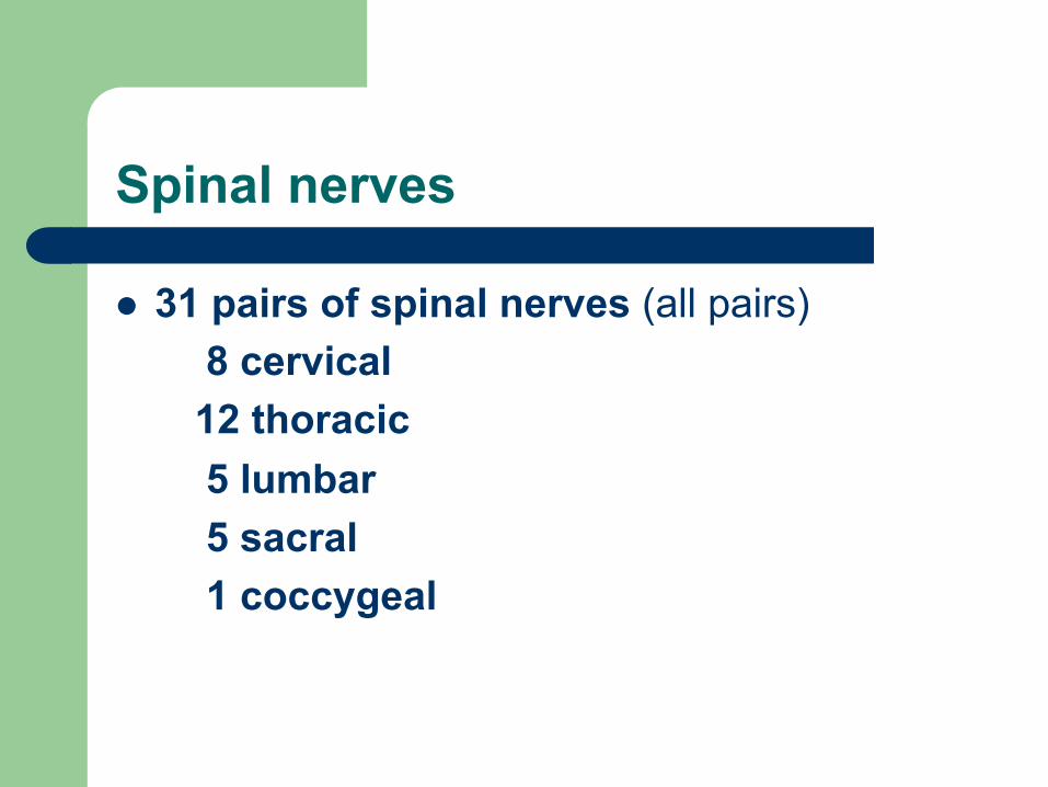
l 31 pairs of spinal nerves (all pairs) 8 cervical 12 thoracic 5 lumbar 5 sacral 1 coccygeal
Spinal Nerve
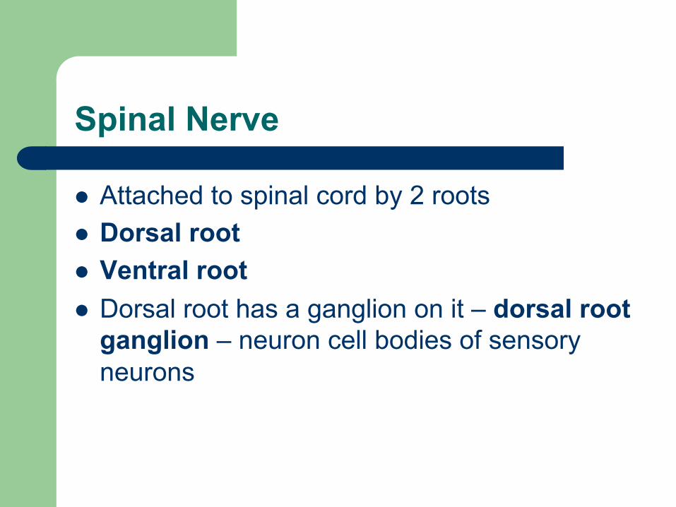
l Attached to spinal cord by 2 roots l Dorsal root l Ventral root l Dorsal root has a ganglion on it – dorsal root
ganglion – neuron cell bodies of sensory neurons
Spinal Nerve
Spinal nerve
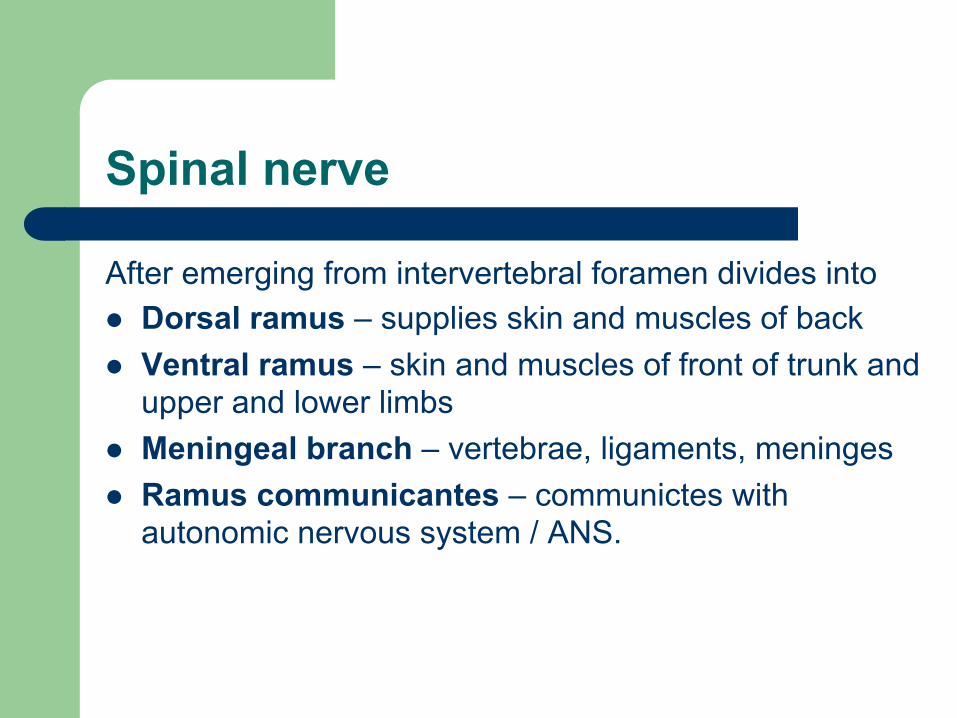
After emerging from intervertebral foramen divides into l Dorsal ramus – supplies skin and muscles of back l Ventral ramus – skin and muscles of front of trunk and
upper and lower limbs l Meningeal branch – vertebrae, ligaments, meninges l Ramus communicantes – communictes with
autonomic nervous system / ANS.
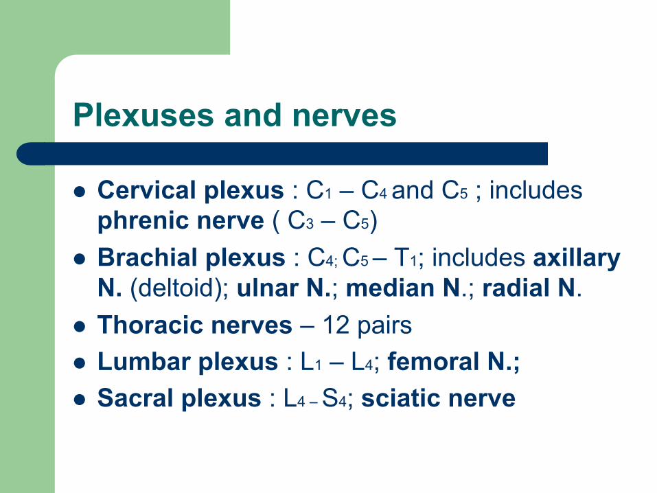
Plexuses and nerves
l Cervical plexus : C1 – C4 and C5 ; includes phrenic nerve ( C3 – C5)
l Brachial plexus : C4; C5 – T1; includes axillary N. (deltoid); ulnar N.; median N.; radial N.
l Thoracic nerves – 12 pairs l Lumbar plexus : L1 – L4; femoral N.; l Sacral plexus : L4 – S4; sciatic nerve