Spinal cord injury: is monitoring from the injury site the ...
9
VIEWPOINT Open Access Spinal cord injury: is monitoring from the injury site the future? Samira Saadoun and Marios C. Papadopoulos * Abstract This paper challenges the current management of acute traumatic spinal cord injury based on our experience with monitoring from the injury site in the neurointensive care unit. We argue that the concept of bony decompression is inadequate. The concept of optimum spinal cord perfusion pressure, which differs between patients, is introduced. Such variability suggests individualized patient treatment. Failing to optimize spinal cord perfusion limits the entry of systemically administered drugs into the injured cord. We conclude that monitoring from the injury site helps optimize management and should be subjected to a trial to determine whether it improves outcome. Keywords: Blood pressure, CNS injury, Clinical trial, Microdialysis, Monitoring, Neurocritical care, Spinal cord injury, Surgery Background Every year, 15–40 people per million suffer a traumatic spinal cord injury (TSCI) [1]. Many TSCI patients are initially admitted to a neurointensive care unit (NICU), but their management is variable. This review introduces novel concepts to aid the management of acute TSCI in NICU based on our findings that: 1) the dura causes spinal cord compression at the injury site; 2) each pa- tient has an optimum spinal cord perfusion pressure; and 3) patient position in bed influences cord perfusion. Patient management Surgical management Some surgeons recommend early bony decompression, but others do not [2, 3]. The Surgical Timing in Acute Spinal Cord Injury Study showed better outcome at 6 months in patients who had decompressive surgery * Correspondence: [email protected] Academic Neurosurgery Unit, St. George’s, University of London, Cranmer Terrace, Tooting, London SW17 0RE, UK within 24 h compared with >24 h after cervical TSCI [4]. This study was underpowered, not randomized, and not blinded. In the UK [3] and internationally [2, 5] there is no consensus on the timing or even the role of surgery for TSCI. Below, we argue that early surgery is controversial because surgeons perform bony decom- pression, but fail to relieve the dural compression. Medical and nursing management There are no drugs that improve outcome after TSCI. The North American Spinal Cord Injury Studies suggested that methylprednisolone given within 8 h after TSCI improves outcome [6, 7], but their findings have been criticized; methylprednisolone is no longer standard of care [3, 8, 9]. The optimum mean arterial pressure (MAP) after TSCI is unknown. The American Association of Neurological Surgeons recommends a MAP of 85–90 mmHg for 7 days with little supporting evidence [10]. In the UK, blood pressure management is variable with 23 % of NICU spe- cialists aiming for MAP >60 mmHg, 54 % >80 mmHg, and 15 % within 20 % of what is considered normal for age [3]. The effects on the injured cord of different anes- thetics, altering arterial pCO 2 , administering vasopressors or mannitol, and patient position in bed are also unclear. In the UK, 96 % of neuroanesthesiologists avoid N 2 O [3] which elevates intracranial pressure (ICP), though its effect on intraspinal pressure (ISP) is unknown. Most surgeons fix the spine and mobilize patients early [2], but in the UK Midlands Spinal Injuries Centre patients were kept flat for 6 weeks after TSCI with an equally good out- comes [11]. Such variability in practice suggests that the optimum medical and nursing management are unknown. Below, we show how neuromonitoring may be used to optimize management. How to optimize management History of monitoring from the injury site In 2009, whilst studying the role of the water channel protein AQP4 in TSCI in mice, Dr Saadoun noticed that the injured cord was compressed against the dura, based © 2016 The Author(s). Open Access This article is distributed under the terms of the Creative Commons Attribution 4.0 International License (http://creativecommons.org/licenses/by/4.0/), which permits unrestricted use, distribution, and reproduction in any medium, provided you give appropriate credit to the original author(s) and the source, provide a link to the Creative Commons license, and indicate if changes were made. The Creative Commons Public Domain Dedication waiver (http://creativecommons.org/publicdomain/zero/1.0/) applies to the data made available in this article, unless otherwise stated. Saadoun and Papadopoulos Critical Care (2016) 20:308 DOI 10.1186/s13054-016-1490-3
Transcript of Spinal cord injury: is monitoring from the injury site the ...

VIEWPOINT Open Access
Spinal cord injury: is monitoring from theinjury site the future?Samira Saadoun and Marios C. Papadopoulos*
Abstract
This paper challenges the current management ofacute traumatic spinal cord injury based on ourexperience with monitoring from the injury site in theneurointensive care unit. We argue that the conceptof bony decompression is inadequate. The concept ofoptimum spinal cord perfusion pressure, which differsbetween patients, is introduced. Such variabilitysuggests individualized patient treatment. Failing tooptimize spinal cord perfusion limits the entry ofsystemically administered drugs into the injured cord.We conclude that monitoring from the injury sitehelps optimize management and should be subjectedto a trial to determine whether it improves outcome.
Keywords: Blood pressure, CNS injury, Clinical trial,Microdialysis, Monitoring, Neurocritical care, Spinalcord injury, Surgery
BackgroundEvery year, 15–40 people per million suffer a traumaticspinal cord injury (TSCI) [1]. Many TSCI patients areinitially admitted to a neurointensive care unit (NICU),but their management is variable. This review introducesnovel concepts to aid the management of acute TSCI inNICU based on our findings that: 1) the dura causesspinal cord compression at the injury site; 2) each pa-tient has an optimum spinal cord perfusion pressure;and 3) patient position in bed influences cord perfusion.
Patient managementSurgical managementSome surgeons recommend early bony decompression,but others do not [2, 3]. The Surgical Timing in AcuteSpinal Cord Injury Study showed better outcome at6 months in patients who had decompressive surgery
* Correspondence: [email protected] Neurosurgery Unit, St. George’s, University of London, CranmerTerrace, Tooting, London SW17 0RE, UK
within 24 h compared with >24 h after cervical TSCI[4]. This study was underpowered, not randomized, andnot blinded. In the UK [3] and internationally [2, 5]there is no consensus on the timing or even the role ofsurgery for TSCI. Below, we argue that early surgery iscontroversial because surgeons perform bony decom-pression, but fail to relieve the dural compression.
Medical and nursing managementThere are no drugs that improve outcome after TSCI. TheNorth American Spinal Cord Injury Studies suggested thatmethylprednisolone given within 8 h after TSCI improvesoutcome [6, 7], but their findings have been criticized;methylprednisolone is no longer standard of care [3, 8, 9].The optimum mean arterial pressure (MAP) after TSCI isunknown. The American Association of NeurologicalSurgeons recommends a MAP of 85–90 mmHg for 7 dayswith little supporting evidence [10]. In the UK, bloodpressure management is variable with 23 % of NICU spe-cialists aiming for MAP >60 mmHg, 54 % >80 mmHg,and 15 % within 20 % of what is considered normal forage [3]. The effects on the injured cord of different anes-thetics, altering arterial pCO2, administering vasopressorsor mannitol, and patient position in bed are also unclear.In the UK, 96 % of neuroanesthesiologists avoid N2O [3]which elevates intracranial pressure (ICP), though itseffect on intraspinal pressure (ISP) is unknown. Mostsurgeons fix the spine and mobilize patients early [2], butin the UK Midlands Spinal Injuries Centre patients werekept flat for 6 weeks after TSCI with an equally good out-comes [11]. Such variability in practice suggests that theoptimum medical and nursing management are unknown.Below, we show how neuromonitoring may be used tooptimize management.
How to optimize managementHistory of monitoring from the injury siteIn 2009, whilst studying the role of the water channelprotein AQP4 in TSCI in mice, Dr Saadoun noticed thatthe injured cord was compressed against the dura, based
© 2016 The Author(s). Open Access This article is distributed under the terms of the Creative Commons Attribution 4.0International License (http://creativecommons.org/licenses/by/4.0/), which permits unrestricted use, distribution, andreproduction in any medium, provided you give appropriate credit to the original author(s) and the source, provide a link tothe Creative Commons license, and indicate if changes were made. The Creative Commons Public Domain Dedication waiver(http://creativecommons.org/publicdomain/zero/1.0/) applies to the data made available in this article, unless otherwise stated.
Saadoun and Papadopoulos Critical Care (2016) 20:308 DOI 10.1186/s13054-016-1490-3

on myelograms and ISP measurements [12]. Shewondered whether the same occurs in TSCI patients. Toinvestigate this, we set up a clinical trial termed InjuredSpinal Cord Pressure Evaluation (ISCoPE), aiming tomonitor ISP from the injury site in humans. By 2014, wehad monitored the ISP in 18 patients with severe TSCIand had shown that ISP is high and potentially detri-mental [13]. A detailed morphological and spectral ana-lysis of the ISP signals followed [14]. Our studies led tothe idea of dural spinal cord compression that causescompartmentalization at the injury site [15–17]. Wethus evaluated the effect of duroplasty after TSCI [18].In 2016, we reviewed all ISCoPE patients and demon-strated the safety and probe placement accuracy of thetechnique [19]. Multi-modality monitoring from theinjury site was introduced in the same year [20], in-cluding a novel analysis technique using Kohonen self-organizing maps [21]. The ISCoPE studies are ongoing.
Brain versus spinal cord injuryNeuromonitoring is the standard of care for acute severetraumatic brain injury (TBI). We use the term neuromo-nitoring to mean monitoring ICP, tissue oxygenation,etc., in the NICU, rather than intraoperative neuro-physiological monitoring. Though a recent study sug-gested that ICP monitoring does not improve outcome[22], the findings are confounded by a delay in the startof monitoring and no rehabilitation after discharge fromthe intensive care unit. Systematic reviews suggest thatICP monitoring in TBI reduces mortality [23] and in-creases the chance of a favorable outcome [24]. Thereare advocates of multi-modality monitoring from the in-jury site including microdialysis (MD), oxygen, bloodflow, and electrocorticography based on the idea thatsecondary damage is multifactorial and arises not onlyfrom ischemia, but also from metabolic disturbance andspreading depolarizations. Probes are routinely intro-duced into the injured brain, but no such monitoringexists for TSCI.Several reasons may account for the lack of neuromo-
nitoring in TSCI. First, there is concern that insertingprobes exacerbates spinal cord damage. Second, spinalprobes require surgery for insertion. Third, many TSCIsare managed by orthopedic surgeons not accustomed toneuromonitoring. Fourth, the clinical impact of elevatedICP is easy to detect in anesthetized brain-injuredpatients as a loss of pupillary light reflex, dilated pupil,cardiovascular instability, and, ultimately, brain death. Incontrast, increased ISP is difficult to detect in anesthe-tized TSCI patients. This requires monitoring of motoror sensory evoked responses, which is not routinelydone. Fifth, CT scans are routinely used in TBI to decidemanagement, e.g., decompressive craniectomy. In con-trast, the injured cord is invisible on CT and, once the
spine is fixed, it is difficult to see on MRI. For these rea-sons, ISP has not attracted the same interest as ICP.
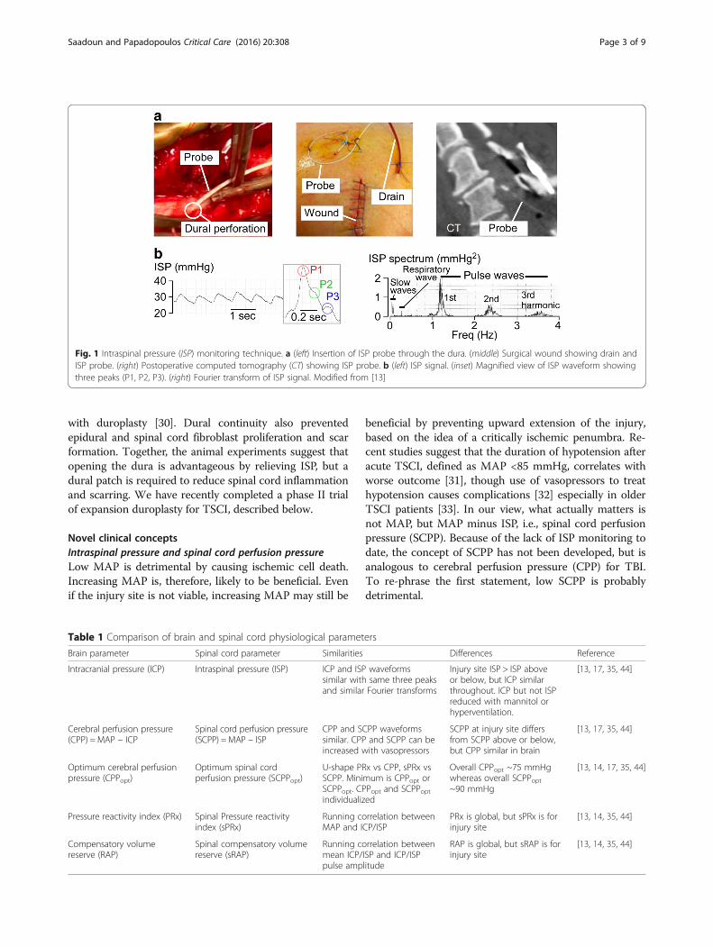
Technique of monitoring from the injury siteThe probe is inserted intradurally during surgery at theinjury site to monitor ISP for a week (Fig. 1a). The probecannot be inserted in the NICU, thus precluding moni-toring non-surgical patients. ISP monitoring is tech-nically simple and safe based on data from 42 patients[19]. The probe is easily removed in the NICU.The ISP and ICP waveforms are similar with three
characteristic peaks, identical Fourier transforms, andidentical shape change as ISP/ICP rise [14] (Fig. 1b).Thus, mathematically, the ISP signal can be subjected tothe same analyses as the ICP signal [25]. Several parame-ters can be computed from these signals that providephysiological information about the injured cord (Table 1).It is important to note that we monitor ISP from the sub-dural space, which is less invasive than intraparenchymalISP. When the injured cord is swollen and compressedagainst the dura, subdural ISP equals intraparenchymalISP at the injury site [13, 17, 26]. After a mild TSCI, thespinal cord may not be compressed against the dura; inthis case, the relation between subdural versus intrapar-enchymal ISP is unknown. To date, we have only moni-tored ISP from patients with severe TSCI.It is also possible to perform multi-modality monitor-
ing by placing ISP and MD probes on the spinal cordsurface at the injury site [20]. In our recent study of 14TSCI patients, we found that surface MD monitoring issafe [20]. There was severe metabolic derangement atthe injury site that correlated with the severity of injury.Below, we discuss how MD can be used to determinethe optimum spinal cord perfusion pressure (SCPPopt)and optimum tissue glucose concentration as well asmaximize the penetration of systemically administereddrugs into the injury site.
Animal modelsAfter TSCI in mice, the injured cord swells and is com-pressed against the dura thus generating high ISP [12].Mice that lack the water channel protein aquaporin-4have reduced spinal cord edema after TSCI with im-proved outcome. Reducing the elevated ISP in a ratcontusion TSCI model also improved outcome [27]. In apig contusion TSCI model, increased thecal sac dimen-sions were associated with reduced cord compression andreduced injury site ISP [28]. Durotomy reduced intrapar-enchymal ISP after TSCI in ex-vivo pig spinal cords [29].In a rat contusion TSCI model, durotomy with duroplastyimproved outcome more than durotomy without duro-plasty [27]. After rat TSCI, durotomy without duroplastywas associated with more macrophage accumulation, cys-tic cavitation, and fibroblast proliferation than durotomy
Saadoun and Papadopoulos Critical Care (2016) 20:308 Page 2 of 9
with duroplasty [30]. Dural continuity also preventedepidural and spinal cord fibroblast proliferation and scarformation. Together, the animal experiments suggest thatopening the dura is advantageous by relieving ISP, but adural patch is required to reduce spinal cord inflammationand scarring. We have recently completed a phase II trialof expansion duroplasty for TSCI, described below.
Novel clinical conceptsIntraspinal pressure and spinal cord perfusion pressureLow MAP is detrimental by causing ischemic cell death.Increasing MAP is, therefore, likely to be beneficial. Evenif the injury site is not viable, increasing MAP may still be
beneficial by preventing upward extension of the injury,based on the idea of a critically ischemic penumbra. Re-cent studies suggest that the duration of hypotension afteracute TSCI, defined as MAP <85 mmHg, correlates withworse outcome [31], though use of vasopressors to treathypotension causes complications [32] especially in olderTSCI patients [33]. In our view, what actually matters isnot MAP, but MAP minus ISP, i.e., spinal cord perfusionpressure (SCPP). Because of the lack of ISP monitoring todate, the concept of SCPP has not been developed, but isanalogous to cerebral perfusion pressure (CPP) for TBI.To re-phrase the first statement, low SCPP is probablydetrimental.
Fig. 1 Intraspinal pressure (ISP) monitoring technique. a (left) Insertion of ISP probe through the dura. (middle) Surgical wound showing drain andISP probe. (right) Postoperative computed tomography (CT) showing ISP probe. b (left) ISP signal. (inset) Magnified view of ISP waveform showingthree peaks (P1, P2, P3). (right) Fourier transform of ISP signal. Modified from [13]
Table 1 Comparison of brain and spinal cord physiological parameters
Brain parameter Spinal cord parameter Similarities Differences Reference
Intracranial pressure (ICP) Intraspinal pressure (ISP) ICP and ISP waveformssimilar with same three peaksand similar Fourier transforms
Injury site ISP > ISP aboveor below, but ICP similarthroughout. ICP but not ISPreduced with mannitol orhyperventilation.
[13, 17, 35, 44]
Cerebral perfusion pressure(CPP) = MAP – ICP
Spinal cord perfusion pressure(SCPP) =MAP – ISP
CPP and SCPP waveformssimilar. CPP and SCPP can beincreased with vasopressors
SCPP at injury site differsfrom SCPP above or below,but CPP similar in brain
[13, 17, 35, 44]
Optimum cerebral perfusionpressure (CPPopt)
Optimum spinal cordperfusion pressure (SCPPopt)
U-shape PRx vs CPP, sPRx vsSCPP. Minimum is CPPopt orSCPPopt. CPPopt and SCPPoptindividualized
Overall CPPopt ~75 mmHgwhereas overall SCPPopt~90 mmHg
[13, 14, 17, 35, 44]
Pressure reactivity index (PRx) Spinal Pressure reactivityindex (sPRx)
Running correlation betweenMAP and ICP/ISP
PRx is global, but sPRx is forinjury site
[13, 14, 35, 44]
Compensatory volumereserve (RAP)
Spinal compensatory volumereserve (sRAP)
Running correlation betweenmean ICP/ISP and ICP/ISPpulse amplitude
RAP is global, but sRAP is forinjury site
[13, 14, 35, 44]
Saadoun and Papadopoulos Critical Care (2016) 20:308 Page 3 of 9
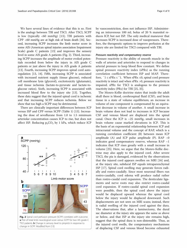
We have several lines of evidence that this is so. Firstis the analogy between TBI and TSCI. After TSCI, SCPPis low (typically <60 mmHg) [13]; TBI patients withCPP <60 mmHg are at high risk of brain death [34]. Sec-ond, increasing SCPP increases the limb motor score insome AIS (American spinal injuries association ImpairmentScale) grade C patients [13] and improves the sensorylevel in some AIS grade A patients (Fig. 2). Third, increas-ing SCPP increases the amplitude of motor evoked poten-tials recorded from below the injury in AIS grade Cpatients or just above the injury in AIS grade A patients[13]. Fourth, increasing SCPP improves spinal cord auto-regulation [13, 14]. Fifth, increasing SCPP is associatedwith increased nutrient supply (tissue glucose), reducedcell membrane lysis (glycerol), excitotoxicity (glutamate),and tissue ischemia (lactate-to-pyruvate and lactate-to-glucose ratios). Sixth, increasing SCPP is associated withincreased blood flow to the injury site [13]. Together,these data suggest that the injured spinal cord is ischemicand that increasing SCPP reduces ischemia. Below weshow that too high a SCPP may be detrimental.There are clinically important differences between ICP
versus ISP and CPP versus SCPP (Table 1) [13]. Increas-ing the dose of sevoflurane from 1.0 to 1.5 minimumarteriolar concentration causes ICP to rise, but does notaffect ISP. Reducing paCO2 by 1 kPa, which reduce ICP
by vasoconstriction, does not influence ISP. Administer-ing an intravenous 100 mL bolus of 20 % mannitol re-duces ICP, but not ISP. The only medical maneuver thatincreases SCPP is increased dose of vasopressors. There-fore, the therapeutic options to improve perfusion at theinjury site are limited for TSCI compared with TBI.
Pressure reactivity and compensatory reservePressure reactivity is the ability of smooth muscle in thewalls of arteries and arterioles to respond to changes inarterial pressure to keep blood flow constant. We definespinal pressure reactivity index (sPRx) as the runningcorrelation coefficient between ISP and MAP. There-fore, –1 ≤ sPRx ≤ +1. When sPRx ≤0, spinal cord pressurereactivity is intact and when sPRx >0, pressure reactivity isimpaired. sPRx for TSCI is analogous to the pressurereactivity index (PRx) for TBI [25, 35].The Monro-Kellie doctrine states that inside the adult
skull there is blood, cerebrospinal fluid (CSF), and braintissue in a state of volume equilibrium; an increase involume of one component is compensated by an equiva-lent decrease in volume of another. A small increase inbrain volume does not lead to increases in ICP becauseCSF and venous blood are displaced into the spinalcanal. Once the ICP is ~25 mmHg, small increases inbrain volume cause marked elevations in ICP. This isthe basis of an exponential relationship between ICP andintracranial volume and the concept of RAP, which is arunning correlation coefficient (R) between mean ICPamplitude (A) and ICP pulse amplitude (P). RAP ~0indicates good compensatory reserve, whereas RAP ~1indicates that ICP rises greatly with a small increase involume [25]. Here, we argue that the Monro-Kellie doc-trine may also apply to the injured cord. After severeTSCI, the pia is damaged, evidenced by the observationsthat the injured cord appears swollen on MRI [16] and,at the injury site, subdural ISP equals intraparenchymalISP [17]. Spinal cord swelling may generate forces radi-ally and rostro-caudally. Since most neuronal fibers runrostro-caudally, cord edema will produce radial ratherthan rostro-caudal cord expansion. The denticulate liga-ments and nerve roots may also restrict rostro-caudalcord expansion. If rostro-caudal spinal cord expansionwere possible, then the spinal cord above the injurywould be displaced upward whereas the spinal cordbelow the injury would be displaced downward. Suchdisplacements are not seen on MRI scans; instead, thereis radial swelling of the injured cord against the dura.The observations that, after a laminectomy, the duralsac diameter at the injury site appears the same as aboveor below, and that ISP at the injury site remains high,suggest that the spinal dura is non-distensible. Thus, asthe injured cord swells, the compensatory mechanismsof displacing CSF and venous blood become exhausted
Fig. 2 Spinal cord perfusion pressure (SCPP) correlates with outcome.a Plot of total limb neurological score versus SCPP for two AIS grade CTSCI patients. b Change in sensory level to pinprick in response tochange in SCPP. Modified from [13]
Saadoun and Papadopoulos Critical Care (2016) 20:308 Page 4 of 9
and ISP rises. This means that the injury site probablyobeys the Monro-Kellie doctrine and the concept ofcompensatory volume reserve likely applies, quantifiedusing spinal RAP (sRAP) by analogy with brain RAP[13, 14]. sRAP ~0 indicates good pressure-volume com-pensatory reserve and, when RAP ~1, the swollen spinalcord is on the steep part of the pressure-volume curve.We showed that as ISP rises >10 mmHg, sRAP increases,and as SCPP rises >40 mmHg, sRAP decreases [13, 14].
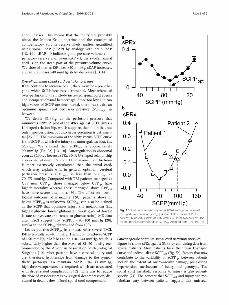
Overall optimum spinal cord perfusion pressureIf we continue to increase SCPP, there must be a point be-yond which SCPP becomes detrimental. Mechanisms ofover-perfusion injury include increased spinal cord edemaand intraparenchymal hemorrhage. Since too low and toohigh values of SCPP are detrimental, there must exist anoptimum spinal cord perfusion pressure (SCPPopt) inbetween.We define SCPPopt as the perfusion pressure that
minimizes sPRx. A plot of the sPRx against SCPP gives aU-shaped relationship, which supports the notion that notonly hypo-perfusion, but also hyper-prefusion is detrimen-tal [25, 35]. The minimum of the sPRx versus SCPP curveis the SCPP at which the injury site autoregulates best, i.e.,SCPPopt. We showed that SCPPopt is approximately90 mmHg (Fig. 3a) [13, 18]. Autoregulation is abnormaleven at SCPPopt because sPRx >0. A U-shaped relationshipalso exists between PRx and CPP in severe TBI. The brainis more extensively vascularized than the spinal cord,which may explain why, in general, optimum cerebralperfusion pressure (CPPopt) is less than SCPPopt at70–75 mmHg. Compared with TBI patients managed atCPP near CPPopt, those managed below CPPopt havehigher mortality whereas those managed above CPPopthave more severe disabilities [36]. The effect on neuro-logical outcome of managing TSCI patients above orbelow SCPPopt is unknown. SCPPopt can also be definedas the SCPP that optimizes injury site metabolism (i.e.,highest glucose, lowest glutamate, lowest glycerol, lowestlactate-to-pyruvate and lactate-to-glucose ratios). MD dataafter TSCI suggest that SCPPopt = 90–100 mmHg [20],similar to the SCPPopt determined from sPRx.Let us put this SCPPopt in context. After severe TSCI,
ISP is typically 20–40 mmHg. Therefore, to achieve SCPPof ~90 mmHg, MAP has to be 110–130 mmHg, which issubstantially higher than the MAP of 85–90 mmHg rec-ommended by the American Association of NeurologicalSurgeons [10]. Most patients have a cervical injury andare, therefore, hypotensive from damage to the sympa-thetic pathways. To maintain MAP 110–130 mmHg,high-dose vasopressors are required, which are associatedwith drug-related complications [32]. One way to reducethe dose of vasopressors is by surgical decompression, dis-cussed in detail below (‘Dural spinal cord compression’).
Patient-specific optimum spinal cord perfusion pressureFigure 3a shows sPRx against SCPP by combining data fromseveral patients. Most patients have their own U-shapedcurve and individualistic SCPPopt (Fig. 3b). Factors that maycontribute to the variability of SCPPopt between patientsinclude the extent of microvascular damage, pre-existinghypertension, mechanism of injury, and genotype. Thespinal cord metabolic response to injury is also patient-specific [21]. The concept that SCPPopt and injury site me-tabolism vary between patients suggests that universal
Fig. 3 Spinal pressure reactivity index (sPRx) and optimum spinalcord perfusion pressure (SCPPopt). a Plot of sPRx versus SCPP for 18patients. b Individual plots of sPRx versus SCPP for two patients. Theminimum value corresponds to SCPPopt. Adapted from [13] and [18]
Saadoun and Papadopoulos Critical Care (2016) 20:308 Page 5 of 9

management protocols are insufficient and that neuromoni-toring is essential to achieve individualized management.
Injury site compartmentalizationBy monitoring two pressures simultaneously [13], or ISPalong the injured cord [17], we showed that three intra-dural compartments form after severe TSCI: 1) abovethe injury; 2) at the injury; and 3) below the injury [15](Fig. 4a). These compartments do not communicate be-tween themselves; therefore, each compartment has adifferent pressure. The highest ISP is at the injury sitewhere the swollen, injured cord is compressed againstthe dura. Because of compartmentalization, monitoringCSF pressure from below the injury does not provide in-formation about ISP at the injury site. Draining CSFwith a lumbar catheter has been proposed as a treatmentfor TSCI [37]. CSF drainage is unlikely to improve SCPPin TSCI because, at the injury site, there is no CSFaround the spinal cord. In 65 patients with TSCI, 63(97 %) had cord compression on MRI; compression wasextradural in 75 % and dural in 25 % [16]. After bony de-compression, most patients still had high ISP [13, 18],suggesting dural cord compression in most. Two otherlines of evidence support the lack of communicationbetween the CSF compartments above and below the in-jury. After TSCI, little CSF can be drained from the lum-bar region and, when the lumbar drain is transduced,
there is a non-pulsatile signal [37]. Draining CSFthrough a lumbar catheter is also potentially detrimentalby causing downward herniation of the injured cord.Compartmentalization may be a feature of the severity
of TSCI and the surrounding CSF space; if TSCI is mildor the CSF space is large, the spinal cord will not becompressed by the dura and, therefore, the three com-partments will not form. It is unknown if, in the firstfew days after TSCI, compartmentalization is constantor dynamic or if compartmentalization is more commonin some TSCI types, e.g., flexion/distraction.
Dural spinal cord compressionWhy is bony decompression for TSCI controversial? Theanswer may be the dura surrounding the damaged cord,which is poorly distensible. Even if early bony decom-pression is performed, the swollen cord remains com-pressed against the dura. Dural spinal cord compressionexplains why ISP at the injury site remains high evenafter realignment of the spinal fracture, fixation, andlaminectomy [13, 18]. The concept of dural compressionis well established for TBI, where decompressive cra-niectomy involves not only removing bone but alsoopening the dura.The idea of dural compression is not appreciated after
TSCI, though compression of the swollen, injured spinalcord against the surrounding dura is evident on
Fig. 4 Expansion duroplasty. a Three intradural compartments form after TSCI—above, at, and below the injury site. b Intraoperative photoshowing duroplasty. D dura, DP dural patch. c Postoperative MRI showing the swollen, injured cord herniating into the duroplasty. d Intraspinalpressure (ISP) and e spinal cord perfusion pressure (SCPP) versus days after injury for 10 patients who had laminectomy and 11 patients who hadlaminectomy + duroplasty. Mean ± standard error. *P < 0.05. Adapted from [13] and [18]
Saadoun and Papadopoulos Critical Care (2016) 20:308 Page 6 of 9

postoperative MR scans [13, 16, 18] (Fig. 4a). The term‘spinal decompression’ refers to removing extraduralcompression such as bone fragments without openingthe dura, whereas the term ‘brain decompression’ refersto removing bone and opening the dura. Surgeonsdeveloped the concept of spinal decompression to treatdegenerative spinal diseases such as canal stenosis whereneural tissue is compressed from outside the dura.Unlike degenerative spinal pathologies, however, in TSCIthe damaged spinal cord itself is swollen against the sur-rounding dura and, therefore, dural spinal cord decom-pression is essential. We, therefore, propose that theconcept of decompression for TSCI be modified to includeopening the dura. Serial MR scans of TSCI patients indi-cate that dural spinal cord compression resolves with ahalf-life of 8.7 days [16]. The slow rate of resolution ofdural spinal cord compression supports early surgicaldecompression to reduce prolonged use of vasopressors.We have recently conducted a phase II non-randomized
trial comparing laminectomy versus laminectomy plusexpansion duroplasty (Fig. 4b–e) [13, 16, 18]. After lamin-ectomy plus expansion duroplasty, the injured cord herni-ates into the extra space that has been created, whichsuggests that the swollen cord was compressed by the dura.Laminectomy plus expansion duroplasty decompressed theinjured cord more effectively than laminectomy withoutduroplasty, as evident on MRI. Laminectomy plus expan-sion duroplasty also reduced ISP, increased SCPP, andlowered the sPRx versus SCPP curve downward with theminimum at 90 mmHg. Duroplasty takes about 10–15 minto perform and is safe; the main side effect is asymptomaticpseudomeningocele in about half of patients that resolveswithin 6 months. This study was powered to detect an in-crease in dural diameter, spinal cord decompression byMRI, a reduction in ISP and sPRx, as well as an increase inSCPP, but was underpowered to detect neurologicalimprovement.
Nursing care: lying supine may be detrimentalIn patients who have had laminectomy, wound compres-sion increases ISP and reduces SCPP due to transmis-sion of the compressive forces to the cord [13]. The ISPrise was up to 20 mmHg, enough to aggravate corddamage. Lying supine is also associated with higher ISP,by 2–4 mmHg, compared with lying laterally [18]. Thispressure difference was more marked for thoracic thancervical TSCI [19], probably because, when supine, thecervical cord is less compressed due to its lordosiswhereas the kyphotic thoracic cord becomes more com-pressed. No difference in ISP or SCPP was observed innon-laminectomized patients. In one thoracic TSCI pa-tient, the difference in ISP between supine and lateralposition was 18 mmHg [19]. These findings suggest that,after TSCI, patients who had laminectomy should be
nursed on their side to avoid aggravating cord damageby increasing ISP and reducing SCPP.
Neuroprotective drug trialsIssues to consider when designing drug trials for TSCI havebeen described elsewhere [38–41]. Patient age, site and se-verity of injury, and rehabilitation influence outcome. Suchheterogeneity means that many patients are required. Theformation of clinical networks may facilitate patient re-cruitment into trials, whereas MR imaging and electro-physiological outcome measures may reduce the number ofpatients required. Another issue is penetration of systemic-ally administered drugs into the injury site, especially sincepatients with severe cervical TSCI are hypotensive withhigh ISP. Though the blood-spinal cord barrier opens afteracute TSCI thus favoring drug entry into the injury site, wehave recently shown that SCPP is also a major, but un-appreciated, determinant of drug entry [20]. An increase inSCPP by 10 mmHg increases dexamethasone penetrationinto the injury site three-fold. Ongoing trials of minocyclineand riluzole [42] as well as completed drug trials of methyl-prednisolone [6, 7] have not optimized SCPP. It is possiblethat drug trials for acute TSCI fail because of inadequatedrug penetration at the injury site.
LimitationsAt present, our unit is the only one that performs neuro-monitoring; it is vital that others reproduce our findings.With our technique, probes are inserted intraoperatively,which limits their use. In the future, the method should berefined to insert probes percutaneously in the NICU. Thiswill allow neuromonitoring to start earlier and widen itsuse to non-TSCI, such as edematous transverse myelitiswhere not only inflammation but also ischemia may play arole [43]. Several of the concepts we introduced, such asSCPPopt, make physiological sense, but are presently theor-etical. Evidence is required that SCPPopt has clinical value,e.g., by showing that patients managed with SCPP close toSCPPopt have better outcomes than those with large SCPPdeviation from SCPPopt. Even if SCPPopt is clinically im-portant, it is computed after the monitoring has finished; acontinuous SCPPopt is required, which is displayed in real-time in the NICU. Another issue is timing of surgery,which is delayed in many units. It is, therefore, unknown ifultra-early bony decompression prevents cord swellingthus precluding the need for ISP monitoring or duroplasty.With further studies, these limitations can be overcome.
ConclusionsWe describe how monitoring from the injury site mayguide the management of TSCI. Ultimately, a randomizedcontrolled trial is required to determine if monitoring andSCPP optimization improve outcome, which can only beachieved by international collaboration.
Saadoun and Papadopoulos Critical Care (2016) 20:308 Page 7 of 9

AbbreviationsAIS: American spinal injuries association Impairment Scale; CPP: Cerebralperfusion pressure; CPPopt: Optimum cerebral perfusion pressure;CSF: Cerebrospinal fluid; ICP: Intracranial pressure; ISCoPE: Injured Spinal CordPressure Evaluation; ISP: Intraspinal pressure; MAP: Mean arterial pressure;MD: Microdialysis; NICU: Neurointensive care unit; PRx: (brain) pressurereactivity index; RAP: (brain) compensatory volume reserve; SCPP: Spinal cordperfusion pressure; SCPPopt: Optimum spinal cord perfusion pressure;sPRx: Spinal pressure reactivity index; sRAP: Spinal compensatory volumereserve; TBI: Traumatic brain injury; TSCI: Traumatic spinal cord injury
AcknowledgementsWe thank the neuroanesthetic, neurointensive care and neurosurgery staff atSt. George’s Hospital for help with patient management. We also thank theorthopedic spinal surgeons at St. George’s Hospital and neurosurgeons atKing’s College Hospital and Hurstwood Park Neurological Centre for helpwith patient recruitment.
FundingSupported by the Wings for Life spinal cord research foundation and theNeurosciences Research Foundation (Fletcher Fund).
Authors’ contributionsBoth authors contributed equally to this manuscript. Both authors read andapproved the final manuscript.
Competing interestsThe authors declare that they have no competing interests.
References1. Lee BB, Cripps RA, Fitzharris M, Wing PC. The global map for traumatic
spinal cord injury epidemiology: update 2011, global incidence rate. SpinalCord. 2014;52:110–6.
2. Fehlings MG, Rabin D, Sears W, Cadotte DW, Aarabi B. Current practice in thetiming of surgical intervention in spinal cord injury. Spine (Phila Pa 1976).2010;35:S166–73.
3. Werndle MC, Zoumprouli A, Sedgwick P, Papadopoulos MC. Variability inthe treatment of acute spinal cord injury in the United Kingdom: results ofa national survey. J Neurotrauma. 2012;29:880–8.
4. Fehlings MG, Vaccaro A, Wilson JR, Singh A, Cadotte WD, Harrop JS, Aarabi B,Shaffrey C, Dvorak M, Fisher C, et al. Early versus delayed decompression fortraumatic cervical spinal cord injury: results of the Surgical Timing in AcuteSpinal Cord Injury Study (STASCIS). PLoS One. 2012;7:e32037.
5. van Middendorp JJ, Hosman AJ, Doi SA. The effects of the timing ofspinal surgery after traumatic spinal cord injury: a systematic review andmeta-analysis. J Neurotrauma. 2013;30:1781–94.
6. Bracken MB, Shepard MJ, Collins WF, Holford TR, Young W, Baskin DS,Eisenberg HM, Flamm E, Leo-Summers L, Maroon J, et al. A randomized,controlled trial of methylprednisolone or naloxone in the treatment ofacute spinal-cord injury. Results of the Second National Acute Spinal CordInjury Study. N Engl J Med. 1990;322:1405–11.
7. Bracken MB, Shepard MJ, Holford TR, Leo-Summers L, Aldrich EF, Fazl M,Fehlings M, Herr DL, Hitchon PW, Marshall LF, et al. Administration ofmethylprednisolone for 24 or 48 hours or tirilazad mesylate for 48 hours inthe treatment of acute spinal cord injury. Results of the Third NationalAcute Spinal Cord Injury Randomized Controlled Trial. Natl Acute SpinalCord Inj Study JAMA. 1997;277:1597–604.
8. Bydon M, Lin J, Macki M, Gokaslan ZL, Bydon A. The current role of steroidsin acute spinal cord injury. World Neurosurg. 2014;82:848–54.
9. Hurlbert RJ, Hamilton MG. Methylprednisolone for acute spinal cord injury:5-year practice reversal. Can J Neurol Sci. 2008;35:41–5.
10. Cozzens JW, Prall JA, Holly L. The 2012 guidelines for the management ofacute cervical spine and spinal cord injury. Neurosurgery. 2013;72 Suppl 2:2–3.
11. Katoh S, el Masry WS. Motor recovery of patients presenting with motorparalysis and sensory sparing following cervical spinal cord injuries.Paraplegia. 1995;33:506–9.
12. Saadoun S, Bell BA, Verkman AS, Papadopoulos MC. Greatly improvedneurological outcome after spinal cord compression injury inAQP4-deficient mice. Brain. 2008;131:1087–98.
13. Werndle MC, Saadoun S, Phang I, Czosnyka M, Varsos GV, Czosnyka ZH,Smielewski P, Jamous A, Bell BA, Zoumprouli A, et al. Monitoring of spinal cordperfusion pressure in acute spinal cord injury: initial findings of the injuredspinal cord pressure evaluation study. Crit Care Med. 2014;42:646–55.
14. Varsos GV, Werndle MC, Czosnyka ZH, Smielewski P, Kolias AG, Phang I,Saadoun S, Bell BA, Zoumprouli A, Papadopoulos MC, et al. Intraspinalpressure and spinal cord perfusion pressure after spinal cord injury: anobservational study. J Neurosurg Spine. 2015;23:763–71.
15. Papadopoulos MC. Intrathecal pressure after spinal cord injury.Neurosurgery. 2015;77:E500.
16. Saadoun S, Werndle MC, Lopez de Heredia L, Papadopoulos MC. The duracauses spinal cord compression after spinal cord injury. Br J Neurosurg.2016;30:582–4.
17. Phang I, Papadopoulos MC. Intraspinal pressure monitoring in a patient withspinal cord injury reveals different intradural compartments: Injured SpinalCord Pressure Evaluation (ISCoPE) study. Neurocrit Care. 2015;23:414–8.
18. Phang I, Werndle MC, Saadoun S, Varsos G, Czosnyka M, Zoumprouli A,Papadopoulos MC. Expansion duroplasty improves intraspinal pressure,spinal cord perfusion pressure, and vascular pressure reactivity index inpatients with traumatic spinal cord injury: injured spinal cord pressureevaluation study. J Neurotrauma. 2015;32:865–74.
19. Phang I, Zoumprouli A, Saadoun S, Papadopoulos MC. Safety profile andprobe placement accuracy of intraspinal pressure monitoring for traumaticspinal cord injury: Injured Spinal Cord Pressure Evaluation study. JNeurosurg Spine. 2016;25:398–405.
20. Phang I, Zoumprouli A, Papadopoulos MC, Saadoun S. Microdialysis tooptimize cord perfusion and drug delivery in spinal cord injury. Ann Neurol.2016. EPub. doi:10.1002/ana.24750.
21. Chen S, Phang I, Zoumprouli A, Papadopoulos MC, Saadoun S. Metabolicprofile of injured human spinal cord determined using surface microdialysis.J Neurochem. 2016, in press.
22. Chesnut RM, Temkin N, Carney N, Dikmen S, Rondina C, Videtta W, PetroniG, Lujan S, Pridgeon J, Barber J, et al. A trial of intracranial-pressuremonitoring in traumatic brain injury. N Engl J Med. 2012;367:2471–81.
23. Yuan Q, Wu X, Cheng H, Yang C, Wang Y, Wang E, Qiu B, Fei Z, Lan Q, Wu S, etal. Is intracranial pressure monitoring of patients with diffuse traumaticbrain injury valuable? An observational multicenter study. Neurosurgery.2016;78:361–8. discussion 368–9.
24. Han J, Yang S, Zhang C, Zhao M, Li A. Impact of intracranial pressuremonitoring on prognosis of patients with severe traumatic brain injury: aPRISMA systematic review and meta-analysis. Medicine (Baltimore).2016;95:e2827.
25. Czosnyka M, Pickard JD. Monitoring and interpretation of intracranialpressure. J Neurol Neurosurg Psychiatry. 2004;75:813–21.
26. Werndle MC, Saadoun S, Phang I, Czosnyka M, Varsos G, Czosnyka Z,Smielewski P, Jamous A, Bell BA, Zoumprouli A, et al. Measurement ofintraspinal pressure after spinal cord injury: technical note from theinjured spinal cord pressure evaluation study. Acta Neurochir Suppl.2016;122:323–8.
27. Smith JS, Anderson R, Pham T, Bhatia N, Steward O, Gupta R. Role of earlysurgical decompression of the intradural space after cervical spinal cordinjury in an animal model. J Bone Joint Surg Am. 2010;92:1206–14.
28. Jones CF, Newell RS, Lee JH, Cripton PA, Kwon BK. The pressure distributionof cerebrospinal fluid responds to residual compression and decompressionin an animal model of acute spinal cord injury. Spine (Phila Pa 1976).2012;37:E1422–31.
29. Awwad W, Bassi M, Shrier I, Al-Ahaideb A, Steele RJ, Jarzem PF. Mitigatingspinal cord distraction injuries: the effect of durotomy in decreasingcord interstitial pressure in vitro. Eur J Orthop Surg Traumatol.2014;24 Suppl 1:S261–7.
30. Iannotti C, Zhang YP, Shields LB, Han Y, Burke DA, Xu XM, Shields CB. Duralrepair reduces connective tissue scar invasion and cystic cavity formationafter acute spinal cord laceration injury in adult rats. J Neurotrauma.2006;23:853–65.
31. Hawryluk G, Whetstone W, Saigal R, Ferguson A, Talbott J, Bresnahan J,Dhall S, Pan J, Beattie M, Manley G. Mean arterial blood pressure correlateswith neurological recovery after human spinal cord injury: analysis of highfrequency physiologic data. J Neurotrauma. 2015;32:1958–67.
32. Inoue T, Manley GT, Patel N, Whetstone WD. Medical and surgicalmanagement after spinal cord injury: vasopressor usage, early surgerys, andcomplications. J Neurotrauma. 2014;31:284–91.
Saadoun and Papadopoulos Critical Care (2016) 20:308 Page 8 of 9

33. Readdy WJ, Whetstone WD, Ferguson AR, Talbott JF, Inoue T, Saigal R,Bresnahan JC, Beattie MS, Pan JZ, Manley GT et al. Complications andoutcomes of vasopressor usage in acute traumatic central cord syndrome. JNeurosurg Spine. 2015;23:574–80.
34. Valadka AB, Robertson CS. Surgery of cerebral trauma and associated criticalcare. Neurosurgery. 2007;61:203–20. discussion 220–1.
35. Czosnyka M, Brady K, Reinhard M, Smielewski P, Steiner LA. Monitoring ofcerebrovascular autoregulation: facts, myths, and missing links. NeurocritCare. 2009;10:373–86.
36. Aries MJ, Czosnyka M, Budohoski KP, Steiner LA, Lavinio A, Kolias AG,Hutchinson PJ, Brady KM, Menon DK, Pickard JD, et al. Continuousdetermination of optimal cerebral perfusion pressure in traumatic braininjury. Crit Care Med. 2012;40:2456–63.
37. Kwon BK, Curt A, Belanger LM, Bernardo A, Chan D, Markez JA, Gorelik S,Slobogean GP, Umedaly H, Giffin M, et al. Intrathecal pressure monitoringand cerebrospinal fluid drainage in acute spinal cord injury: a prospectiverandomized trial. J Neurosurg Spine. 2009;10:181–93.
38. Fawcett JW, Curt A, Steeves JD, Coleman WP, Tuszynski MH, Lammertse D,Bartlett PF, Blight AR, Dietz V, Ditunno J, et al. Guidelines for the conduct ofclinical trials for spinal cord injury as developed by the ICCP panel:spontaneous recovery after spinal cord injury and statistical power neededfor therapeutic clinical trials. Spinal Cord. 2007;45:190–205.
39. Steeves JD, Lammertse D, Curt A, Fawcett JW, Tuszynski MH, Ditunno JF,Ellaway PH, Fehlings MG, Guest JD, Kleitman N, et al. Guidelines for theconduct of clinical trials for spinal cord injury (SCI) as developed by theICCP panel: clinical trial outcome measures. Spinal Cord. 2007;45:206–21.
40. Tuszynski MH, Steeves JD, Fawcett JW, Lammertse D, Kalichman M, Rask C,Curt A, Ditunno JF, Fehlings MG, Guest JD, et al. Guidelines for the conduct ofclinical trials for spinal cord injury as developed by the ICCP Panel: clinical trialinclusion/exclusion criteria and ethics. Spinal Cord. 2007;45:222–31.
41. Lammertse D, Tuszynski MH, Steeves JD, Curt A, Fawcett JW, Rask C,Ditunno JF, Fehlings MG, Guest JD, Ellaway PH, et al. Guidelines for theconduct of clinical trials for spinal cord injury as developed by the ICCPpanel: clinical trial design. Spinal Cord. 2007;45:232–42.
42. Nagoshi N, Fehlings MG. Investigational drugs for the treatment of spinalcord injury: review of preclinical studies and evaluation of clinical trials fromPhase I to II. Expert Opin Investig Drugs. 2015;24:645–58.
43. Lassmann H. Hypoxia-like tissue injury as a component of multiple sclerosislesions. J Neurol Sci. 2003;206:187–91.
44. Vender J, Waller J, Dhandapani K, McDonnell D. An evaluation andcomparison of intraventricular, intraparenchymal, and fluid-coupledtechniques for intracranial pressure monitoring in patients with severetraumatic brain injury. J Clin Monit Comput. 2011;25:231–6.
Saadoun and Papadopoulos Critical Care (2016) 20:308 Page 9 of 9