SPINAL BIOMECHANICS POSTURE ANALYSIS. POSTURE Keep in mind the spine is found at the posterior...
53
SPINAL BIOMECHANICS POSTURE ANALYSIS
-
Upload
vernon-burke -
Category
Documents
-
view
226 -
download
0
Transcript of SPINAL BIOMECHANICS POSTURE ANALYSIS. POSTURE Keep in mind the spine is found at the posterior...
SPINAL BIOMECHANICS
POSTURE ANALYSIS

POSTURE
• Keep in mind the spine is found at the posterior aspect of the body, behind the center of gravity
• Center of gravity lies:– Through the atlanto-occipital joint– Tragus of the ear– Anterior humeral head– Anterior-inferior edge of T11– Greater trochanter– Just behind the patella– Through the lateral malleoli
DURING POSTURAL ANALYSIS…
• Usually stance is asymmetrical if not intentional.• The weight of the body is borne by the skeleton aided by the
action of intrinsic back muscles• Sway occurs during stance.• Postural sway of the vertebral column on the pelvis is
controlled by the erector spinae, and the rectus abdominis. • 80% of the contraction occurs in the E.S., whereas only 20% of
contraction occurs in the abdominals, as confirmed by EMG studies.
• In scoliosis, E.S. contraction is higher on the convex side.

AFFECTS OF AXIAL COMPRESSIVE FORCES
• Increases from the C/S to the L/S
• Lumbar problems are common--#1 reason to see a Chiropractor
HOW DO MUSCLES BECOME IMBALANCED?
• Skeletal misalignment- triggers other muscles to be recruited to restore normal posture
• Joint pain or malformation- imbalance in stance and gait
• Ligamentous injury/instability- recruits muscles to support the joint
• Muscle fatigue- recruits other muscles to contract to accomplish the same movement, often resulting in myofascial trigger points

BEGINNING POSTURE ANALYSIS
• Work from the “ground-up”:
– Check for any lower extremity deformity that may be creating imbalance above
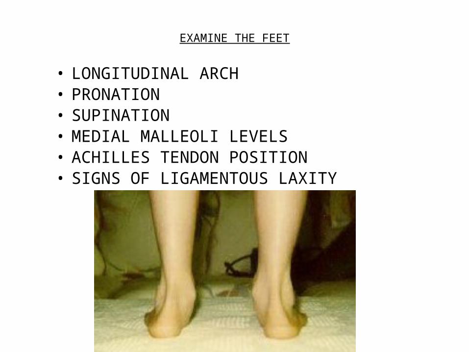
EXAMINE THE FEET
• LONGITUDINAL ARCH• PRONATION• SUPINATION• MEDIAL MALLEOLI LEVELS• ACHILLES TENDON POSITION• SIGNS OF LIGAMENTOUS LAXITY

Pes Cavus
Pes Planus

REASONS BEHIND TOE-IN & TOE-OUT
• TOE-IN– INTERNAL TIBIAL ROTATION– TIBIA VARUS– INCREASED INTERNAL ROTATION of FEMUR —often due to
muscular contraction/imbalance
• TOE-OUT– BILATERAL- SACRAL ANTERIORITY– UNILATERAL- PELVIC ANTERIORITY – INCREASED EXTERNAL ROTATION of FEMUR—often due to
muscular contraction/imbalance
Blount's disease
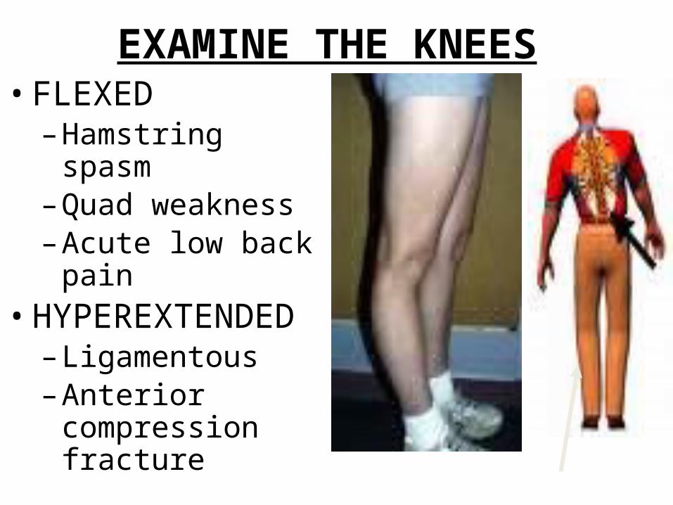
EXAMINE THE KNEES • FLEXED–Hamstring spasm–Quad weakness–Acute low back pain
• HYPEREXTENDED– Ligamentous –Anterior compression
fracture
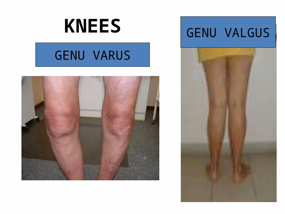
KNEESGENU VARUS
GENU VALGUS

Q – ANGLE(Quadriceps)
An abnormally high Q-Angle can cause stress on the entire kinetic chain of the lower extremity causing many conditions from low back pain to foot pain. D. Robert Kuhn, DC, Terry R. Yochum, DC, Anton R. Cherry, Sean S.Rodgers
• Wide Hips (female runners)• Knock Knees (·Genu
valgum)• Pronation of the feet• Subluxating Patella• High riding patella (patella
alta)• Weak Vastus Medialis

Imbalance of Hip Rotators
• Leg length discrepancies and foot pronation may lead to:
• Iliotibial band syndrome • Piriformis syndrome
• Recurrent muscle strains (hamstring and groin pulls) can be an indicator of asymmetry in structural alignment.

HIP MUSCLES…• Transfer ground-reaction forces from legs to
trunk during gait • Supply coordinated propulsion• Provide balanced stability for the pelvis and
spine • Through repetitive use patterns and after
injuries, hip muscles may become shortened and/or weak [1] Kim D. Christensen, DC, CCSP, DACRB
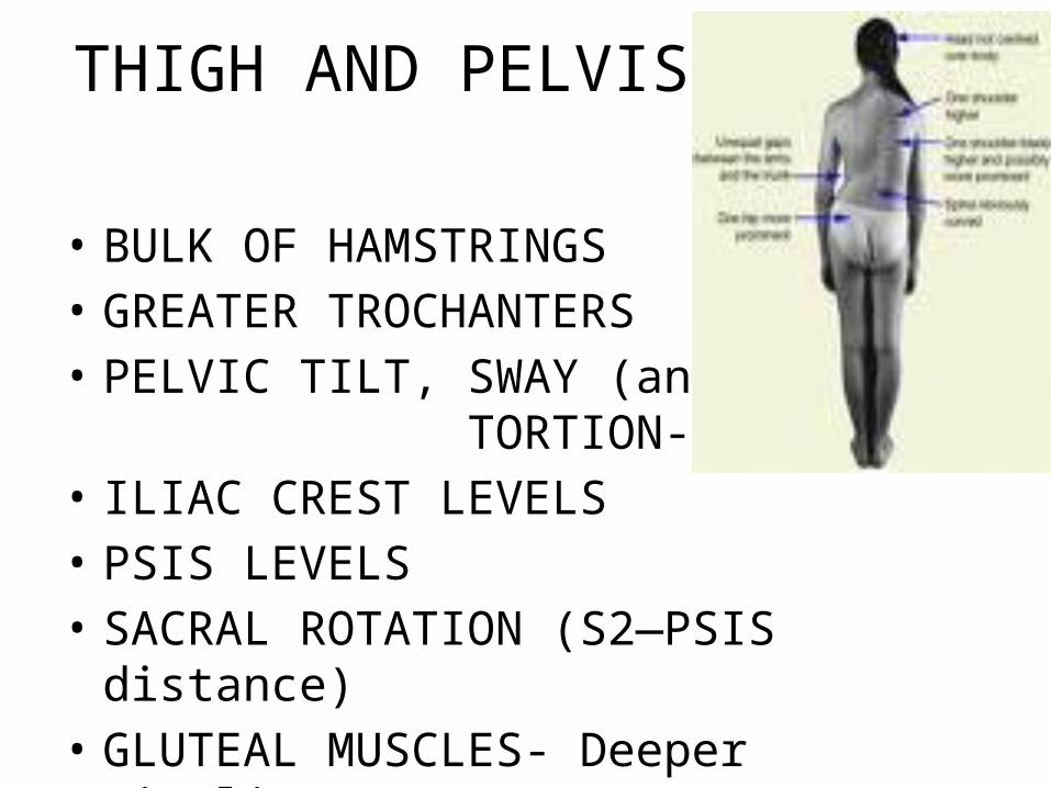
THIGH AND PELVIS
• BULK OF HAMSTRINGS• GREATER TROCHANTERS• PELVIC TILT, SWAY (antalgia),
TORTION- AS or PI• ILIAC CREST LEVELS• PSIS LEVELS• SACRAL ROTATION (S2—PSIS distance)• GLUTEAL MUSCLES- Deeper Dimpling

POSTURAL ANALYSIS P-A View
• Sacral Base-– Level– Held in place by innominate bones– Dependant upon equal leg lengths
• What can go wrong?– Sacral deformity-
» Transitional segment» Plateau base
– Anatomical short leg» Congenital» Acquired
– Functional» Due to muscle imbalance» Due to pelvic distortion

BODY RESPONDS IN A PREDICTABLE MANNER
• Attempts to restore balance:– Eyes on horizontal plane “Righting Reflex”– Equally distributing weight to center of gravity

VERTICAL PLANE of LUMBAR SPINE
• SPINOUS ALIGNMENT • SECTIONAL TOWERING• CURVATURE • LORDOSIS• PARASPINAL MUSCLE TONICITY• SKIN DISCOLORATION
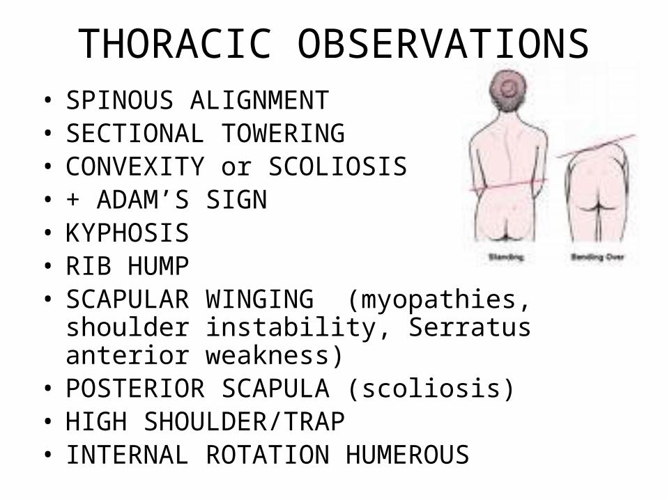
THORACIC OBSERVATIONS• SPINOUS ALIGNMENT • SECTIONAL TOWERING • CONVEXITY or SCOLIOSIS• + ADAM’S SIGN• KYPHOSIS • RIB HUMP• SCAPULAR WINGING (myopathies, shoulder
instability, Serratus anterior weakness)• POSTERIOR SCAPULA (scoliosis)• HIGH SHOULDER/TRAP• INTERNAL ROTATION HUMEROUS

NECK and HEAD OBSERVATIONS
• C2 SPINOUS ALIGNS WITH S2 TUBERCLE?• MASTOID PROCESS LEVELS• HEAD TILT OR ROTATION• ANTERIOR HEAD CARRIAGE• LORDOSIS• MUSCLE TONE

LATERAL VIEW• SACRUM: Inclines from 26-56º from horizontal– LUMBAR: Levels off at L4 superior body surface
(Apex), continues posteriorly in upper L/S– THORACIC: Gradual reversal of curve: body wedging
to create kyphosis at apex (T4-T6)– CERVICAL: Curve reverses again: apex (C4)
• What constitutes postural abnormality?– Any Variation in the AP or Lat
» Pelvic unleveling» Spinal segment unleveling
• Produces imbalance & altered weight imposition

Kendall, et. al.

FROM THE SIDEKYPHOSIS LORDOSIS
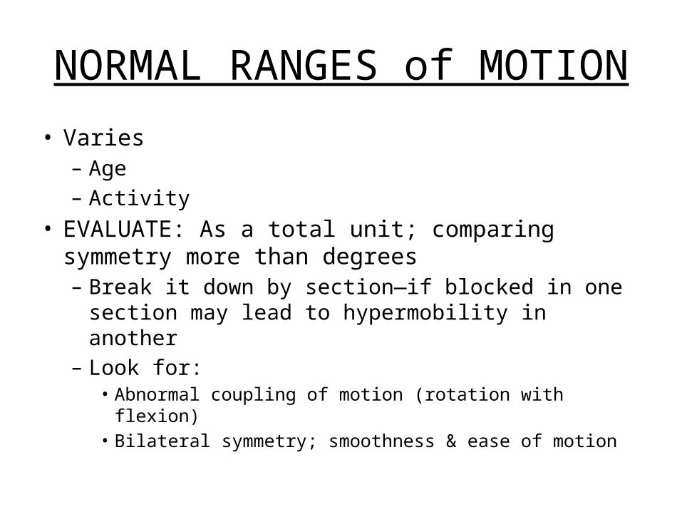
NORMAL RANGES of MOTION
• Varies– Age– Activity
• EVALUATE: As a total unit; comparing symmetry more than degrees– Break it down by section—if blocked in one section
may lead to hypermobility in another– Look for:
• Abnormal coupling of motion (rotation with flexion)• Bilateral symmetry; smoothness & ease of motion

MOTION T/L C
FLEXION 90 60
EXTENSION 40 50
UNILAT ROTATION 30 80
UNILAT LAT
FLEXION
35 45
SACROILIAC KINETICS
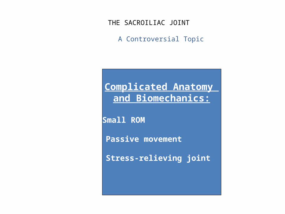
THE SACROILIAC JOINT
A Controversial Topic
Complicated Anatomy and Biomechanics:
1. Small ROM
2. Passive movement
3. Stress-relieving joint

MOTION IN THE S/I JOINT• No gross excursion (except due to severe trauma) Movement: Normal physiological effect of shock
absorption– Obvious movement during ambulation-Sacral nutation
• Clear osseous limitation-– Interlocking ridges & grooves– Strong reinforcing ligaments– Key-stone in arch stability
• Age Factors in degree of motion:– Flexible—to—Ankylosis

Gillett’s test …
Demonstrates pelvic motion by comparing PSIS motion B/L:
• Fixation• Pseudo-ankylosis• Fusion• Lumbar or hip muscle hypertonicity

Pelvis Tips and Rotates in Accommodation… A response to dysfunction
above or below• Leads to:• Abnormal: unequal weight into each S/I joint
leading to…
• Pelvic distortion
• Eccentric weight imposition into each S/I joint
• Abnormal posture
• Abnormal gait

PELVIC DISTORTION IS PREDICTABLE…•Predictable patterns of accommodation have been demonstrated as a response to imbalance both above and below.•Therefore, pelvic distortion is often not a primary subluxation, but a compensatory, secondary distortion

PRIMARY SUBLUXATION IN THE LUMBAR SPINE
(Secondary S/I Dysfunction) IVD HERNIATION
CURVATURE OR SCOLIOSIS
TRANSITIONAL SEGMENT
ALTERED SAGITTAL CURVE
FUNCTIONAL: GROSS MUSCULAR
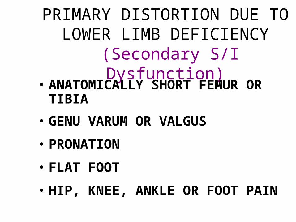
PRIMARY DISTORTION DUE TO LOWER LIMB DEFICIENCY
(Secondary S/I Dysfunction)• ANATOMICALLY SHORT FEMUR OR TIBIA
• GENU VARUM OR VALGUS
• PRONATION
• FLAT FOOT
• HIP, KNEE, ANKLE OR FOOT PAIN

PRIMARY Sacroiliac Fixation
• Chronic stress to the S/I joints leads to:
– Repetitive microtraumas
– Gross muscular compensation—holding joint in the fixed malposition
• May eventually lead to :– Sclerotic changes
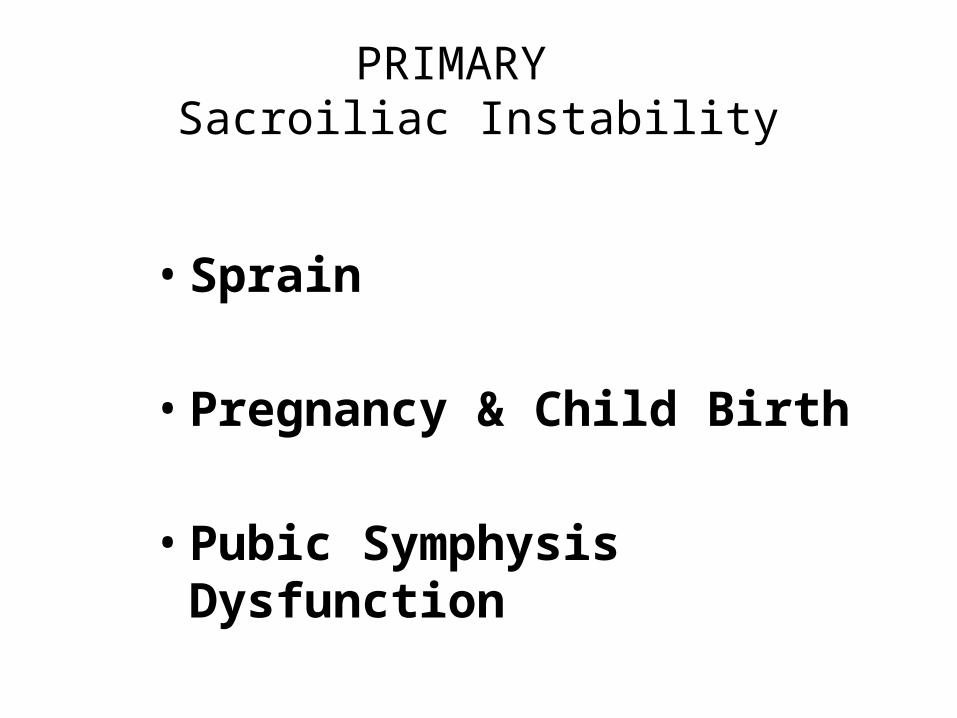
PRIMARY Sacroiliac Instability
• Sprain
• Pregnancy & Child Birth
• Pubic Symphysis Dysfunction

CHARACTERISTICS OF S/I PAIN
• Painful to walk• Ascending or descending stairs• Standing from a sitting position• Hopping or standing on involved leg• Sharp pain that awakens the patient
from sleep upon turning in bed

What Research Has Shown
• L/S may refer pain to S/I• S/I ROM: – Decreases with age– Minimal compared to spine
• Pain can=– 1° Fixation, Instability or – 2° Accommodation

CONTINUED S/I JOINT STRESS…
• May lead to true fixation in its misalignment—becoming a primary subluxation– Prolonged accommodation to chronic spinal
subluxation and postural abnormality or leg deficiency may lead to• Fixation• Gross muscular change• Sclerosis
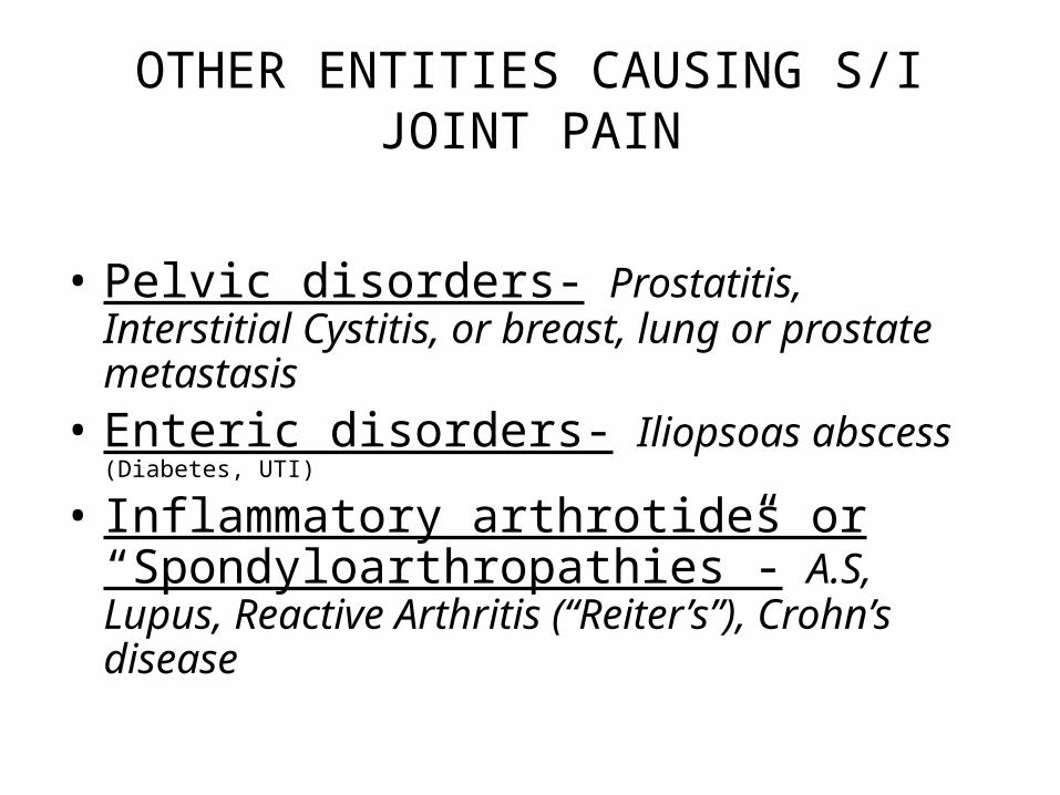
OTHER ENTITIES CAUSING S/I JOINT PAIN
• Pelvic disorders- Prostatitis, Interstitial Cystitis, or breast, lung or prostate metastasis
• Enteric disorders- Iliopsoas abscess (Diabetes, UTI)
• Inflammatory arthrotides or “Spondyloarthropathies”- A.S, Lupus, Reactive Arthritis (“Reiter’s”), Crohn’s disease

EXAMINATION
1. Observation2. Primary Stress Tests3. Leg Length Tests4. Weight Bearing Kinetic Tests5. Secondary Stress Tests6. Orthopedic Tests

OBSERVATION
I. OBSERVATION Pages 88-90
1. Postural Analysis: 1. Pelvic tilt (Anterior or Posterior)2. Lateral pelvic tilt3. Any structural asymmetry
2. Check for landmark:1. Alignment2. Tenderness
3. Belt Test: Test to R/O lumbar involvement
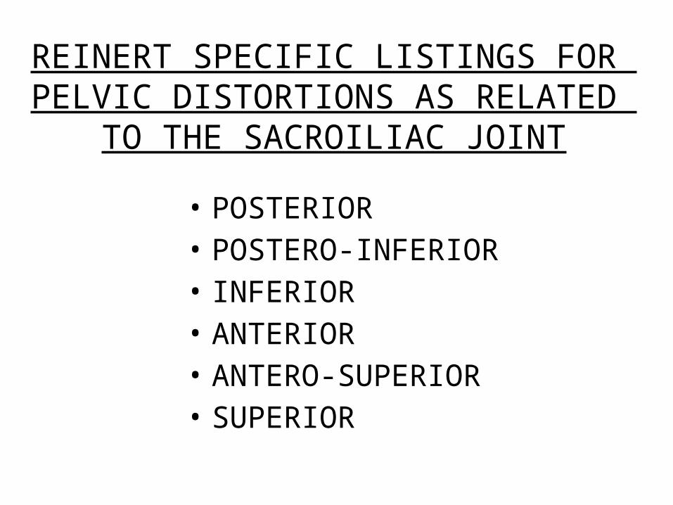
REINERT SPECIFIC LISTINGS FOR PELVIC DISTORTIONS AS RELATED TO THE
SACROILIAC JOINT
• POSTERIOR• POSTERO-INFERIOR• INFERIOR• ANTERIOR• ANTERO-SUPERIOR• SUPERIOR

II. PRIMARY STRESS TESTS

LEG LENGTH
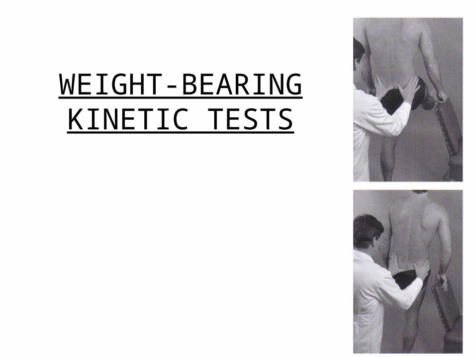
WEIGHT-BEARING KINETIC TESTS

Sacral Compression Test
Forced Counternutation

GAENSLEN’S TEST
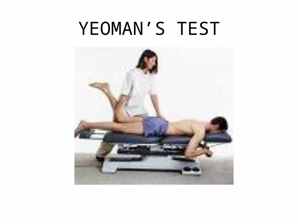
YEOMAN’S TEST

FABER PATRICK’S TEST

HIBB’S TEST

PRONE PALPATION
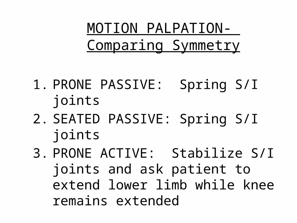
MOTION PALPATION- Comparing Symmetry
1. PRONE PASSIVE: Spring S/I joints2. SEATED PASSIVE: Spring S/I joints3. PRONE ACTIVE: Stabilize S/I joints and ask
patient to extend lower limb while knee remains extended

CONCLUSION
• Determine if S/I pain is 1° or 2 °
• Once this is achieved, the doctor can determine the appropriate treatment


![FIELD DOCTOR APPLICATION - Become a Chiropractor · FIELD DOCTOR APPLICATION CHECK LIST ... NUCCA [] Posture Analysis [ ] ... [ ] Biomechanics/CBP [] Heat / Ice [ ] Other: ...](https://static.fdocuments.us/doc/165x107/5b2787347f8b9a4e0e8b707a/field-doctor-application-become-a-field-doctor-application-check-list-.jpg)