Spinal anatomy final
90
HOSAM M ATEF;MD 1H Spinal Anatomy
-
Upload
hosam-atef -
Category
Education
-
view
107 -
download
0
Transcript of Spinal anatomy final

HOSAM M ATEF;MD
1H
Spinal Anatomy

H 2
Overview: Gross Spinal Anatomy
• Bony elements• Spinal ligaments and joints• Intervertebral discs• Neural elements• Blood supply


Spinal Column

H 7
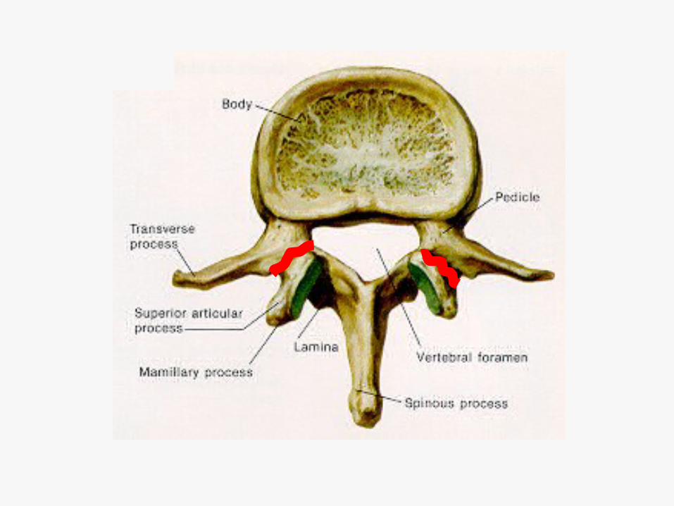
Vertebral Column
• 24 separate vertebrae plustwo composite vertebrae:– 7 cervical– 12 thoracic– 5 lumbar– Composites:
• Sacrum: 5 pieces• Coccyx: 4 pieces

Lumbar Vertebral Body

Spinous process


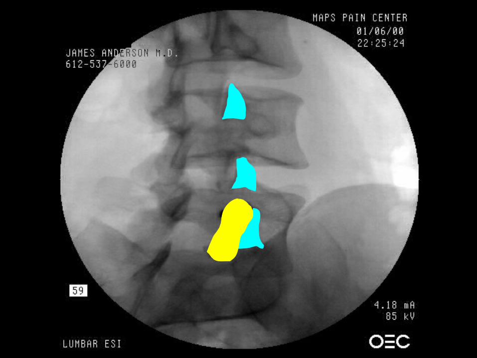
Pars Defect:Spondylolysis and Spondylolisthesis


Thoracic Vertebral Body

Spinous Process
Vertebral body
Transverse
costal facet

Vertebral costal facet

Cervical Vertebral Column

Spinous process

C1

C2

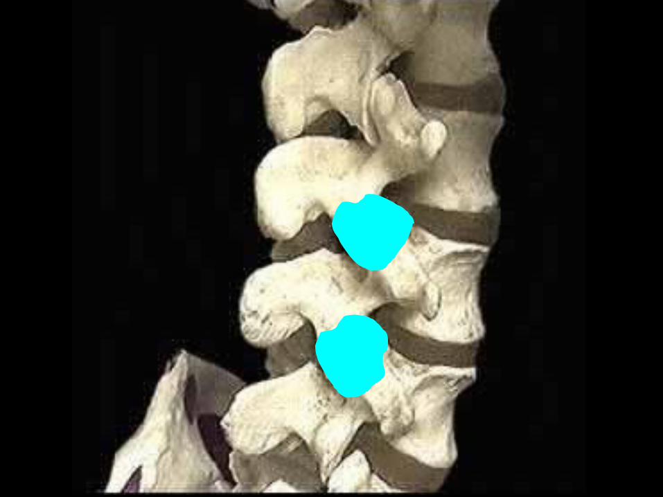
Spinal Motion Segment


Ligamentsand
Spinal Joints

PosteriorLongitudinalligament
Supraspinousligament
Interspinousligament
Ligamentumflavum

PosteriorLongitudinal
Ligament


Zygapophyseal Joints

Zygapophyseal joints
• True synovial joints:– Covered by fibrous capsule– Filled with synovial fluid– Hyaline cartilage lining bony surfaces– Innervated with nociceptive fibers
• Posterior spinal structures– Nerve root exits anterior to joint
through foramen
H 34

H
Zygapophyseal joints• Lumbar joints:
– oriented obliquely at L5-S1– Oriented increasingly more in the sagittal
plane up to thoracic levels
• Cervical joints are tilted cephalocaudad
• Joints are curved structures

HERNIATING DISC VIDEO.AVI


Spinous processes
Zygapophysealjoints
Articular pillars
Intervertebralforamina
Rib articulations

Atlanto-axialand
Atlanto-occipital Joints

H 42
Atlanto-axial and atlanto-occipital joints
• Anterior spinal structures– Nerve root exits posterior to joint– No associated neural foramen
• Do not have typical medial branch innervation
• True synovial joints

Sacroiliac Joint


Neural Elements

H 48
Spinal cord
• Begins at C1 and ends at L1-2• Transmits bilateral, segmental
spinal nerves:– 8 cervical– 12 thoracic– 5 lumbar– 5 sacral

L1
Filum terminale
Cervical expansion
Lumbar expansion
S2Cauda equina
Dura

Filum terminaleLumbar expansion
Cauda equina
Termination ofdural sac
Cervical expansion

Spinal Nerves

H 52
Spinal nerve
• Originate from a dorsal and ventral root– Roots originate intra-durally, exit
through dural root sleeve
• Give rise to dorsal and ventralprimary rami


Sympathetic Nervous System

Ventral ramus
Ventral root
Filum terminale
Dorsal root ganglion
Dorsal root

Whiterami
Grayrami

Nerve Roots


H 62
Cervical nerve roots• C5 contacts the disc in the proximal
foramen
• C6 and C7 have considerable contact with their respective discs in the middle foramen
• C8 has little contact with the C7-T1 disc

H 63
Neural foramina
• Transmit spinal nerves• First and second spinal nerves (C1,
C2) do not exit via a foramen• First neural foramen is C2-3 which
transmits the C3 spinal nerve• There are 8 cervical nerves with C8
exiting out the C7-T1 foramen

Uncovertebral joint
Uncinate process
Superiorand
inferiorarticular
processes
Foramen transversarium
Neural foramen
Disc

H 65
Numbering of spinal nerve roots• In the cervical spine, the last number
of the foramen names the exiting nerve:– C3 exits the C2-3 foramen– C7 exits the C6-7 foramen
• C8 exits the C7-T1 foramen• From T1-2 down, the first number of
the foramen names the exiting nerve:– L3 exits the L3-4 foramen

C5-6, C6-7 joints
C4, C5 and C6articular pillars
C2 and C3spinous processes
C2-3 and C3-4foramina
C4
C3
C5
C6
C8
C7C6, C7 vertebral
bodies


Dorsal Primary RamiAnd
Medial Branches


C5-6 and C6-7zygapophysealjoints
C4, C5 and C6articular pillars
C2 and C3spinous processes
C2-3 and C3-4foramina
TON
Medial branch of C3
Medial branch of C4
Medial branch of C5
Medial branch of C8
C4
C3
C5
C6
C8
C7

Intervertebral Discs

Lumbar Disc


phospholipase A2
prostaglandin E
histamine
inflammatory polypeptides
Internal Disc Disruption

Cervical Disc


Vascular Elements

Course of the Vertebral Artery

Pleura
Stellate ganglion
Vertebral artery
Subclavianartery


Posterior View of Brainstem
Basilar artery
Vertebralarteries Anterior
spinal artery

Spinal Cord Blood Supply

great radicular artery of Adamkiewicz
thoracic radicular artery
Cervical radicular arteries
L and R vertebral arteries
Ascending sacral radicular artery

Meningesof the
Brain and Spinal Cord

H87
Meninges
• The dura mater surrounds the brain and spinal cord
• The arachnoid lies just beneath the dura
• Pia mater covers and is adherent to the brain and spinal cord
• CSF flows between the arachnoid and the pia mater

Arachnoidgranulations
Dura mater
Arachnoid
Choroid plexusSubarachnoi
dspace
Pia mater


End