Špero M., Brkljačić B., Kolarić B., Marotti M. (2010...
28
Središnja medicinska knjižnica Špero M., Brkljačić B., Kolarić B., Marotti M. (2010) Preoperative staging of renal cell carcinoma using magnetic resonance imaging: comparison with pathological staging. Clinical Imaging, 34 (6). pp. 441-7. ISSN 0015-0282 http://www.elsevier.com/locate/issn/08997071 http://www.sciencedirect.com/science/journal/08997071 http://dx.doi.org/10.1016/j.clinimag.2009.09.005 http://medlib.mef.hr/1419 University of Zagreb Medical School Repository http://medlib.mef.hr/
Transcript of Špero M., Brkljačić B., Kolarić B., Marotti M. (2010...

Središnja medicinska knjižnica
Špero M., Brkljačić B., Kolarić B., Marotti M. (2010) Preoperative
staging of renal cell carcinoma using magnetic resonance imaging:
comparison with pathological staging. Clinical Imaging, 34 (6). pp.
441-7. ISSN 0015-0282
http://www.elsevier.com/locate/issn/08997071 http://www.sciencedirect.com/science/journal/08997071 http://dx.doi.org/10.1016/j.clinimag.2009.09.005 http://medlib.mef.hr/1419
University of Zagreb Medical School Repository
http://medlib.mef.hr/

Spero, Brkljacic
Authors of the manuscript:
Martina Spero, Boris Brkljacic, Branko Kolaric, Miljenko Marotti
Martina Spero, MD, specialist in radiology
Boris Brkljacic, MD, PhD
Professor of radiology, Medical School University of Zagreb, Croatia
Branko Kolaric, MD, PhD, specialist in epidemiology
Miljenko Marotti, MD, PhD
Professor of radiology, Medical School University of Zagreb, Croatia
Title of the manuscript:
PREOPERATIVE STAGING OF RENAL CELL CARCINOMA USING
MAGNETIC RESONANCE IMAGING: COMPARISON WITH PATHOLOGIC
STAGING
Abbreviated title: MRI preoperative staging of renal cell carcinoma

Spero, Brkljacic
The institution where the work was performed:
University Hospital Sestre milosrdnice
Department of Radiology
Vinogradska cesta 32, 10 000 Zagreb, Croatia
The reprint address:
Martina Spero, MD, specialist in radiology
University Hospital Dubrava
Department of Radiology
Avenija Gojka Suska 6, 10 000 Zagreb, Croatia
Phone: ++ 385 1 290 32 55
Fax: ++ 385 1 290 32 55
E-mail addresses: [email protected] or [email protected]

Spero, Brkljacic
ABSTRACT
We have retrospectively assessed the accuracy of our MRI protocol on 1.0 T MRI system
for preoperative staging of renal cell carcinoma using the 2002 TNM staging system, and
pathological staging as the gold standard.
Medical records of 48 patients, mean age 56.28 years, with 57 renal tumors were
reviewed: 52 malignant renal tumors were found, most of the patients were staged
T1N0M0. In our study, kappa test revealed excellent agreement between all three classes
of the TNM staging system.
Index terms: renal cell carcinoma, magnetic resonance imaging, TNM staging.
Spero, Brkljacic
INTRODUCTION
Renal cell carcinoma (RCC) is the most common primary malignant neoplasm of
the kidney and accounts for 2% of all cancer diagnosed(1). Its incidence is constantly
increasing over the last decades: incidence of RCC is 150,000 cases per year in the
world(1), an annual incidence in the United States is 35,000 cases with more than 12 000
deaths per year(2). Recent trends indicate an increase in frequency in younger
individuals, particularly females and including adolescents(3). It is due primarily to the
increased use of cross-sectional imaging, mainly ultrasound (US) and computed
tomography (CT), for investigating renal and non-renal symptoms. The percentage of
incidentaly discovered RCC ranges from 15% to 60%(4): these tumors are generally
smaller, with a lower tumor stage, and therefore a better prognosis. In turn, this has led to
the increased use of minimally invasive techniques, e.g. partial nephrectomy,
laparoscopic resection, radiofrequency ablation and cryotherapy(5).
Surgery is currently the only curative therapeutic approach to RCC, and accurate
preoperative staging is mandatory for surgical planning and assessing prognosis that
depends on the extent of the tumor and its metastasis at the time of its primary diagnosis
as well as on other factors including the patient's age, general state of health, and
comorbidities, which may influence life expectancy in general(6).
Cross-sectional imaging, in particular multidetector-row computed tomography
(MDCT) and magnetic resonance imaging (MRI), have considerably improved
preoperative staging. Magnetic resonance imaging is one of the most attractive
Spero, Brkljacic
approaches: the technology is widely available, it is not associated with the exposure to
ionizing radiation, and does not require the injection of iodinated contrast agent(7).
The Robson staging system has been replaced by the TNM staging system
developed by the American Joint Committee on Cancer (AJCC) which provides a more
detailed description and demonstrates more capability to grow and change with
increasing knowledge in the diagnosis and treatment of RCC.
The aim of our study was to assess the accuracy of MRI for preoperative staging
of renal cell carcinoma using the 2002 TNM staging system, with pathological staging as
the gold standard.
MATERIAL AND METHODS
Patient Population
After institutional review board approval, we have reviewed medical records of
48 patients, 23 male and 25 female, mean age 56.28 years (31-88), with renal neoplasm
who underwent MRI for preoperative staging in our institution, between January 2006
and December 2007. Renal neoplasm was suspected or diagnosed at first during the
ultrasound examination for renal or non-renal symptoms in our hospital (Figure 1 and 2)
or in other medical institutions. Preoperative evaluation with MRI and surgical treatment
were conducted in our institution for all patients.
Five patients had more than one tumor treated as separate lesions: two patients
had unilateral tumors – one had two and the other had three tumors of one kidney, while

Spero, Brkljacic
three patients had bilateral renal tumors – one had two lesions, the other had three
lesions, and the third one had four lesions. Two patients with bilateral multiple tumors
had von Hipple-Lindau disease. All together 57 tumors were analyzed.
MR Imaging Protocol
MRI studies were performed on 1.0 T MR imaging system (25 mT/m, Magnetom
Harmony, Siemens, Erlangen, Germany) with body phased-array coil. Sequences were
entirely breath-hold and imaging protocol included unenhanced:
1. axial T1-weighted in-phase gradient echo image: TR 201.65 msec, TE 6.3 msec,
φ (flip angle) 70º, slice thickness 10 mm, FOV (field of view) 350x350;
2. axial T1W-weighted out-of-phase gradient echo image: TR 185.2 msec, TE 40
msec, φ 70º, slice thickness 10 mm, FOV 350x350;
3. axial fat saturated HASTE T2-weighted image: TR 1200 msec, TE 55 msec, TI
150 msec, φ 180º, slice thickness 8 mm, FOV 390x390;
4. coronal True-FISP T2-weighted image: TR 7.08 msec, TE 3.54 msec, φ 80º, slice
thickness 6 mm, FOV 390x390;
5. axial fat saturated T1-weighted image: TR 157.6 msec, TE 4.8 msec, φ 75º, slice
thickness 8 mm, FOV 350x350.
Unenhanced sequences were followed by gadolinium-enhanced imaging:
1. axial T1-weighted in-phase gradient echo image: TR 201.65 msec, TE 6.3 msec,
φ (flip angle) 70º, slice thickness 10 mm, FOV (field of view) 350x350: 0 sec, 40
sec, 80 sec and 120 sec after the contrast administration;

Spero, Brkljacic
2. coronal fat saturated T1-weighted image: TR 157.6 msec, TE 4.8 msec, φ 75º,
slice thickness 8 mm, FOV 350x350, immediately after contrast dynamic
imaging, in sagittal plane in case vascular thrombosis;
3. axial T1W-weighted out-of-phase gradient echo image: TR 185.2 msec, TE 40
msec, φ 70º, slice thickness 10 mm, FOV 350x350, 10 minutes after contrast
dynamic imaging.
Gadopentetate dimeglumine (Magnevist, Schering AG, Germany) was used at the
dose of 0.1mmol/kg of body weight, or a contrast volume of 20mL if the patient's weight
was unknown. Gadolinium was administered through a cannula placed in the antecubital
fossa, by hand injection with firm push in a bolus. The arterial phase aquisition was timed
using carebolus (Siemens Medical Systems).
All MRI studies were evaluated by one of three experienced MR radiologist with
special interest in urologic imaging: they were introduced with patients' clinical histories.
MR Staging of RCC
The preoperative staging was based on the 2002 TNM staging system (Table 1).
MR Image Analysis
The tumor diameter was measured in three planes, and the largest one was chosen
to represent the tumor size.
Perinephric fat invasion was diagnosed if the tumor-fat interface was irregular or
indistinct, if thick (> 5 mm) perirenal soft tissue streaks and/or nodules (≥ 5 mm)
surrounded the tumor and extending into the perirenal fat, if capsular integrity was lost –

Spero, Brkljacic
disruption of the hypointense line around the kidney on T1 and T2-weighted images, and
if the pseudocapsule was present around the tumor.
Involvement of the adjacent suprarenal gland was considered when the gland was
enlarged and enhancing.
Tumor thrombosis was diagnosed in case of direct continuity with the renal mass,
high signal intensity, and signal heterogeneity compared with skeletal muscle on T2-
weighted imaging, and contrast enhancement.
Lymphadenopathy was diagnosed if there were regional lymph nodes (nodes
along the renal arteries, para-caval nodes for right-sides and para-aortic nodes for left-
sided RCC) greater than 1 cm in short axis, and/or contrast enhancement of the enlarged
lymph nodes.
Distant metastases were limited to the abdomen and visualized portions of the
lung bases and axial skeleton. Metastatic disease in the chest was evaluated by a chest X-
rays and if suspected chest CT was performed.
Statistical Analysis
MRI staging was compared with pathological staging as the gold standard, and
agreement between the two staging systems was determined using the kappa statistic
(0.0-0.2, poor; 0.2-0.4, fair; 0.4-0.6, moderate; 0.6-0.8, good; 0.8-1.0, excellent).
Agreement was calculated for all renal tumors and separately for malignant tumors.
Spero, Brkljacic
RESULTS
Forty-eight patients with fifty-seven renal tumors staged preoperatively by MRI
were submitted to the surgical treatment: 33/57 tumors were treated by total
nephrectomy, while 24/57 was treated by partial nephrectomy. Two patients with
bilateral multiple tumors were treated by bilateral partial nephrectomies, one patient with
two tumors of one kidney and one tumor of the other kidney was treated with
combination of total nephrectomy of the kidney with two tumors and partial nephrectomy
of the other kidney. One patient with two tumors of the same kidney was treated by total
nephrectomy, while patient with three very small tumors of one kidney was treated by
partial nephrectomy.
According to the MRI, there were suspected 45 (78.9%) solid renal tumors, 7
(12.3%) cystic tumors, 3 (5.3%) complex cysts, while in 2 (3.5%) patients we had doubt
weather the renal lesion was a complex cyst or a cystic form of malignant tumor.
According to the pathological findings there were 52 (91.2%) malignant renal
tumors and 5 (8.8%) benign tumors, four oncocytoma and one complex cyst. From 52
malignant renal cell tumors 33(63.5%) were clear cell RCC, 7(13.5%) multilocular cystic
clear cell RCC, 5 (9.6%) chromophobe RCC, 3(5.7%) were papillary RCC with two type
1(basophilic) and one type 2 (eosinophilic) papillary RCC, 1(1.9%) tumor were
unclassified, sarcomatoid RCC. In two patients renal adenocarcinomas were mixed
tumors containing cells with clear and chromophobe-eosinophilic cells, while in one
patient carcinoma contained cells of uroepithelial origin. Regarding nuclear grade, 10
(19.2%) tumors were grade 1, 30 (57.7%) were grade 2, 11 (21.2%) were grade 3, while
one tumor (1.9%) was grade 4.

Spero, Brkljacic
Regarding tumor size, according to MRI tumor mean size was 43.9 mm (10 mm –
150 mm), while according macroscopic tumor size measured by pathologist tumor mean
size was 43.4 mm (10 mm – 140 mm).
According to the TNM staging, MRI staged 48(84.2%) tumors in T1 stage, 4 (7%)
tumors in T2 stage and 5(8.8%) tumors in T3 stage: 2/5 staged T3a, and 3/5 staged T3b
with two tumors in renal vein and one tumor invading vena cava below diaphragm.
Regarding T3a stage in one tumor perirenal fat infiltration was suspected, while in
another patient the involvement of suprarenal gland was suspected. Fifty-five (96.5%)
tumors did not showed enlarged regional lymph nodes and were staged N0, while in two
cases (3.5%) more than one lymph node was enlarged and therefore were staged N2. One
case (1/57) was staged M1 by MRI because MRI showed distant metastasis in liver. In all
other cases of renal tumors (56/57) MRI did not showed distant metastasis and were
staged M0.
According to the pathological staging, 47/52 (90.4%) tumors were staged as T1,
2/52 (3.8%) tumor was as staged T2, 3/52 (5.8%) tumors were staged as T3. In case of 33
tumors, pathological specimen contained regional lymph nodes: 32/33 tumors were
staged N0, and 1/33 was staged N2. Twenty-nine specimens contained suprarenal gland:
28/29 was staged M0 and 1/29 was staged M1. In four cases suprarenal gland was not
found in perirenal fat. MRI and pathological T staging results are shown in Table 1.
The results for agreement between MRI staging and pathologic staging are shown
in Table 3.
Spero, Brkljacic
DISCUSSION
During the last two to three decades, constant development and the wide use of
modern cross-sectional imaging methods, US, CT and MRI, resulted in considerable
change of epidemiologic and clinical characteristics of RCC with the increasing number
of incidentally found tumors presenting with lower stage, grade and proportion of
metastases. Results of our study are in agreement with these observations; the mean size
of renal tumors in this study was about 43 mm, and most of the patients were staged
T1NOMO.
Accurate preoperative staging of RCC is important for choosing the appropriate
surgical approach, total or partial nephrectomy, and for predicting prognosis and survival.
It is determined by the T-staging as the most important part of the TNM staging system.
The partial nephrectomy, or nephron-sparing surgery (NSS) is considered the standard
surgical treatment of small renal tumors(8). RCC of TNM class T1a without evidence of
metastasis at primary staging, is considered a small renal tumor: the oncologic efficacy
and safety of NSS for the treatment is equivalent to radical nephrectomy(8).
Both MDCT and MRI perform highly in T-staging of local tumor extent and M-
staging of distant metastasis, but perform poorly in N-staging(6). In our study, kappa test
revealed excellent agreement between all three classes of the TNM staging system which
is consistent with the results of Kamel and co-workers(9) who reported 80-82% accuracy
of the MRI in staging organ-confined renal cell carcinoma, and Ergen and co-
workers(10) who reported good agreement between MRI and pathological staging for T
and M staging and poor for N staging.

Spero, Brkljacic
The T-staging is determined by the tumor size and extent, including the
possibility of venous involvement. For only malignant tumors percentage of agreement in
our study is excellent for T-staging: it is the best in T2, followed by T3 and T1. Trying to
be very precise, in few cases radiologists overmeasured tumor’s diameter when the value
was adjacent to 40 or 70 mm which resulted in overstaging tumor as T1b instead of T1a,
or T2 instead of T1b. At the end, it was not clinically so important because small tumors
staged T1b, sized a little bit bigger than 40 mm and confined to kidney without metastasis
at MRI our urologists decided to treat with partial nephrectomy.
According to the MRI findings, radiologists overstaged two T2 tumors as T3a: in
one patient involvement of perirenal fat was suspected, while in the case of bigger tumor
(diameter 65 mm) in the upper pole of a kidney with enlarged, enhancing suprarenal
gland, its invasion was suspected on MRI. In both cases, pathologist did not confirm the
MRI finding: in the former pathologist staged tumor as T1b, while in the latter the
pathological specimen of suprarenal gland showed malignant cells but without direct
organ invasion so the tumor was staged T1bM1. Those were attributed to the moderately
large tumors which compressed the perirenal fat and obscure the renal capsule with
indistinct interface between the tumor and adjacent suprarenal gland that made it difficult
to exclude capsular invasion and invasion of the enlarged, enhancing suprarenal gland.
Clinically it was not so important because both tumors were treated with total
nephrectomy when kidney and the perirenal fat were removed en bloc. Involvement of
perirenal fat is clinically important when NSS is considered as a surgical method for the
treatment of small tumors.

Spero, Brkljacic
Distinction between RCC with and without confinement to the renal capsule
challenges cross-sectional imaging: Catalano and co-workers(11) have diagnosed
perirenal fat infiltration by MDCT on 1-mm scans with 96% sensitivity, 93% specificity
and 95% accuracy, Roy and co-workers(12) reported 84% sensitivity, 95% specificity
and 91% accuracy in T3a staging by MRI, while Ergen and co-worker(10) conclude that
MRI is a reliable method for preoperative staging of RCC. Such results are probably due
to patient selection, imaging methods and techniques, image interpretation, or work-up
and interpretation of surgical pathological specimens, but in the end it appears that
radiological distinction between confinement of RCC to the true renal capsule and
extension in the perirenal fat is currently not fully reliable. Therefore, at present, surgical
treatment planning should be individually reviewed for each patient whose cross-
sectional imaging results leave any doubt as to the involvement of the renal capsule and
the perirenal fat layer (6, 10) .
Involvement of the renal vein (RV) and/or the inferior vena cava (IVC) (TNM
stage T3b and T3c) occurs in 4–10% of patients with RCC. It is important to
preoperatively detect the presence and the extent of RV and/or IVC tumor thrombus as
well as the invasion of the IVC wall for planning subsequent treatment and choosing the
appropriate surgical approach. Small study conducted by Aslam Sohaib and co-
workers(13), reported 100% sensitivity and 89% sensitivity of MRI in the detection of the
IVC wall involvement: the most reliable sign of IVC wall invasion was tumor signal both
inside and outside the vessel wall, while altered signal in the vessel wall and its
enhancement were nonspecific. In our study all three tumors, two with VR involvement
and one with IVC involvement below the diaphragm were correctly assessed by MRI.

Spero, Brkljacic
Regional nodal involvement, classified as N-classes of the TNM system, is one of
the major factors influencing the prognosis of patients with RCC: incidence of the
metastasis in regional lymph nodes without distant metastasis at the same time is 10-15%,
while 5-year survival rate with lymph node involvement is 8-35%. Whether with MDCT
or MRI, usual criteria for lymph node metastases remain limited to size assessment (14).
On histopathology regional lymph nodes can be enlarged because of hyperplasic or
inflammatory change related to the RCC. Specificity of cross-sectional imaging for
regional lymph node involvement is poor: the use of contrast agents can improve
situation. Gadolinium chelates in MRI reach lymph nodes directly via their feeding
arteries: enlarged regional lymph nodes because of metastases show contrast
enhancement. Ultrasmall superparamagnetic iron oxide particles (USPIO) are described
as a negative contrast agent for the detection of small lymph node metastasis: the USPIO
particles are ingested by macrophages through phagocytosis, accumulate in healthy
lymph nodes and cause a decrease in signal intensity on T2- and T2*-weighted images.
Lymph node metastases displace the macrophages in the lymph node and therefore do not
show the loss in signal intensity seen in normal lymph nodes(15). Only one patient (1/33)
in this study had enlarged and enhancing regional lymph nodes and was staged N2 by
MRI which was confirmed by histopathology. The rest of 32 patients with regional
lymphadenectomy performed did not have enlarged and/or enhancing lymph nodes on
MRI, and but on the histopathology in 5 of 32 patients follicular hyperplasia vas observed
in lymph nodes. Those data are consistent with the data from the literature.
The M-classes of the TNM system describes distant metastasis of the RCC which
includes all metastases that are either extra- nodal or involve non-regional lymph nodes.
Spero, Brkljacic
Any organ can be involved with metastases from the RCC, but the lung (31%), bone
(15%), brain (8%) and liver (5%) are the most frequently involved. MRI performs highly
in M-staging of distant metastasis. During MRI examination, search for distant
metastases is limited to the abdomen and visualized portions of the lung bases and axial
skeleton. In preoperative staging of the RCC, every patient should have chest X-rays and
if lung metastasis is suspected chest CT should be performed(16). If bone metastases are
suspected bone scintigraphy should be performed as well as cerebral CT or MRI if brain
metastases are suspected(16). In this study, MRI revealed a small liver metastasis in one
patient who was staged M1.
Nephrogenic systemic fibrosis (NSF) is an important delayed adverse reaction to
some less stable gadolinium based contrast agents, particularly gadodiamide(17).
Recently it has been shown that gadopentate dimeglumine may also trigger NSF, but not
with the same high frequency as gadodiamide(17). Patients at higher risk are those with
CKD 4 and 5 (GRF <30 ml/min per 1.73 m2), those on haemodialysis or peritoneal
dialysis and patients with reduced renal function who have had or are awaiting liver
transplantation (17-19). Patients at lower risk are those with CKD 3 (GRF 30-60 ml/min
per 1.73 m2). There were no NSF cases reported in the literature in patients with normal
renal function or CKD 1 and 2 (GRF >60 ml/min per 1.73 m2)(17). Few patients in our
study had CKD 1 and 2, and did not develop the NSF.
This study has limitations in the sample size (57 patients) with the most cases
staged T1 and T2, but this reflects the current trend of early RCC detection by cross-
sectional imaging. Although 1.5T MR imaging systems are usually used for preoperative
staging, we have managed to reveal very good results in preoperative staging of the RCC

Spero, Brkljacic
using 1T MR imaging system with excellent correlation between MRI and pathological
findings even with our 57 patient samples.
Until recent times, MRI has been considered as an alternative modality to CT
investigation for cases in which there are contraindications to CT or the CT findings were
inconclusive. However, cumulative experience has demonstrated that MRI surpasses CT
in several respects: (1) avoids the significant health risk associated with radiation dose
from a CT procedure, (2) provides inherently superior soft-tissue contrast even before the
administration of intravenous contrast, (3) a greater patient safety achieved with
gadolinium-based contrast agents used in MRI compared to the iodine-based contrast
agents used in CT, and (4) more different types of tissue can be interrogated during a
single MRI examination. Health-care costs and the ordering practice of referring
physicians were impediments to a wide-spread preference for MRI over CT in the US
medical community. Until recently, MRI has been considered an expensive diagnostic
test. The actual costs of MRI equipment, service, and site expenses have plummeted over
the past 20 years by factors approximately 65%, 55%, and 80%, respectively. The
average MRI technical fee in 2004 had fallen by 70% compared to the average fee in
1985. Further, the trend of decreasing costs for MRI is expected to continue over next 20
years. The charges for MRI studies are generally about 20% higher than those for CT
studies (hospital charges)(20).
In conclusion, the role of MRI in renal imaging has changed over time and it has
become an important modality for evaluating renal masses and for staging of patients
with RCC. Therefore it has become equivalent to CT in preoperative diagnosis and
staging of RCC. MRI can have additional diagnostic value in the evaluation of lesions

Spero, Brkljacic
with minimal amounts of fat or with intracellular fat(15, 21), and higher sensitivity in
evaluating complicated cysts(15).

Spero, Brkljacic
REFERENCES
1. Godley P, Kim S. Renal cell carcinoma. Curr Opin Oncol. 2002;14:280-5.
2. Jemal A, Murray T, Ward E (eds). Cancer statistic, 2005. CA Cancer J Clin.
2005;55:10-30.
3. Tuite DJ, Geoghegan T, McCauley G, Govender P, Browne RJF, Torreggiani
WC. Three-dimensional gadolinium-enhanced magnetic resonance breath-hold FLASH
imaging in the diagnosis and staging of renal cell carcinoma. Clin Radiol. 2006;61:23-30.
4. Touloupidis D, Papathanasion A, Kalaitzis C, Fatles G, Manavis I, Rombis V.
Renal cell carcinoma: The influence of new diagnostic imaging techniques on the size
and stage of tumors diagnosed over the past 26 years. International Urology and
Nephrology 2006;38:193-7.
5. Coll DM, Smith RC. Update on radiological imaging of renal cell carcinoma. BJU
International. 2007;99:1217-22.
6. Mueller-Lisse UG, Mueller-Lisse UL, Meindl T, Coppenrath E, Degenhart C,
Graser A, Scherr M, Reiser MF. Staging of renal cell carcinoma. Eur Radiol.
2007;17:2268-77.
7. Kirova G. The MR imaging as a one-way shopping tool for detecting and staging
renal tumours Radiol Oncol. 2005;39(1):23-35.
8. Shuch B, Lam JS, Belldegrun AS. Open partial nephrectomy for the treatment of
renal cell carcinoma. Cur Urol Rep. 2006;7:31-8.

Spero, Brkljacic
9. Kamel IR, Hochman MG, Keogan MT, Eng J, Longmaid HE, DeWolf W,
Edelman RR. Accuracy of Breath-Hold Magnetic resonance Imaging in Preoperative
Staging of Organ-Confined Renal Cell Carcinoma. J Comput Assist Tomogr.
2004;28:327-32.
10. Ergen FB, Hussain HK, Caoili EM, Korobkin M, Carlos RC, Weadock W,
Johnson TD, Shah R, Hayasaka S, Francis IR. MRI for preoperative staging of renal cell
carcinoma using the 1997 TNM classification: comparison with surgical and pathologic
staging. AJR. 2004;182:217-25.
11. Catalano C, Fraioli F, Laghi A, Napoli A, Pediconi F, Danti M, Nardis P,
Passariello R. High-resolution multidetector CT in preoperative evaluation of patients
with renal cell carcinoma. AJR. 2003;180:1271-7.
12. Roy C Sr., El Ghali S, Buy X, Lindner V, Lang H, Saussine C, Jacqmin D.
Significance of the Pseudocapsule on MRI of Renal Neoplasms and Its Potential
Application for Local Staging: A Retrospective Study. AJR. 2005;184:113-20.
13. Aslam Sohaib SA, Teh J, Nargund VH, Lumley JS, Hendry WF, Reznek RH.
Assessment of tumor invasion of the vena caval wall in renal cell carcinoma cases by
magnetic resonance imaging. J Urol. 2002;167:1271-5.
14. Luciani A, Itti E, Rahmouni A, Meignan M, Clement O. Lymph node imaging:
Basic principles. Eur J Radiol. 2006;58:338-44.
15. Nikken JJ, Krestin G. MRI of the kidney - state of the art. Eur Radiol.
2007;17:2780-93.
16. Heidenreich A, Ravery V. Preoperative imaging in renal cell cancer World J Urol.
2004;22:307-15.

Spero, Brkljacic
17. Thomsen HS, Marckmann P, Logager VB. Nephrogenic systemic fibrosis (NSF):
a late adverse reaction to some of the gadolinium based contrast agents. Cancer Imaging.
2007;7:130-7.
18. Kurtkoti J, Snow T, Hiremagalur B. Gadolinium and nephrogenic systemic
fibrosis: Association or causation. Nephrology. 2008;13:235-41.
19. Thomsen HS, Marckmann P. Extracellular Gd-CA: Differences in prevalence of
NSF. Eur J Radiol. 2008;66:180-3.
20. Semelka RC, Armao DM, Elias Junior J, Huda W. Imaging Strategies to Reduce
the Risk of Radiation in CT Studies, Including Selective Substitution With MRI. J Magn
Reson Imaging. 2007;25:900-9.
21. Silverman SG, Koenraad JM, Tuncali K, Jinzaki M, Cibas ES. Hyperattenuating
Renal Masses: Etiologies, Pathogenesis, and Imaging Evaluation. RadioGraphics.
2007;27:1131-43.

Spero, Brkljacic
Table 1 TNM classification system of RCC (AJCC, 2002)
Classification Description
T-classification
T1 limited to the kidney
T1a ≤ 4cm
T1b ≤ 7cm
T2 limited to the kidney, > 7 cm
T3 confined to Gerota's fascia
T3a: extending to ipsilateral adrenal or perirenal fat
T3b: extending to RV or IVC below diaphragm
T3c: extending to IVC above diaphragm
T4 extending beyond Gerota's fascia
N-classification
N0 no regional lymph node metastasis
N1 metastasis in one regional lymph node
N2 metastasis in more than one regional lymph node
Nx regional lymph nodes can not be evaluated
M-classification
M0 no distant metastasis
M1 distant metastasis
Mx distant metastasis can not be evaluated

Spero, Brkljacic
Table 2 MRI and pathologic T staging
T stage MRI
T staging
PATHOLOGIC
T staging
all tumors (n=57) malignant tumors (n=52) malignant tumors (n=52)
T1 48 (84.2%) 44 (84.6%) 47 (90.4%)
T1a 29 (60.4%) 25 (56.8%) 31 (66%)
T1b 19 (39.6%) 19 (43.2%) 16 (34%)
T2 4 (7%) 3 (5.8%) 2 (3.8%)
T3 5 (8.8%) 5 (9.6%) 3 (5.8%)
T3a 2/5 2/5 0
T3b 3/5 3/5 3

Spero, Brkljacic
Table 3 Agreement between MRI and pathologic TNM staging
T1 T2 T3 T4 N0 N1 N2 M0 M1
kappaa
(St.Error)
0.558
(0.148)
0.650
(0.228)
0.732
(0.179)
/
/
0.186
(0.202)
/
/
0.655
(0.320)
/
/
/
/
kappab
(St.Error)
0.738
(0.142)
0.790
(0.203)
0.731
(0.180)
/
/
0.653
(0.321)
/
/
0.653
(0.321)
/
/
/
/
Percentage of
agreementa
87.7 96.5 96.5 100 84.2 100 97.3 82.4 94.1
Percentage of
agreementb
94.2 98.0 96.2 100 96.9 100 96.9 96.6 96.6
aRefers to all tumors;
bOnly malignant tumors
Spero, Brkljacic
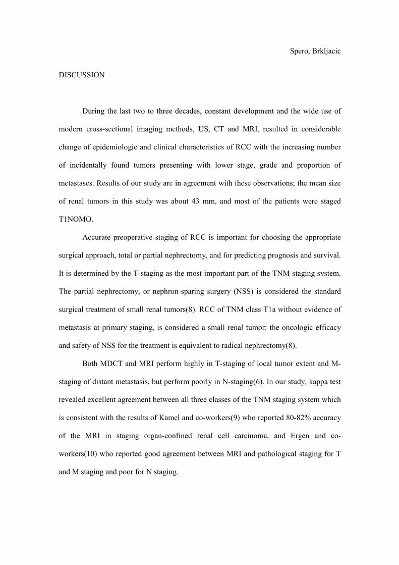
Figure 1 38-years old male patient, asymptomatic. (A) Abdominal ultrasound revealed
expansile mass with cystic parts in the lower pole of the left kidney. (B) Axial T1-
weighted out-of-phase image showed hypointense mass with compressed adjacent
parenchyma, while (C) coronal True-FISP T2-weighted image showed the mass consists
of multiple hyperintense cysts with irregular, thick septa that enhanced on post-contrast
axial T1-weighted image (D). Pathological finding: multi-locular cyst.

Spero, Brkljacic

Spero, Brkljacic
Figure 2 47-years old male patient with right-sided blunt pain in his lower back. (A)
Abdominal ultrasound revealed tumor in the lower pole of the right kidney. MRI showed
solid mass discretely hypointense on axial T1-weighted in-phase image (B) and almost
iso-intense to adjacent parenchyma on axial fat saturated HASTE T2-weighted image
(C), sharply delineated, without perinephric fat invasion, enlargement of the adjacent
suprarenal gland, regional lymphadenopathy and liver metastasis. Axial post-contrast T1-
weighted image (D) showed inhomogeneous enhancement of the tumor. MRI stage:
T1bN0M0. Total nephrectomy was preformed: pathological staging was consistent with
MRI stage (pathological finding: cromophobe renal cell carcinoma, eosinophilic variant,
grade 2).

Spero, Brkljacic