Sperm ultra-structure of Odontosyllis ctenostoma ... · Sperm ultra-structure of Odontosyllis...
7
Sperm ultra-structure of Odontosyllis ctenostoma (Polychaeta: Syllidae) with inferences on syllid phylogeny and reproductive biology LUIGI MUSCO 1 , ADRIANA GIANGRANDE 1 , MIRIAM GHERARDI 2 , ELENA LEPORE 2 , MARIA MERCURIO 2 and MARGHERITA SCISCIOLI 2 1 Dipartimento di Scienze e Tecnologie Biologiche ed Ambientali, Università del Salento, CoNISMa, 73100 Lecce, Italy. E-mail: [email protected] 2 Dipartimento di Zoologia, Università di Bari, 70125 Bari, Italy. SUMMARY: The analysis of the complex reproductive patterns of Syllidae may drastically change the taxonomic hierarchy of the family. To further contribute to the knowledge of Syllidae we have described the sperm ultra-structure and some steps of spermiogenesis of Odontosyllis ctenostoma Claparède, 1868, the first non interstitial eusylline investigated. The mature sperm has a bell-shaped acrosome and contains electron-dense granular material and thin filaments. The barrel-shaped nu- cleus bears two depressions: one anterior facing the acrosome and the other posterior partially containing the distal centriole and up to six mitochondria. Odontosyllis ctenostoma spermatozoa can be ascribed to the ect-aquasperm type typical of spe- cies practising external fertilisation. This morphology is not in complete accordance either with the particular brood protec- tion reported for the species or the egg size. The sperm is similar to those of the Syllinae species thus far investigated, but the acrosome resembles that of the exogonine spermatid. Some authors consider Odontosyllis to be phylogenetically closer to Syllinae, though it shares epigamy with Exogoninae. Others have hypothesised that the exogonine sperm could have been derived from sylline sperm by simplification. In our hypothesis this could have happened through a gradual passage from an Odontosyllis-like eusylline ancestor. Keywords: sperm morphology, ultrastructure, Polychaeta, Eusyllinae, reproduction, phylogeny. RESUMEN: Ultra-estructura de los espermatozoides de ODONTOSYLLIS CTENOSTOMA (Polychaeta: Syllidae) y su aplicación a la filogenia y biología reproductiva de los Sílidos. – La jerarquía taxonómica actualmente aceptada de la familia Syllidae podría cambiar drásticamente después de un profundo análisis de las modalidades reproductivas; especialmente para la subfamilia Eusyllinae que es considerada parafilética. Con el objetivo de dar una mayor contribución al conocimiento de la biología de la familia Syllidae, en este trabajo se ha estudiado la ultraestructura del espermatozoide y algunas fases de la espermatogénesis de Odontosyllis ctenostoma Claparède, 1868. El espermatozoide maduro presenta un acrosoma en forma de campana, cuya parte basal contiene material electrón denso donde se originan filamentos. El núcleo es en forma de barril con dos depresiones: una en la base del acrosoma y otra posterior que contiene seis mitocondrias que circundan los centriolos. El espermatozoide de O. ctenostoma puede ser adscrito al tipo “ect-aquasperma” que es típico en aquellas especies que tienen una fecundación externa. Esta morfología no concuerda con su biología reproductiva y con el tamaño de los huevos. El espermatozoide es muy similar a los de especies de la subfamilia Syllinae estudiadas hasta el mo- mento, si bien el acrosoma es más similar al acrosoma del espermatidio de Exogoninae. Según algunos autores Odontosyllis es filogenéticamente similar a la subfamilia Syllinae si bien comparte la epigamia con la subfamilia Exogoninae. En otros trabajos se ha formulado la hipótesis que el espermatozoide maduro de algunas especies de Exogoninae podría derivar para simplificación del espermatozoide de Syllinae. Se formula la hipótesis que un ancestro similar a Odontosyllis para un trayec- to gradual podría ser la conexión de uno con otro. Palabras clave: morfología espermatozoides, ultraestructura, Polychaeta, Eusyllinae, reproducción, filogenia. SCIENTIA MARINA 72(3) September 2008, 421-427, Barcelona (Spain) ISSN: 0214-8358
Transcript of Sperm ultra-structure of Odontosyllis ctenostoma ... · Sperm ultra-structure of Odontosyllis...

Sperm ultra-structure of Odontosyllis ctenostoma (Polychaeta: Syllidae) with inferences on syllid
phylogeny and reproductive biology
Luigi Musco 1, AdriAnA giAngrAnde 1, MiriAM gherArdi 2, eLenA Lepore 2, MAriA Mercurio 2 and MArgheritA sciscioLi 2
1 dipartimento di scienze e tecnologie Biologiche ed Ambientali, università del salento, conisMa, 73100 Lecce, italy. e-mail: [email protected]
2 dipartimento di Zoologia, università di Bari, 70125 Bari, italy.
suMMArY: the analysis of the complex reproductive patterns of syllidae may drastically change the taxonomic hierarchy of the family. to further contribute to the knowledge of syllidae we have described the sperm ultra-structure and some steps of spermiogenesis of Odontosyllis ctenostoma claparède, 1868, the first non interstitial eusylline investigated. the mature sperm has a bell-shaped acrosome and contains electron-dense granular material and thin filaments. the barrel-shaped nu-cleus bears two depressions: one anterior facing the acrosome and the other posterior partially containing the distal centriole and up to six mitochondria. Odontosyllis ctenostoma spermatozoa can be ascribed to the ect-aquasperm type typical of spe-cies practising external fertilisation. this morphology is not in complete accordance either with the particular brood protec-tion reported for the species or the egg size. the sperm is similar to those of the syllinae species thus far investigated, but the acrosome resembles that of the exogonine spermatid. some authors consider Odontosyllis to be phylogenetically closer to syllinae, though it shares epigamy with exogoninae. others have hypothesised that the exogonine sperm could have been derived from sylline sperm by simplification. in our hypothesis this could have happened through a gradual passage from an Odontosyllis-like eusylline ancestor.
Keywords: sperm morphology, ultrastructure, polychaeta, eusyllinae, reproduction, phylogeny.
resuMen: Ultra-estructura de los espermatozoides de OdOntOsyllis ctenOstOma (Polychaeta: Syllidae) y su aplicación a la filogenia y biología reproductiva de los Sílidos. – La jerarquía taxonómica actualmente aceptada de la familia syllidae podría cambiar drásticamente después de un profundo análisis de las modalidades reproductivas; especialmente para la subfamilia eusyllinae que es considerada parafilética. con el objetivo de dar una mayor contribución al conocimiento de la biología de la familia syllidae, en este trabajo se ha estudiado la ultraestructura del espermatozoide y algunas fases de la espermatogénesis de Odontosyllis ctenostoma claparède, 1868. el espermatozoide maduro presenta un acrosoma en forma de campana, cuya parte basal contiene material electrón denso donde se originan filamentos. el núcleo es en forma de barril con dos depresiones: una en la base del acrosoma y otra posterior que contiene seis mitocondrias que circundan los centriolos. el espermatozoide de O. ctenostoma puede ser adscrito al tipo “ect-aquasperma” que es típico en aquellas especies que tienen una fecundación externa. esta morfología no concuerda con su biología reproductiva y con el tamaño de los huevos. el espermatozoide es muy similar a los de especies de la subfamilia syllinae estudiadas hasta el mo-mento, si bien el acrosoma es más similar al acrosoma del espermatidio de exogoninae. según algunos autores Odontosyllis es filogenéticamente similar a la subfamilia syllinae si bien comparte la epigamia con la subfamilia exogoninae. en otros trabajos se ha formulado la hipótesis que el espermatozoide maduro de algunas especies de exogoninae podría derivar para simplificación del espermatozoide de syllinae. se formula la hipótesis que un ancestro similar a Odontosyllis para un trayec-to gradual podría ser la conexión de uno con otro.
Palabras clave: morfología espermatozoides, ultraestructura, polychaeta, eusyllinae, reproducción, filogenia.
Scientia Marina 72(3)september 2008, 421-427, Barcelona (spain)
issn: 0214-8358

422 • L. Musco et al.
sci. MAr., 72(2), september 2008, 421-427. issn 0214-8358
introduction
the reproductive biology of syllidae includes a great diversity of phenomena within the different subfamilies (daly, 1975; schroeder and hermans, 1975; Franke and pfannenstiel, 1984; garwood, 1991; Bührmann et al., 1996; Franke, 1999; Lepore et al., 2006), with epigamy typical of eusyllinae and exogoninae, and stolonisation (schizogamy) typical of syllinae and Autolytinae (nygren, 1999). Varia-tions exist in both reproductive modes. For example, epigamy is accompanied by external gestation in ex-ogoninae, while, except for some interstitial species, eusyllinae develop from a free-swimming larva (san Martín, 2003). From a phylogenetic point of view, various authors consider the general reproduction pattern of syllids to be conservative within sub-fami-lies (garwood, 1991; Franke, 1999; nygren, 1999). in this case the sperm morphology can also probably help in the phylogenetic reconstruction.
the great diversity of reproductive phenomena is connected to an abundant variety of sperm types. Following the terminology of Jamieson and rouse (1989), the syllid sperms described up to now can morphologically be ascribed in accordance with the reproductive strategies to the ent-aquasperm type such as Salvatoria (= Grubea) clavata (Franzén, 1974), Autolytus sp. (Franzén, 1982), Exogone naidina, E. dispar (giangrande et al., 2002) and (possibly) Sphaerosyllis hystrix (Franzén, 1956), or to the intro-sperm type, which seems to be present in some interstitial taxa such as Neopetitia (= Petitia) amphophthalma (Bührmann et al., 1996), Sphaer-osyllis hermaphrodita (Kuper and Westheide, 1997) and in some parasite species whose taxonomic posi-tion within syllidae is under taxonomic debate: Ca-lamyzas sp. (Jamieson and rouse, 1989) and Ase-tocalamyzas laonicola (Vortsepneva et al., 2006). ect-aquasperms were found in the three examined species of the genus Syllis (= Typosyllis) (Jamieson and rouse, 1989; heacox and schroeder, 1981a; Lepore et al., 2006).
taking into account the high species diversity of the family (about 670 species) (san Martín, 2003), sperm ultrastructure studies are limited to only a small percentage of syllids. this is a gap of knowl-edge also considering that the sperm structure may be useful for the reconstruction of the phylogenetic patterns of the family (Franke, 1999). Moreover, the analysis of the reproductive patterns, which seem quite fixed at subfamily level, could drastically
change the currently accepted taxonomic hierarchy of the group. however, the few analysed species can already give us some information. in fact, apart from interstitial forms whose shared intro-sperm type could be considered a homoplasy (Lepore et al., 2006), all the epigamic forms show ent-aquasperm while the schizogamic syllinae show ect-aquasperm and external fertilisation. Moreover, the acrosome morphology could also be indicative of phylog-eny within species sharing the same morphological sperm type (Lepore et al., 2006). the acrosome mor-phology was also helpful in clarifying the phylogeny within sabellidae (patti et al., 2003).
therefore, an increase in knowledge on sperm morphology is needed, especially within the very poorly screened eusyllinae (only the interstitial Ne-opetitia amphophthalma), which is the most prob-lematic subfamily from a taxonomic point of view. on the basis of epigamy and brooding type the eu-syllinae are considered paraphyletic by several au-thors (nygren, 1999; san Martín, 2003), and pos-sibly include some taxa more closely related to other subfamilies. since the beginning of the last century pierantoni (1903) considered brood protection of some Pionosyllis to be an atypical character that may be sufficient to consider them as belonging to a different genus in eusyllinae. Moreover, as pointed out by nygren (1999), due to the ambiguous taxo-nomic position of the genus Pionosyllis, the analy-sis of reproductive patterns in syllidae could help to drastically change the currently accepted taxonomic hierarchy of the family.
in the present paper the sperm ultra-structure of the eusylline Odontosyllis ctenostoma claparède, 1868, is studied and compared with previous corre-spondent analyses of the other syllid species.
MAteriAL And Methods
Target species and study area
samplings were performed in the Mar piccolo of taranto (northern ionian sea; 40°28.4’n-17°16.4’e) during May 2006, in order to collect ripe syllid speci-mens. the sampling station is located near the punta penna bridge, along a cement quay of several km in length, situated on the northwestern coast of the Mar piccolo first inlet. A rich hard bottom macrozoo-benthic community lives on the vertical submerged part of the quay (mean depth 50-70 cm). samples

MALe gAMete uLtrA-structure in OdONTOSylliS CTENOSTOmA • 423
sci. MAr., 72(2), september 2008, 421-427. issn 0214-8358
were collected by scraping off a surface of 400 cm2. soon after collection the samples were transported in cooled bags to the laboratories of the Zoology department of the university of Bari for examina-tion of live specimens. polychaetes were carefully sorted using a stereomicroscope; some specimens of O. ctenostoma were then examined to evaluate their degree of sexual maturity. the species is common in shallow waters of the Mediterranean and eastern Atlantic areas, particularly inhabiting photophilic in-fra-littoral hard substrates and Posidonia oceanica rhizomes. Odontosyllis ctenostoma is a relatively large, dioecious, epigamic syllid (up to 20 mm for 100 chaetigers) that is currently placed within the subfamily eusyllinae (san Martín, 2003).
Electron microscopy
For transmission electron microscopic investi-gations, specimens were fixed in a mixture of 2.5% glutaraldehyde, cacodylate buffer (0.4 M) and fil-tered sea water (ph 7.4). subsequently, they were postfixed for 1h in 1.0% oso4 in sea water at 4°c, rinsed in sea water, dehydrated with increasing ace-tone, and embedded in Araldite (taab, Aldermaston, england). ultrathin sections were cut with an LKB ultratome, stained with 5% uranyl acetate in 50% ethanol and lead citrate in distilled water and exam-ined under a philips eM 208 microscope. semi-thin sections (1 µm thick) were heat-stained with toluid-ine blue borate (Millonig, 1976).
resuLts
Apart from a larger number of chaetae per para-podium (c.a 20 vs 12) and a slight dorsal-ventral gradation of their length, the specimens of O. cten-ostoma examined herein (Fig. 1) fit the available descriptions of the species (see san Martín, 2003), particularly in the chaetal and typical acicular shape (Fig. 1B, c), nuchal lobe and prostomial organisa-tion (Fig. 1A).
the length of the 10 mature specimens (6 males and 4 females) examined ranged from 12 mm to 14 mm for 60-70 chaetigers. the sexually ripe region of the body extended from about chaetiger 30 to the posterior end. the chaetigers of ripe males and, par-ticularly females were wider and no elongated swim-ming chaetae were observed. the mature males were distinguishable from the mature females by the dif-
ferent coloration of the chaetigers containing gam-etes. the ripe region was orange in males, but ol-ive-green in females. in ripe specimens the germinal cells entirely filled the coelomic cavity, compressing and reducing the gut lumen.
the coelomic cavity of the ripe females was filled with large oocytes measuring about 100 µm in diameter.
As regards the males, some specimens showed many spermatocytes, others few spermatocytes and numerous spermatids together with a few sperms, and others only sperms (Fig. 2A, B).
the spermatocytes had a large and elliptic nu-cleus with scattered condensations of chromatin and scarce cytoplasm (Fig. 2c). they appeared as closely packed and irregularly shaped cells, joined by desmosomes (Fig. 2d).
Fig. 1. – Odontosyllis ctenostoma. A, prostomial region, dorsal view, scale bar: 2 mm; B compound chaetae from mid-body, scale
bar: 0.05 mm; c, aciculae from mid-body, scale bar: 0.05 mm.

424 • L. Musco et al.
sci. MAr., 72(2), september 2008, 421-427. issn 0214-8358
the passage from spermatocytes to early sperma-tids was marked by a progressive condensation of the chromatin from the periphery of the nucleus to-ward the centre. the early spermatids formed tetrads of cells connected by cytoplasmic bridges (Fig. 2e).
in the late spermatids the condensed chromatin had a granular structure and in some of them some central electron-lucent areas were visible (Fig. 3A, B). small electron-dense granules and mitochondria already placed at the base of the nucleus were ob-servable (Fig. 3A). in the later stages the amount of heterochromatin increased gradually and finally the nucleus was completely electron-dense and homo-
geneous. the shape of the nucleus changed from an irregular structure to an elongated cylindrical body. then the spermatozoon had a strongly condensed, lengthened nucleus measuring about 3.5 µm in length and 2 µm in diameter. the nucleus had two depressions, one anterior at the base of the acro-some and the other posterior containing the proxi-mal centriole and in part the mitochondria (Fig. 3B). the acrosome was bell-shaped and contained a large sub-acrosomal space with fibrillar material (Fig. 3B, c). A superior narrower part measuring 0.5 µm and a wide base measuring 0.8 µm were distinguished in the acrosome. the height of the acrosome was 1 µm.
Fig. 2. – A and B, semi-thin sections; c, d and e, ultra-thin sections. A, coelomic cavity with male germinal elements at different phases of maturation, scale bar: 9 µm; B, coelomic cavity of a ripe specimen mainly containing spermatozoa, scale bar: 9 µm; c, spermatocytes (spt) with partially condensed chromatin and spermatozoa (sp), n, nucleus, scale bar: 2 µm; d, portions of two spermatocytes (spt) joined by desmosomes (arrowhead), n, nucleus, scale bar: 0.4 µm; e, early spermatids (spd) joined by cytoplasmic bridges and spermatozoa (sp), n,
nucleus, scale bar: 3 µm.
MALe gAMete uLtrA-structure in OdONTOSylliS CTENOSTOmA • 425
sci. MAr., 72(2), september 2008, 421-427. issn 0214-8358
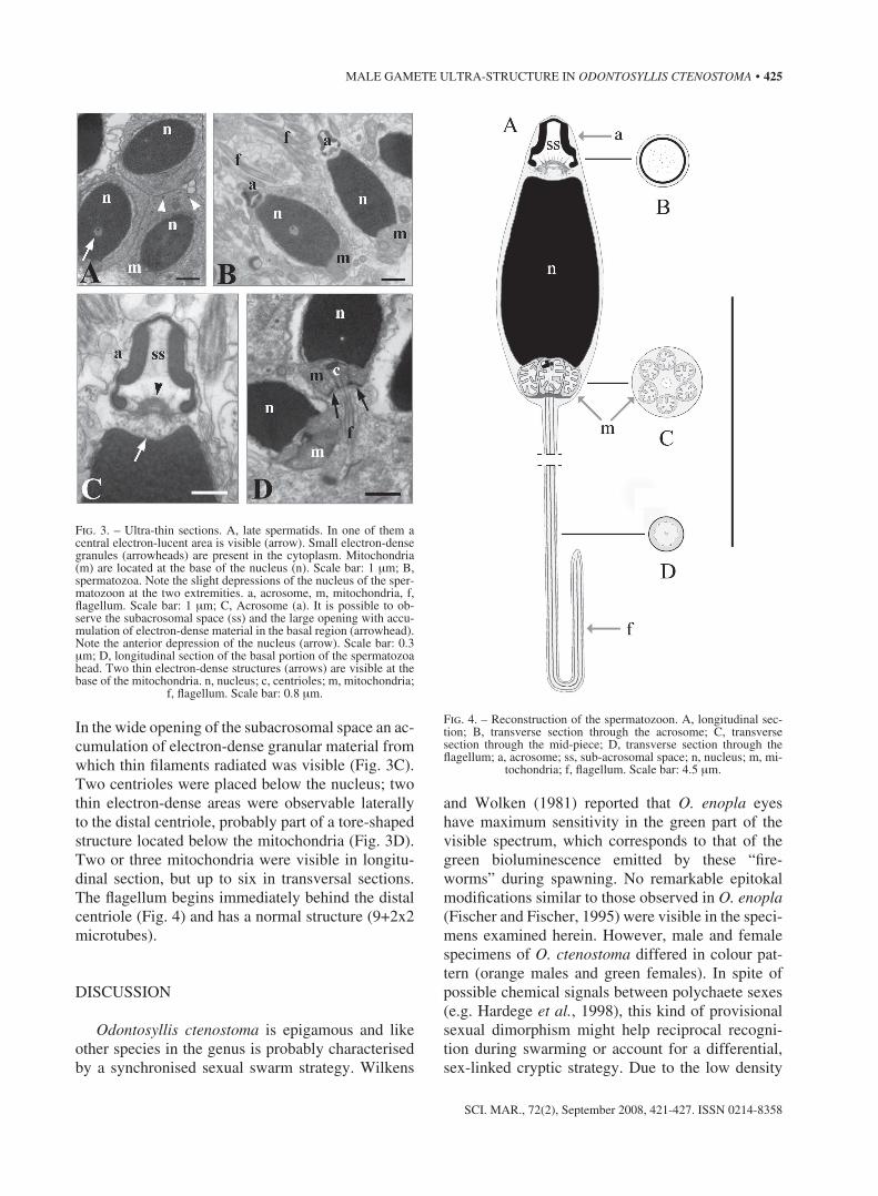
in the wide opening of the subacrosomal space an ac-cumulation of electron-dense granular material from which thin filaments radiated was visible (Fig. 3c). two centrioles were placed below the nucleus; two thin electron-dense areas were observable laterally to the distal centriole, probably part of a tore-shaped structure located below the mitochondria (Fig. 3d). two or three mitochondria were visible in longitu-dinal section, but up to six in transversal sections. the flagellum begins immediately behind the distal centriole (Fig. 4) and has a normal structure (9+2x2 microtubes).
discussion
Odontosyllis ctenostoma is epigamous and like other species in the genus is probably characterised by a synchronised sexual swarm strategy. Wilkens
and Wolken (1981) reported that O. enopla eyes have maximum sensitivity in the green part of the visible spectrum, which corresponds to that of the green bioluminescence emitted by these “fire-worms” during spawning. no remarkable epitokal modifications similar to those observed in O. enopla (Fischer and Fischer, 1995) were visible in the speci-mens examined herein. however, male and female specimens of O. ctenostoma differed in colour pat-tern (orange males and green females). in spite of possible chemical signals between polychaete sexes (e.g. hardege et al., 1998), this kind of provisional sexual dimorphism might help reciprocal recogni-tion during swarming or account for a differential, sex-linked cryptic strategy. due to the low density
Fig. 3. – ultra-thin sections. A, late spermatids. in one of them a central electron-lucent area is visible (arrow). small electron-dense granules (arrowheads) are present in the cytoplasm. Mitochondria (m) are located at the base of the nucleus (n). scale bar: 1 µm; B, spermatozoa. note the slight depressions of the nucleus of the sper-matozoon at the two extremities. a, acrosome, m, mitochondria, f, flagellum. scale bar: 1 µm; c, Acrosome (a). it is possible to ob-serve the subacrosomal space (ss) and the large opening with accu-mulation of electron-dense material in the basal region (arrowhead). note the anterior depression of the nucleus (arrow). scale bar: 0.3 µm; d, longitudinal section of the basal portion of the spermatozoa head. two thin electron-dense structures (arrows) are visible at the base of the mitochondria. n, nucleus; c, centrioles; m, mitochondria;
f, flagellum. scale bar: 0.8 µm.
Fig. 4. – reconstruction of the spermatozoon. A, longitudinal sec-tion; B, transverse section through the acrosome; c, transverse section through the mid-piece; d, transverse section through the flagellum; a, acrosome; ss, sub-acrosomal space; n, nucleus; m, mi-
tochondria; f, flagellum. scale bar: 4.5 µm.

426 • L. Musco et al.
sci. MAr., 72(2), september 2008, 421-427. issn 0214-8358
of the sampled population, the complete series of events leading to mature sperms and eggs in O. cten-ostoma was not fully sequenced. only large oocytes, which appear morphologically similar to the oocytes of Syllis pulchra described by heacox and schroeder (1981b), were observed in females. Moreover, only some phases of spermatogenesis in males were de-scribed. however, the data are sufficient for inter-specific comparisons and some general phylogenetic inferences.
the sperm structure observed herein, resembling the ect-aquasperm type, does not completely fit the reproductive strategy reported for O. ctenostoma. A particular brood protection with temporary mainte-nance of fertilised eggs in a mass on the ventrum of swarming females is in fact traditionally reported (gravier and dantan, 1928). A similar reproductive strategy has been confirmed in myrianida (= Autoly-tus) edwarsi (schiedges, 1979), and the sole exam-ined sperm morphology from an autolytine showed an ent-aquasperm type (Franzén, 1982). Brood pro-tection is also common in other eusyllinae species, such as some small Pionosyllis (pierantoni, 1903), but it is typical of small-sized epigamic exogoninae having ent-aquasperm (Mastrodonato et al., 2003). thus, it can be hypothesised that brood protection is an adaptive convergence in several small-sized species. By contrast, O. ctenostoma is a large syllid. therefore, it can be assumed that the O. ctenostoma specimens described by gravier and dantan (1928) as brooders belonged to a different and possibly smaller species. on the other hand, on the basis of the intrafamiliar phylogenetic constraints observed in polychaetes (giangrande, 1997), if compared to the egg size range in syllidae, the size of O. cten-ostoma eggs (c.a 100 µm) could support direct de-velopment and a brood protection strategy. Further studies are therefore needed.
As stated before, the sperm structure of O. cten-ostoma fits the ect-aquasperm type, with a classical mid-piece with rounded mitochondria, and a fairly elongate barrel-shaped nucleus. the acrosome ultra-structure and its internal organisation are interesting, particularly because of the presence of fibrillar elec-tron-dense material in the sub-acrosomal space. the basal invagination of the acrosomal vesicle forming the sub-acrosomal space is common in polychaete sperms (Jamieson and rouse, 1989; rice, 1992). in S. krohni (Lepore et al., 2006), as well as in the ect-aquasperm of Syllis sp (Jamieson and rouse, 1989) and S. pigmentata (heacox and schroeder, 1981a),
only a small opening is retained between the sub-acrosomal space and the underlying nucleus, and the sub-acrosomal material within the enclosed cavity is differentiated as an ellipsoid with a number of elec-tron-dense thick filaments. this space is more devel-oped in S. krohni (Lepore et al., 2006), as in the case of O. ctenostoma. however, the mature acrosome of the currently investigated species particularly re-sembled the up-side-down cup-shaped acrosome of the late spermatids of Exogone naidina and E. dis-par (giangrande et al., 2002), albeit with a reduced apical region that made it appear bell-shaped rather than cup-shaped. With maturation, however, the sub-acrosomal space in Exogone species becomes narrower and the sub-acrosomal material condenses and finally transforms into an electron-opaque mesh-work within the acrosome. this has been seen as a simplification and possibly indicative of evolution-ary derivation from the syllinae ect-aquasperm type (giangrande et al., 2002). the similarity between the Odontosyllis mature sperm and Exogone spermatids could be indicative of a close phylogenetic relation-ship. completely different late spermatids have up to now been found in the genus Syllis, which in the phylogenetic hypothesis of nygren (1999) is more closely related to Odontosyllis than exogoninae are. in fact, the subfamily eusyllinae is considered para-phyletic in nygren’s (1999) cladistic analysis, and the genus Odontosyllis in particular is closer to the syllinae but resembles the exogoninae in the shared epigamy.
it is well known that syllinae and Autolytinae are generally characterised by schizogamy and eu-syllinae and exogoninae by epigamy; however, as stressed by nygren (1999), it is uncertain which of the two is the plesiomorphic reproductive mode. Following our hypothesis, if the exogonine sperm was derived from simplification of a sylline sperm through a gradual passage from an Odontosyllis-like eusylline ancestor, it could be equally possible that epigamy is derived from schizogamy, Odontosyllis (or an Odontosyllis-like ancestor) representing the linkage between the two. Further studies on sperm morphology will likely contribute to a better under-standing of syllid phylogeny.
AcKnoWLedgeMents
We would like to thank the anonymous referee who gave us constructive suggestions. the work of

MALe gAMete uLtrA-structure in OdONTOSylliS CTENOSTOmA • 427
sci. MAr., 72(2), september 2008, 421-427. issn 0214-8358
the authors A. giangrande and L. Musco was car-ried out within the MarBeF network of excellence “Marine Biodiversity and ecosystem Functioning”, which is funded in the european union’s sixth Framework programme (contract no. goce-ct-2003-505446). the research of the other authors was supported by grants from the italian Murst (scien-tific research Ateneo of Bari university).
reFerences
Bührmann, c., W. Westheide and g. purschke. – 1996. sperma-togenesis and sperm ultra-structure in the interstitial syllid Petitia amphophtalma (Annelida, polychaeta). Ophelia, 45: 201-211.
daly, J.M. – 1975. reversible epitoky in the life history of the poly-chaete Odontosyllis polycera (schmarda, 1861). J. mar. Biol. Ass. UK. 55: 327-344.
Fischer, A. and u. Fischer. – 1995. on the life-style and the life-cycle of the luminescent polychaete Odontosyllis enopla (An-nelida: polychaeta). invertebr. Biol., 114(3): 236-247.
Franke, h. d. – 1999. reproduction of syllidae (Annelida: polycha-eta). Hydrobiologia, 402: 39-55.
Franke, h.d. and h.d. pfannenstiel. – 1984. some aspects of en-docrine control of polychaete reproduction. in: A. Fischer and h.d. pfannenstiel (eds.), Polychaete Reproduction. Fortschr. Zool., 29: 53-72.
Franzén, Å. – 1956. on spermiogenesis, morphology and the sper-matozoon, and biology of fertilisation among invertebrates. Zool. Bijdr., 31: 355-482.
Franzén, Å. – 1974. sperm structure in some polychaeta. in: B.A. Afzelius (ed.), The Functional Anatomy of the Spermatozoon, pp. 267-278. pergamon press, oxford.
Franzén, Å. – 1982. ultra-structure of spermatids and spermatozoa in three polychaetes with modified biology of reproduction: Autolytus sp, Chitinopoma serrula and Capitella capitata. int. J. invertebr. Reprod., 5: 185-200.
garwood, p.r. – 1991. reproduction and the classification of the family syllidae (polychaeta). Ophelia, suppl., 5: 81-87.
giangrande, A. – 1997. polychaete reproductive patterns, life cycle and life histories: an overview. Oceanogr. mar. Biol. Annu. Rev., 35: 323-386.
giangrande, A., M. sciscioli, e. Lepore, M. Mastrodonato, p. Lupetti and r. dallai. – 2002. sperm ultra-structure and spermiogenesis in two Exogone species (polychaeta, syllidae, exogoninae). in-vertebr. Biol., 121(4): 339-349.
gravier c. and J.L. dantan. – 1928. peches nocturnes a la lumière dans la Baie d’Alger. Annélides polychètes. Ann. inst. Océa-nogr. Paris Nouv. ser., 5: 1-187
hardege, J.d., Müller, c., Beckmann, M., h.d. Bartels-hardege and M.g. Bentley. – 1998. timing of reproduction in marine poly-chaetes: the role of sex pheromones. Ecoscience, 5: 395-404.
heacox, A.e. and p.c. schroeder. – 1981a. A light- and electron-microscopic investigation of gametogenesis in Typosyllis pul-chra (Berkeley and Berkeley) (polychaeta: syllidae) i. gonad structure and spermatogenesis. Cell Tissue Res., 218: 623-639.
heacox, A.e. and p.c. schroeder. – 1981b. A light- and electron-mi-croscopic investigation of gametogenesis in Typosyllis pulchra (Berkeley and Berkeley) (polychaeta: syllidae) ii. oogenesis. Cell Tissue Res., 218: 641-658.
Jamieson, B.g.M. and g.W. rouse. – 1989. the spermatozoa of the polychaeta (Annelida): an ultrastructural review. Biol. Rev. Camb. Philos. Soc., 64: 93-157.
Kuper, M. and W. Westheide. – 1997. sperm ultra-structure and spermatogenesis in the interstitial polychaete Sphaerosyllis her-maphrodita (syllidae: exogoninae). invertebr. Reprod. dev., 32(3): 189-200.
Lepore, e., sciscioli M, Mastrodonato M., gherardi M., A. gian-grande and L. Musco. – 2006. sperm ultra-structure and sper-miogenesis in Syllis krohni (polychaeta: syllidae), with some observations on its reproductive biology. Sci. mar., 70(4): 585-592.
Mastrodonato, M., M. sciscioli, e. Lepore, M. gherardi, A. gian-grande, d. Mercati, r. dallai and p. Lupetti. – 2003. external gestation of Exogone naidina oersted, 1845 (polychaeta, syl-lidae): ventral attachment of eggs and embryos. Tissue Cell., 35(4): 297-305.
Millonig, g. – 1976. laboratory manual of biological electron mi-croscopy. edizioni saviolo. Vercelli.
nygren, A. – 1999. phylogeny and reproduction in syllidae (poly-chaeta). Zool. J. linn. Soc., 126(3): 365-386.
patti, p., M.c. gambi and A. giangrande. – 2003. preliminary study on the systematic relationships of sabellinae (polychaeta: sabellidae), based on the c1 domain of the 28s rdnA, with discussion of reproductive features. ital. J. Zool., 70: 269-278.
pierantoni, u. – 1903. La gestazione esterna (contributo alla biolo-gia e all’embriologia dei sillidi). Arch. Zool. ital., 1: 231-252.
rice, s.A. – 1992. polychaeta. spermatogenesis and sperm structure. in: harrison F. W. and gardiner s. (eds.), microcopic Anatomy of invertebrates Vol. Vii (Annelida), pp. 129-151. Wiley-Liss inc., Wilmington.
san Martín, g. – 2003. Annelida, polychaeta ii: syllidae. in: M.A. ramos sánchez, J. Alba tercedor, X. Bellés i ros, J. gosálbez i noguera, A. guerra sierra, e. Macpherson Mayol, F. Mar-tín piera, J. serrano Marino and J. templado gonzález (eds.), Fauna ibérica, Vol 21, pp. 1-554. Museo nacional de ciencias naturales. csic, Madrid.
schiedges, K.L. – 1979. reproductive Biology and ontogenesis in the polychaete genus Autolytus (Annelida: syllidae): ob-servations on Laboratory cultured individuals. mar. Biol., 54: 239-250.
schroeder, p. and c.o. hermans. – 1975. Annelida: polychaeta. in: A.c. giese and J.s. pearse (eds.), Reproduction of marine invertebrates. Vol. iii. Annelids and Echiurans, pp. 1-213. Aca-demic press, new York.
Vortsepneva, e.V., A.e. Zhadan and A.B. tzetlin. – 2006. spermio-genesis and sperm ultrastructure of Asetocalamyzas laonicola tzetlin, 1985 (polychaeta), an ectoparasite of the large spionid Scolelepis cf. matsugae sikorsfi, 1994, from the White sea. Sci. mar., 70s3: 343-350.
Wilkens, L.A. and J.J. Wolken. – 1981. electroretinograms from Odontosyllis enopla (polychaeta; syllidae): initial observations on the visual system of the bioluminescent fireworm of Ber-muda. mar. Behav. Physiol., 8: 55-66.
scient. ed.: r. sardà.received April 17, 2007. Accepted February 27, 2008.published online May 7, 2008.