Sperm Morphology Assessment in the Era of Intracytoplasmic ...
14
Received: Mar 29, 2021 Revised: Apr 21, 2021 Accepted: May 9, 2021 Published online Jun 17, 2021 Correspondence to: Ashok Agarwal https://orcid.org/0000-0003-0585-1026 Andrology Center and American Center for Reproductive Medicine, Cleveland Clinic, Mail Code X-11, 10681 Carnegie Avenue, Cleveland, OH 44195, USA. Tel: +1-216-444-9485, Fax: +1-216+445-6049, E-mail: [email protected], Website: www.Clevelandclinic.org/ReproductiveResearchCenter Copyright © 2021 Korean Society for Sexual Medicine and Andrology Sperm Morphology Assessment in the Era of Intracytoplasmic Sperm Injection: Reliable Results Require Focus on Standardization, Quality Control, and Training Ashok Agarwal 1 , Rakesh Sharma 1 , Sajal Gupta 1 , Renata Finelli 1 , Neel Parekh 1 , Manesh Kumar Panner Selvam 1,2 , Ralf Henkel 1,3,4,5 , Damayanthi Durairajanayagam 6 , Camila Pompeu 7 , Sarah Madani 8 , Andrea Belo 9 , Neha Singh 10 , Simryn Covarrubias 11 , Sara Darbandi 12 , Raha Sadeghi 11 , Mahsa Darbandi 12 , Paraskevi Vogiatzi 13 , Florence Boitrelle 14,15 , Mara Simopoulou 16 , Ramadan Saleh 17 , Mohamed Arafa 1,18,19 , Ahmad Majzoub 1,19 , Hussein Kandil 20 , Armand Zini 21 , Edmund Ko 22 , Juan G. Alvarez 23 , Marlon Martinez 24 , Jonathan Ramsay 25 , Sunil Jindal 26 , Gian Maria Busetto 27 , Hassan Sallam 28 , Israel Maldonado 29 , Christina Anagnostopoulou 30 , Marco G. Alves 31 , Pallav Sengupta 32 , Kambiz Gilany 33 , Donald P. Evenson 34 , Sheena E.M. Lewis 35,36 , Jaime Gosalvez 37 , Rafael F. Ambar 38 , Rupin Shah 39 1 American Center for Reproductive Medicine, Cleveland Clinic, Cleveland, OH, 2 Department of Urology, Tulane University Health Sciences Center, New Orleans, LA, USA, 3 Department of Metabolism, Digestion and Reproduction, Imperial College London, London, UK, 4 Department of Medical Bioscience, University of the Western Cape, Bellville, South Africa, 5 LogixX Pharma, Theale, Reading, Berkshire, UK, 6 Department of Physiology, Faculty of Medicine, Universiti Teknologi MARA, Sungai Buloh Campus, Selangor, Malaysia, 7 Instituto Ideia Fèrtil, Santo Andrè, Brazil, 8 Department of Biology and Physiology of Organisms, Faculty of Biological Sciences, University of Science and Technnology, Houari Boumedien, Algiers, Algeria, 9 Huntington Centro de Medicina Reproditiva S/A, Sao Paulo, São Paulo, Brazil, 10 NOVA IVF, Gorakhpur, India, 11 Department of Physiology, University of San Francisco, CA, USA, 12 Fetal Health Research Center, Hope Generation Foundation, Tehran, Iran, 13 Andromed Health & Reproduction, Fertility Diagnostics Laboratory, Maroussi, Athens, Greece, 14 Reproductive Biology, Fertility Preservation, Andrology, CECOS, Poissy Hospital, Poissy, 15 Paris Saclay University, UVSQ, INRAE, BREED, Jouy-en-Josas, France, 16 Department of Experimental Physiology, School of Health Sciences, Faculty of Medicine, National and Kapodistrian University of Athens, Athens, Greece, 17 Department of Dermatology, Venereology and Andrology, Faculty of Medicine, Sohag University, Sohag, Egypt, 18 Department of Urology, Hamad Medical Corporation, 19 Department of Urology, Weill Cornell Medicine-Qatar, Doha, Qatar, 20 Fakih IVF Fertility Center, Abu Dhabi, UAE, 21 Department of Surgery, McGill University, Montreal, QC, Canada, 22 Department of Urology, Loma Linda University Health, Loma Linda, CA, USA, 23 Centro ANDROGEN, La Coruña, Spain, 24 Section of Urology, University of Santo Tomas Hospital, Manila, Philippines, 25 Department of Andrology, Hammersmith Hospital, London, UK, 26 Department of Andrology and Reproductive Medicine, Jindal Hospital, Meerut, India, 27 Department of Urology and Renal Transplantation, University of Foggia Policlinico Riuniti of Foggia, Foggia, Italy, 28 Department of Obstetrics and Gynaecology, Alexandria University Faculty of Medicine, Alexandria, Egypt, 29 Citmer Reproductive Medicine, IVF LAB, Mexico City, Mexico, 30 IVF Clinic “Akeso-Embryo ART”, Athens, Greece, 31 Department of Anatomy, Unit for Multidisciplinary Research in Biomedicine (UMIB), Institute of Biomedical Sciences Abel Salazar (ICBAS), University of Porto, Porto, Portugal, 32 Department of Physiology, Faculty of Medicine, MAHSA University, Selangor, Malaysia, 33 Reproductive Biotechnology Research Center, Avicenna Research Institute, ACECR, Tehran, Iran, 34 SCSA Diagnostics, Brookings, SD, USA, 35 Queens University Belfast, 36 Examenlab Ltd., Weavers Court, Belfast, Northern Ireland, UK, 37 Genetic Unit, Department of Biology, Universidad Autónoma de Madrid, Madrid, Spain, 38 Department of Urology, Centro Universitario em Saude do ABC/Andrology Group at Ideia Fertil Institute of Human Reproduction, Santo André, Brazil, 39 Department of Urology, Lilavati Hospital and Research Centre, Mumbai, India Review Article pISSN: 2287-4208 / eISSN: 2287-4690 World J Mens Health Published online Jun 17, 2021 https://doi.org/10.5534/wjmh.210054 Male reproductive health and infertility
Transcript of Sperm Morphology Assessment in the Era of Intracytoplasmic ...
Received: Mar 29, 2021 Revised: Apr 21, 2021 Accepted: May 9, 2021 Published online Jun 17, 2021Correspondence to: Ashok Agarwal https://orcid.org/0000-0003-0585-1026 Andrology Center and American Center for Reproductive Medicine, Cleveland Clinic, Mail Code X-11, 10681 Carnegie Avenue, Cleveland, OH 44195, USA.Tel: +1-216-444-9485, Fax: +1-216+445-6049, E-mail: [email protected], Website: www.Clevelandclinic.org/ReproductiveResearchCenter
Copyright © 2021 Korean Society for Sexual Medicine and Andrology
Sperm Morphology Assessment in the Era of Intracytoplasmic Sperm Injection: Reliable Results Require Focus on Standardization, Quality Control, and Training
Ashok Agarwal1 , Rakesh Sharma1 , Sajal Gupta1 , Renata Finelli1 , Neel Parekh1 , Manesh Kumar Panner Selvam1,2 , Ralf Henkel1,3,4,5 , Damayanthi Durairajanayagam6 , Camila Pompeu7 , Sarah Madani8 , Andrea Belo9 , Neha Singh10 , Simryn Covarrubias11 , Sara Darbandi12 , Raha Sadeghi11 , Mahsa Darbandi12 , Paraskevi Vogiatzi13 , Florence Boitrelle14,15 , Mara Simopoulou16 , Ramadan Saleh17 , Mohamed Arafa1,18,19 , Ahmad Majzoub1,19 , Hussein Kandil20 , Armand Zini21 , Edmund Ko22 , Juan G. Alvarez23 , Marlon Martinez24 , Jonathan Ramsay25 , Sunil Jindal26 , Gian Maria Busetto27 , Hassan Sallam28 , Israel Maldonado29 , Christina Anagnostopoulou30 , Marco G. Alves31 , Pallav Sengupta32 , Kambiz Gilany33 , Donald P. Evenson34 , Sheena E.M. Lewis35,36 , Jaime Gosalvez37 , Rafael F. Ambar38 , Rupin Shah39
1American Center for Reproductive Medicine, Cleveland Clinic, Cleveland, OH, 2Department of Urology, Tulane University Health Sciences Center, New Orleans, LA, USA, 3Department of Metabolism, Digestion and Reproduction, Imperial College London, London, UK, 4Department of Medical Bioscience, University of the Western Cape, Bellville, South Africa, 5LogixX Pharma, Theale, Reading, Berkshire, UK, 6Department of Physiology, Faculty of Medicine, Universiti Teknologi MARA, Sungai Buloh Campus, Selangor, Malaysia, 7Instituto Ideia Fèrtil, Santo Andrè, Brazil, 8Department of Biology and Physiology of Organisms, Faculty of Biological Sciences, University of Science and Technnology, Houari Boumedien, Algiers, Algeria, 9Huntington Centro de Medicina Reproditiva S/A, Sao Paulo, São Paulo, Brazil, 10NOVA IVF, Gorakhpur, India, 11Department of Physiology, University of San Francisco, CA, USA, 12Fetal Health Research Center, Hope Generation Foundation, Tehran, Iran, 13Andromed Health & Reproduction, Fertility Diagnostics Laboratory, Maroussi, Athens, Greece, 14Reproductive Biology, Fertility Preservation, Andrology, CECOS, Poissy Hospital, Poissy, 15Paris Saclay University, UVSQ, INRAE, BREED, Jouy-en-Josas, France, 16Department of Experimental Physiology, School of Health Sciences, Faculty of Medicine, National and Kapodistrian University of Athens, Athens, Greece, 17Department of Dermatology, Venereology and Andrology, Faculty of Medicine, Sohag University, Sohag, Egypt, 18Department of Urology, Hamad Medical Corporation, 19Department of Urology, Weill Cornell Medicine-Qatar, Doha, Qatar, 20Fakih IVF Fertility Center, Abu Dhabi, UAE, 21Department of Surgery, McGill University, Montreal, QC, Canada, 22Department of Urology, Loma Linda University Health, Loma Linda, CA, USA, 23Centro ANDROGEN, La Coruña, Spain, 24Section of Urology, University of Santo Tomas Hospital, Manila, Philippines, 25Department of Andrology, Hammersmith Hospital, London, UK, 26Department of Andrology and Reproductive Medicine, Jindal Hospital, Meerut, India, 27Department of Urology and Renal Transplantation, University of Foggia Policlinico Riuniti of Foggia, Foggia, Italy, 28Department of Obstetrics and Gynaecology, Alexandria University Faculty of Medicine, Alexandria, Egypt, 29Citmer Reproductive Medicine, IVF LAB, Mexico City, Mexico, 30IVF Clinic “Akeso-Embryo ART”, Athens, Greece, 31Department of Anatomy, Unit for Multidisciplinary Research in Biomedicine (UMIB), Institute of Biomedical Sciences Abel Salazar (ICBAS), University of Porto, Porto, Portugal, 32Department of Physiology, Faculty of Medicine, MAHSA University, Selangor, Malaysia, 33Reproductive Biotechnology Research Center, Avicenna Research Institute, ACECR, Tehran, Iran, 34SCSA Diagnostics, Brookings, SD, USA, 35Queens University Belfast, 36Examenlab Ltd., Weavers Court, Belfast, Northern Ireland, UK, 37Genetic Unit, Department of Biology, Universidad Autónoma de Madrid, Madrid, Spain, 38Department of Urology, Centro Universitario em Saude do ABC/Andrology Group at Ideia Fertil Institute of Human Reproduction, Santo André, Brazil, 39Department of Urology, Lilavati Hospital and Research Centre, Mumbai, India
Review Article
pISSN: 2287-4208 / eISSN: 2287-4690World J Mens Health Published online Jun 17, 2021https://doi.org/10.5534/wjmh.210054
Male reproductive health and infertility
https://doi.org/10.5534/wjmh.210054
2 www.wjmh.org
INTRODUCTION
Infertility is defined as the inability to achieve a suc-cessful pregnancy after 12 months or more of regular, unprotected sexual intercourse [1]. The World Health Organization (WHO) demographics estimate that about 200 million people are affected by infertility globally [2,3], with male factor contributing to 50% of infertility cases [4]. A meta-analysis published in 1992 reported a decline in sperm quality over a period of 50 years (1938 to 1991) [5] while, more recently, Levine et al [6] re-ported a 50% to 60% decline in sperm count from 1973 to 2011 across North America, Europe, New Zealand, and Australia. Furthermore, other researchers have reported a significant decline in sperm concentration, total count, morphology, and motility between 2000 to 2017 [7]. A negative association is also reported between poor morphology and gonadotropins [8]. As the analysis of sperm parameters is the primary approach for iden-tifying and diagnosing male infertility, semen analysis is of particular importance since it defines fertility status and potential, as well as the course of natural or assisted reproduction [9]. In this framework, sperm morphology is established as the most prominent com-ponent, as this parameter cannot be surpassed even by the most invasive interventions such as intracytoplas-mic sperm injection (ICSI).
Interestingly, there have been significant changes in the classification of sperm morphology over the years. Normal sperm morphology reference values have been revised dramatically from ≥80.5% reported in the 1st
edition of the WHO manual [10,11] to ≥14% in the 4th edition [12], and even lower to ≥4% in the 5th and most recent edition [13]. The ‘strict criteria’ for assess-ing sperm morphology classify semen as normal if the percentage of normal sperm is ≥14% [14,15]. Based on this classification, abnormal sperm morphology has been associated with poor fertilization and clinical outcomes after assisted reproductive technology (ART), thereby establishing sperm morphology as a predictor of ART outcomes [16-18]. The cut-off for sperm mor-phology based on strict criteria was further revised in the 5th edition of the WHO manual [13] which uses the 5th centiles (and their 95% confidence intervals [CIs]) as the lowest reference limit (≥4% cutoff for normal sperm morphology). The lowest limit for morphologi-cally normal forms of spermatozoa is 4% (95% CI, 3%–4%) and all borderline forms are considered ab-normal. This classification serves as a surrogate tool to choose the most appropriate type of ART procedure for infertile couples [19]; if the percentage of normal forms is ≥4%, the probability of fertilization with ART is high and procedures such as intrauterine insemination (IUI) or in vitro fertilization (IVF) may be selected. In specimens with otherwise normal count and motility, poor morphology may itself be a determinant in decid-ing whether or not to proceed with ICSI. When the percentage of sperm with normal morphology is <4%, fertilization with IUI and IVF is poor, and ICSI should be preferred [20].
The predictive value of sperm morphology in ART continues to be a matter of debate. Recent studies sug-
Semen analysis is the first, and frequently, the only step in the evaluation of male fertility. Although the laboratory procedures are conducted according to the World Health Organization (WHO) guidelines, semen analysis and especially sperm mor-phology assessment is very difficult to standardize and obtain reproducible results. This is mainly due to the highly subjective nature of their evaluation. ICSI is the choice of treatment when sperm morphology is severely abnormal (teratozoospermic). Hence, the standardization of laboratory protocols for sperm morphology evaluation represents a fundamental step to ensure reliable, accurate and consistent laboratory results that avoid misdiagnoses and inadequate treatment of the infertile patient. This article aims to promote standardized laboratory procedures for an accurate evaluation of sperm morphology, including the establishment of quality control and quality assurance policies. Additionally, the clinical importance of sperm morphol-ogy results in assisted reproductive outcomes is discussed, along with the clinical management of teratozoospermic patients.
Keywords:Keywords: Abnormality, teratozoospermia; Morphology, stain; Sperm
This is an Open Access article distributed under the terms of the Creative Commons Attribution Non-Commercial License (http://creativecommons.org/licenses/by-nc/4.0) which permits unrestricted non-commercial use, distribution, and reproduction in any medium, provided the original work is properly cited.
Ashok Agarwal, et al: Standardization of Semen Morphology Assessment
3www.wjmh.org
gest that morphology may not be a good predictor of fertilization or pregnancy outcome in ICSI [21-25]. Ab-normal fertilization following ICSI has been associated with abnormal parameters in semen analysis. Oligo-asthenozoospermia and oligoasthenoteratozoospermia have been suggested to be further associated with lower cleavage and blastocyst formation rates [26]. A careful assessment is critical to identify the underlying cause of infertility, as this can serve as a tool to choose the most appropriate type of ART method [27]. Howev-er, heterogeneity of study groups, differences in stain-ing methods, intra- and inter-laboratory variations, differences in the scoring classifications, and manual versus computer assisted semen analyzer (CASA) scor-ing may be among the many contributory factors for the lack of robust predictive power [19,28,29].
Diagnostic and predictive value of sperm morphology can be improved by uniform and consistent application of the defined strict criteria for scoring sperm morphol-ogy, standardization of the staining methods, internal and external quality controls (QCs), and training of the laboratory personnel [11,19,30].
In this review, we aim to: a) summarize standardized laboratory procedures for a proper evaluation of sperm morphology; b) highlight the importance of QC and quality assurance (QA) in laboratory assessment of sperm morphology; c) discuss the association between abnormal sperm morphology (teratozoospermia) and ART outcomes; and d) review the clinical management of men with abnormal sperm morphology.
MORPHOLOGICAL ASSESSMENT OF SPERM BY IMPLEMENTATION OF STANDARDIZATION AND QUALITY CONTROL
1. Preparation of semen sampleSemen sample is collected in a sterile collection con-
tainer according to the recommendations provided by the WHO (2010) and incubated at 37°C for 30 minutes to allow liquefaction [13]. If the ejaculate is viscous, pro-teolytic enzymes such as α-chymotrypsin or bromelain can be added to the sample and incubated at 37°C for an additional 10 minutes [13,31]. The liquefied sample is vortexed for 10 seconds, and a 10 µL aliquot is quickly removed. If the sperm concentration is <2×10⁶/mL, the sample is centrifuged at 600 g for 10 minutes to re-move most of the supernatant, leaving about 100 µL
of seminal plasma in the underlying concentrate. The pellet is resuspended by gentle pipetting to redilute the sample, not exceeding 50×106/mL. Centrifugation may affect sperm morphology and should be recorded in the patient’s worksheet [32].
2. Preparing a smear for sperm morphologyOn a clean frosted slide with the patient identifiers,
10 µL of well-mixed semen aliquot is added on one end of the slide. A second slide is used with an angle of 45° to quickly spread the drop of semen along the frosted slide, forming a smooth and even smear. The slides are prepared in duplicate and then air-dried before stain-ing.
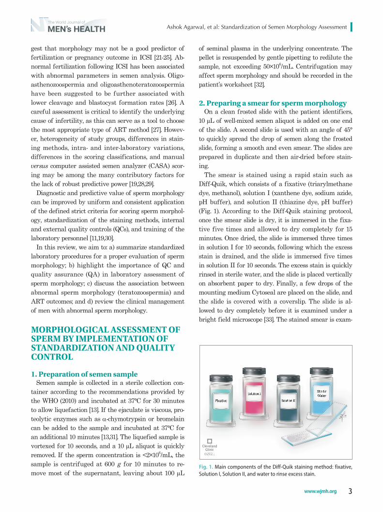
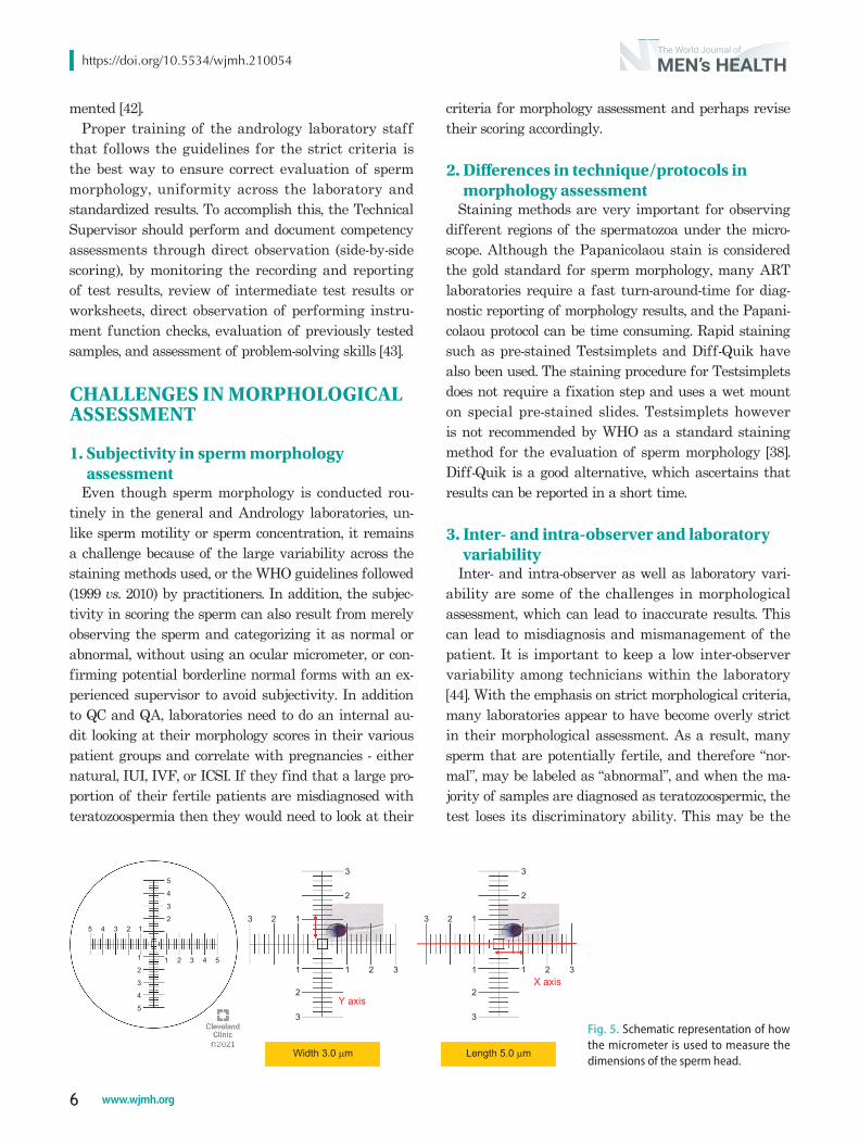
The smear is stained using a rapid stain such as Diff-Quik, which consists of a fixative (triarylmethane dye, methanol), solution I (xanthene dye, sodium azide, pH buffer), and solution II (thiazine dye, pH buffer) (Fig. 1). According to the Diff-Quik staining protocol, once the smear slide is dry, it is immersed in the fixa-tive five times and allowed to dry completely for 15 minutes. Once dried, the slide is immersed three times in solution I for 10 seconds, following which the excess stain is drained, and the slide is immersed five times in solution II for 10 seconds. The excess stain is quickly rinsed in sterile water, and the slide is placed vertically on absorbent paper to dry. Finally, a few drops of the mounting medium Cytoseal are placed on the slide, and the slide is covered with a coverslip. The slide is al-lowed to dry completely before it is examined under a bright field microscope [33]. The stained smear is exam-
Fig. 1. Main components of the Diff-Quik staining method: fixative, Solution I, Solution II, and water to rinse excess stain.
https://doi.org/10.5534/wjmh.210054
4 www.wjmh.org
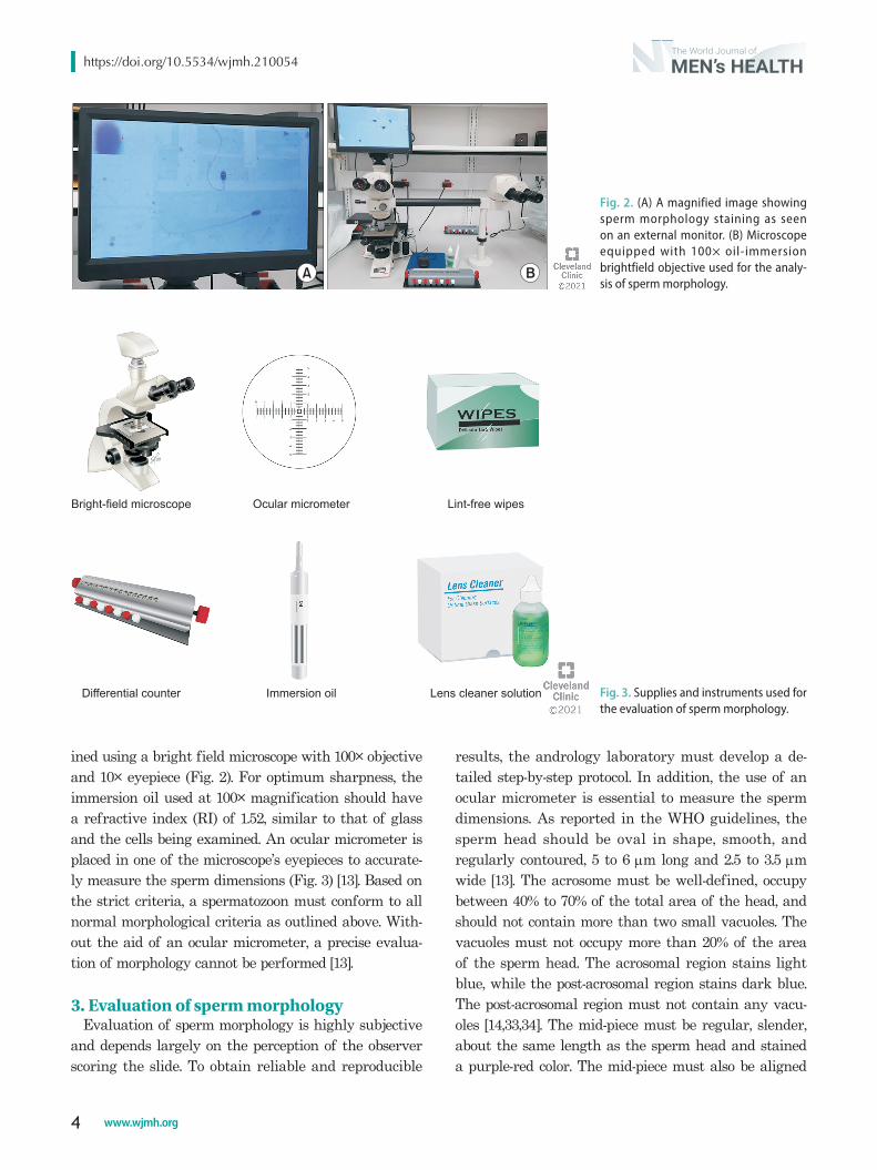
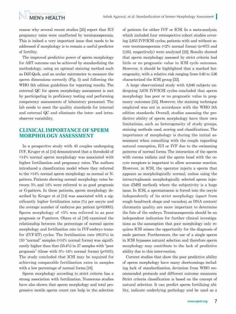
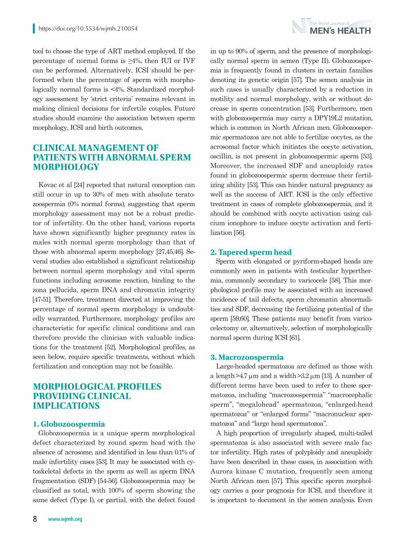
ined using a bright field microscope with 100× objective and 10× eyepiece (Fig. 2). For optimum sharpness, the immersion oil used at 100× magnification should have a refractive index (RI) of 1.52, similar to that of glass and the cells being examined. An ocular micrometer is placed in one of the microscope’s eyepieces to accurate-ly measure the sperm dimensions (Fig. 3) [13]. Based on the strict criteria, a spermatozoon must conform to all normal morphological criteria as outlined above. With-out the aid of an ocular micrometer, a precise evalua-tion of morphology cannot be performed [13].
3. Evaluation of sperm morphologyEvaluation of sperm morphology is highly subjective
and depends largely on the perception of the observer scoring the slide. To obtain reliable and reproducible
results, the andrology laboratory must develop a de-tailed step-by-step protocol. In addition, the use of an ocular micrometer is essential to measure the sperm dimensions. As reported in the WHO guidelines, the sperm head should be oval in shape, smooth, and regularly contoured, 5 to 6 μm long and 2.5 to 3.5 μm wide [13]. The acrosome must be well-defined, occupy between 40% to 70% of the total area of the head, and should not contain more than two small vacuoles. The vacuoles must not occupy more than 20% of the area of the sperm head. The acrosomal region stains light blue, while the post-acrosomal region stains dark blue. The post-acrosomal region must not contain any vacu-oles [14,33,34]. The mid-piece must be regular, slender, about the same length as the sperm head and stained a purple-red color. The mid-piece must also be aligned
A B
Fig. 2. (A) A magnified image showing sperm morphology staining as seen on an external monitor. (B) Microscope equipped with 100× oil-immersion brightfield objective used for the analy-sis of sperm morphology.
Bright-field microscope Ocular micrometer Lint-free wipes
Differential counter Immersion oil Lens cleaner solution Fig. 3. Supplies and instruments used for the evaluation of sperm morphology.
Ashok Agarwal, et al: Standardization of Semen Morphology Assessment
5www.wjmh.org
with the axis of the head of the sperm. If a residual cytoplasm larger than one-third of the area of the head is present, the sperm should be considered as abnor-mal [35]. If present, excess residual cytoplasm may be seen around the midpiece and is be stained pink/red or reddish orange depending on the type of stain used. The tail should be approximately 45 μm long, uniform along its length, appear thinner than the mid-piece, and stained a blue or reddish color. At least 2 replicates of 100 spermatozoa must be scored, with all borderline forms being considered as abnormal. When this strict classification is followed, the reference threshold is ≥4% for morphologically normal forms [13].
QUALITY CONTROL AND QUALITY ASSURANCE IN SPERM MORPHOLOGY ASSESSMENT
Sperm morphology remains one of the most con-troversial semen parameters, as an incorrect catego-rization can lead to lack of predictive value for ART outcomes, as reported in several recent publications, al-though sperm morphology has shown strong predictive values in the early nineties [21-25].
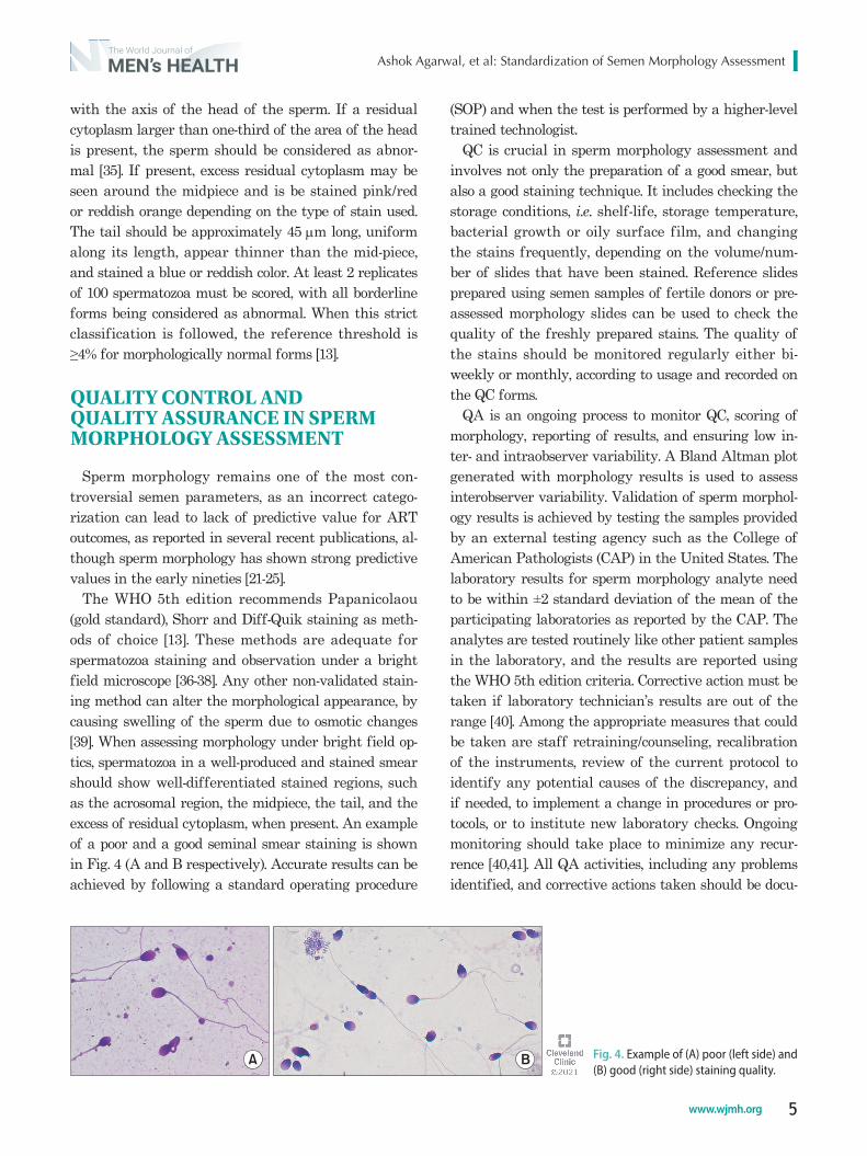
The WHO 5th edition recommends Papanicolaou (gold standard), Shorr and Diff-Quik staining as meth-ods of choice [13]. These methods are adequate for spermatozoa staining and observation under a bright field microscope [36-38]. Any other non-validated stain-ing method can alter the morphological appearance, by causing swelling of the sperm due to osmotic changes [39]. When assessing morphology under bright field op-tics, spermatozoa in a well-produced and stained smear should show well-differentiated stained regions, such as the acrosomal region, the midpiece, the tail, and the excess of residual cytoplasm, when present. An example of a poor and a good seminal smear staining is shown in Fig. 4 (A and B respectively). Accurate results can be achieved by following a standard operating procedure
(SOP) and when the test is performed by a higher-level trained technologist.
QC is crucial in sperm morphology assessment and involves not only the preparation of a good smear, but also a good staining technique. It includes checking the storage conditions, i.e. shelf-life, storage temperature, bacterial growth or oily surface film, and changing the stains frequently, depending on the volume/num-ber of slides that have been stained. Reference slides prepared using semen samples of fertile donors or pre-assessed morphology slides can be used to check the quality of the freshly prepared stains. The quality of the stains should be monitored regularly either bi-weekly or monthly, according to usage and recorded on the QC forms.
QA is an ongoing process to monitor QC, scoring of morphology, reporting of results, and ensuring low in-ter- and intraobserver variability. A Bland Altman plot generated with morphology results is used to assess interobserver variability. Validation of sperm morphol-ogy results is achieved by testing the samples provided by an external testing agency such as the College of American Pathologists (CAP) in the United States. The laboratory results for sperm morphology analyte need to be within ±2 standard deviation of the mean of the participating laboratories as reported by the CAP. The analytes are tested routinely like other patient samples in the laboratory, and the results are reported using the WHO 5th edition criteria. Corrective action must be taken if laboratory technician’s results are out of the range [40]. Among the appropriate measures that could be taken are staff retraining/counseling, recalibration of the instruments, review of the current protocol to identify any potential causes of the discrepancy, and if needed, to implement a change in procedures or pro-tocols, or to institute new laboratory checks. Ongoing monitoring should take place to minimize any recur-rence [40,41]. All QA activities, including any problems identified, and corrective actions taken should be docu-
A BFig. 4. Example of (A) poor (left side) and (B) good (right side) staining quality.
https://doi.org/10.5534/wjmh.210054
6 www.wjmh.org
mented [42].Proper training of the andrology laboratory staff
that follows the guidelines for the strict criteria is the best way to ensure correct evaluation of sperm morphology, uniformity across the laboratory and standardized results. To accomplish this, the Technical Supervisor should perform and document competency assessments through direct observation (side-by-side scoring), by monitoring the recording and reporting of test results, review of intermediate test results or worksheets, direct observation of performing instru-ment function checks, evaluation of previously tested samples, and assessment of problem-solving skills [43].
CHALLENGES IN MORPHOLOGICAL ASSESSMENT
1. Subjectivity in sperm morphology assessment
Even though sperm morphology is conducted rou-tinely in the general and Andrology laboratories, un-like sperm motility or sperm concentration, it remains a challenge because of the large variability across the staining methods used, or the WHO guidelines followed (1999 vs. 2010) by practitioners. In addition, the subjec-tivity in scoring the sperm can also result from merely observing the sperm and categorizing it as normal or abnormal, without using an ocular micrometer, or con-firming potential borderline normal forms with an ex-perienced supervisor to avoid subjectivity. In addition to QC and QA, laboratories need to do an internal au-dit looking at their morphology scores in their various patient groups and correlate with pregnancies - either natural, IUI, IVF, or ICSI. If they find that a large pro-portion of their fertile patients are misdiagnosed with teratozoospermia then they would need to look at their
criteria for morphology assessment and perhaps revise their scoring accordingly.
2. Differences in technique/protocols in morphology assessment
Staining methods are very important for observing different regions of the spermatozoa under the micro-scope. Although the Papanicolaou stain is considered the gold standard for sperm morphology, many ART laboratories require a fast turn-around-time for diag-nostic reporting of morphology results, and the Papani-colaou protocol can be time consuming. Rapid staining such as pre-stained Testsimplets and Diff-Quik have also been used. The staining procedure for Testsimplets does not require a fixation step and uses a wet mount on special pre-stained slides. Testsimplets however is not recommended by WHO as a standard staining method for the evaluation of sperm morphology [38]. Diff-Quik is a good alternative, which ascertains that results can be reported in a short time.
3. Inter- and intra-observer and laboratory variability
Inter- and intra-observer as well as laboratory vari-ability are some of the challenges in morphological assessment, which can lead to inaccurate results. This can lead to misdiagnosis and mismanagement of the patient. It is important to keep a low inter-observer variability among technicians within the laboratory [44]. With the emphasis on strict morphological criteria, many laboratories appear to have become overly strict in their morphological assessment. As a result, many sperm that are potentially fertile, and therefore “nor-mal”, may be labeled as “abnormal”, and when the ma-jority of samples are diagnosed as teratozoospermic, the test loses its discriminatory ability. This may be the
5 4 3 2 1
54321
5
4
3
2
5
4
3
2
1
3
2
1 2 3
3 2 1
1
2
3
3
2
1 2 3
3 2 1
1
2
3
Y axis
X axis
Width 3.0 m� Length 5.0 m�
Fig. 5. Schematic representation of how the micrometer is used to measure the dimensions of the sperm head.
Ashok Agarwal, et al: Standardization of Semen Morphology Assessment
7www.wjmh.org
reason why several recent studies [25] report that IUI pregnancy rates were unaffected by teratozoospermia. This is indeed a very important issue that needs to be addressed if morphology is to remain a useful predictor of fertility.
The improved predictive power of sperm morphology for ART outcome can be achieved by standardizing the methodology, using an optimal staining method such as Diff-Quik, and an ocular micrometer to measure the sperm dimensions correctly (Fig. 5) and following the WHO 5th edition guidelines for reporting results. The external QC for sperm morphology assessment is met by participating in proficiency testing and performing competency assessments of laboratory personnel. The lab needs to meet the quality standards for internal and external QC and eliminate the inter- and intra-observer variability.
CLINICAL IMPORTANCE OF SPERM MORPHOLOGY ASSESSMENT
In a prospective study with 45 couples undergoing IVF, Kruger et al [14] demonstrated that a threshold of >14% normal sperm morphology was associated with higher fertilization and pregnancy rates. The authors introduced a classification model where they referred to the >14% normal sperm morphology as normal or N-pattern. Patients showing normal morphology rates be-tween 5% and 14% were referred to as good prognosis or G-pattern. In these patients, sperm morphology de-scribed by Kruger et al [14] was associated with a sig-nificantly higher fertilization rates (%) per oocyte and the average number of embryos per patient (p=0.0001). Sperm morphology of <5% was referred to as poor prognosis or P-pattern. Obara et al [18] examined the relationship between the percentage of normal sperm morphology and fertilization rate in IVF-embryo trans-fer (IVF-ET) cycles. The fertilization rate (80.5%) in 110 “normal” samples (>14% normal forms) was signifi-cantly higher than that (55.4%) in 27 samples with “poor prognosis” (those with 5%–14% normal forms) (p<0.01). The study concluded that ICSI may be required for achieving comparable fertilization rates in samples with a low percentage of normal forms [18].
Sperm morphology according to strict criteria has a strong association with fertilization. Numerous studies have also shown that sperm morphology and total pro-gressive motile sperm count can help in the selection
of patients for either IVF or ICSI. In a meta-analysis, which included four retrospective cohort studies cover-ing 2,853 IVF/ICSI cycles, patients with and without se-vere teratozoospermia (<2% normal forms) (n=673 and 2,183, respectively) were analyzed [22]. Results showed that sperm morphology assessed by strict criteria had little or no prognostic value in ICSI cycle outcomes. However, it should be highlighted that a marked het-erogeneity, with a relative risk ranging from 0.40 to 3.36 characterized the ICSI group [22].
A large observational study with 8,846 subjects un-dergoing 3,676 IVF/ICSI cycles concluded that sperm morphology has poor or no prognostic value in preg-nancy outcomes [23]. However, the staining technique employed was not in accordance with the WHO 5th edition standards. Overall, studies assessing the pre-dictive ability of sperm morphology have their own limitations, such as heterogeneity of study groups, staining methods used, scoring and classifications. The importance of morphology is during the initial as-sessment when consulting with the couple regarding natural conception, IUI or IVF due to the swimming patterns of normal forms. The interaction of the sperm with corona radiata and the sperm head with the oo-cyte receptors is important to allow acrosome reaction. However, in ICSI, the operator injects a sperm that appears as morphologically normal, unless using the intracytoplasmic morphologically selected sperm injec-tion (IMSI method), where the subjectivity is a huge issue. In ICSI, a spermatozoon is forced into the oocyte independently of its strict morphology (apart from rough head/neck shape and vacuoles), as DNA content/chromatin quality are more important to determine the fate of the embryo. Teratozoospermia should be an independent indication for further clinical investiga-tions as the assumption that poor morphology only re-quires ICSI misses the opportunity for the diagnosis of male partner. Furthermore, the use of a single sperm in ICSI bypasses natural selection and therefore sperm morphology may contribute to the lack of predictive ability due to this intervention.
Current studies that show the poor predictive ability of sperm morphology have many shortcomings includ-ing lack of standardization, deviation from WHO rec-ommended protocols and different outcome measures. Strict criteria classification is based on the concept of natural selection. It can predict sperm fertilizing abi-lity, indicate underlying pathology and be used as a
https://doi.org/10.5534/wjmh.210054
8 www.wjmh.org
tool to choose the type of ART method employed. If the percentage of normal forms is ≥4%, then IUI or IVF can be performed. Alternatively, ICSI should be per-formed when the percentage of sperm with morpho-logically normal forms is <4%. Standardized morphol-ogy assessment by ‘strict criteria’ remains relevant in making clinical decisions for infertile couples. Future studies should examine the association between sperm morphology, ICSI and birth outcomes.
CLINICAL MANAGEMENT OF PATIENTS WITH ABNORMAL SPERM MORPHOLOGY
Kovac et al [24] reported that natural conception can still occur in up to 30% of men with absolute terato-zoospermia (0% normal forms), suggesting that sperm morphology assessment may not be a robust predic-tor of infertility. On the other hand, various reports have shown significantly higher pregnancy rates in males with normal sperm morphology than that of those with abnormal sperm morphology [27,45,46]. Se-veral studies also established a significant relationship between normal sperm morphology and vital sperm functions including acrosome reaction, binding to the zona pellucida, sperm DNA and chromatin integrity [47-51]. Therefore, treatment directed at improving the percentage of normal sperm morphology is undoubt-edly warranted. Furthermore, morphology profiles are characteristic for specific clinical conditions and can therefore provide the clinician with valuable indica-tions for the treatment [52]. Morphological profiles, as seen below, require specific treatments, without which fertilization and conception may not be feasible.
MORPHOLOGICAL PROFILES PROVIDING CLINICAL IMPLICATIONS
1. GlobozoospermiaGlobozoospermia is a unique sperm morphological
defect characterized by round sperm head with the absence of acrosome, and identified in less than 0.1% of male infertility cases [53]. It may be associated with cy-toskeletal defects in the sperm as well as sperm DNA fragmentation (SDF) [54-56]. Globozoospermia may be classified as total, with 100% of sperm showing the same defect (Type I), or partial, with the defect found
in up to 90% of sperm, and the presence of morphologi-cally normal sperm in semen (Type II). Globozoosper-mia is frequently found in clusters in certain families denoting its genetic origin [57]. The semen analysis in such cases is usually characterized by a reduction in motility and normal morphology, with or without de-crease in sperm concentration [53]. Furthermore, men with globozoospermia may carry a DPY19L2 mutation, which is common in North African men. Globozoosper-mic spermatozoa are not able to fertilize oocytes, as the acrosomal factor which initiates the oocyte activation, oscillin, is not present in globozoospermic sperm [53]. Moreover, the increased SDF and aneuploidy rates found in globozoospermic sperm decrease their fertil-izing ability [53]. This can hinder natural pregnancy as well as the success of ART. ICSI is the only effective treatment in cases of complete globozoospermia, and it should be combined with oocyte activation using cal-cium ionophore to induce oocyte activation and ferti-lization [56].
2. Tapered sperm headSperm with elongated or pyriform-shaped heads are
commonly seen in patients with testicular hyperther-mia, commonly secondary to varicocele [58]. This mor-phological profile may be associated with an increased incidence of tail defects, sperm chromatin abnormali-ties and SDF, decreasing the fertilizing potential of the sperm [59,60]. These patients may benefit from varico-celectomy or, alternatively, selection of morphologically normal sperm during ICSI [61].
3. MacrozoospermiaLarge-headed spermatozoa are defined as those with
a length >4.7 μm and a width >3.2 μm [13]. A number of different terms have been used to refer to these sper-matozoa, including “macrozoospermia” “macrocephalic sperm”, “megalohead” spermatozoa, “enlarged-head spermatozoa” or “enlarged forms” “macronuclear sper-matozoa” and “large head spermatozoa”.
A high proportion of irregularly shaped, multi-tailed spermatozoa is also associated with severe male fac-tor infertility. High rates of polyploidy and aneuploidy have been described in these cases, in association with Aurora kinase C mutation, frequently seen among North African men [57]. This specific sperm morphol-ogy carries a poor prognosis for ICSI, and therefore it is important to document in the semen analysis. Even
Ashok Agarwal, et al: Standardization of Semen Morphology Assessment
9www.wjmh.org
in cases of a moderate percentage of large-head sperm, genetic analysis such as sperm FISH could be useful to determine the ploidy of spermatozoa [62].
4. Pin headA pin head is a spermatozoon lacking chromatin
with only a tail [63]. This defect results from errors in the formation of the connecting piece during spermio-genesis. Recent genetic studies related the defect to a mutation in the Sad1 and UNC84 domain containing 5 (SUN5) gene, which is normally responsible for the formation of complexes linking the nucleus to the cyto-skeleton [64]. Using pin head sperm in ICSI may result in fertilization but with no progression to cleavage-stage embryo [65,66].
5. Residual cytoplasmic dropletNormal mature spermatozoa do not have a cytoplas-
mic droplets around the midpiece [67]. In contrast, im-mature, abnormal sperm have excess cytoplasm that is not removed during spermiogenesis. These are referred to as cytoplasmic droplets and reflect defective matura-tion processes, and epididymal function associated with infertility [68]. Cytoplasmic droplets are considered an anomaly when they exceed one third of the sperm head size, and they are prevalent in the semen of infertile patients with varicocele [35,69]. Smoking is reported to be associated with increased incidence of cytoplasmic droplets [70]. Antioxidant therapy, varicocelectomy along with lifestyle modification may be beneficial in restoring fertility in such types of sperm morphological abnormalities [71].
6. Tail defectsTail defects are usually associated with astheno-
zoospermia. Stump tails and coiled tails are linked to toxic and chemicals substances’ exposure [72]. In cases of primary ciliary dyskinesia, sperm tails may appear normal despite total sperm immotility. This is caused by the dynein arm defect in the sperm tail [73]. It is usually associated with chronic respiratory problems and with situs inversus in some cases (Kartagener’s syndrome). ICSI can be performed in these cases using ejaculated sperm or testicular sperm. Additional testing such as the hypoosmotic swelling test should be per-formed to identify viable sperm. Studies have shown that immotile testicular sperm show a better prognosis in ICSI than in the immotile ejaculated sperm [74-76].
GENERAL TREATMENT MODALITIES FOR ABNORMAL SPERM MORPHOLOGY
1. Lifestyle modification and limitation of occupational exposure
Overall, certain lifestyle habits and occupational exposures are known to affect semen quality and ferti-lity potential. However, few studies have investigated the impact of these risk factors on sperm morphology, revealing contradictory results. Pacey et al [77] indi-cated that modifiable lifestyle factors such as obesity, smoking or alcohol intake/ consumption have little ef-fect on the percentage of sperm morphology. Similarly, Cherry et al [78] examined the occupational exposures of 2,011 men attending infertility clinics across the UK and failed to find any significant relation with sperm morphology. Several other studies reported a significant negative impact of alcohol and smoking on various semen parameters including sperm morphol-ogy [79-81]. Similarly, other studies reported a negative impact of exposure to heavy metals (lead, cadmium), bisphenol A, pesticides, radiation, and excessive heat on sperm morphology [82-85]. Regardless of whether life-style modification can affect semen quality, adopting healthy habits and avoiding environmental pollutants can positively influence overall health and longevity.
2. Antioxidant supplementationAntioxidant supplementation is commonly used in
the treatment of male infertility to alleviate the det-rimental effects of oxidative stress on sperm produc-tion [86]. Although of low quality, recent evidence has shown a significant benefit for antioxidant use on conventional semen parameters [86,87]. A systematic review identified the antioxidants vitamin E, N-acetyl cysteine, lycopene, selenium, and zinc to be particularly useful when attempting to address abnormal sperm morphology [88].
3. VaricocelectomyVaricocele is the most common correctable cause of
male infertility. Its detrimental effects on various se-men parameters have been established. Treatment is indicated in men with clinically palpable disease in the presence of abnormal semen parameters, inclu-ding sperm morphology. The current evidence suggests that varicocele ligation significantly improves semen
https://doi.org/10.5534/wjmh.210054
10 www.wjmh.org
parameters including morphology. It is also associated with an improvement in fertility potential [89].
4. Assisted reproductionThe use of morphologically normal sperm during
ICSI has been shown to overcome most of the sperm abnormalities resulting in better reproductive out-comes. Certain morphological abnormalities such as globozoospermia and primary ciliary dyskinesia re-quire the use of ICSI. Sperm selected for ICSI using ultra-high magnification are reportedly associated with better fertilization and pregnancy rates in couples with male factor infertility or those with a history of ART failure [90].
CONCLUSIONS
In conclusion, evaluation of sperm morphology re-quires laboratory skills with consistent training and monitoring and the use of an ocular micrometer for accurate measurement of sperm dimensions. Sources of error should be promptly identified, and rectified. QC and QA for sperm morphology are crucial for labora-tory accreditation and practices for dependable results. Further, proficiency testing is an external QC tool for reporting accurate morphology results. Although the oocyte and the embryo are key players in ART, sper-matozoa equally contribute to a successful outcome. Therefore, the interpretation of sperm morphology is important in the clinical management of infertile couples and in their choice of ART.
ACKNOWLEDGEMENTS
Authors are thankful to the artists from the Cleve-land Clinic’s Center for Medical Art & Photography for their help with the illustrations. The study was support-ed by the American Center for Reproductive Medicine.
Conflict of Interest
The authors have nothing to disclose.
Author Contribution
Conceptualization: AA, RSharma, SG. Writing – original draft: All the authors. Writing – review & editing: All the au-thors.
REFERENCES
1. Zegers-Hochschild F, Adamson GD, Dyer S, Racowsky C, de Mouzon J, Sokol R, et al. The international glossary on infer-tility and fertility care, 2017. Hum Reprod 2017;32:1786-801.
2. Kumar N, Singh AK. Trends of male factor infertility, an important cause of infertility: a review of literature. J Hum Reprod Sci 2015;8:191-6.
3. Rutstein SO, Iqbal HS. Infecundity, infertility, and childless-ness in developing countries. DHS comparative reports no. 9. Calverton (MD): ORC Macro; 2004. Co-published by the World Health Organization.
4. Agarwal A, Mulgund A, Hamada A, Chyatte MR. A unique view on male infertility around the globe. Reprod Biol Endo-crinol 2015;13:37.
5. Carlsen E, Giwercman A, Keiding N, Skakkebaek NE. Evi-dence for decreasing quality of semen during past 50 years. BMJ 1992;305:609-13.
6. Levine H, Jørgensen N, Martino-Andrade A, Mendiola J, Weksler-Derri D, Mindlis I, et al. Temporal trends in sperm count: a systematic review and meta-regression analysis. Hum Reprod Update 2017;23:646-59.
7. Mínguez-Alarcón L, Williams PL, Chiu YH, Gaskins AJ, Nassan FL, Dadd R, et al.; Earth Study Team. Secular trends in semen parameters among men attending a fertility center between 2000 and 2017: identifying potential predictors. En-viron Int 2018;121(Pt 2):1297-303.
8. Trussell JC, Coward RM, Santoro N, Stetter C, Kunselman A, Diamond MP, et al.; Reproductive Medicine Network. As-sociation between testosterone, semen parameters, and live birth in men with unexplained infertility in an intrauterine insemination population. Fertil Steril 2019;111:1129-34.
9. Agarwal A, Baskaran S, Parekh N, Cho CL, Henkel R, Vij S, et al. Male infertility. Lancet 2021;397:319-33.
10. Comhaire FH. Towards more objectivity in diagnosis and management of male infertility: results of a World Health Organization multicentre study. Oxford; Blackwell Scientific; 1987.
11. Menkveld R. Clinical significance of the low normal sperm morphology value as proposed in the fifth edition of the WHO laboratory manual for the examination and processing of human semen. Asian J Androl 2010;12:47-58.
12. World Health Organization (WHO). WHO laboratory man-ual for the examination of human semen and sperm-cervical mucus interaction. 4th ed. Cambridge: Cambridge University Press; 1999.
13. World Health Organization (WHO). WHO laboratory man-ual for the examination and processing of human semen. 5th
Ashok Agarwal, et al: Standardization of Semen Morphology Assessment
11www.wjmh.org
ed. Geneva: World Health Organization; 2010.14. Kruger TF, Menkveld R, Stander FS, Lombard CJ, Van der
Merwe JP, van Zyl JA, et al. Sperm morphologic features as a prognostic factor in in vitro fertilization. Fertil Steril 1986;46:1118-23.
15. Menkveld R, Stander FS, Kotze TJ, Kruger TF, van Zyl JA. The evaluation of morphological characteristics of human spermatozoa according to stricter criteria. Hum Reprod 1990;5:586-92.
16. Kruger TF, Acosta AA, Simmons KF, Swanson RJ, Matta JF, Oehninger S. Predictive value of abnormal sperm morphol-ogy in in vitro fertilization. Fertil Steril 1988;49:112-7.
17. Enginsu ME, Pieters MH, Dumoulin JC, Evers JL, Geraedts JP. Male factor as determinant of in-vitro fertilization out-come. Hum Reprod 1992;7:1136-40.
18. Obara H, Shibahara H, Tsunoda H, Taneichi A, Fujiwara H, Takamizawa S, et al. Prediction of unexpectedly poor fertil-ization and pregnancy outcome using the strict criteria for sperm morphology before and after sperm separation in IVF-ET. Int J Androl 2001;24:102-8.
19. Gatimel N, Moreau J, Parinaud J, Léandri RD. Sperm mor-phology: assessment, pathophysiology, clinical relevance, and state of the art in 2017. Andrology 2017;5:845-62.
20. Zhu DL, Zhang HG, Wang RX, Jiang YT, Liu RZ. Re-evalu-ation of the value of sperm morphology in classical in vitro fertilization in a Northeastern Chinese population. J Int Med Res 2019;47:4134-42.
21. French DB, Sabanegh ES Jr, Goldfarb J, Desai N. Does severe teratozoospermia affect blastocyst formation, live birth rate, and other clinical outcome parameters in ICSI cycles? Fertil Steril 2010;93:1097-103.
22. Hotaling JM, Smith JF, Rosen M, Muller CH, Walsh TJ. The relationship between isolated teratozoospermia and clinical pregnancy after in vitro fertilization with or without intra-cytoplasmic sperm injection: a systematic review and meta-analysis. Fertil Steril 2011;95:1141-5.
23. van den Hoven L, Hendriks JC, Verbeet JG, Westphal JR, Wetzels AM. Status of sperm morphology assessment: an evaluation of methodology and clinical value. Fertil Steril 2015;103:53-8.
24. Kovac JR, Smith RP, Cajipe M, Lamb DJ, Lipshultz LI. Men with a complete absence of normal sperm morphology exhib-it high rates of success without assisted reproduction. Asian J Androl 2017;19:39-42.
25. Kohn TP, Kohn JR, Ramasamy R. Effect of sperm morphol-ogy on pregnancy success via intrauterine insemination: a systematic review and meta-analysis. J Urol 2018;199:812-22.
26. Pantos K, Sfakianoudis K, Maziotis E, Rapani A, Karantzali E,
Gounari-Papaioannou A, et al. Abnormal fertilization in ICSI and its association with abnormal semen parameters: a ret-rospective observational study on 1855 cases. Asian J Androl 2021. doi: 10.4103/aja.aja_84_20 [Epub].
27. Jedrzejczak P, Taszarek-Hauke G, Hauke J, Pawelczyk L, Dule-ba AJ. Prediction of spontaneous conception based on semen parameters. Int J Androl 2008;31:499-507.
28. Danis RB, Samplaski MK. Sperm morphology: history, chal-lenges, and impact on natural and assisted fertility. Curr Urol Rep 2019;20:43.
29. Talarczyk-Desole J, Berger A, Taszarek-Hauke G, Hauke J, Pawelczyk L, Jedrzejczak P. Manual vs. computer-assisted sperm analysis: can CASA replace manual assessment of hu-man semen in clinical practice? Ginekol Pol 2017;88:56-60.
30. Carrell DT, De Jonge CJ. The troubling state of the semen analysis. Andrology 2016;4:761-2.
31. Schallmoser A, Bakjaji F, Königsberger S, John J, Färber C, Schmidt E, et al. Effect of mild α-chymotrypsin treatment of highly viscous semen samples on fertilization rates. Transl Androl Urol 2021;10:448-54.
32. Zhu WJ. Preparation and observation methods can produce misleading artefacts in human sperm ultrastructural mor-phology. Andrologia 2018;50:e13043.
33. Agarwal A, Gupta S, Sharma R. Sperm morphology stain (Diff-Quik®). In: Agarwal A, Gupta S, Sharma R, editors. Andrological evaluation of male infertility: a laboratory guide. Cham: Springer; 2016;79-82.
34. Menkveld R, Wong WY, Lombard CJ, Wetzels AM, Thomas CM, Merkus HM, et al. Semen parameters, including WHO and strict criteria morphology, in a fertile and subfertile pop-ulation: an effort towards standardization of in-vivo thresh-olds. Hum Reprod 2001;16:1165-71.
35. Mortimer D, Menkveld R. Sperm morphology assessment-- historical perspectives and current opinions. J Androl 2001; 22:192-205.
36. Singh S, Sharma S, Jain M, Chauhan R. Importance of papani-colaou staining for sperm morphologic analysis: comparison with an automated sperm quality analyzer. Am J Clin Pathol 2011;136:247-51.
37. Oral E, Yetis O, Elibol F, Senol H, Irez T, Aksu FM. Assess-ment of human sperm morphology by strict criteria: com-parison of wet preparation versus stained with the modified Diff-Quik method. Arch Androl 2002;48:307-14.
38. Henkel R, Schreiber G, Sturmhoefel A, Hipler UC, Zermann DH, Menkveld R. Comparison of three staining methods for the morphological evaluation of human spermatozoa. Fertil Steril 2008;89:449-55.
39. Björndahl L, Mortimer D, Barratt CLR, Castilla JA, Menkveld
https://doi.org/10.5534/wjmh.210054
12 www.wjmh.org
R, Kvist U, et al. A practical guide to basic laboratory androl-ogy. Cambridge: Cambridge University Press; 2010.
40. College of American Pathologists. Commission on laboratory accreditation reproductive laboratory checklist: laboratory accreditation program. Northfield (IL): College of American Pathologists; 2007;1-70.
41. Brazil C, Swan SH, Tollner CR, Treece C, Drobnis EZ, Wang C, et al.; Study for Future Families Research Group. Quality control of laboratory methods for semen evaluation in a mul-ticenter research study. J Androl 2004;25:645-56.
42. Keel BA. Quality control, quality assurance, and proficiency testing in the andrology laboratory. Arch Androl 2002;48:417-31.
43. U.S. Departments and Agencies of the Federal Government. 42 CFR Ch. IV. Part 493 - laboratory requirements. In: U.S. Government, editor. Code of Federal Regulations. Washing-ton, D.C.: U.S. Government; 2011;516-637.
44. Comhaire F, Schoonjans F, Vermeulen L, De Clercq N. Methodological aspects of sperm morphology evaluation: comparison between strict and liberal criteria. Fertil Steril 1994;62:857-61.
45. Sripada S, Townend J, Campbell D, Murdoch L, Mathers E, Bhattacharya S. Relationship between semen parameters and spontaneous pregnancy. Fertil Steril 2010;94:624-30.
46. Kruger TF, Lacquet FA, Sarmiento CA, Menkveld R, Ozgür K, Lombard CJ, et al. A prospective study on the predictive value of normal sperm morphology as evaluated by computer (IVOS). Fertil Steril 1996;66:285-91.
47. Majzoub A, Arafa M, Mahdi M, Agarwal A, Al Said S, Al-Emadi I, et al. Oxidation-reduction potential and sperm DNA fragmentation, and their associations with sperm morpho-logical anomalies amongst fertile and infertile men. Arab J Urol 2018;16:87-95.
48. Liu DY, Baker HW. Disordered acrosome reaction of sperma-tozoa bound to the zona pellucida: a newly discovered sperm defect causing infertility with reduced sperm-zona pellucida penetration and reduced fertilization in vitro. Hum Reprod 1994;9:1694-700.
49. Liu DY, Baker HW. Calcium ionophore-induced acrosome reaction correlates with fertilization rates in vitro in patients with teratozoospermic semen. Hum Reprod 1998;13:905-10.
50. Abu Hassan Abu D, Franken DR, Hoffman B, Henkel R. Ac-curate sperm morphology assessment predicts sperm func-tion. Andrologia 2012;44 Suppl 1:571-7.
51. Brahem S, Mehdi M, Elghezal H, Saad A. Detection of DNA fragmentation and meiotic segregation in human with iso-lated teratozoospermia. J Assist Reprod Genet 2011;28:41-8.
52. Auger J, Jouannet P, Eustache F. Another look at human
sperm morphology. Hum Reprod 2016;31:10-23.53. Fesahat F, Henkel R, Agarwal A. Globozoospermia syndrome:
an update. Andrologia 2020;52:e13459.54. Vozdova M, Rybar R, Kloudova S, Prinosilova P, Texl P, Rubes
J. Total globozoospermia associated with increased frequency of immature spermatozoa with chromatin defects and aneu-ploidy: a case report. Andrologia 2014;46:831-6.
55. Han F, Liu C, Zhang L, Chen M, Zhou Y, Qin Y, et al. Glo-bozoospermia and lack of acrosome formation in GM130-deficient mice. Cell Death Dis 2017;8:e2532.
56. Eskandari N, Tavalaee M, Zohrabi D, Nasr-Esfahani MH. As-sociation between total globozoospermia and sperm chroma-tin defects. Andrologia 2018;50:e12843.
57. Ounis L, Zoghmar A, Coutton C, Rouabah L, Hachemi M, Martinez D, et al. Mutations of the aurora kinase C gene caus-ing macrozoospermia are the most frequent genetic cause of male infertility in Algerian men. Asian J Androl 2015;17:68-73.
58. Parikh FR, Kamat SA, Kodwaney GG, Balaiah D. Computer-assisted semen analysis parameters in men with varicocele: is surgery helpful? Fertil Steril 1996;66:440-5.
59. Lee JD, Kamiguchi Y, Yanagimachi R. Analysis of chromo-some constitution of human spermatozoa with normal and aberrant head morphologies after injection into mouse oo-cytes. Hum Reprod 1996;11:1942-6.
60. Prisant N, Escalier D, Soufir JC, Morillon M, Schoevaert D, Misrahi M, et al. Ultrastructural nuclear defects and increased chromosome aneuploidies in spermatozoa with elongated heads. Hum Reprod 2007;22:1052-9.
61. Prasivoravong J, Marcelli F, Lemaître L, Pigny P, Ramdane N, Peers MC, et al. Beneficial effects of varicocele embolization on semen parameters. Basic Clin Androl 2014;24:9.
62. Guthauser B, Pollet-Villard X, Boitrelle F, Vialard F. Is intra-couple assisted reproductive technology an option for men with large-headed spermatozoa? A literature review and a decision guide proposal. Basic Clin Androl 2016;26:8.
63. Ray PF, Toure A, Metzler-Guillemain C, Mitchell MJ, Arnoult C, Coutton C. Genetic abnormalities leading to qualita-tive defects of sperm morphology or function. Clin Genet 2017;91:217-32.
64. Yassine S, Escoffier J, Abi Nahed R, Pierre V, Karaouzene T, Ray PF, et al. Dynamics of Sun5 localization during sper-matogenesis in wild type and Dpy19l2 knock-out mice indi-cates that Sun5 is not involved in acrosome attachment to the nuclear envelope. PLoS One 2015;10:e0118698.
65. Chemes HE, Puigdomenech ET, Carizza C, Olmedo SB, Zanchetti F, Hermes R. Acephalic spermatozoa and abnormal development of the head-neck attachment: a human syn-
Ashok Agarwal, et al: Standardization of Semen Morphology Assessment
13www.wjmh.org
drome of genetic origin. Hum Reprod 1999;14:1811-8.66. Saïas-Magnan J, Metzler-Guillemain C, Mercier G, Carles-
Marcorelles F, Grillo JM, Guichaoua MR. Failure of pregnan-cy after intracytoplasmic sperm injection with decapitated spermatozoa: case report. Hum Reprod 1999;14:1989-92.
67. Huszar G, Vigue L. Spermatogenesis-related change in the synthesis of the creatine kinase B-type and M-type isoforms in human spermatozoa. Mol Reprod Dev 1990;25:258-62.
68. Gomez E, Buckingham DW, Brindle J, Lanzafame F, Irvine DS, Aitken RJ. Development of an image analysis system to monitor the retention of residual cytoplasm by human sper-matozoa: correlation with biochemical markers of the cyto-plasmic space, oxidative stress, and sperm function. J Androl 1996;17:276-87.
69. Zini A, Defreitas G, Freeman M, Hechter S, Jarvi K. Vari-cocele is associated with abnormal retention of cytoplasmic droplets by human spermatozoa. Fertil Steril 2000;74:461-4.
70. Mak V, Jarvi K, Buckspan M, Freeman M, Hechter S, Zini A. Smoking is associated with the retention of cytoplasm by hu-man spermatozoa. Urology 2000;56:463-6.
71. Rengan AK, Agarwal A, van der Linde M, du Plessis SS. An investigation of excess residual cytoplasm in human sperma-tozoa and its distinction from the cytoplasmic droplet. Re-prod Biol Endocrinol 2012;10:92.
72. Dávila Garza SA, Patrizio P. Reproductive outcomes in pa-tients with male infertility because of Klinefelter's syndrome, Kartagener's syndrome, round-head sperm, dysplasia fibrous sheath, and 'stump' tail sperm: an updated literature review. Curr Opin Obstet Gynecol 2013;25:229-46.
73. Sha YW, Ding L, Li P. Management of primary ciliary dyski-nesia/Kartagener's syndrome in infertile male patients and current progress in defining the underlying genetic mecha-nism. Asian J Androl 2014;16:101-6.
74. Nijs M, Vanderzwalmen P, Vandamme B, Segal-Bertin G, Lejeune B, Segal L, et al. Fertilizing ability of immotile sper-matozoa after intracytoplasmic sperm injection. Hum Reprod 1996;11:2180-5.
75. Westlander G, Barry M, Petrucco O, Norman R. Different fer-tilization rates between immotile testicular spermatozoa and immotile ejaculated spermatozoa for ICSI in men with Karta-gener's syndrome: case reports. Hum Reprod 2003;18:1286-8.
76. Esteves SC, Sánchez-Martín F, Sánchez-Martín P, Schneider DT, Gosálvez J. Comparison of reproductive outcome in oligozoospermic men with high sperm DNA fragmentation undergoing intracytoplasmic sperm injection with ejaculated and testicular sperm. Fertil Steril 2015;104:1398-405.
77. Pacey AA, Povey AC, Clyma JA, McNamee R, Moore HD, Baillie H, et al.; Participating Centres of Chaps-UK. Modifi-
able and non-modifiable risk factors for poor sperm mor-phology. Hum Reprod 2014;29:1629-36.
78. Cherry N, Povey AC, McNamee R, Moore H, Baillie H, Cly-ma JA, et al.; participating centres of CHAPS-UK. Occupation exposures and sperm morphology: a case-referent analysis of a multi-centre study. Occup Environ Med 2014;71:598-604.
79. Boeri L, Capogrosso P, Ventimiglia E, Pederzoli F, Cazzaniga W, Chierigo F, et al. Heavy cigarette smoking and alcohol consumption are associated with impaired sperm parameters in primary infertile men. Asian J Androl 2019;21:478-85.
80. Gaur DS, Talekar M, Pathak VP. Effect of cigarette smok-ing on semen quality of infertile men. Singapore Med J 2007;48:119-23.
81. Gaur DS, Talekar MS, Pathak VP. Alcohol intake and cigarette smoking: impact of two major lifestyle factors on male fertil-ity. Indian J Pathol Microbiol 2010;53:35-40.
82. Zamkowska D, Karwacka A, Jurewicz J, Radwan M. Envi-ronmental exposure to non-persistent endocrine disrupting chemicals and semen quality: an overview of the current epidemiological evidence. Int J Occup Med Environ Health 2018;31:377-414.
83. Wijesekara GU, Fernando DM, Wijerathna S, Bandara N. Environmental and occupational exposures as a cause of male infertility. Ceylon Med J 2015;60:52-6.
84. Pollard SH, Cox KJ, Blackburn BE, Wilkins DG, Carrell DT, Stanford JB, et al. Male exposure to bisphenol A (BPA) and semen quality in the home observation of periconceptional exposures (HOPE) cohort. Reprod Toxicol 2019;90:82-7.
85. Meeker JD, Rossano MG, Protas B, Diamond MP, Puscheck E, Daly D, et al. Cadmium, lead, and other metals in rela-tion to semen quality: human evidence for molybdenum as a male reproductive toxicant. Environ Health Perspect 2008;116:1473-9.
86. Agarwal A, Leisegang K, Majzoub A, Henkel R, Finelli R, Panner Selvam MK, et al. Utility of antioxidants in the treat-ment of male infertility: clinical guidelines based on a sys-tematic review and analysis of evidence. World J Mens Health 2021;39:233-90.
87. Arafa M, Agarwal A, Majzoub A, Panner Selvam MK, Bas-karan S, Henkel R, et al. Efficacy of antioxidant supplementa-tion on conventional and advanced sperm function tests in patients with idiopathic male infertility. Antioxidants (Basel) 2020;9:219.
88. Majzoub A, Agarwal A. Systematic review of antioxidant types and doses in male infertility: benefits on semen param-eters, advanced sperm function, assisted reproduction and live-birth rate. Arab J Urol 2018;16:113-24.
89. Agarwal A, Deepinder F, Cocuzza M, Agarwal R, Short RA,

https://doi.org/10.5534/wjmh.210054
14 www.wjmh.org
Sabanegh E, et al. Efficacy of varicocelectomy in improving semen parameters: new meta-analytical approach. Urology 2007;70:532-8.
90. Karabulut S, Aksunger O, Korkmaz O, Eren Gozel H, Keskin I. Intracytoplasmic morphologically selected sperm injection, but for whom? Zygote 2019;27:299-304.