
Spectra of water dimer from a new ab initio potential with ...
18
Spectra of water dimer from a new ab initio potential with flexible monomers Claude Leforestier, Krzysztof Szalewicz, and Ad van der Avoird Citation: J. Chem. Phys. 137, 014305 (2012); doi: 10.1063/1.4722338 View online: http://dx.doi.org/10.1063/1.4722338 View Table of Contents: http://jcp.aip.org/resource/1/JCPSA6/v137/i1 Published by the American Institute of Physics. Additional information on J. Chem. Phys. Journal Homepage: http://jcp.aip.org/ Journal Information: http://jcp.aip.org/about/about_the_journal Top downloads: http://jcp.aip.org/features/most_downloaded Information for Authors: http://jcp.aip.org/authors
Transcript of Spectra of water dimer from a new ab initio potential with ...

Spectra of water dimer from a new ab initio potential with flexiblemonomersClaude Leforestier, Krzysztof Szalewicz, and Ad van der Avoird Citation: J. Chem. Phys. 137, 014305 (2012); doi: 10.1063/1.4722338 View online: http://dx.doi.org/10.1063/1.4722338 View Table of Contents: http://jcp.aip.org/resource/1/JCPSA6/v137/i1 Published by the American Institute of Physics. Additional information on J. Chem. Phys.Journal Homepage: http://jcp.aip.org/ Journal Information: http://jcp.aip.org/about/about_the_journal Top downloads: http://jcp.aip.org/features/most_downloaded Information for Authors: http://jcp.aip.org/authors
THE JOURNAL OF CHEMICAL PHYSICS 137, 014305 (2012)
Spectra of water dimer from a new ab initio potentialwith flexible monomers
Claude Leforestier,1,a) Krzysztof Szalewicz,2,b) and Ad van der Avoird3,c)
1Institut Charles Gerhardt (CTMM)-UMR 5253, CC 1501, Université Montpellier II-CNRS,34095 Montpellier Cedex 05, France2Department of Physics and Astronomy, University of Delaware, Newark, Delaware 19716, USA3Theoretical Chemistry, Institute for Molecules and Materials, Radboud University Nijmegen,Heyendaalseweg 135, 6525 AJ Nijmegen, The Netherlands
(Received 29 March 2012; accepted 11 May 2012; published online 5 July 2012)
We report the definition and testing of a new ab initio 12-dimensional potential for the water dimerwith flexible monomers. Using our recent accurate CCpol-8s rigid water pair potential [W. Cencek,K. Szalewicz, C. Leforestier, R. van Harrevelt, and A. van der Avoird, Phys. Chem. Chem. Phys. 10,4716 (2008)] as a reference for the undistorted monomers’ geometries, a distortion correction hasbeen added, which was taken from a former flexible-monomer ab initio potential. This correctionallows us to retrieve the correct binding energy De = 21.0 kJ mol−1, and leads to an equilibrium ge-ometry in close agreement with the one obtained from benchmark calculations. The kinetic energyoperator describing the flexible-monomer water dimer has been expressed in terms of Radau coor-dinates for each monomer and a recent general cluster polyspherical formulation describing theirrelative motions. Within this formulation, an adiabatic scheme has been invoked in order to decouplefast (intramolecular) modes and slow (intermolecular) ones. Different levels of approximation weretested, which differ in the way in which the residual potential coupling between the intramolecularmodes located on different monomers and the dependence of the monomer rotational constants onthe dimer geometry are handled. Accurate calculations of the vibration-rotation-tunneling levels of(H2O)2 and (D2O)2 were performed, which show the best agreement with experiments achieved sofar for any water potential. Intramolecular excitations of the two monomers were calculated withintwo limiting cases, to account for the lack of non-adiabatic coupling between intramolecular modesdue to the intermolecular motion. In the first model, the excitation was assumed to stay either onthe donor or the acceptor molecule, and to hop between the two moieties upon donor-acceptor in-terchange. In the second model, the excitation remains on the same molecule whatever is the dimergeometry. Marginal frequency differences, less than 2 cm−1, were obtained for all modes, and the re-sulting infrared shifts are in good agreement with experiments. © 2012 American Institute of Physics.[http://dx.doi.org/10.1063/1.4722338]
I. INTRODUCTION
Water dimer is the subject of continuous theoretical andexperimental interest.1 This is due to both the importance ofthe isolated dimer, in particular for atmospheric science,2, 3
and to the fact that the dimer interactions constitute the lead-ing and most important term in a many-body expansion forwater clusters and condensed phases of water.1, 4 The mainsource of experimental information on water dimer comesfrom spectral measurements.5–9 The quest to reproduce the in-termolecular rovibrational transitions began in the late 1990swhen six-dimensional (6D) “exact” quantum nuclear dynam-ics calculations became possible.10–13 In such calculations,the monomers are kept rigid, preferably at their averageground rovibrational state configuration.14, 15 The vibrationsand hindered rotations of the monomers in the water dimerhave large amplitudes and are highly anharmonic, so thatnearly exact quantum dynamics is absolutely necessary to
a)Electronic mail: [email protected])Electronic mail: [email protected])Electronic mail: [email protected].
quantitatively describe the transitions between the intermolec-ular rovibrational levels. In addition, these levels are split byquantum tunneling and the harmonic approximation fails togive even a qualitatively correct spectrum. The standard meth-ods improving upon this approximation predict transitionswith large errors. It has been found in the early calculationsthat even if fully anharmonic quantum dynamics is used, noneof the existing water dimer potentials gives even a qualita-tively correct description of the measured spectrum. However,it was possible to fit a potential to spectral data and to modelthe spectrum well.11 The first ab initio potential to achieve afairly accurate description of the spectrum was the SAPT-5spotential developed in Refs. 13, 16, and 17. With two recentwater pair potentials,18, 19 it became possible to reach nearlyperfect agreement of the intermolecular mode frequenciesand tunneling splittings with the measured high-resolutionspectra.
The next challenge in investigations of water dimerwas to go beyond the rigid-monomer approximation. Thisextension allows in particular the computation of the shiftsof intra-monomer fundamental infrared (IR) vibrational
0021-9606/2012/137(1)/014305/17/$30.00 © 2012 American Institute of Physics137, 014305-1

014305-2 Leforestier, Szalewicz, and van der Avoird J. Chem. Phys. 137, 014305 (2012)
transitions. These shifts are subjects of numerous measure-ments for hydrogen-bonded dimers and constitute a signatureof the presence of hydrogen bonds. Although some of the IRshifts in the water dimer have been measured more than twodecades ago,5, 20, 21 new experimental data are still becomingavailable9 and part of the existing data have been reassigned.The agreement between the previously calculated frequencyshifts and the experimental data is still not as good as it is forthe intermolecular modes. Whereas “exact” 12-dimensional(12D) calculations are still not possible, a [6+6]D adiabaticdecoupling method developed by one of the present authorsand co-workers22 is sufficiently accurate for the water dimer.This method was initially used to fit an empirical potentialto the spectra.22 The only flexible-monomer water-dimerpotentials available until the early 2000s were empirical onesfitted to bulk water properties with the inter-intramolecularcouplings achieved only by the “atom-following” approachin atom-atom potential functional forms, i.e., by allowingthe monomers to deform without changing the interatomicparameters. Such an approach gives only a qualitativedescription of the couplings.23, 24 The first fully ab initio 12-dimensional water pair potentials were developed in Refs. 25and 26. The potential in Ref. 25 was based on interaction en-ergies calculated on a grid of almost a quarter million flexible-monomer geometries using symmetry-adapted perturbationtheory (SAPT).27, 28 The level of SAPT and the basis set wasthe same as used to develop the SAPT-5s potential13, 16, 17
and the two developed 12-dimensional potentials were calledSAPT-5s′f and SAPT-5s′fIR, where “f” stands for “flexiblemonomer” and the prime indicates a modification of the rigid-monomer functional form relative to the original SAPT-5spotential. The SAPT-5s′fIR potential differed from SAPT-5s′fby using in the fitting procedure the Hessian of the potentialat the dimer equilibrium. This Hessian was computed from abinitio interaction energies by numerical differentiation. TheSAPT-5s′fIR performed much better in spectral predictionsthan SAPT-5s′f, and only this potential will be consideredhere. For intermonomer transitions, the predictions fromflexible-monomer calculations25 were very similar to thosefrom rigid-monomer ones, which shows that the effects ofmonomer flexibility on these transitions are small. The predic-tions for the intra-monomer IR shifts were in good agreementwith experiment, with a root-mean-square deviation (rmsd) ofonly 8 cm−1. This result can be compared to the best ab initioharmonic calculations of the shifts (from numerical Hessians)which gave errors of 26 cm−1 (Ref. 29) or 12 cm−1 (Ref. 30).One should mention here that simple harmonic calculationspredict IR frequency shifts reasonably well, in contrast to pre-dictions of the intermonomer transition frequencies. Inciden-tally, the harmonic shifts computed directly from numericalSAPT Hessians and the harmonic SAPT-5s′fIR shifts gave thesmallest rmsd of 6 cm−1. The differences between harmonicand anharmonic SAPT-5s′fIR shifts ranged between −9 and11 cm−1, and provided the first estimate of the importance ofanharmonic effects in calculations of IR shifts. However, partof this effect is due to the inadequacy of the potential fit, as thedifferences between SAPT-5s′fIR and SAPT harmonic fre-quencies ranged from −4 to 18 cm−1. The flexible-monomerwater dimer potential developed by Huang et al.26 (we will
denote this potential as HBB0) was based on 19 805 ab initiopoints computed at the coupled-cluster level with single, dou-ble, and noniterative triple excitations [CCSD(T)] in an aug-mented triple-zeta quality basis set. Due to the larger basis setthan used in the development of SAPT-5s′fIR, the interactionenergies of Huang et al. are more accurate, but the number ofgrid points is much smaller than in the case of SAPT-5s′fIR.An improved version of HBB0 (denoted as HBB), based on30 000 grid points, was used in Ref. 19 in its rigid-monomerversion to compute intermonomer spectral transitions.The agreement with experiment was very good, slightlybetter than that given earlier by the CCpol-5s potential ofRefs. 31–33 but not as good as that given by the CCpol-8spotential.18 The IR shifts predicted by the HBB potential,calculated in Ref. 34, were relatively poor, with a rmsd of15 cm−1.
In the present paper, we combine the currently most ac-curate rigid-monomer water dimer potential, CCpol-8s, withSAPT-5s′fIR. Such a combination is possible since SAPT-5s′fIR was constructed in such a way that if both monomersare in their average ground vibrational state geometries,SAPT-5s′fIR becomes identical with the SAPT-5s′ rigid-monomer potential. Thus, if one subtracts SAPT-5s′ and addsCCpol-8s, such a potential extends CCpol-8s to 12 dimen-sions by including monomer deformation effects at the levelof SAPT-5s′fIR. The new potential has been used by us tocompute the spectra of water dimer.
The outline of this paper is as follows. In Sec. II, webriefly recall our previous high accuracy rigid CCpol-8s andflexible-monomer SAPT-5s′fIR potentials which we combinehere to define a new flexible-monomer potential. Section IIIintroduces the adiabatic formulation which allows us to han-dle flexible monomers. Section IV presents the resulting mi-crowave (MW) and far infrared spectra, as well as the infraredshifts, and comparison to experiments. Finally, some conclu-sions are drawn in Sec. V.
II. A FLEXIBLE CCPOL-8S POTENTIAL
Recently, Cencek et al.18 proposed an accurate analyticalrepresentation of the water pair potential, denoted CCpol-8s.This rigid-monomer potential was obtained from a refitting ofprevious supermolecular calculations that used second-orderperturbation theory with the Møller-Plesset decomposition ofthe Hamiltonian in combination with the CCSD(T) method.31
The functional form of this potential is given by
V (2) =sites∑a∈A
sites∑b∈B
uab(rab) + V ind2 (A,B), (1)
where
V ind2 (A,B) = −1
2
{μind
A · FA + μindB · FB
}(2)
represents the induction term with the induced dipole mo-ments μind
A and μindB and electric fields FA and FB on the
monomers. The fields are produced by only the static (perma-nent) fractional point charges. In contrast, the induced dipolemoments are fully iterated, i.e., are proportional to the elec-tric fields produced by both the static charges and the induced

014305-3 Leforestier, Szalewicz, and van der Avoird J. Chem. Phys. 137, 014305 (2012)
dipole on the interacting partner. The site-site potentials uab
depend only on the distances rab and have the following gen-eral form:
uab(rab) = f1(δ
(1)ab , rab
)qa qb
rab
+ e−βabrab
3∑m=0
c(m)ab rm
ab
+∑
n=6,8,10
fn
(δ
(n)ab , rab
)C
(n)ab r−n
ab , (3)
with fn(δ, r) representing Tang-Toennies35 damping functions.The improvement with respect to the CCpol-5s potential31, 32
came from using the same site-site form interaction poten-tial but with eight symmetry-nonequivalent sites on eachmonomer, instead of five previously, and a better optimiza-tion of the locations of these sites. As a result, the root-mean-square error of the fit with respect to the ab initio data de-creased from 0.089 to 0.010 kcal/mol for the data points withnegative interaction energies. This new rigid-monomer poten-tial, based on vibrationally averaged monomer geometries,produced a dimer vibration-rotation-tunneling (VRT) spec-trum in substantially better agreement with high-resolutionexperimental data than the already quite accurate one pre-dicted by CCpol-5s. Although CCpol-8s is close to the limitthat might be expected for a rigid monomer potential, thelack of monomer flexibility precludes consideration of theIR frequency shifts of the monomer vibrations which con-stitute a signature of hydrogen bonding in the water dimer.Also other properties, such as the second virial coefficient,3
are sensitive to flexibility as it changes the bindingenergy De.
Previously, we obtained a flexible-monomer water dimerpotential from ab initio calculations performed with SAPTat about a quarter million grid points with non-rigidmonomers.25 A flexible-monomer 12-dimensional potential,denoted SAPT-5s′fIR, was fitted to these points by means ofa similar site-site form as in Eqs. (1)–(3), but using only fivenonequivalent sites on each monomer and a perturbative (non-iterated) induction term
V ind2 (A,B) = −1
2{αA|FA|2 + αB |FB |2}f6(δ(6)
d , R), (4)
with αA and αB being the molecular polarizabilities and Rthe distance between polarization centers. This induction termcontains a similar type of damping function as used in theother long range terms, while the CCpol-8s potential has anundamped but fully iterated induction term. To account forthe energy variation upon distortion of the monomers, all theparameters (qa, qb, δ
(i)ab, βab, c
(m)ab , C
(i)ab ) entering Eqs. (1), (3)
and (4) were made dependent on the distortion qX − qref ofeach monomer. The symbol qX stands for the set of three in-ternal coordinates of monomer X(= A, B), and qref refers to areference geometry, for which we take the vibrationally aver-aged structure. The quality of this flexible-monomer potentialsurface can be checked, among other things, by comparingthe characteristics of the global minimum with the benchmarkcalculations of Tschumper et al.30 Table I shows (the dimerinteratomic distances and angles are defined in Fig. 1) that in-termonomer parameters are reproduced by SAPT-5s′fIR witha few-percent errors: the binding energy De with 5.2%, the
TABLE I. Binding energy De and fully optimized minimum geometry (seeFig. 1) as given by the benchmark calculations of Tschumper et al.,30 theSAPT-5s′fIR and CCpol-8sf potentials. The subscripts d and a denote thedonor and acceptor H2O molecule, the subscripts b and f refer to the boundand free H atom of the donor, respectively.
Ref. 30 SAPT-5s′fIR CCpol-8sf
De 21.0 19.9 21.0 kJ/molOd–Hf 0.958 0.956 0.957 ÅOd–Hb 0.966 0.961 0.962 ÅOa–H 0.960 0.958 0.959 ÅOa–Hb 1.948 2.006 1.956 ÅHfOdHb angle 104.45 104.77 104.69◦
HOaH angle 104.58 104.99 104.68◦
OdHbOa angle 172.92 171.64 171.74◦
HOaOd angle 110.50 112.36 109.67◦
equilibrium distance Oa–Hb with 3.0%, and the angle HOaOd
with 1.7% error. It was demonstrated by Bukowski et al.32
that deficiencies of SAPT-5s compared to CCpol-5s resultmainly from the smaller basis set used to develop the formerpotential.
In order to add flexibility to the recent very accurateCCpol-8s potential, we used the following simple prescrip-tion to define the pair potential V
(2)CCpol-8sf :
V(2)
CCpol-8sf (d)=V(2)
CCpol-8s(u)+V(2)
SAPT-5s′f IR(d)−V(2)
SAPT-5s′ (u),
(5)
where d and u refer to geometries with distorted and the corre-sponding undistorted monomers, respectively. The latter im-plies that both monomers are at their reference geometry qref.Merging the two potentials as indicated above is actually pos-sible because the qref geometry defined in SAPT-5s′fIR cor-responds to the same vibrationally averaged H2O geometryas used for the rigid monomers in CCpol-8s. The definitiongiven above relies on the assumption that the intrinsic er-rors of SAPT-5s′fIR, due to the lower accuracy level of themethod, are similar for distorted and undistorted geometries.The difference VSAPT-5s′f IR(d) − VSAPT-5s′ (u) thus representsthe flexibility correction to be applied to the VCCpol-8s(u) po-tential obtained for rigid monomers.
In the bound-state calculations, the 12D geometry of thesystem is described by two sets of coordinates: (i) the inter-molecular coordinates, denoted by Q = (R,�A,�B), whichdetermine the center-of-mass separation R and the relativeorientations of the two monomer-fixed frames, and (ii) theintramolecular ones (qA, qB) that specify the geometry of
Od OaHb
Hf
HH
za
FIG. 1. Equilibrium geometry of the water dimer and naming conventionsused in the text: d means the donor molecule, and a the acceptor one; Hf andHb correspond, respectively, to the free and bound hydrogens in the donormolecule.

014305-4 Leforestier, Szalewicz, and van der Avoird J. Chem. Phys. 137, 014305 (2012)
each monomer. These two sets of coordinates are not in-dependent as the Euler angles �X that determine the ori-entation of monomer X with respect to a dimer frame de-pend on the embedding of the monomer frame in the (vibra-tionally distorted) monomer. The SAPT-5s′fIR potential de-pends only on the Cartesian coordinates of all atoms whichuniquely define the 12D geometry of the system. This 12Dgeometry can then be represented by a set of inter- andintra-monomer coordinates based on arbitrary embedding.Thus, we first associate with any 12D distorted geometry(d), specified by Cartesian coordinates of all atoms, the set(qA, qB, Q) based on some assumed embedding. The undis-torted geometry is then (u) ≡ (qref, qref, Q), where qref cor-responds to the vibrationally averaged geometry mentionedbefore. Equation (5) can thus be rewritten more explicitlyas
V(2)
CCpol-8sf (qA, qB, Q) = V(2)
CCpol-8s(Q)
+V(2)
SAPT-5s′f IR(qA, qB, Q)
−V(2)
SAPT-5s′ (Q). (6)
Note that V(2)
SAPT-5s′ (Q) = V(2)
SAPT-5s′f IR(qref, qref, Q). As theabove definition refers to the two-body term, it must be sup-plemented by the one-body terms V (1)(qX) to obtain the fullpotential energy of the water dimer
VCCpol-8sf (qA, qB, Q) = V (1)(qA) + V (1)(qB)
+V(2)
CCpol-8sf (qA, qB, Q). (7)
For the one-body terms, we used the spectroscopically accu-rate water PJT2 potential of Polyansky et al.36 It should benoted that these one-body terms were already included in thefitting of the flexible-monomer SAPT-5s′fIR potential to theset of ab initio points.
Table I shows that this new flexible-monomer potentialgives significantly better characteristics of the global mini-mum than SAPT-5s′fIR. In particular, the 5.2% error in thebinding energy De is reduced to zero and the errors of inter-monomer coordinates are all below 1%.
III. VRT LEVELS FROM A FLEXIBLE-MONOMERPOTENTIAL
In this section, we first define the coordinates used todescribe the fully flexible water dimer and the resultingHamilton operator. We then explain the adiabatic decouplingbetween the fast (intramolecular) and slow (intermolecular)modes, and the different approximations which can be in-voked depending on the level of accuracy needed. In par-ticular, we will consider how the time-consuming calcula-tion of the adiabatic potential can be drastically acceler-ated by neglecting a part of the potential coupling betweenthe intramolecular modes located on different monomers.We then discuss two limiting cases for the definition ofintramolecular excitations of the two monomers to ac-count for the breakdown of the adiabatic separation be-tween the intramolecular modes on different monomers.
Finally, it is shown that this adiabatic scheme allows usto recast the calculation into a pseudo-rigid monomer oneusing an adiabatic potential energy surface, except for therotational constants which depend on the intermoleculargeometry.
A. Full flexible-monomer Hamiltonian
As formulated in a previous paper,22 the exact Hamilto-nian for the fully flexible water dimer is
H = − ¯2
2μABR∂2
∂R2R + T A
V R + T BV R + V (qA, qB, Q)
+ 1
2μABR2{ J
2 + j 2AB − 2jAB · J}, (8)
where jAB = jA + jB with jA and jB the internal rotationangular momenta of the monomers, J = jAB + L is the to-tal angular momentum with L being the end-over-end angularmomentum of the monomers’ centers of mass and μAB is thereduced mass of the dimer. The operator T X
V R represents thevibration-rotation kinetic energy operator (KEO) of monomerX, to be specified in Sec. III C. In order to proceed with thederivations, we re-express this KEO in matrix form37 (sup-pressing the X label)
TV R = 1
2[ p† j †]
(� σ
σ t �
) [pj
], (9)
with p = (p1, p2, p3) being the momenta conjugate to the in-ternal coordinates (q1, q2, q3) and j = (jx, jy, jz) the rota-tional angular momentum components. The matrices �, �,and σ are defined in Sec. III C.
An exact quantum calculation explicitly treating these12 internal degrees of freedom would probably be possiblenowadays, due to the huge computing power of massivelyparallel machines. However, one can resort to an adiabaticseparation between the “fast” intramolecular coordinates {qA,qB} and the “slow” intermolecular coordinates Q, as appliedbefore by Klopper, Quack, and Suhm38, 39 in a [4 + 2]D treat-ment of the HF dimer, and more recently by one of us andco-workers for the water dimer.22
B. Adiabatic approximation
To define here such a [6+6]D formulation, i.e., to de-couple the intramolecular coordinates from the intermolecularones, we make two basic approximations:
(i) we set the intramolecular vibration-rotation couplingmatrix σ to zero, so that
TV R � T(ad)V R = T
(ad)V + T
(ad)R , (10)
which will be justified later on. This allows a partitioning ofthe total Hamiltonian according to
H (ad) = Hinter + Hintra(Q), (11)
with
Hinter = TR + TA(ad)R + T
B(ad)R + TCC, (12)

014305-5 Leforestier, Szalewicz, and van der Avoird J. Chem. Phys. 137, 014305 (2012)
Hintra(Q) = TA(ad)V + T
B(ad)V + V (qA, qB ; Q), (13)
where TR stands for the first term of Eq. (8) and TCC
(centrifugal and Coriolis coupling) for the last term in thisequation.
(ii) the Q-dependence of the intramolecular modes istreated in the “clamped Q” approximation, nonadiabatic cou-pling terms are neglected. The calculations of the intramolec-ular modes proceed as presented below.
C. 6D intramolecular calculations
The intramolecular (adiabatic) eigenfunctions �N(qA,qB; Q) at fixed Q are obtained from the eigenvalueproblem
{T
A(ad)V + T
B(ad)V + V (qA, qB, Q)
}�N(qA, qB ; Q)
= V(ad)
N (Q)�N(qA, qB ; Q), (14)
with the intramolecular Hamiltonian of Eq. (13). The sub-script N is a composite index representing the six vibrationalquantum numbers associated with the two monomers, as willbe discussed in Sec. III D.
For efficient convergence, we express the TX(ad)V opera-
tors in terms of Radau’s coordinates (r1, r2, β) (Ref. 40)
T(ad)V = 1
2p† � p
= 1
2[p†
r1, p†
r2, p
†β]
⎛⎜⎜⎝
1mH
0 0
0 1mH
0
0 0 1mH r2
1+ 1
mH r22
⎞⎟⎟⎠
⎡⎢⎢⎣
pr1
pr2
pβ
⎤⎥⎥⎦ ,
(15)
where mH is the mass of hydrogen atom which are ideallysuited to the H2O molecule as shown by Bacic et al.41 Weuse the f-embedding formulation of Wei and Carrington,42, 43
with the value of the f parameter equal to 1, i.e., choosingthe z axis of each monomer frame as the vector that bisectsthe angle between the two Radau vectors. This results in a �
matrix given by
�= 1
2mH
⎛⎜⎜⎜⎜⎜⎝
12
[1r2
1+ 1
r22
]0 0
0 11−c
[1r2
1+ 1
r22
]1√
1−c2
[1r2
2− 1
r21
]0 1√
1−c2
[1r2
2− 1
r21
]1
1+c
[1r2
1+ 1
r22
]
⎞⎟⎟⎟⎟⎟⎠ ,
(16)
where c = cos β. The resulting Coriolis matrix σ enteringEq. (9) has only zero elements except for
σβx = σxβ = 1
2mH
√1 − c2
[1
r21
− 1
r22
], (17)
which vanish at equilibrium (r1 = r2), and stay very smallexcept for highly distorted monomer geometries. In the workpresented here, this term was neglected, which allowed forthe decoupling of the intermolecular calculations from the in-tramolecular ones. Actually, this intramolecular Coriolis cou-pling gives rise to the terms
p†β σβx jx + j †
x σxβ pβ,
which would lead to a correction that vanishes in first-orderperturbation theory. This is easily shown by expressing jx interms of ladder operators jx = 1
2 (j+ + j−) that have vanish-ing diagonal matrix elements in the asymmetric rotor eigen-function basis of the H2O monomers (see Sec. III E).
The adiabatic intermolecular potential V(ad)
N (Q) resultsfrom a six-dimensional calculation of Eq. (14), to be per-formed for every six-dimensional intermolecular geometry(�106 Q-points). In order to make these calculations efficient,we used the following two-step procedure at each geometryQ:
(i) We first optimize the intramolecular geometry (qAopt, qB
opt)for a given geometry Q, so that
∂V (qA, qB, Q)
∂qX
∣∣∣∣qX
opt
= 0 (X = A,B). (18)
We use the Powell method44 which does not require ana-lytic energy derivatives. The associated potential energyis Vopt(Q) = V (qA
opt, qBopt, Q).
(ii) We then expand the intramolecular potential around theminimum
V (qA, qB ; Q) = V (qAopt, qB
opt; Q) (≡ Vopt(Q))
+V (qA, qBopt; Q) − Vopt(Q) (≡ V A(qA; Q))
+V (qAopt, qB ; Q) − Vopt(Q) (≡ V B(qB ; Q))
+V (qA, qB ; Q) − V A(qA; Q) − V B(qB ; Q) − Vopt(Q) (≡ V AB(qA, qB ; Q)), (19)
014305-6 Leforestier, Szalewicz, and van der Avoird J. Chem. Phys. 137, 014305 (2012)
which allows one to define single-monomer termsV X(qX; Q) at fixed optimized geometry of the othermonomer and a residual coupling term V AB(qA, qB ; Q)between the two monomers.
(iii) The vibrational states of monomer X are exactly com-puted in the field of the other monomer frozen at its op-timized geometry, by means of a sequential truncation-reduction scheme45, 46{T
X(ad)V + V X(qX; Q)
}ϕnX
(qX; Q) = EXnX
(Q)ϕnX(qX; Q).
(20)
The eigenvalue EXnX
(Q) represents the energy of modenX with respect to the instantaneous energy minimum.The six-dimensional zero-order intramolecular states�
(0)N (qA, qB ; Q) are then defined as
�(0)N (qA, qB ; Q) = ϕnA
(qA; Q) ϕnB(qB ; Q), (21)
with associated energies
V[3+3]D
N (Q) = Vopt(Q) + EAnA
(Q) + EBnB
(Q) − 2E∞0 ,
(22)
where E∞0 is the zero-point energy of an isolated
monomer. This approach corresponds to the [3+3]D adi-abatic formulation in which one ignores the potentialcoupling between intramolecular modes. The groundstate energy V
[3+3]D0 (Q) reflects the change in the zero-
point energies of both monomers when the intermolec-ular geometry Q varies. With this definition, the disso-ciation limit V
[3+3]D0 (R → ∞) equals zero provided that
limR→∞ Vopt(Q) = 0.(iv) The remaining term V AB(qA, qB ; Q) can then be taken
into account either by perturbation theory
V6Dp
N (Q) = Vopt(Q) + EAnA
(Q) + EBnB
(Q) − 2E∞0
+〈ϕAnA
(Q)ϕBnB
(Q)|V AB |ϕAnA
(Q)ϕBnB
(Q)〉,(23)
which we call the 6Dp adiabatic formulation, or within afull variational treatment in which the potentials V 6D
N (Q)are defined as the eigenvalues of the Hamiltonian matrix
Hn′An′
B;nAnB
= [Vopt(Q) + EA
nA(Q) + EB
nB(Q) − 2E∞
0
]δn′
AnAδn′
BnB
+〈ϕA
n′A
(Q)ϕB
n′B
(Q)|V AB |ϕAnA
(Q)ϕBnB
(Q)〉, (24)
which is named the 6D adiabatic formulation.
These three different definitions of the adiabatic potentialwill be compared further on.
D. Excited intramolecular states
In our [6+6]D adiabatic model for the vibrational groundstate of the monomers, the 6D adiabatic intermolecular poten-tial has eight equivalent minima. Each of these minima corre-sponds to a hydrogen-bonded structure in which one of themonomers is the donor in the hydrogen bond and the other
monomer is the acceptor. In four of the minima, monomerA is the donor and B the acceptor, in the other four min-ima the monomers are interchanged, so that B is the donorand A the acceptor. The intermolecular VRT states calculatedon the ground state potential also correspond to hydrogen-bonded structures; they are adapted to the irreducible rep-resentations (irreps) of the full permutation-inversion groupG16. The states that carry the one-dimensional irreps of G16
are either symmetric or antisymmetric with respect to themonomer interchange operator PAB, which leads to a small(≈1 cm−1) interchange tunneling splitting of the intermolec-ular rovibrational levels. The two components of the statesthat carry the two-dimensional G16 irreps are interchanged bythe action of PAB and the corresponding energy levels are notsplit by interchange tunneling.
If one of the monomers is vibrationally excited, the sit-uation is more complicated. If the excitation is localized onone of the monomers, say A, the corresponding adiabatic in-termolecular potential is no longer symmetric under donor-acceptor interchange PAB. An equivalent adiabatic state existswith monomer B excited, which is not symmetric under PAB
either, but which may be obtained from the adiabatic statein which A is excited by acting on it with PAB. The inter-change tunneling splitting in the monomer excited state canbe calculated from a two-state model in which the two equiv-alent adiabatic potentials and the non-adiabatic coupling areincluded, as it was done, for example, by Fraser.47 The excitedstate interchange tunneling splitting is smaller by an order ofmagnitude than the corresponding ground state splitting5 be-cause the monomer excitation is localized either on the donoror on the acceptor, and donor-acceptor interchange also in-volves hopping of the excitation between the monomers Aand B. So, it is clear that our adiabatic [6+6]D model wouldhave to be extended to compute reliable values of the excitedstate interchange tunneling splitting. This is not a goal of thepresent paper, however. Here, we perform monomer excitedstate calculations to obtain the infrared frequency shifts of themonomer vibrations in the dimer. In a previous paper,34 twoof us discussed and applied two approximate methods to dothis, while maintaining the [6+6]D adiabatic model.
(i) One option is to identify the donor (D) and the accep-tor (A) monomers at each intermolecular geometry Q,and to define donor- or acceptor-excited adiabatic po-tentials. The acceptor can, for example, be identified asthe monomer displaying the shortest dO···H distance be-tween its oxygen atom and a hydrogen atom located onthe other monomer. Such a definition retains the full G16
symmetry of the adiabatic intermolecular potentials, andwe will therefore call it the G16 model. It implies thatupon donor-acceptor interchange (which corresponds tobringing the complex from one minimum to an equiva-lent one) the excitation energy simultaneously migratesfrom one monomer to the other. The interchange tun-neling splitting that would result from this model forthe excited state would be roughly of the same sizeas the ground state interchange tunneling splitting, incontrast with high-resolution spectra5 which show thatthe excited state tunneling splitting is reduced by one

014305-7 Leforestier, Szalewicz, and van der Avoird J. Chem. Phys. 137, 014305 (2012)
order of magnitude with respect to the ground statesplitting.
(ii) A second possibility is to stipulate that, whatever is theintermolecular geometry Q, the excitation energy stayson the same monomer, say A. In that case, the excita-tion does not migrate upon donor-acceptor interchange.The symmetry of the adiabatic potentials is broken be-cause the minima at geometries Qeq differ slightly in en-ergy from those at PABQeq. As the interchange PAB is nolonger a symmetry operation, the G16 symmetry of theintermolecular VRT states is lost and one must use thesubgroup G8 instead. If this scheme, which we call the G8
model, were used to compute excited state interchangetunneling splittings, the resulting splittings would be toosmall.
In Sec. IV, we will present results corresponding to thesetwo extreme possibilities, and show that they produce nearlyidentical infrared frequency shifts of the monomer vibrationsin the water dimer.
E. 6D intermolecular calculations
The full dimer wave function N(qA, qB, Q) is a solutionof the equation
(Hinter + Hintra(Q))N = ENN, (25)
with the operators Hinter and Hintra(Q) defined inEqs. (12) and (13). Within the adiabatic approximation,the full dimer wave function is written as
N(qA, qB, Q) = �N(qA, qB ; Q) ψN(Q). (26)
Neglecting all non-adiabatic coupling terms, the Schrödingerequation to be solved in the coordinates {qA, qB, Q} thusreads as{
TR + TA(ad)R + T
B(ad)R + TCC + V
(ad)N (Q)
}�N ψN
= EN�N ψN. (27)
The operators that differentiate with respect to the intermolec-ular coordinates Q act only on ψN and not on the function�N, as implied by the adiabatic approximation. The adiabaticpotential V
(ad)N (Q) in this equation was obtained by solving
the eigenvalue problem, Eq. (14), in the intramolecular coor-dinates qA, qB with clamped Q. Projecting Eq. (27) onto theadiabatic eigenfunction �N(qA, qB; Q), one obtains the inter-molecular equation{TR+T
A(N)R +T
B(N)R +TCC + V
(ad)N (Q)
}ψN(Q)=ENψN(Q).
(28)
The notation TX(N)R , with the operators T
X(ad)R defined in
Eqs. (9) and (10), implies averaging over the adiabaticmonomer states �N(qA, qB; Q), i.e., integration over themonomer coordinates q ≡ {qA, qB}
TX(N)R = 1
2jX† 〈�N|�X|�N〉q jX ≡ 1
2jX† �X(N) jX. (29)
In Sec. IV, we will also present calculations in which this termwas obtained without averaging, but instead evaluated for the
optimized intramolecular coordinates qopt, see Eq. (18),
TX(opt)R = 1
2jX† �X(opt) jX, (30)
with �X(opt) = �X(qXopt). Such term is denoted as “opti-
mized” in contrast to the “averaged” version of Eq. (29). Inboth cases, the operator TR contains off-diagonal terms, cf.Eqs. (9) and (16)
�yz{j †y jz + j †
z jy}. (31)
These terms will be neglected as �yz is much smaller than thediagonal elements in Eq. (16) when the monomer geometriesare not too strongly distorted and r1 ≈ r2. Moreover, theircontribution vanishes in first-order perturbation theory due tothe fact that jy only contains step-up and step-down operators.
Equation (28) formally corresponds to the formulation ofBrocks et al.48 for rigid monomers, but it uses an adiabatic po-tential which reflects the variation of the monomer zero-pointenergies as a function of the intermolecular geometry Q. Asecond, important difference stems from the Q-dependence ofthe rotational constant matrices �X(N) of the monomers. Thecoordinates (r1, r2, cos β) entering Eq. (16) correspond to ei-ther the optimized values qopt(Q) given by Eq. (18) or theyare averaged over Q-dependent adiabatic monomer eigen-functions as shown in Eq. (29). Explicitly handling these Q-dependent terms makes the energy levels calculation aboutone order of magnitude more expensive than in the rigid case.Therefore, we have investigated the approximation of makingthe �X(N) (or �X(opt)) matrices independent of the Euler angles�A and �B through the averaging procedure
�X(N)
(R) =∫
d�A
∫d�B�X(N)(R,�A,�B)
× exp(−α
[V
(ad)N (R,�A,�B) − Vmin
]), (32)
while explicitly retaining the R-dependence. In Eq. (32), Vmin
is the minimum value of the potential and α is some constant(≈102 Hartree−1). This approximation allows us to recast thefully flexible formulation into a rigid one, except for the rota-
tional matrices �X(N)
which only depend on the separation R.Within this averaging approximation, which yields
TX(N)R = 1
2jX† �
X(N)(R) jX, (33)
we will consider the two possibilities of either ignoring theangular dependence of the �X(N) matrices{TR+T
A(N)R +T
B(N)R + TCC + V
(ad)N (Q)
}ψN
n (Q) = ENn ψN
n (Q)
(34)
or subsequently retrieving this dependence with
�X(N)(Q) = �X(N)(Q) − �X(N)
(R) (35)
by first order perturbation theory
ENn = 1
2〈ψN
n |jA† �A(N) jA + jB† �B(N) jB |ψNn 〉.
(36)We will show in Sec. IV that this latter correction essentiallygives the exact transition energies obtained from Eq. (28).
014305-8 Leforestier, Szalewicz, and van der Avoird J. Chem. Phys. 137, 014305 (2012)
A detailed description of the implementation of the cal-culation of the intermolecular VRT states has been givenpreviously.12, 49 For the sake of clarity, we briefly recall it.The intermolecular KEO of Eq. (28) leads to simple matrixelements in the overall basis set
B = {|n〉} ⊗ {|jA, kA,mA〉} ⊗ {|jB, kB,mB〉} ⊗ {|J,K,M〉}(mA + mB = K), (37)
where {|n〉} is an appropriate basis in the interfragmentdistance coordinate R, the Wigner functions 〈�|j, k,m〉=
√(2j + 1)/8π2 D
j∗mk(�) form a basis for the rotation of
each monomer with the Euler angles �X = {ϕX, θX, χX},and {|J, K, M〉} is the Wigner basis associated with the over-all rotation of the complex. The basis B can be projected ontothe different irreps � of the molecular symmetry group G16
(or G8) used to label the energy levels50
B =⊕
�
{|n〉 ⊗ |ν; �〉},
where ν stands for the combination of all angular quantumnumbers. The most compact representation for the adiabaticpotential energy V
(ad)N (R,�A,�B) is the six-dimensional
direct-product grid {ϕ × χA × χB × θA × θB × R}, wherethe symbols ϕ, etc., denote the grids in the respective co-ordinates and the coordinate ϕ = ϕA − ϕB. This grid isrestricted to points where the potential energy is lowerthan some threshold value Vmax, and only non-symmetry-equivalent points are computed, which typically correspondsto a dimension of about 106. As the six-dimensional in-tramolecular eigenvalue problem has to be solved for eachpoint of this grid, this is an expensive step of the whole cal-culation. It is straightforwardly parallelized by means of theOpen MP protocol. More specifically, the inner do-loop run-ning over the ϕ, χA, and χB grid points was declared to run inparallel. On a 12-core processor, a speed up factor of 5.9 wasobtained, close to the best performance expected due to thelimited size of the common cache memory. Energy levels arecomputed from an iterative Lanczos procedure,51 parallelizedon the outer do-loop running over the radial basis set index.A similar speed up factor of 6.8 was obtained for this step.When needed, intermolecular eigenvectors |ψN
α 〉 were com-puted afterwards from the previously stored Lanczos vectors{|un〉}
|ψNα 〉 =
∑n
Tnα|un〉, (38)
where T corresponds to the eigenvector matrix resulting fromthe diagonalization of the tridiagonal Hamiltonian matrixbuilt by the Lanczos scheme. Transformations between thespectral and grid representations,12, 49 as required to act withthe potential energy operator on the successive Lanczos vec-tors, are performed by means of a six-dimensional pseudo-spectral scheme involving a three-dimensional fast Fouriertransform. Depending on the method, [3+3]D, 6Dp, or 6D,used to compute the adiabatic potential, the whole calcu-lation will be termed [6+[3+3]]D, [6D+6Dp] or [6+6]D,respectively, as the intermolecular calculation is effectivelysix-dimensional.
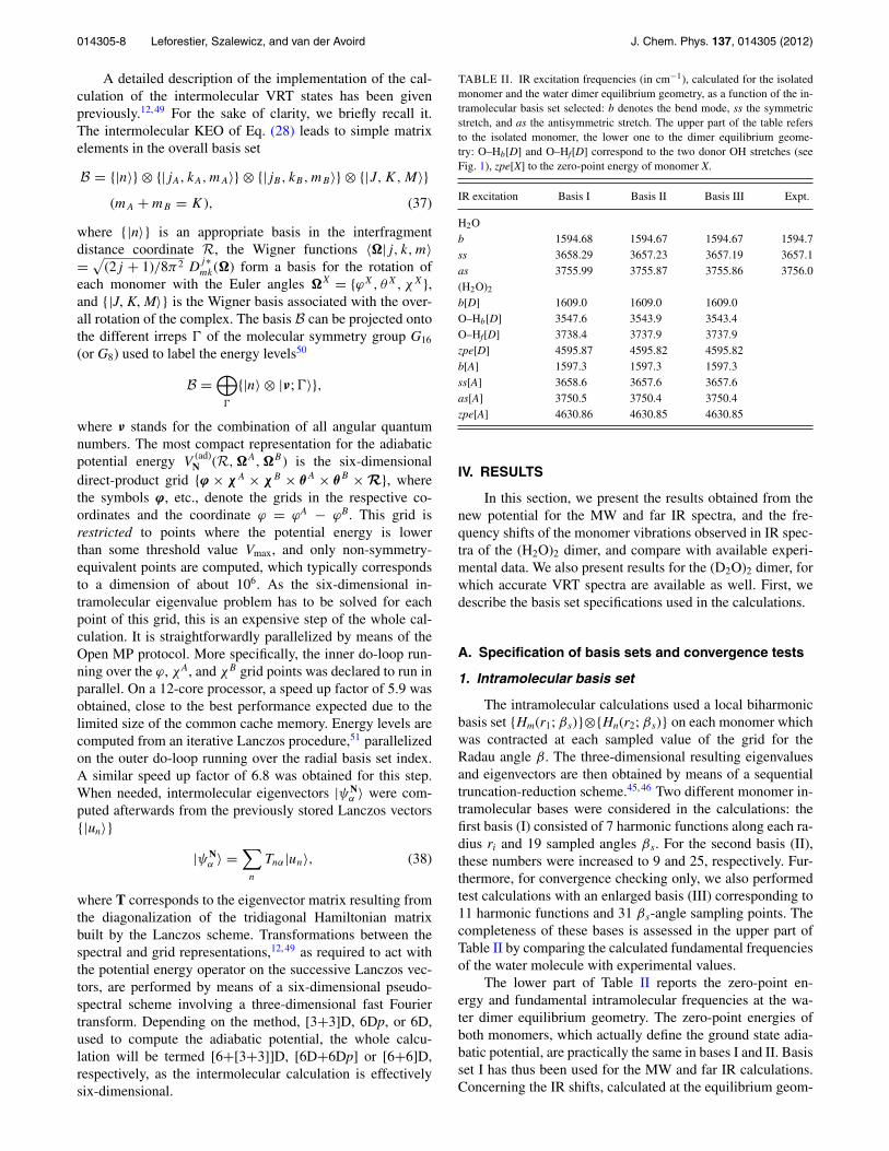
TABLE II. IR excitation frequencies (in cm−1), calculated for the isolatedmonomer and the water dimer equilibrium geometry, as a function of the in-tramolecular basis set selected: b denotes the bend mode, ss the symmetricstretch, and as the antisymmetric stretch. The upper part of the table refersto the isolated monomer, the lower one to the dimer equilibrium geome-try: O–Hb[D] and O–Hf[D] correspond to the two donor OH stretches (seeFig. 1), zpe[X] to the zero-point energy of monomer X.
IR excitation Basis I Basis II Basis III Expt.
H2Ob 1594.68 1594.67 1594.67 1594.7ss 3658.29 3657.23 3657.19 3657.1as 3755.99 3755.87 3755.86 3756.0(H2O)2
b[D] 1609.0 1609.0 1609.0O–Hb[D] 3547.6 3543.9 3543.4O–Hf[D] 3738.4 3737.9 3737.9zpe[D] 4595.87 4595.82 4595.82b[A] 1597.3 1597.3 1597.3ss[A] 3658.6 3657.6 3657.6as[A] 3750.5 3750.4 3750.4zpe[A] 4630.86 4630.85 4630.85
IV. RESULTS
In this section, we present the results obtained from thenew potential for the MW and far IR spectra, and the fre-quency shifts of the monomer vibrations observed in IR spec-tra of the (H2O)2 dimer, and compare with available experi-mental data. We also present results for the (D2O)2 dimer, forwhich accurate VRT spectra are available as well. First, wedescribe the basis set specifications used in the calculations.
A. Specification of basis sets and convergence tests
1. Intramolecular basis set
The intramolecular calculations used a local biharmonicbasis set {Hm(r1; βs)}⊗{Hn(r2; βs)} on each monomer whichwas contracted at each sampled value of the grid for theRadau angle β. The three-dimensional resulting eigenvaluesand eigenvectors are then obtained by means of a sequentialtruncation-reduction scheme.45, 46 Two different monomer in-tramolecular bases were considered in the calculations: thefirst basis (I) consisted of 7 harmonic functions along each ra-dius ri and 19 sampled angles βs. For the second basis (II),these numbers were increased to 9 and 25, respectively. Fur-thermore, for convergence checking only, we also performedtest calculations with an enlarged basis (III) corresponding to11 harmonic functions and 31 βs-angle sampling points. Thecompleteness of these bases is assessed in the upper part ofTable II by comparing the calculated fundamental frequenciesof the water molecule with experimental values.
The lower part of Table II reports the zero-point en-ergy and fundamental intramolecular frequencies at the wa-ter dimer equilibrium geometry. The zero-point energies ofboth monomers, which actually define the ground state adia-batic potential, are practically the same in bases I and II. Basisset I has thus been used for the MW and far IR calculations.Concerning the IR shifts, calculated at the equilibrium geom-

014305-9 Leforestier, Szalewicz, and van der Avoird J. Chem. Phys. 137, 014305 (2012)
K=0 K=1
Exp. CCpol-8s Exp. CCpol-8s
GS(1) 0.00(0.75) / 0.00(0.79)
(2) 11.18(0.65) / 12.31(0.67)(2) 11.66(0.54) / 12.21(0.56)
(1) 14.39(0.70) / 15.22(0.74)
DT(2) 64.52(2.54) / 61.58(2.76)
(1) / 113.19(6.46)(1) 87.75(1.11) / 86.21(1.41)
(2) / 92.05(3.75)
AW(1) 107.93(2.95) / 109.37(3.89)
(2) 108.89(0.02) / 109.57(0.03)(1) 109.98(5.24) / 109.43(5.69)
(2) 123.56(3.41) / 123.60(4.10)
AT(2) 120.19(9.39) / 117.86(9.83)
(1) / 132.11(1.02)(2) / 137.07(5.46)
(1) / 143.15(4.72)
DT2(1) / 128.12(10.9)
(2) / 185.38(19.6)(1)
(2)
OO(1) / 147.61(2.50)
(2) 153.62(1.88) / 153.32(1.98)(2) / 157.26(4.00)
(1) / 155.50(2.60)
FIG. 2. Experimental6, 8, 54, 70, 71 and calculated (CCpol-8s) VRT levels andtunneling splittings of (H2O)2: ground state (GS), donor torsion (DT), ac-ceptor wag (AW), acceptor twist (AT), donor torsion overtone (DT2), andintermolecular stretch (OO). The energies given (in cm−1) correspond tothe origins o1(K) and o2(K) of the levels (1) and (2) with quantum numbersK = 0 and K = 1, respectively, as defined in Eq. (39); the values in paren-theses are the interchange tunneling splittings i1(K) and i2(K), also defined inthe text.
etry, one can observe a notable change δ( ω) = −3.7 cm−1
for the O-Hb stretch which constitutes the most anharmonicvibration as it is associated to the hydrogen bond. A furtherincrease in the basis set size (basis III) reveals that no furtherchange appears in the IR shifts, except for a very small one of−0.5 cm−1 for the O-Hb stretch. We thus systematically usedbasis II for calculating the IR shifts.
2. Intermolecular basis set
Convergence with respect to the intermolecular basis set,explicited in Eq. (37), was assessed by systematically increas-ing its size. For each intermolecular vibration considered inthis work, convergence was tested on both the subfork posi-tions and their associated splittings, as depicted in Fig. 2.
For the (H2O)2 dimer, we used a Wigner basis up tomaximum values of jA, jB = 11 on the monomers, and aprimitive radial basis set of 20 sine functions spanning therange [4.2 ≤ R ≤ 10]a0, contracted to 9 functions by meansof the Harris, Engerholm, and Gwinn procedure.52 A furtherincrease in the Wigner bases, up to jA = jB = 12, leads tochanges less than 0.08 (0.025) cm−1 for the subfork positions(splittings). For the ground state results, these values are re-duced to 0.06 (0.002) cm−1.
Concerning the (D2O)2 dimer, a larger intermolecular ba-sis (with maximum jA, jB = 13 values) had to be used in or-der to converge the tunneling splittings, which in (D2O)2 aresmaller than in (H2O)2 by an order of magnitude. Increas-ing the basis to j = 14 changes the excited subfork positions(splittings) by less than 0.14 (0.01) cm−1. These values arereduced to 0.06 (0.001) cm−1 for the ground state.
The intramolecular calculations were systematically con-ducted in the 6D full variational approach, which takes into
account the vibrational coupling between monomers andleads to the global [6+6]D formulation. We will, however,mention how the [6+[3+3]]D and [6D+6Dp] approxima-tions compare with it.
B. (H2O)2 intermolecular VRT levels and splittings
1. Rigid-monomer results
In order to assess the changes due to monomer flexibil-ity, we first show in Fig. 2 the experimental energy levels,53, 54
and those obtained with the rigid version of the CCpol-8spotential.18 The latter energies were also calculated earlierin Ref. 18 and the two sets are essentially the same. A dif-ference with respect to our previous work is that the energylevels formerly assigned to the donor torsion overtone (DT2)are reassigned to the O–O stretch. From the associated eigen-states ψ0
n (Q), we computed the mean radial kinetic energy〈ψ0
n |TR|ψ0n〉Q. For most of the states, this quantity ranges
from 30 to 45 cm−1, for both states that we now assign to theO–O stretch the radial kinetic energy is about 80−95 cm−1.
In this diagram, the indices (1) and (2) refer to theA±
1 , B±1 and A±
2 , B±2 subforks, respectively, that occur for
each value of the projection quantum number K. The sym-bols A±
1 , B±1 and A±
2 , B±2 refer to the one-dimensional irreps
of the molecular symmetry group G16 of the water dimer (seeRef. 18 for a detailed description). The splittings between the(1) and (2) levels are due to acceptor tunneling. As the wa-ter dimer is a prolate near symmetric top, the origins o1 ando2 of the subforks are customarily defined according to theconvention
Ei(J,K) = oi(K) + B + C
2[J (J + 1) − K2], (39)
where J is the total dimer angular momentum quantum num-ber and Ei(J, K) means the average energy of the A
+/−i and
B+/−i levels pertaining to each subfork. The origin o1(K =
0) is set to zero, so that all other values of oi(K) should beconsidered as excitation energies. The values given in paren-theses correspond to the splittings ii(K) = |A+/−
i − B+/−i |
between the levels within each subfork which are due todonor-acceptor interchange tunneling. The acceptor tunnelingsplitting ai(K) between the subforks (1) and (2) is defined as|o1(K) − o2(K)|.
2. Test of some approximations for flexible monomers
We have shown in Sec. III B that, within the adiabaticapproximation, one can recast the 12-dimensional calcula-tion for flexible monomers into a series of calculations ofthe intramolecular vibrations on a grid of intermolecular co-ordinates Q, which yields a set of adiabatic potentials, and apseudo-rigid monomer calculation of the intermolecular VRTstates on one of these adiabatic potentials. However, the re-sulting Q-dependence of the monomer rotational constant ma-trices � renders the Lanczos iterative diagonalization schemeabout one order of magnitude more costly. The reason is thatone has to switch from the spectral to the grid representationmany times in order to evaluate the effect of the monomer

014305-10 Leforestier, Szalewicz, and van der Avoird J. Chem. Phys. 137, 014305 (2012)
rotation operators T XR . The perturbative approach defined by
Eqs. (32)−(36) allows one to retrieve the exact transition en-ergies and splittings to within 0.03%. If we ignore the finalperturbation step in Eq. (36), the errors increase to about 1%.
The second series of tests concerns the calculation ofthe adiabatic potential V
(ad)N (Q) as detailed in Sec. III C. We
compared the results for the MW and far IR transitions ob-tained, respectively, in the [6+[3+3]]D approximation, i.e.,completely neglecting the six-dimensional V AB(qA, qB ; Q)correction term of Eq. (19), and in the full variational treat-ment [6+6]D of this term by means of Eq. (24).
We found that, for excited states, the transition ener-gies oi(K) are lower in the [6+6]D treatment than in the[6+[3+3]]D approximation, but the decrease does not ex-ceed 1 cm−1. The associated interchange splittings ii(K) dif-fer by 0.5 cm−1 at most. For the ground state, these valuesare 0.25 cm−1 and 0.036 cm−1, respectively. The calculationspresented in the following were systematically conducted inthe full variational [6+6]D treatment. The results correspond-ing to the [6D + 6Dp] perturbative approach, not shown here,are essentially indistinguishable from those obtained withinthe [6+6]D variational one.
Finally, we compare the results obtained from either q-averaging the rotational constant matrices �X(0) as given byEq. (29), or using their optimized values �X(opt) from Eq. (30).It should be kept in mind that the adiabatic potentials are iden-tical in both cases, only the rotational constant matrices �X
differ. For the excited states, the differences in the oi(K) lev-els are marginal, between 0 and 0.6 cm−1 (≤0.5%), smallerthan the deviation from experimental results when available.The corresponding splittings ii(K) show changes of about 4%,with the exception of the very small acceptor wag i2(0) value.For the ground state, the changes are about 1% for the energylevels oi(K). The associated splittings ii(K) are systematicallydecreased by 0.02 cm−1 when moving from optimized to av-eraged rotational constants.
In Table III, we show the comparison between optimizedqopt and vibrationally averaged qavg internal monomer coor-dinates at the water dimer equilibrium geometry Qeq. Alsogiven are the values qref for the monomer reference geome-try obtained by averaging over the isolated monomer groundstate vibrational wave functions and used in the rigid CCpol-8s potential. Not surprisingly, the optimized values of r1 andr2 are systematically smaller than all the values obtained byvibrational averaging. Among the latter, we observe that ther1 and r2 values are larger when averaged over the adiabaticmonomer wave functions in the dimer than when averagedover the isolated monomer wave functions. The lower partof the table lists the associated monomer rotational constants�X
αα(Qeq). With the exception of �zz, the adiabatically aver-aged constants are closer to the reference values.
3. Flexible-monomer results
In Fig. 3, we report the energy levels, i.e., the originso1(K) and o2(K) for K = 0 and K = 1, and the interchange tun-neling splittings i1(K) and i2(K) calculated with the full flex-ible potential in the [6+6]D adiabatic approximation. Alsothe available experimental data are included in this figure.
TABLE III. Optimized qopt [Eq. (18)] and averaged qavg [Eq. (29)] inter-nal coordinates (r1, r2, β) (in a0 and radians) of the acceptor (A) and donor(D) moieties at the water dimer equilibrium geometry; qref refers to the fixed(isolated monomer averaged) values used in the rigid CCpol-8s potential. Thelower part of the table reports the corresponding ground-state rotational con-stants �X
αα in cm−1. The constants in the last column are those denoted as
�X(0)α,α in the text.
q qref qopt qavg
rA1 1.836 1.812 1.845
rA2 1.836 1.812 1.845
θA 1.827 1.827 1.827
rD1 1.836 1.818 1.855
rD2 1.836 1.809 1.841
θD 1.827 1.827 1.828
�Axx 9.2778 9.4914 9.2937
�Ayy 27.8806 27.4240 28.0047
�Azz 14.5216 14.5151 14.4274
�Dxx 9.2778 9.4775 9.2646
�Dyy 27.8806 27.4055 27.9094
�Dzz 14.5216 14.4872 14.3773
When comparing this figure with Fig. 2, we can observe theeffects of using flexible monomers, rather than rigid ones.The agreement with the experimental data, which was alreadyvery good for the rigid CCpol-8s potential, remains aboutequally good for the flexible-monomer potential CCpol-8sf.Only for the O–O stretch mode, the result slightly deteri-orates by including monomer flexibility: the frequency ob-tained from CCpol-8sf is lower than the experimental valueby about 4 cm−1, while the frequency obtained from CCpol-8s agreed to 0.3 cm−1. This might be related to the fact that, asshown in Table I, the SAPT-5s′fIR hydrogen bond length (Oa-Hb) is too large by about 0.06 Å as compared to the bench-mark calculations of Tschumper et al.30 Although the Oa-Hb
hydrogen bond length from the CCpol-8sf potential agreesmuch better with the corresponding result of Tschumper et al.,
K=0 K=1
Exp. CCpol-8sf (avg) Exp. CCpol-8sf(avg)
GS(1) 0.00(0.75) / 0.00(0.72)
(2) 11.18(0.65) / 12.75(0.61)(2) 11.66(0.54) / 12.36(0.51)
(1) 14.39(0.70) / 15.45(0.67)
DT(2) 64.52(2.54) / 61.33(2.48)
(1) / 113.35(5.91)(1) 87.75(1.11) / 86.37(1.32)
(2) / 92.18(3.34)
AW(1) 107.93(2.95) / 109.23(3.29)
(2) 108.89(0.02) / 107.82(0.10)(1) 109.98(5.24) / 108.28(4.76)
(2) 123.56(3.41) / 123.12(3.16)
AT(2) 120.19(9.39) / 117.50(8.67)
(1) / 132.10(1.48)(2) / 136.52(4.66)
(1) / 142.42(4.04)
DT2(1) / 128.22(9.19)
(2) / 184.57(18.3)(1)
(2)
OO(1) / 143.20(3.27)
(2) 153.62(1.88) / 149.63(1.23)(2) / 153.54(2.54)
(1) / 152.07(1.48)
FIG. 3. Comparison of experimental VRT levels and tunneling splittingswith results calculated with the CCpol-8sf potential using averaged valuesof the �X rotational matrices. For definitions, see Fig. 2.

014305-11 Leforestier, Szalewicz, and van der Avoird J. Chem. Phys. 137, 014305 (2012)
K=0 K=1
Exp. CCpol-8s Exp. CCpol-8s
GS (1) 0.00(0.039) / 0.00(0.038)(2) 1.77(0.036) / 1.78(0.034)
(2) 4.74(0.033) / 4.69(0.032)(1) 5.36(0.036) / 5.28(0.034)
DT (2) 59.59(0.20) / 57.53(0.20)(1) 75.38(0.33) / 73.21(0.33)
(1) 68.27(0.13) / 66.25(0.14)(2) 71.81(0.26) / 69.53(0.26)
AW (1) 82.64(0.13) / 81.43(0.12)(2) 84.40(0.11) / 83.24(0.11)
(1) 85.57(0.40) / 84.10(0.39)(2) 89.56(0.17) / 88.34(0.16)
AT (2) 90.37(0.44) / 88.13(0.73)(1) 92.91(0.43) / 91.16(0.39)
(2) / 94.13(0.56)(1) / 96.97(0.44)
DT2(1) 104.24(0.78) /101.59(0.82)(2) /132.97(1.36)
(2) /115.76(0.03)(1) /122.72(0.60)
TW (1) /132.67(0.07)(2) /136.68(0.86)
OO (1) /145.60(1.00)(2) /150.50(0.38)
(2) /147.61(0.39)(1) /149.91(0.77)
FIG. 4. Comparison of (D2O)2 experimental VRT levels and tunneling split-tings with values obtained from the rigid CCpol-8s potential. For definitions,see Fig. 2. The symbol TW stands for a combination of the donor torsion andacceptor wag mode.
the hydrogen bond (or O–O) stretch motion might be less welldescribed by the monomer flexibility correction in CCpol-8sf,which was taken from SAPT-5s′fIR, cf. Eq. (6). The O–Ostretch frequency difference of 4 cm−1 amounts to a relativeerror of only 2.7%, however, so this deviation may also beaccidental.
As a final test, we compare the dissociation energy D0
= 1108.2 cm−1 obtained from the CCpol-8sf potential withthe experimental value 1105 ± 10 cm−1 recently measured byRocher-Casterline and co-workers55 by velocity map imag-ing. The calculated value is off by only 3 cm−1, well withinthe experimental error bars, which tends to validate boththis new potential and the method used to perform the VRTlevel calculations. It should be noted that the rigid-monomerCCpol-8s potential leads to a dissociation energy D0
= 1094 cm−1, demonstrating the need for a flexible-monomerdescription in order to precisely reproduce some of the prop-erties of the dimer.
C. Intermolecular VRT levels for (D2O)2
We first present in Fig. 4 the results obtained from therigid-monomer CCpol-8s potential. These calculations wereconducted with a larger intermolecular basis (with maximumjA, jB = 13) as mentioned previously. Convergence tests, de-scribed in Sec. IV A 2, show that further changes with respectto the basis set size are smaller than the deviations from theavailable experimental results. The results differ slightly fromthe corresponding results in Ref. 18, because the basis in thatstudy was smaller. The O–O stretch levels were assigned onthe basis of a considerably higher radial kinetic energy, just asthe corresponding levels in (H2O)2.
For the flexible-monomer calculations, we first comparedthe averaged and optimized � matrices using the same, jA, jB≤ 13, intermolecular basis in the [6+[3+3]]D approximation.It was found that the energies oi(K) changed by 0.036 at most(≤1.6%) and by 0.30 cm−1 (≤0.4%) for the ground and ex-cited states, respectively. For the interchange tunneling split-
K=0 K=1
Exp. CCpol-8sf Exp. CCpol-8sf
GS (1) 0.00(0.039) / 0.00(0.037)(2) 1.77(0.036) / 2.20(0.034)
(2) 4.74(0.033) / 4.95(0.031)(1) 5.36(0.036) / 5.69(0.034)
DT (2) 59.59(0.20) / 56.35(0.19)(1) 75.38(0.33) / 74.51(0.33)
(1) 68.27(0.13) / 65.72(0.13)(2) 71.81(0.26) / 69.46(0.24)
AW (1) 82.64(0.13) / 80.99(0.11)(2) 84.40(0.11) / 82.46(0.09)
(1) 85.57(0.40) / 83.49(0.36)(2) 89.56(0.17) / 88.46(0.15)
AT (2) 90.37(0.44) / 88.22(0.71)(1) 92.91(0.43) / 91.66(0.32)
(2) / 94.49(0.52)(1) / 97.46(0.41)
DT2(1) 104.24(0.78) /100.78(0.81)(2) /132.35(1.99)
(2) /116.33(0.01)(1) /123.98(0.59)
TW (1) /132.42(0.42)(2) /138.04(1.08)
OO (1) /142.29(1.67)(2) /148.58(0.11)
(2) /144.27(0.23)(1) /147.40(0.61)
FIG. 5. Comparison of (D2O)2 experimental VRT levels and tunneling split-tings with values obtained from the flexible-monomer CCpol-8sf potential.For definitions, see Figs. 2 and 4.
tings ii(K), these values are reduced to 0.001 and 0.018 cm−1,respectively (that is, 2%−3%). All these differences are con-siderably smaller than the corresponding deviations from theavailable experimental results. Hence, the effect of using av-eraged or optimized � matrices becomes much less importantupon deuteration, which is due to the more localized nature ofthe intramolecular wave function for the deuterated species.
The larger intermolecular basis set used renders the adi-abatic potential calculation step much costlier, as the associ-ated intermolecular coordinate grid has to be larger; it spans2 × 106 non-symmetry-equivalent points at which the in-tramolecular eigenvalue problem has to be solved. Hence,we checked that for a smaller, jA, jB ≤ 11, basis perturba-tive [6D+6Dp] and full variational [6+6]D calculations gavenearly identical results. For the ground state, the energiesoi(K) changed by 10−3 cm−1 at most, while the interchangetunneling splittings ii(K) varied by less than 10−3 cm−1. Forexcited states, these changes corresponded to 10−2 at mostand 10−3 cm−1, respectively. In all cases, these variations arewell below the intrinsic convergence limit for the intermolec-ular basis with jA, jB ≤ 13 retained for the calculations. Theflexible-monomer calculations presented in Fig. 5 were thusperformed with the jA, jB ≤ 13 basis within the perturbative[6D+6Dp] treatment, using averaged � matrices.
The inclusion of the correlation V AB(qA, qB ; Q) betweenthe intramolecular vibrations, Eq. (19), by perturbation the-ory, cf. Eq. (23), essentially left the ground state charac-teristics unchanged, while the energies of the excited stateschanged at most by a few times 0.1 cm−1 and the splittingsby a few times 0.01 cm−1. Again, this effect can be under-stood from the increased localization of the intramolecularwave functions of D2O, relative to H2O.
Figure 5 presents a comparison of the VRT levels andsplittings calculated on the flexible-monomer CCpol-8sf po-tential with the experimental data.8, 53, 54, 56–58 No significantdifferences are observed with respect to the rigid CCpol-8sresults in Fig. 4, the good agreement with the available exper-imental data remains.

014305-12 Leforestier, Szalewicz, and van der Avoird J. Chem. Phys. 137, 014305 (2012)
TABLE IV. Frequency shifts of the monomer vibrations (in cm−1) in thewater dimer. For definitions, see Table II. The G16 and G8 models are definedin the text. Except for the rightmost column obtained with optimized valuesof the � matrices, the calculations were performed with the averaged ones.
[6+6]D [6+[3+3]]D
Intramolecular mode G16 G16 G8 G16[opt]
b[D] +13.4 +13.7 +14.8 +9.1O–Hb[D] −61.6 −57.9 −59.0 −55.1O–Hf[D] −13.5 −17.6 −18.9 −14.7b[A] +7.3 +9.6 +7.5 +4.4ss[A] −7.1 −8.6 −8.7 −6.2as[A] −6.6 −10.4 −9.0 −7.2
D. Frequency shifts of the monomer modes in (H2O)2
In Sec. III D we explained that, in contrast to the groundstate, the adiabatic [6+6]D approximation is not completelyvalid if one of the monomers is vibrationally excited. We pro-posed two approximate adiabatic models, called G16 and G8,that should give a good approximation to the more rigorous“two-state” non-adiabatic model that is appropriate for the vi-brationally excited states. It may be expected that a compari-son of the results of the G16 and G8 models for the vibrationalfrequency shifts will give us a good indication of the accuracyof the shifts calculated by either model.
The frequency shift of each monomer vibration was ob-tained by subtracting the energy of the dimer ground statelevel computed on the lowest adiabatic 6D potential (withboth monomers in their ground state) from the dimer groundstate level computed on the excited adiabatic potential ob-tained for the corresponding monomer vibration. In the G8
model, one can choose which monomer is excited, A or B;the results are the same. Since a given monomer excited stategives rise to two dimer excitations in the G8 model, one onthe donor and one on the acceptor, we had to consider thelowest two eigenvalues from each calculation. The acceptorcan be identified a posteriori as the monomer that has itsO atom in the shortest hydrogen bond Oa···Hb. A compari-son of the results from the G16 and G8 models is given inTable IV. Furthermore, we show in this table what is the effectof simplifying the full adiabatic [6+6]D approach to the com-putationally cheaper [6+[3+3]]D approximation, as well asthe effect of taking monomer rotational constant matrices �X
calculated at the optimized monomer geometries, instead ofusing the values averaged over the adiabatic monomer wavefunctions.
Before discussing the results, we must also explain thesymmetry of the intramolecular vibrations and the appropri-ate selection rules. The water dimer in its equilibrium geom-etry has a mirror plane, point group Cs. The donor monomer(D) lies in the plane of reflection and its symmetric and asym-metric OH stretch modes become localized. One of them isthe O–H stretch mode of the H atom involved in the hydro-gen bond, O–Hb, the other one is the O–H stretch mode ofthe free H atom, O–Hf. Both modes have A′ symmetry in Cs.The two H atoms of the acceptor monomer (A) stick out ofthe symmetry plane; they are interchanged by the reflection
symmetry operator. The acceptor symmetric O–H stretch (ss)mode has A′ and the asymmetric O–H stretch (as) mode hasA′′ symmetry. The HOH bend modes of both monomers haveA′ symmetry. Since the water dimer tunnels between eightequivalent equilibrium geometries of this type, we must usethe molecular symmetry group G16, instead of the point groupCs. The intramolecular vibrations are all of A+
1 symmetry,except for the as[A] mode which is of A+
2 symmetry. Actu-ally, if the as[A] mode were adapted to the full G16 symme-try, one would obtain A+
2 and B+2 symmetry components, but
we already explained above that the adiabatic separation ofthe monomer vibrational coordinates and the intermolecularcoordinates does not fully apply to the excited intramolecu-lar vibrations. Hence, it is reasonable to apply the selectionrules of the subgroup G8, as it was done in the interpretationof the high-resolution spectra in Ref. 5. In G8 symmetry, theG16 irreps A+
2 and B+2 become equivalent. If one (or more)
of the intermolecular (tunneling or vibrational) modes is ex-cited, their symmetries should be combined with those of theintramolecular modes, as well as with those of the overall ro-tation functions of the dimer. The transition dipole momentoperator has A−
1 symmetry, which tells us that transitions areallowed between irreps of the same type, except for the ± la-bels which must be reversed. This is a strict selection rule.In addition, there are approximate selection rules based onthe separability between the intra- and intermolecular vibra-tions. Furthermore, there may be approximate selection rulesif the vibrations have small amplitudes and can be well sep-arated from the overall rotations. This does not apply to theintermolecular vibrations,59 but it holds for the intramolec-ular modes which obey the selection rules of the pointgroup Cs.
All fundamental intramolecular modes are allowed in thewater dimer, as they are in the free monomers. For all modesexcept as[A], we report in Table IV the shifts associatedwith the A−
1 [J ′ = 1,K ′ = 0] ← A+1 [J ′′ = 0,K ′′ = 0] transi-
tions belonging to the lower acceptor tunneling components.These transitions may be considered as pure intramolecularvibrational fundamentals, accompanied only by a change J= 1 in total angular momentum that does not alter theinternal motions in the dimer. For the as[A] mode, such transi-tions are forbidden; the excitation of this mode is only allowedin combination with a change in the projection quantum num-ber K. As one can see in Figs. 2 and 3, the VRT levels for K= 0 are quite different from the levels for K = 1, so itis clear that the change of K affects the internal motionsof the monomers in the dimer. In Table IV, we report theallowed A+
1 [J ′ = 1,K ′ = 1] ← A−2 [J ′′ = 0,K ′′ = 0] transi-
tion for the as[A] mode, which was also measured.5
Within the [6+[3+3]]D approximation (rightmostcolumns in the table), we first assessed the changes in thefrequency shifts upon using averaged or optimized � matricesin the G16 model. It can be seen that the shifts are consid-erably modified, in some cases by as much as 5 cm−1. Theexplanation of these relatively large differences is that theseshifts result from transitions to excited adiabatic potentialswhich correspond to more delocalized intramolecular wavefunctions than the ground state. Therefore, the averaging cansubstantially modify the � matrices and we used it in all

014305-13 Leforestier, Szalewicz, and van der Avoird J. Chem. Phys. 137, 014305 (2012)
TABLE V. Calculated frequency shifts of the monomer vibrations in (H2O)2
for J = 0 → 1 and J = 1 → 0 dimer transitions. The symmetries indicatedrefer to the intermolecular VRT levels that produce allowed transitions incombination with the intramolecular mode. For the K = 1 → 0 transitions inthe last column, the symmetries of the initial and final states are reversed.
Transition Frequency shift (cm−1)
Intramolecular mode Symmetry K = 0 → 0 K = 0 → 1 K = 1 → 0
b[D] A+1 → A−
1 13.39 29.60 –4.22B+
1 → B−1 11.84 28.12 –2.72
A−2 → A+
2 14.45 12.81 12.94B−
2 → B+2 13.13 11.62 14.16
O–Hb[D] A+1 → A−
1 –61.63 –47.17 –78.89B+
1 → B−1 –62.83 –48.33 –77.75
A−2 → A+
2 –63.15 –62.96 –64.37B−
2 → B+2 –64.17 –63.91 –63.45
O–Hf[D] A+1 → A−
1 –13.47 1.20 –30.91B+
1 → B−1 –14.85 –0.13 –29.58
A−2 → A+
2 –14.42 –14.42 –15.81B−
2 → B+2 –15.61 –15.52 –14.72
b[A] A+1 → A−
1 7.26 22.84 –10.37B+
1 → B−1 5.68 21.34 –8.85
A−2 → A+
2 7.55 6.60 6.07B−
2 → B+2 6.26 5.40 7.26
ss[A] A+1 → A−
1 –7.06 7.84 –24.64B+
1 → B−1 –8.59 6.38 –23.17
A−2 → A+
2 –7.49 –7.94 –8.95B−
2 → B+2 –8.76 –9.12 –7.79
as[A] A+1 → A−
2 3.48 –21.57B+
1 → B−2 3.22 –21.43
A−2 → A+
1 –6.65 –12.73B−
2 → B+1 –6.65 –12.82
subsequent calculations. We then considered the two limitingmodels, G16 and G8. The results from these two models arequite similar, the largest difference being about 2 cm−1. Thesmallest difference one might expect is about half of the sizeof the ground state interchange tunneling splitting i1(K), cf.Fig. 3, since this splitting is given by the G16 calculations butnot by the G8 model.
The first column in Table IV provides the calculatedshifts obtained within the [6+6]D full variational approach,using averaged � matrices. Comparison with the second col-umn allowed us to estimate the importance of the residualcoupling term V AB(qA, qB ; Q) between the two monomers,cf. Eq. (19). This coupling is particularly important for thedonor O–H stretches and the acceptor asymmetric stretchmodes, resulting in changes as large as 4 cm−1. In all furthercalculations, we used the full [6+6]D adiabatic approach andthe G16 model.
The frequency shifts of the allowed J = 0 → 1 and J= 1 → 0 transitions are listed in Table V for all six funda-mental intramolecular modes. It is assumed that only differ-ent intermolecular tunneling components of the ground andexcited states take part in these transition, and that the inter-molecular vibrations are not excited. It should be kept in mindthat for (H2O)2 the A±
1 , B±1 , A±
2 , and B±2 levels have nuclear
spin statistical weights 1, 0, 3, and 6, respectively. In addi-tion, there are transitions between the levels of E± symmetrywith weight 3 which are not shown; their shifts are intermedi-
ate between those involving the A±1 , A±
2 and B±1 , B±
2 levels.As it was already mentioned above, the interchange splittingsbetween the A±
1 and B±1 components and between the A∓
2 andB∓
2 components are exaggerated in our G16 model calcula-tions, because the excited state interchange splittings are ac-tually much smaller. The energy gaps between the A+
1 , B+1
levels and the A−2 , B−
2 levels, caused by acceptor tunneling,are much larger. These gaps are not reflected in the frequencyshifts of the K = 0 → 0 transitions because these transitionsoccur from the lower to the lower or from the upper to the up-per acceptor tunneling levels. Such K = 0 → 0 transitions arenot allowed for the acceptor asymmetric stretch mode, dueto the A+
2 symmetry of this mode. The acceptor tunnelingsplitting is considerably smaller for K = 1 than for K = 0and the order of the tunneling levels is reversed, hence, theK = 0 → 1 and K = 1 → 0 transitions have rather differentfrequency shifts for transitions involving the A+
1 , B+1 levels or
the A−2 , B−
2 levels.Although experimental data from IR spectroscopy are
available for each of the (H2O)2 monomer modes, it is notstraightforward to compare our calculated frequency shiftswith these data. The measured frequency shifts originate fromvery different sources: high-resolution molecular beam IRspectra5, 20 only for the as[A] mode, cavity ringdown spectra60
for the bend modes, size-selected cluster molecular beamspectra,61, 62 matrix spectra in solid noble gas matrices,21, 63
and matrix spectra in very cold helium droplets9 for all theO–H stretch modes. In the lower resolution spectra, the bandsare quite broad and not resolved, so it is not clear which of thecalculated tunneling components and rotational transitions ac-tually correspond to the frequency shifts extracted from thesespectra. In matrix spectra, the frequencies are shifted by anunknown amount due to the effect of the surrounding matrix,although such shifts are probably small in the case of heliumdroplets. In Table VI, we summarize the experimental dataand compare them with the calculated results with which theyshould most probably be associated.
The most detailed and best defined information is avail-able for the acceptor asymmetric stretch mode. In the high-resolution spectrum of this mode measured by Huang andMiller,5 they observed three different bands corresponding tothe K = 1 → 0, K = 0 → 1, and K = 1 → 2 transitions; manyrotational lines in these bands were assigned and fitted. Thefrequencies obtained for the K = 1 → 0 and K = 0 → 1transitions of A−
2 → A+2 symmetry are 3738.4 and 3752.6
cm−1, respectively, which corresponds to shifts of –17.4 and–3.2 cm−1 relative to the H2O monomer asymmetric stretchfrequency. The same mode was observed by Kuyanov, Choi,and Vilesov9 for the water dimer in helium nanodroplets ina molecular beam setup. These authors reported two peakswith frequency shifts of –3.4 and 4.4 cm−1. One of the peaksmay correspond to the same K = 0 → 1 transition observedby Huang and Miller, but the other peak is not the K = 1→ 0 transition, since the energy gap between the two peaksis smaller by nearly a factor of two than the energy differ-ence between the K = 0 → 1 and K = 1 → 0 transitions inRef. 5 and it is believed that the K = 1 levels are not populatedat the temperature of 0.4 K of the He droplets. Our calcula-tions show that the two peaks in the spectrum of Ref. 9 are
014305-14 Leforestier, Szalewicz, and van der Avoird J. Chem. Phys. 137, 014305 (2012)
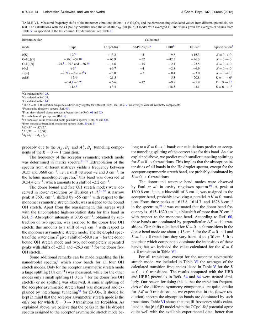
TABLE VI. Measured frequency shifts of the monomer vibrations (in cm−1) in (H2O)2 and the corresponding calculated values from different potentials, seetext. The calculations with the CCpol-8sf potential used the adiabatic G16 full [6+6]D model with averaged �. The values given are averages of values fromTable V, as specified in the last column. For definitions, see Table II.
Intramolecular Calculated
mode Expt. CCpol-8sf SAPT-5s′fIRa HBBb HBB2c Specificationd
b[D] +20e +13.2 +5 +9.6 +16.3 K = 0 → 0O–Hb[D] −56,f −59.0g − 62.9 −52 − 42.5 − 46.3 K = 0 → 0O–Hf[D] −21,f −25.3 and −26.3g − 14.6 −15 − 2.1 − 23.5 K = 0 → 0b[A] +6e +6.7 +4 +2.8 +6.9 K = 0 → 0ss[A] −2.2g (−2 to +3h) − 8.0 −7 − 0.4 − 3.0 K = 0 → 0as[A] −17.4i − 21.5 − 5.5 − 20.8 K = 1 → 0j
−3.4,g −3.2i − 6.6 −12 +9.8 − 5.9 K = 0 → 1k
+4.4g +3.4 +18.5 +3.1 K = 0 → 1l
aCalculated in Ref. 25.bCalculated in Ref. 34.cCalculated in Ref. 64.dThe K = 0 → 0 transition frequencies differ only slightly for different irreps, see Table V; we averaged over all symmetry components.eFrom cavity ringdown spectra (Ref. 60).fFrom size-selected cluster molecular beam spectra (Refs. 61 and 62).gFrom helium droplet spectra (Ref. 9).hExtrapolated value from solid noble gas matrix spectra (Refs. 21 and 63).iFrom molecular beam high-resolution spectra (Refs. 20 and 5).jA−
2 /B−2 → A+
1 /B+1 .
kA−2 /B−
2 → A+1 /B+
1 .lA+
1 /B+1 → A−
2 /B−2 .
probably due to the A−2 , B−
2 and A+1 , B+
1 tunneling compo-nents of the K = 0 → 1 transition.
The frequency of the acceptor symmetric stretch modewas determined in matrix spectra.21, 63 Extrapolation of thespectra from different matrices yields a frequency between3655 and 3660 cm−1, i.e., a shift between –2 and 3 cm−1. Inthe helium nanodroplet spectra,9 this band was observed at3654.4 cm−1, which amounts to a shift of –2.2 cm−1.
The donor bound and free OH stretch modes were ob-served in lower resolution by Huisken et al.61, 62 A narrowpeak at 3601 cm−1, shifted by –56 cm−1 with respect to themonomer symmetric stretch mode, was assigned to the boundOH stretch. Apart from the reassignment, this agrees wellwith the (incomplete) high-resolution data for this band inRef. 5. Absorption intensity at 3735 cm−1, obtained by sub-traction of two spectra, was ascribed to the donor free OHstretch; this amounts to a shift of –21 cm−1 with respect tothe monomer asymmetric stretch mode. The He droplet spec-tra of the water dimer9 give a shift of –59.0 cm−1 for the donorbound OH stretch mode and two, not completely separatedpeaks with shifts of –25.3 and –26.3 cm−1 for the donor freeOH stretch.
Some additional remarks can be made regarding the Henanodroplet spectra,9 which show bands for all four OHstretch modes. Only for the acceptor asymmetric stretch modea large splitting (7.8 cm−1) was measured, while for the othermodes only a small splitting (1.0 cm−1 for the donor free OHstretch) or no splitting was observed. A similar splitting ofthe acceptor asymmetric stretch band was measured and ex-plained by interchange tunneling58 for (D2O)2. It should bekept in mind that the acceptor asymmetric stretch mode is theonly one for which K = 0 → 0 transitions are forbidden. Asexplained above, we believe that the peaks in the He dropletspectra assigned to the acceptor asymmetric stretch mode be-
long to a K = 0 → 1 band; our calculations predict an accep-tor tunneling splitting of the correct size for this band. As alsoexplained above, we predict much smaller tunneling splittingsfor K = 0 → 0 transitions. This implies that the absorption in-tensities of all bands in the He droplet spectra, except for theacceptor asymmetric stretch band, are probably dominated byK = 0 → 0 transitions.
The donor and acceptor bend modes were observedby Paul et al. in cavity ringdown spectra.60 A peak at1600.6 cm−1, i.e, a blueshift of 6 cm−1, was assigned to theacceptor bend, probably involving a parallel K = 0 transi-tion. From three peaks at 1613.8, 1614.7, and 1628.6 cm−1
in the spectrum,60 it was estimated that the donor bend fre-quency is 1615–1620 cm−1, a blueshift of more than 20 cm−1
with respect to the monomer bend. According to Ref. 60,these bands are dominated by perpendicular K = ±1 tran-sitions. Our shifts calculated for K = 0 → 0 transitions in thedonor bend mode are about +13 cm−1, for the K = 0 → 1 andK = 1 → 0 transitions they vary from –4 to +30 cm−1. It isnot clear which components dominate the intensities of thesebands, but we included the value calculated for the K = 0→ 0 transition in Table VI.
For all transitions, except for the acceptor asymmetricstretch mode, we included in Table VI the averages of thecalculated transition frequencies listed in Table V for the K= 0 → 0 transitions. The results computed with the HBBand HBB2 potentials in Refs. 34 and 64 were treated simi-larly. Our reason for doing this is that the transition frequen-cies of the different symmetry components are quite similarfor K = 0 transitions, so we expect that in the (lower res-olution) spectra the absorption bands are dominated by suchtransitions. Table VI shows that the IR frequency shifts calcu-lated in the [6+6]D model with the CCpol-8sf potential agreequite well with the available experimental data, better than
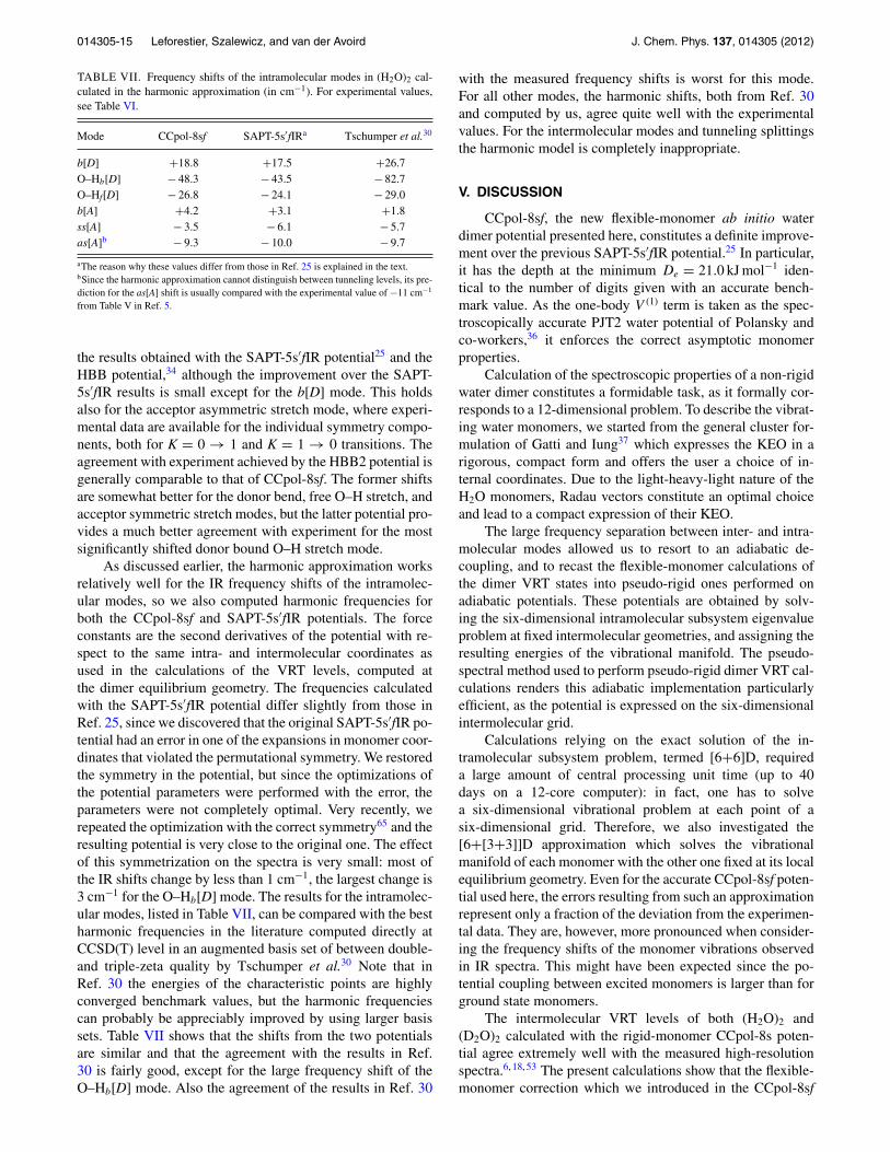
014305-15 Leforestier, Szalewicz, and van der Avoird J. Chem. Phys. 137, 014305 (2012)
TABLE VII. Frequency shifts of the intramolecular modes in (H2O)2 cal-culated in the harmonic approximation (in cm−1). For experimental values,see Table VI.
Mode CCpol-8sf SAPT-5s′fIRa Tschumper et al.30
b[D] +18.8 +17.5 +26.7O–Hb[D] − 48.3 − 43.5 − 82.7O–Hf[D] − 26.8 − 24.1 − 29.0b[A] +4.2 +3.1 +1.8ss[A] − 3.5 − 6.1 − 5.7as[A]b − 9.3 − 10.0 − 9.7
aThe reason why these values differ from those in Ref. 25 is explained in the text.bSince the harmonic approximation cannot distinguish between tunneling levels, its pre-diction for the as[A] shift is usually compared with the experimental value of −11 cm−1
from Table V in Ref. 5.
the results obtained with the SAPT-5s′fIR potential25 and theHBB potential,34 although the improvement over the SAPT-5s′fIR results is small except for the b[D] mode. This holdsalso for the acceptor asymmetric stretch mode, where experi-mental data are available for the individual symmetry compo-nents, both for K = 0 → 1 and K = 1 → 0 transitions. Theagreement with experiment achieved by the HBB2 potential isgenerally comparable to that of CCpol-8sf. The former shiftsare somewhat better for the donor bend, free O–H stretch, andacceptor symmetric stretch modes, but the latter potential pro-vides a much better agreement with experiment for the mostsignificantly shifted donor bound O–H stretch mode.
As discussed earlier, the harmonic approximation worksrelatively well for the IR frequency shifts of the intramolec-ular modes, so we also computed harmonic frequencies forboth the CCpol-8sf and SAPT-5s′fIR potentials. The forceconstants are the second derivatives of the potential with re-spect to the same intra- and intermolecular coordinates asused in the calculations of the VRT levels, computed atthe dimer equilibrium geometry. The frequencies calculatedwith the SAPT-5s′fIR potential differ slightly from those inRef. 25, since we discovered that the original SAPT-5s′fIR po-tential had an error in one of the expansions in monomer coor-dinates that violated the permutational symmetry. We restoredthe symmetry in the potential, but since the optimizations ofthe potential parameters were performed with the error, theparameters were not completely optimal. Very recently, werepeated the optimization with the correct symmetry65 and theresulting potential is very close to the original one. The effectof this symmetrization on the spectra is very small: most ofthe IR shifts change by less than 1 cm−1, the largest change is3 cm−1 for the O–Hb[D] mode. The results for the intramolec-ular modes, listed in Table VII, can be compared with the bestharmonic frequencies in the literature computed directly atCCSD(T) level in an augmented basis set of between double-and triple-zeta quality by Tschumper et al.30 Note that inRef. 30 the energies of the characteristic points are highlyconverged benchmark values, but the harmonic frequenciescan probably be appreciably improved by using larger basissets. Table VII shows that the shifts from the two potentialsare similar and that the agreement with the results in Ref.30 is fairly good, except for the large frequency shift of theO–Hb[D] mode. Also the agreement of the results in Ref. 30
with the measured frequency shifts is worst for this mode.For all other modes, the harmonic shifts, both from Ref. 30and computed by us, agree quite well with the experimentalvalues. For the intermolecular modes and tunneling splittingsthe harmonic model is completely inappropriate.
V. DISCUSSION
CCpol-8sf, the new flexible-monomer ab initio waterdimer potential presented here, constitutes a definite improve-ment over the previous SAPT-5s′fIR potential.25 In particular,it has the depth at the minimum De = 21.0 kJ mol−1 iden-tical to the number of digits given with an accurate bench-mark value. As the one-body V (1) term is taken as the spec-troscopically accurate PJT2 water potential of Polansky andco-workers,36 it enforces the correct asymptotic monomerproperties.
Calculation of the spectroscopic properties of a non-rigidwater dimer constitutes a formidable task, as it formally cor-responds to a 12-dimensional problem. To describe the vibrat-ing water monomers, we started from the general cluster for-mulation of Gatti and Iung37 which expresses the KEO in arigorous, compact form and offers the user a choice of in-ternal coordinates. Due to the light-heavy-light nature of theH2O monomers, Radau vectors constitute an optimal choiceand lead to a compact expression of their KEO.
The large frequency separation between inter- and intra-molecular modes allowed us to resort to an adiabatic de-coupling, and to recast the flexible-monomer calculations ofthe dimer VRT states into pseudo-rigid ones performed onadiabatic potentials. These potentials are obtained by solv-ing the six-dimensional intramolecular subsystem eigenvalueproblem at fixed intermolecular geometries, and assigning theresulting energies of the vibrational manifold. The pseudo-spectral method used to perform pseudo-rigid dimer VRT cal-culations renders this adiabatic implementation particularlyefficient, as the potential is expressed on the six-dimensionalintermolecular grid.
Calculations relying on the exact solution of the in-tramolecular subsystem problem, termed [6+6]D, requireda large amount of central processing unit time (up to 40days on a 12-core computer): in fact, one has to solvea six-dimensional vibrational problem at each point of asix-dimensional grid. Therefore, we also investigated the[6+[3+3]]D approximation which solves the vibrationalmanifold of each monomer with the other one fixed at its localequilibrium geometry. Even for the accurate CCpol-8sf poten-tial used here, the errors resulting from such an approximationrepresent only a fraction of the deviation from the experimen-tal data. They are, however, more pronounced when consider-ing the frequency shifts of the monomer vibrations observedin IR spectra. This might have been expected since the po-tential coupling between excited monomers is larger than forground state monomers.
The intermolecular VRT levels of both (H2O)2 and(D2O)2 calculated with the rigid-monomer CCpol-8s poten-tial agree extremely well with the measured high-resolutionspectra.6, 18, 53 The present calculations show that the flexible-monomer correction which we introduced in the CCpol-8sf

014305-16 Leforestier, Szalewicz, and van der Avoird J. Chem. Phys. 137, 014305 (2012)
potential did not further improve the agreement with exper-iment for the intermolecular VRT levels; it remains aboutequally good. Essentially the same conclusion was reached34
in calculations with the flexible-monomer HBB potential ofBowman and co-workers:26 when the VRT levels were com-puted from this potential in [6+6]D calculations with flexi-ble monomers, some of the levels agree slightly better withthe experimental data than in 6D calculations with rigidmonomers, for other levels the agreement became slightlyworse. Only the ground state acceptor tunneling splittingwas systematically improved for the HBB potential whenthe monomers were kept flexible. The present results com-puted with the CCpol-8sf potential may also be comparedwith similar results obtained recently64 with the newer HBB2version66, 67 of the HBB potential. The flexible-monomerHBB2 potential produces slightly better agreement with ex-periment for the acceptor tunneling splitting in the groundstate of the dimer, but for the excited states and for the groundstate interchange tunneling splittings the CCpol-8sf results areslightly superior. Disregarding these subtle differences, it maybe stated that the potentials discussed above produced VRTlevels that agree very well with the MW and far IR data. Ear-lier calculations of the VRT levels of (H2O)2 and (D2O)2 withthe various available water potentials have shown, however,that this is certainly not the case for all water potentials. Infact, in addition to the SAPT, CCpol, and HBB potentials,only the empirical VRT(ASP) potentials fitted to dimer spec-tra are in same league in terms of agreement with experiment.It may be reiterated that the comparison of accurately calcu-lated VRT levels of the water dimer with high-resolution ex-perimental data constitutes a very critical test of the quality ofthe intermolecular potential, a test that is failed by most of theavailable water potentials.
Table VI shows that the frequency shifts of the monomervibrations obtained with the CCpol-8sf potential constitute adefinite improvement when compared to the results in Ref. 34computed with the HBB potential. They are also slightly bet-ter than the shifts obtained25 from the SAPT-5s′fIR potential.The frequency shifts obtained in Ref. 64 with the HBB2 po-tential agree about equally well with experiment as the shiftsobtained here with the CCpol-8sf potential. The shifts fromthe HBB2 potential are somewhat better for the donor bend,free O–H stretch, and acceptor symmetric stretch modes, theCCpol-8sf potential produces a much better shift for the donorbound O–H stretch mode.
Three possible explanations can be given for the remain-ing discrepancies between theory and experiment:
1. Most of the intramolecular modes (an exception is theacceptor asymmetric stretch mode) were only observedin lower resolution molecular beam spectra with ratherbroad bands, in which the fine structure due to rotationsand tunneling was not resolved, or in matrix spectra.The experimental frequencies extracted from these spec-tra mostly correspond to some maximum in the absorp-tion bands, sometimes after subtraction of other bands.Therefore, it is uncertain what these frequencies actuallycorrespond to, and to which of the calculated transitionfrequencies they should be compared.
2. The accuracy of the [6+6]D adiabatic model used forthe water dimer with flexible monomers has not yet beenevaluated. This is particularly relevant for the excited in-tramolecular vibrations since, as explained above, a for-mally correct treatment of the monomer excited stateswould require a “two-state” non-adiabatic model. Theshifts computed by the more approximate adiabaticmodels, called G16 and G8, may be amenable to im-provement.
3. One can estimate68 that residual inaccuracies of poten-tial surfaces due to post-CCSD(T) excitations, basis settruncations, and fit imperfections are of the order of 1%of interaction energy, i.e., about 20 cm−1 in the mini-mum region. This value is larger than most discrepancieswith experimental spectra, mainly due to the fact thatmost spectral values correspond to energy differenceswhich are usually more accurate due to cancellations oferrors. Recently, calculations beyond the CCSD(T) levelimproved agreement with experiment for the spectra ofH2–CO by nearly an order of magnitude69 and similarimprovements are probably possible for the water dimer.
Summarizing, we may conclude that, as reportedpreviously,18 the CCpol-8s potential still constitutes the bestavailable rigid-monomer potential for the description of theVRT levels of both (H2O)2 and (D2O)2. The VRT levels fromthe flexible-monomer CCpol-8sf potential are, on average,equally good. Also the frequency shifts of the monomer vi-brations calculated with the CCpol-8sf potential agree wellwith the experimental data extracted from IR spectra (withsome uncertainties, as indicated).
ACKNOWLEDGMENTS
Dr. F. Gatti is acknowledged for helpful discussions. K.Sz. was partly supported by the National Science Foundation(NSF) Grant No. CHE-0848589.
1K. Szalewicz, C. Leforestier, and A. van der Avoird, Chem. Phys. Lett. 482,1 (2009).
2H. A. Gebbie, W. J. Burroughs, J. Chamberlain, J. E. Harries, and R. G.Jones, Nature (London) 221, 143 (1969).
3Y. Scribano, N. Goldman, R. J. Saykally, and C. Leforestier, J. Phys. Chem.A 110, 5411 (2006).
4U. Gora, R. Podeszwa, W. Cencek, and K. Szalewicz, J. Chem. Phys. 135,224102 (2011).
5Z. S. Huang and R. E. Miller, J. Chem. Phys. 91, 6613 (1989).6L. B. Braly, K. Liu, M. G. Brown, F. N. Keutsch, R. S. Fellers, and R. J.Saykally, J. Chem. Phys. 112, 10314 (2000).
7N. Goldman, R. S. Fellers, M. G. Brown, L. B. Braly, C. J. Keoshian, C.Leforestier, and R. J. Saykally, J. Chem. Phys. 116, 10148 (2002).
8F. N. Keutsch, N. Goldman, H. A. Harker, C. Leforestier, and R. J.Saykally, Mol. Phys. 101, 3477 (2003).
9K. Kuyanov-Prozument, M. Y. Choi, and A. F. Vilesov, J. Chem. Phys. 132,014304 (2010).
10H. Chen, S. Liu, and J. C. Light, J. Chem. Phys. 110, 168 (1999).11R. S. Fellers, C. Leforestier, L. B. Braly, M. G. Brown, and R. J. Saykally,
Science 284, 945 (1999).12R. S. Fellers, C. Leforestier, L. B. Braly, M. G. Brown, and R. J. Saykally,
J. Chem. Phys. 110, 6306 (1999).13G. C. Groenenboom, E. M. Mas, R. Bukowski, K. Szalewicz, P. E. S.
Wormer, and A. van der Avoird, Phys. Rev. Lett. 84, 4072 (2000).14E. M. Mas and K. Szalewicz, J. Chem. Phys. 104, 7606 (1996).15M. Jeziorska, P. Jankowski, K. Szalewicz, and B. Jeziorski, J. Chem. Phys.
113, 2957 (2000).

014305-17 Leforestier, Szalewicz, and van der Avoird J. Chem. Phys. 137, 014305 (2012)
16E. M. Mas, R. Bukowski, K. Szalewicz, G. C. Groenenboom, P. E. S.Wormer, and A. van der Avoird, J. Chem. Phys. 113, 6687 (2000).
17G. C. Groenenboom, P. E. S. Wormer, A. van der Avoird, E. M. Mas, R.Bukowski, and K. Szalewicz, J. Chem. Phys. 113, 6702 (2000).
18W. Cencek, K. Szalewicz, C. Leforestier, R. van Harrevelt, and A. van derAvoird, Phys. Chem. Chem. Phys. 10, 4716 (2008).
19X. Huang, B. J. Braams, J. M. Bowman, R. E. A. Kelly, J. Tennyson, G. C.Groenenboom, and A. van der Avoird, J. Chem. Phys. 128, 034312 (2008).
20Z. S. Huang and R. E. Miller, J. Chem. Phys. 88, 8008 (1988).21B. Nelander, J. Chem. Phys. 88, 5254 (1988).22C. Leforestier, F. Gatti, R. S. Fellers, and R. J. Saykally, J. Chem. Phys.
117, 8710 (2002).23G. Murdachaew and K. Szalewicz, Faraday Discuss. 118, 121 (2001).24G. Murdachaew, R. Bukowski, and K. Szalewicz, Phys. Rev. Lett. 88,
123202 (2002).25K. Szalewicz, G. Murdachaew, R. Bukowski, O. Akin-Ojo, and C. Lefor-
estier, in Lecture Series on Computer and Computational Science: IC-CMSE 2006, edited by G. Maroulis, and T. Simos (Brill Academic, Leiden,2006), Vol. 6, pp. 482–491.
26X. Huang, B. J. Braams, and J. M. Bowman, J. Phys. Chem. A 110, 445(2006).
27B. Jeziorski, R. Moszynski, and K. Szalewicz, Chem. Rev. 94, 1887(1994).
28K. Szalewicz, K. Patkowski, and B. Jeziorski, Struct. Bonding (Berlin) 116,43 (2005).
29S. S. Xantheas and T. Dunning, J. Chem. Phys. 99, 8774 (1993).30G. S. Tschumper, M. L. Leininger, B. C. Hoffman, E. F. Valeev, H. F.
Schaefer III, and M. Quack, J. Chem. Phys. 116, 690 (2002).31R. Bukowski, K. Szalewicz, G. C. Groenenboom, and A. van der Avoird,
Science 315, 1249 (2007).32R. Bukowski, K. Szalewicz, G. C. Groenenboom, and A. van der Avoird,
J. Chem. Phys. 128, 094313 (2008).33R. Bukowski, K. Szalewicz, G. C. Groenenboom, and A. van der Avoird,
J. Chem. Phys. 128, 094314 (2008).34C. Leforestier, R. van Harrevelt, and A. van der Avoird, J. Phys. Chem. A
113, 12285 (2009).35K. T. Tang and J. P. Toennies, J. Chem. Phys. 80, 3726 (1984).36O. L. Polyansky, P. Jensen, and J. Tennyson, J. Chem. Phys. 105, 6490
(1996).37F. Gatti and C. Iung, Phys. Rep. 484, 1 (2009).38W. Klopper, M. Quack, and M. A. Suhm, Chem. Phys. Lett. 261, 35 (1996).39W. Klopper, M. Quack, and M. A. Suhm, J. Chem. Phys. 108, 10096
(1998).40F. T. Smith, Phys. Rev. Lett. 45, 1157 (1980).41Z. Bacic, D. Watt, and J. C. Light, J. Chem. Phys. 89, 947 (1988).42H. Wei and T. Carrington, J. Chem. Phys. 107, 2813 (1997).43H. Wei and T. Carrington, Chem. Phys. Lett. 287, 289 (1998).
44W. H. Press, B. P. Flannery, S. A. Teukolsky, and W. T. Vetterling, Numer-ical Recipes (Cambridge University Press, Cambridge, 1986).
45Z. Bacic and J. C. Light, J. Chem. Phys. 85, 4594 (1986).46Z. Bacic and J. C. Light, J. Chem. Phys. 87, 4008 (1987).47G. T. Fraser, J. Chem. Phys. 90, 2097 (1989).48G. Brocks, A. van der Avoird, B. T. Sutcliffe, and J. Tennyson, Mol. Phys.
50, 1025 (1983).49C. Leforestier, L. B. Braly, K. Liu, M. J. Elrod, and R. J. Saykally, J. Chem.
Phys. 106, 8527 (1997).50T. R. Dyke, J. Chem. Phys. 66, 492 (1977).51C. Lanczos, J. Res. Natl. Bur. Stand. 45, 255 (1950).52D. O. Harris, G. G. Engerholm, and W. D. Gwinn, J. Chem. Phys. 43, 1515
(1965).53L. B. Braly, J. D. Cruzan, K. Liu, R. S. Fellers, and R. J. Saykally, J. Chem.
Phys. 112, 10293 (2000).54F. N. Keutsch, L. B. Braly, M. G. Brown, H. A. Harker, P. B. Petersen, C.
Leforestier, and R. J. Saykally, J. Chem. Phys. 119, 8927 (2003).55B. E. Rocher-Casterline, L. C. Ch’ng, A. K. Mollnera, and H. Reisler,
J. Chem. Phys. 134, 211101 (2011).56E. Zwart, J. J. ter Meulen, and W. L. Meerts, Chem. Phys. Lett. 166, 500
(1990).57E. N. Karyakin, G. T. Fraser, and R. D. Suenram, Mol. Phys. 78, 1179
(1993).58J. B. Paul, R. A. Provencal, and R. J. Saykally, J. Phys. Chem. A 102, 3279
(1998).59M. J. Smit, G. C. Groenenboom, P. E. S. Wormer, A. van der Avoird, R.
Bukowski, and K. Szalewicz, J. Phys. Chem. A 105, 6212 (2001).60J. B. Paul, R. A. Provencal, C. Chapo, K. Roth, R. Casaes, and R. J.
Saykally, J. Phys. Chem. A 103, 2972 (1999).61F. Huisken, M. Kaloudis, and A. Kulcke, J. Chem. Phys. 104, 17 (1996).62U. Buck and F. Huisken, Chem. Rev. 100, 3863 (2000).63J. Ceponkus, P. Uvdal, and B. Nelander, J. Phys. Chem. A 112, 3921
(2008).64C. Leforestier, Phil. Trans. R. Soc. London, Ser. A 370, 2675 (2012).65G. Murdachaew, R. Bukowski, P. Jankowski, O. Akin-Ojo, C. Leforestier,
and K. Szalewicz, “Water pair potential with flexible monomers fromsymmetry-adapted perturbation theory” (to be published).
66A. Shank, Y. Wang, A. Kaledin, B. J. Braams, and J. M. Bowman, J. Chem.Phys. 130, 144314 (2009).
67Y. Wang and J. M. Bowman, Chem. Phys. Lett. 419, 1 (2010).68R. Podeszwa, K. Patkowski, and K. Szalewicz, Phys. Chem. Chem. Phys.
12, 5974 (2010).69P. Jankowski, A. R. W. McKellar, and K. Szalewicz, Science 336, 1147
(2012).70E. Zwart, J. J. ter Meulen, W. L. Meerts, and L. H. Coudert, J. Mol. Spec-
trosc. 147, 27 (1991).71G. T. Fraser, Int. Rev. Phys. Chem. 10, 189 (1991).