Species diversity of the subgenus Amiota (s. str). Loew, 1862 (Diptera… · 2015-07-22 · Species...
46
Species diversity of the subgenus Amiota (s. str.) Loew, 1862 (Diptera, Drosophilidae) in southern China HONG-WEI CHEN 1,2 , HIDE-AKI WATABE 3 , JIAN-JUN GAO 4 , HISAKI TAKAMORI 5 , YA-PING ZHANG 4,6 , & TADASHI AOTSUKA 2 1 Department of Biology, Shenyang Normal University, 253rd Huanghe North Street, Shenyang, 110034 China; e-mail: [email protected], 2 Department of Biology, Tokyo Metropolitan University, 1-1 Minami-Ohsawa, Hachioji-shi, 192-0397 Japan, 3 Biological Laboratory, Hokkaido University of Education, 5-3-1 Ainosato, Sapporo, 002-8502 Japan, 4 Laboratory of Molecular Evolution and Genome Diversity, Kunming Institute of Zoology, Chinese Academy of Sciences, 32nd Jiaochang Donglu, Kunming, Yunnan, 650223 China, 5 Biological Laboratory, Tokyo Gakugei University, 4-1-1 Nukuikitamachi, Koganei, Tokyo, 184-8501 Japan, and 6 Laboratory for Conservation and Utilization of Bio-resource, Yunnan University, Kunming, 650223 China (Accepted 15 September 2003) Abstract A total of 38 Amiota (s. str.) species (about 40% of the world total) are reported from southern China, with descriptions of 23 new species, i.e. sinuata species-group: aculeata Chen and Aotsuka, sp. nov., subsinuata Chen and Aotsuka, sp. nov., xishuangbanna Chen and Aotsuka, sp. nov.; basdeni species- group: brevipartita Chen and Gao, sp. nov., curvispina Chen and Gao, sp. nov., lipingae Chen and Gao, sp. nov., huae Chen and Gao, sp. nov., longispina Chen and Gao, sp. nov.; taurusata species- group: asymmetrica Chen and Takamori, sp. nov.; femorata Chen and Takamori, sp. nov., yixiangensis Chen and Takamori, sp. nov.; alboguttata species-group: ailaoshanensis Chen and Watabe, sp. nov., arcuata Chen and Watabe, sp. nov., dehiscentia Chen and Watabe, sp. nov., jizushanensis Chen and Watabe, sp. nov., latitabula Chen and Watabe, sp. nov., luguhuensis Chen and Watabe, sp. nov., nozawai Chen and Watabe, sp. nov., paraspinata Chen and Watabe, sp. nov., shangrila Chen and Watabe, sp. nov.; and ungrouped: fuscicata Chen and Zhang, sp. nov., wangi Chen and Zhang, sp. nov., wuyishanensis Chen and Zhang, sp. nov. A key to all species from southern China is provided. The Amiota fauna of southern China at the species-group level is compared with that of six geographic regions. The subgenus Amiota is assumed to have originated and produced many species-groups in the Oriental region of East Asia, and then the basdeni, alboguttata and rufescens species-groups might have spread to Europe and North–Central America throughout the Palearctic region of East Asia and both the apodemata, sinuata and nagatai species-groups to tropical regions of South-East Asia. Keywords: Taxonomy, Diptera, Drosophilidae, Amiota (s. str.), new species, adaptive radiation, southern China Introduction The subgenus Amiota (s. str.) Loew, 1862 is a large taxon within the subfamily Steganinae, and a total of 71 species have hitherto been reported from the world (Chen and Toda Journal of Natural History, 2005; 39(3): 265–310 ISSN 0022-2933 print/ISSN 1464-5262 online # 2005 Taylor & Francis Ltd DOI: 10.1080/00222930310001657883
Transcript of Species diversity of the subgenus Amiota (s. str). Loew, 1862 (Diptera… · 2015-07-22 · Species...

Species diversity of the subgenus Amiota (s. str.) Loew,1862 (Diptera, Drosophilidae) in southern China
HONG-WEI CHEN1,2, HIDE-AKI WATABE3, JIAN-JUN GAO4,
HISAKI TAKAMORI5, YA-PING ZHANG4,6, & TADASHI AOTSUKA2
1Department of Biology, Shenyang Normal University, 253rd Huanghe North Street, Shenyang,
110034 China; e-mail: [email protected], 2Department of Biology, Tokyo Metropolitan
University, 1-1 Minami-Ohsawa, Hachioji-shi, 192-0397 Japan, 3Biological Laboratory, Hokkaido
University of Education, 5-3-1 Ainosato, Sapporo, 002-8502 Japan, 4Laboratory of Molecular
Evolution and Genome Diversity, Kunming Institute of Zoology, Chinese Academy of Sciences, 32nd
Jiaochang Donglu, Kunming, Yunnan, 650223 China, 5Biological Laboratory, Tokyo Gakugei
University, 4-1-1 Nukuikitamachi, Koganei, Tokyo, 184-8501 Japan, and 6Laboratory for
Conservation and Utilization of Bio-resource, Yunnan University, Kunming, 650223 China
(Accepted 15 September 2003)
AbstractA total of 38 Amiota (s. str.) species (about 40% of the world total) are reported from southern China,with descriptions of 23 new species, i.e. sinuata species-group: aculeata Chen and Aotsuka, sp. nov.,subsinuata Chen and Aotsuka, sp. nov., xishuangbanna Chen and Aotsuka, sp. nov.; basdeni species-group: brevipartita Chen and Gao, sp. nov., curvispina Chen and Gao, sp. nov., lipingae Chen andGao, sp. nov., huae Chen and Gao, sp. nov., longispina Chen and Gao, sp. nov.; taurusata species-group: asymmetrica Chen and Takamori, sp. nov.; femorata Chen and Takamori, sp. nov., yixiangensisChen and Takamori, sp. nov.; alboguttata species-group: ailaoshanensis Chen and Watabe, sp. nov.,arcuata Chen and Watabe, sp. nov., dehiscentia Chen and Watabe, sp. nov., jizushanensis Chen andWatabe, sp. nov., latitabula Chen and Watabe, sp. nov., luguhuensis Chen and Watabe, sp. nov.,nozawai Chen and Watabe, sp. nov., paraspinata Chen and Watabe, sp. nov., shangrila Chen andWatabe, sp. nov.; and ungrouped: fuscicata Chen and Zhang, sp. nov., wangi Chen and Zhang, sp.nov., wuyishanensis Chen and Zhang, sp. nov. A key to all species from southern China is provided.The Amiota fauna of southern China at the species-group level is compared with that of six geographicregions. The subgenus Amiota is assumed to have originated and produced many species-groups inthe Oriental region of East Asia, and then the basdeni, alboguttata and rufescens species-groups mighthave spread to Europe and North–Central America throughout the Palearctic region of East Asia andboth the apodemata, sinuata and nagatai species-groups to tropical regions of South-East Asia.
Keywords: Taxonomy, Diptera, Drosophilidae, Amiota (s. str.), new species, adaptive radiation,southern China
Introduction
The subgenus Amiota (s. str.) Loew, 1862 is a large taxon within the subfamily Steganinae,
and a total of 71 species have hitherto been reported from the world (Chen and Toda
Journal of Natural History, 2005; 39(3): 265–310
ISSN 0022-2933 print/ISSN 1464-5262 online # 2005 Taylor & Francis Ltd
DOI: 10.1080/00222930310001657883

2001). More than half of the species (about 40 species) were recorded from the Japanese
islands and its neighbouring districts (Okada 1960, 1968, 1971; Takada et al. 1979; Maca
and Lin 1993; Toda et al. 1996; Zhang et al. 1996). Thus, this subgenus is mainly
distributed in temperate regions of the Northern Hemisphere and is especially abundant in
East Asia. However, our knowledge on subgenus Amiota in the mainland of China,
especially in its southern districts is not sufficient. So far, faunal surveys of Amiota in
China have been performed mainly in north-eastern regions and information on southern
China was limited (Toda and Peng 1992; Chen and Toda 1998a, 1998b, 2001).
Since 2000, we have engaged in faunal studies of drosophilid flies in southern China
covering Hunan, Fujian, Guangdong, Guangxi, Guizhou and Yunnan Provinces, and
found 15 known and 23 new species of the subgenus Amiota (Table I). This paper describes
23 new species, and provides geographic information and a key to all 38 species of the
subgenus Amiota from southern China. Based upon the faunal comparisons between
southern China and surrounding regions, we discussed the origin of Amiota (s. str.).
Materials and methods
Collection and observation. Most of the flies examined were collected around human eyes, and
are not mentioned in each description. Several flies were obtained from tree trunks or by banana
traps. All specimens were preserved in 70% ethanol. External morphology was observed under
a stereoscopic microscope, and metric characters were measured with an ocular micrometer.
Male terminalia were detached from the body, treated with 10% KOH solution at around
100uC for several minutes, and observed in a droplet of glycerol under a compound light
microscope. Drawings were made with an ocular mesh micrometer and section paper.
Type depositories. Type specimens were dried, pinned and deposited in the following
institutions: Department of Biology, Shenyang Normal University, Shenyang, China
(DBSU); Kunming Institute of Zoology, Chinese Academy of Sciences (KIZ), Kunming,
China; Systematic Entomology, The Hokkaido University Musemu, Hokkaido University,
Sapporo, Japan (SEHU).
Terminology and indices. For morphological terminology and indices for the definitions, see
Chen and Toda (2001) or Chen and Aotsuka (2003).
Description
Subgenus Amiota Loew
Amiota Loew, 1862: 229. Type-species: Amiota leucostoma Loew, 1862.
Amiota (s. str.): Wheeler, 1952: 166; Okada, 1960: 89; 1968: 303; 1971: 82; Maca, 1980:
328; Maca and Lin, 1993: 1; Chen and Toda: 2001: 1517.
Diagnosis. Face, postpronotal lobe and wing base each with distinctly milky white spot;
clypeus dark brown to black; aedeagal apodeme broad, dorso-ventrally flattened; a few
anterior sensilla of medial cibarials short, somewhat peg-like (Chen and Toda 2001).
Description. Eyes dark reddish brown. Ocellar triangle dark brown to black. Frontal vitta
brownish orange to dark brown, slightly pollinose, with a few minute, interfrontal setulae.
Anterior reclinate orbital slightly shorter than other orbitals. Pedicel and first flagellomere
266 H.-W. Chen et al.

Table I. A list of 15 known and 23 new species of the subgenus Amiota from southern China, with information on collection records and geographical distributions.
Localities Yunnan Hunan Guizhou Guangxi Fujian Guangdong Hainan
Lake Lugu,
Ninglang
Mt
Gaoligong,
Pianma
Mt Jizu,
Binchuan
Bamboo
Temple,
Kunming
Mts Ailao
and Wuliang,
Jingdong
Manxi and
Yixiang,
Simao
Xishuang-
bana (1),
Yexianggu,
Mengyang
Xishuangbana
(2),
Wangtiangshu,
Mengla
Mt.
Badagong,
Sangzhi
Longgong,
Anshun
Nonggang,
Longzhou Mt Wuyi
Nanling,
Ruyuan
Mt Dinghu,
Zhaoqing
Jianfeng,
Ledong
Altitude (m) 1800–2700 2300 2150 2100 1700–2450 1100–1400 700–800 600–800 700 700 500 700 500 500 200
Latitude and
longitude
27u38’N,
100u47’E
26u01’N,
98u37’E
26u00’N,
100u21’E
25u02’N,
102u43’E
24u32’N,
101u01’E
22u47’N,
101u02’E
22u20’N,
100u51’E
21u28’N,
101u38’E
28u25’N,
109u54’E
26u05’N,
105u58’E
22u22’N,
106u51’E
27u43’N,
117u57’E
25u00’N,
113u24’E
23u10’N,
112u34’E
18u41’N,
108u52’E
Species-groups
apodemata apodemata +
sinuata pengi +
sinuata * * +
aculeata +
neosinuata +
xishuangbana +
nagatai nagatai * * +
okinawana +
basdeni curvistyla *
macai *
onchopyga *
palpifera +
xianggelila + +
curvispina +
Linpingae +
huae +
longispina +
taurusata asymmetrica + +
femorata + +
yixiangensis +
alboguttata cuii * +
ailaoshanensis +
arcuata +
dehiscentia +
latitabula + +
Species
div
ersityof
the
subgen
us
Am
iota
267

Localities Yunnan Hunan Guizhou Guangxi Fujian Guangdong Hainan
Lake Lugu,
Ninglang
Mt
Gaoligong,
Pianma
Mt Jizu,
Binchuan
Bamboo
Temple,
Kunming
Mts Ailao
and Wuliang,
Jingdong
Manxi and
Yixiang,
Simao
Xishuang-
bana (1),
Yexianggu,
Mengyang
Xishuangbana
(2),
Wangtiangshu,
Mengla
Mt.
Badagong,
Sangzhi
Longgong,
Anshun
Nonggang,
Longzhou Mt Wuyi
Nanling,
Ruyuan
Mt Dinghu,
Zhaoqing
Jianfeng,
Ledong
Altitude (m) 1800–2700 2300 2150 2100 1700–2450 1100–1400 700–800 600–800 700 700 500 700 500 500 200
Latitude and
longitude
27u38’N,
100u47’E
26u01’N,
98u37’E
26u00’N,
100u21’E
25u02’N,
102u43’E
24u32’N,
101u01’E
22u47’N,
101u02’E
22u20’N,
100u51’E
21u28’N,
101u38’E
28u25’N,
109u54’E
26u05’N,
105u58’E
22u22’N,
106u51’E
27u43’N,
117u57’E
25u00’N,
113u24’E
23u10’N,
112u34’E
18u41’N,
108u52’E
luguhuensis +
jizushanensis +
nozawai +
pseudospinata + +
shangrila +
rufescens magniflava *
Ungrouped acuta *
dentata + +
furcata + + + * * *
subfurcata + + + + + + * *
fuscicata + +
wangi +
wuyishanensis + + +
*New records.
Table I. (Continued).
268
H.-W
.C
hen
etal.

almost greyish yellow; arista plumose, without terminal fork. Face brown. Palpus larger in
female than in male, greyish yellow, with several stout setae on lateral margin. Vibrissa
prominent; other orals small. Gena and postgena brown to brownish black. Occiput glossy
black.
Thorax slightly glossy, usually dark brown to black, except for a small number of species
with yellow thorax. Postpronotal seta 1. Acrostichal setulae in ca 12 irregular rows.
Prescutellar setae present (absent only in sinuata species-group). Scutellum unicolorous.
Basal scutellar setae divergent; apical ones cruciate.
Wing hyaline. Veins greyish yellow; crossveins clear. Basal medial-cubital crossvein present.
Ventral surface of costal vein between R2+3 and R4+5 usually with more than 18 minute, peg-
like spinules; such spinules more distinct and bigger in apodemata, sinuata and nagatai
species-groups (spinules ca 14–15) than in other groups. C1 setae two, less differentiated. R2+3
slightly curved to costa at tip; R4+5 and M1 converged distally. Halteres white.
Legs usually yellow, exceptionally dark brown in a few species. Apical seta present on mid
tibia; preapical dorsal setae present on all tibiae. Fore femur with two or three irregular,
posterior rows of setae. Mid tarsus with one or two (usually one) row(s) of minute cuneiform
setulae ventrally; hind tarsus with one row of such setulae. Fore and hind first tarsomeres each
usually slightly shorter than remaining tarsomeres combined; mid first tarsomere as long as
remaining ones combined; second tarsomere usually about twice as long as width.
Abdominal tergites glossy, usually brown to brownish black; first and second medially
paler. Sternites pale greyish yellow; first small, lacking pubescence; second to sixth („) or
seventh (R) pubescent and setigerous.
Male terminalia: epandrium with a number of setae near posterior to ventral margins;
apodeme less-developed. Surstylus with a row of prensisetae on distal margin and several
setae apically. Tenth sternite laterally fused to surstylus and with one pair of lobe-like
processes, contiguous dorsally to cercus and ventrally to gonopod. Cercus separated from
epandrium, entirely pubescent and setigerous. Membrane between epandrium and cercus
pubescent. Hypandrium usually narrow and arcuate. Gonopods almost fused to each other,
forming postero-median plate, anteriorly forming vertical process, postero-laterally
contiguous to posterior ends of hypandrium and antero-ventral corners of epandrium.
Parameres usually basally contiguous to arms of aedeagal apodeme. Aedeagus usually
sclerotized and distinguishable from other structures; outer membrane less-developed;
apodeme with one pair of arms.
Female terminalia: seventh tergite usually separated mid-dorsally into lateral lobes.
Eighth sternite (oviscapt) not bilobed. Pregenital lamella present posteriorly to eighth
sternite, usually sclerotized, partly bilobed. Spermatheca dark brown, usually with
numerous, minute, apically round, mould-like processes over outer surface; duct not
introverted into capsule.
The above characters commonly seen in all species examined are not referred to in the
description of each species.
1. The apodemata species-group
Amiota (Amiota) apodemata species-group, Chen and Toda, 1998a: 271.
Diagnosis. Fifth tergite with dark-coloured strips laterally; sixth tergite very small, pointed
laterally and not reaching ventral margin of fifth tergite; aedeagus basally fused to apodeme.
Distribution. Oriental region.
Species diversity of the subgenus Amiota 269

Amiota (Amiota) apodemata Gupta and Panigrahy
Amiota (Amiota) apodemata Gupta and Panigrahy, 1987: 57; Chen and Toda, 1998a: 272;
2001: 1524.
Specimens examined. Hainan Is: Jianfeng, 2„, 21–22 September 1993, ex tree trunks, M. J.
Toda leg.
Distribution. China (Hainan Is), India.
2. The sinuata species-group
Amiota (Amiota) sinuata species-group, Chen and Toda, 1998b: 409; 2001: 1527.
Diagnosis. Prescutellar setae usually absent; hypandrium anteriorly connected with ventro-
medial portion of parameres by articulating plate; parameres fused to each other baso-
medially; aedeagal apodeme slightly curved.
Distribution. Oriental region and Australian region (North).
Amiota (Amiota) pengi Chen and Toda
Amiota (Amiota) pengi Chen and Toda, 1998b: 413; 2001: 1527.
Specimens examined. China: paratypes, 6„, Jianfeng, Hainan Is, 21 Sepember 1993, M. J.
Toda leg. (SEHU).
Distribution. China (Hainan Is).
Amiota (Amiota) sinuata Okada
Amiota (Amiota) sinuata Okada, 1968: 305; Chen and Toda, 1998b: 410; 2001: 1528.
Specimens examined. Yunnan: 1„, Wangtianshu, ex banana trap, 7 November 2001, H.
Watabe leg.; 1„, Manxi, 9 November 2001, L.-P. He leg.
Distribution. China (Guangdong, Hainan Is, Yunnan), Japan (Yakushima Is, Ryukyu Is),
Myanmar (Yangon).
Amiota (Amiota) aculeata Chen and Aotsuka, sp. nov.
(Figure 1)
Diagnosis. Prescutellar setae present; vertical lobe of gonopod apically with one pair of
elongated processes, which is much sclerotized apically and contiguous to well-developed
outer membrane of aedeagus with fine serration (Figure 1D).
Description
Thorax. Thorax and scutellum dark yellow; pleura nearly brown.
Wing. Ventral surface of costal vein with ca 14–15 distinct, peg-like spinules between R2+3
and R4+5.
270 H.-W. Chen et al.

Abdominal tergites. Abdominal tergites black; first and second tergites yellow medially.
Male terminalia. Epandrium with ca 12–13 setae near posterior margin and at postero-
ventral corner (Figure 1A). Surstylus with ca eight or nine prensisetae (Figure 1B).
Paramere slightly curved basally in lateral view, medially with ca 14–15 sensilla, distally
with ca four or five minute sensilla. Aedeagal apodeme large, broadened, sclerotized
(Figure 1C, D). Ejaculatory apodeme with ca six pits on each lateral margin; stalk ca 2.2
times as long as the apical plate (Figure 1E).
Measurements. BL52.93 mm; ThL51.40 mm; WL52.50 mm; WW51.25 mm.
Indices. arb56/2, avd50.90, adf52.50, flw52.20, FW/HW50.35, ch/o50.06, prorb50.90,
rcorb50.65, vb50.35, dcl50.60, sctl51.30, sterno50.90, orbito52.10, dcp50.25,
sctlp51.00, C51.23, 4c51.87, 4v52.30, 5x51.50, ac56.14, M50.78, C3F50.81.
HOLOTYPE: „, Yunnan: Wangtianshu, 11 September 2002, ex tree trunks, H.-W. Chen
leg. (KIZ).
Distribution. China (Yunnan).
Relationship. This species is very characteristic in having prescutellar setae, although it
surely belongs to the sinuata species-group by other diagnostic characters (body colour,
length of aristal branches, broad hypandrium, shaped of paramere, etc.). This suggests a
taxonomic relationship between sinuata and apodemata groups.
Figure 1. Amiota (Amiota) aculeata Chen and Aotsuka, sp. nov. „. (A) Epandrium (epand) and cercus (cerc),
lateral view; (B) surstylus (sur) and tenth sternite (st 10), ventral view; (C) parameres (pm) and aedeagal apodeme
(aed a), ventral view; (D) hypandrium (hypd), gonopod (gon), parameres and aedeagal apodeme, lateral view; (E)
ejaculatory apodeme. Scale bars: 0.1 mm.
Species diversity of the subgenus Amiota 271

Etymology. Referring to the outer membrane of aedeagus with fine serration.
Amiota (Amiota) subsinuata Chen and Aotsuka, sp. nov.
(Figure 2)
Diagnosis. Vertical lobe of gonopod extremely elongated basally, forming one pair of strongly
curved, rod-shaped processes with one sickle-like projection submedially (Figure 2C, D).
Description
Thorax. Thorax dark yellow, medially with one broadened, brown stripe, and laterally with
two thin versicolour stripes; pleura yellow, with brown patch on upper half; scutellum dark
yellow, with brown margin.
Wing. Ventral surface of costal vein with ca 14–15 distinct, peg-like spinules between R2+3
and R4+5.
Abdominal tergites. Abdominal tergites dark brown; second yellow medially; third and
fourth with yellow stripe medially; fifth and sixth entirely black.
Male terminalia. Epandrium with ca 10–11 setae near posterior margin and at postero-ventral
corner (Figure 2A). Surstylus with ca seven prensisetae (Figure 2B). Hypandrial apodeme
with two pairs of sharply pointed projections subbasally and distally. Paramere basally curved
Figure 2. Amiota (Amiota) subsinuata Chen and Aotsuza, sp. nov. „. (A) Epandrium and cercus, lateral view; (B)
surstylus and tenth sternite, ventral view; (C, D) hypandrium, gonopod, parameres and aedeagal apodeme: (C)
ventral view, (D) lateral view; (E) ejaculatory apodeme. Scale bars: 0.1 mm.
272 H.-W. Chen et al.

triangularly in lateral view, with a row of ca eight or nine sensilla submedially to apically.
Aedeagal apodeme very small, less sclerotized (Figure 2C, D). Ejaculatory apodeme with
ca six pits on each lateral margin; stalk slightly longer than the apical plate (Figure 2E).
Measurements. BL53.03 mm; ThL51.40 mm; WL52.47 mm; WW51.00 mm.
Indices. arb56/3, avd50.85, adf52.10, flw52.00, FW/HW50.35, ch/o50.06, prorb50.90,
rcorb50.65, vb50.35, dcl50.55, sctl51.20, sterno50.90, orbito52.30, dcp50.25,
sctlp51.00, C51.23, 4c52.10, 4v52.85, 5x51.50, ac56.00, M50.90, C3F50.83.
HOLOTYPE: „, Yunnan: Yexianggu, 14 September 2002, H.-W. Chen leg. (KIZ).
Distribution. China (Yunnan).
Relationship. This species closely resembles A. (A.) sinuata in the general morphology of
male terminalia, but can be distinguished from it by the vertical lobe of gonopod being
elongated basally (not elongated basally in sinuata).
Etymology. In reference to a close relationship to sinuata.
Amiota (Amiota) xishuangbanna Chen and Aotsuka, sp. nov.
(Figure 3)
Diagnosis. Paramere slender; distal part longer than basal one, apically slightly broadened
in lateral view, with ca five or six minute sensilla on distal margin (Figure 3D).
Description
Thorax. Thorax yellow, with dull, brown patches and stripes; scutellum yellow, brown on
margin; pleura brown.
Wing. Ventral surface of costal vein with ca 14–15 distinct, peg-like spinules between R2+3
and R4+5.
Abdominal tergites. Abdominal tergites black; first to third yellow medially.
Male terminalia. Epandrium with ca 12 setae near posterior margin and at postero-ventral
corner (Figure 3A). Surstylus with ca eight prensisetae (Figure 3B). Paramere acutely
curved basally and formed triangular corner in lateral view, with ca 15–16 sensilla
submedially and subapically (Figure 3D). Aedeagal apodeme less-sclerotized (Figure 3C,
D). Ejaculatory apodeme with ca five pits on each lateral margin; stalk as long as the apical
plate (Figure 3E).
Measurements. BL52.55 mm in holotype (range in 3„ paratypes: 2.48–2.65);
ThL51.00 mm (1.00–1.15); WL52.00 mm (1.90–2.00), WW50.96 mm (0.88–0.90).
Indices. arb54/3 (4–5/2–3), avd51.00 (0.85–1.00), adf52.10 (2.00–2.20), flw52.00 (1.80–
2.00), FW/HW50.35 (0.30–0.35), ch/o50.06 (0.06), prorb51.00 (1.00–1.10), rcorb50.80
Species diversity of the subgenus Amiota 273

(0.70–0.80), vb50.30 (0.30–0.35), dcl50.55 (0.50–0.60), sctl51.20 (1.20–1.30),
sterno50.85 (0.80–0.95), orbito52.00 (2.00–2.20), dcp50.20 (0.20), sctlp51.00 (1.00),
C51.18 (1.14–1.22), 4c52.00 (1.85–2.20), 4v52.50 (2.40–2.65), 5x51.88 (1.44–1.80),
ac55.70 (4.11–5.75), M50.75 (0.64–0.73), C3F50.75 (0.76–0.81).
HOLOTYPE:„, Yunnan: Yexianggu,13 September 2002, ex tree trunks, H.-W.Chen leg. (KIZ).
PARATYPES: 4„, Yunnan: same data as the holotype (KIZ, DBSU and SEHU).
Distribution. China (Yunnan).
Relationship. This species resembles A. (A.) sinuata in the general morphology of male
terminalia, but can be distinguished from the latter by the apically broadened paramere
(not broadened in sinuata).
Etymology. In reference to the type locality.
3. The nagatai species-group
Amiota (Amiota) nagatai species-group, Chen and Toda, 2001: 1529.
Diagnosis. Costal vein with ca 14–15 distinct, peg-like spinules on ventral surface between
R2+3 and R4+5; aedeagus separated into one pair of processes shorter than paramere.
Distribution. Oriental region.
Figure 3. Amiota (Amiota) xishuangbanna Chen and Aotsuka, sp. nov. „ (see Figure 2 for further explanation).
274 H.-W. Chen et al.
Amiota (Amiota) nagatai Okada
Amiota (Amiota) nagatai Okada, 1971: 97 [Amiota (Amiota) alboguttata, forma nagatai
Okada, 1960: 96]; Toda and Peng, 1992: 202; Chen and Toda, 2001: 1529.
Specimens examined. Fujian: 1„, Mt Wuyi, 15 August 2001, ex tree trunk, H.-W. Chen leg.
Guizhou: 1„, Longgong, 21 July 2000, J.-J. Gao leg.
Distribution. China (Fujian, Guangdong, Guizhou), Japan (Kyushu, Ryukyu Is).
Amiota (Amiota) okinawana Okada
Amiota (Amiota) okinawana Okada, 1971: 86; Toda and Peng, 1992: 202; Maca and Lin,
1993: 2; Chen and Toda, 2001: 1529.
Specimens examined. Guangdong: 6„, Mt Dinghu, 31 August to 6 September 1988, ex
banana traps, T.-X. Peng leg.
Distribution. China (Taiwan, Guangdong), Japan (Ryukyu Is).
4. The basdeni species-group
Amiota (Amiota) basdeni species-group, Chen and Toda, 2001: 1531.
Diagnosis. Surstylus with one to five aristate processes separated from or fused to each other on
mesal surface; vertical lobe of gonopod with two sclerotized, basally fused M-shaped processes.
Distribution. Oriental region (southern China) and Palearctic region.
Amiota (Amiota) curvistyla Okada
Amiota (Amiota) curvistyla Okada, 1971: 86; Chen and Toda, 2001: 1531.
Specimens examined. Fujian: 3„, Mt Wuyi, 15–19 August 2001, ex tree trunks, H.-W. Chen leg.
Distribution. China (Fujian), Japan (Honshu).
Amiota (Amiota) macai Chen and Toda
Amiota (Amiota) macai Chen and Toda, 2001: 1535.
Specimens examined. Yunnan: 10„, Luguhu, 22–27 July 2001, H. Watabe, J.-J. Gao and
L.-P. He leg.
Distribution. China (Hubei, Yunnan).
Amiota (Amiota) onchopyga Nishiharu
Amiota (Amiota) onchopyga Nishiharu: 1979: 39.
Specimens examined. Fujian: 7„, Mt Wuyi, 15–19 August 2001, ex tree trunks, M. Nozawa,
H. Watabe and H.-W. Chen leg.
Distribution. China (Fujian), Japan (Honshu).
Species diversity of the subgenus Amiota 275

Amiota (Amiota) palpifera Okada
Amiota (Amiota) palpifera Okada, 1971: 89; Chen and Toda, 2001: 1533.
Specimens examined. Yunnan: 1„, Jizushan, 17 August 2000, H.-W. Chen leg.
Distribution. Russia (Amur region, Khabarovsk region, Ussuri region), China (Jilin,
Yunnan), Japan (Honshu).
Amiota (Amiota) brevipartita Chen and Gao, sp. nov.
(Figure 4)
Diagnosis. Paramere less sclerotized, broadened distally in lateral view, with ca five to seven
sensilla and with several tiny setulae submedially, basally fused to each other (Figure 4D,
E); aedeagus half as long as paramere, much sclerotized, with one pointed process apically
(Figure 4D, E).
Description
Wing. Wing with ca 18–20 minute, peg-like spinules on ventral surface of costal vein
between R2+3 and R4+5.
Male terminalia. Epandrium constricted heavily (about one-half width of mid-dorsal), with
ca 15 setae near posterior to ventral margins (Figure 4A). Surstylus entirely pubescent,
basally with two aristate processes fused to each other and forming palm-like lobe on
mesal surface, finger-like process at postero-ventral corner, ca nine long prensisetae on
distal margin, and a few stout, spine-like setae on inner surface (Figure 4B). Anterior
portion of hypandrium slightly broadened at middle. Vertical lobe of gonopod with two
sclerotized basally fused M-shaped processes (Figure 4C). Paramere articulated with
aedeagus at base (Figure 4D). Aedeagus separated into two triangular-like processes
(Figure 4E); outer membrane high and erected basally. Ejaculatory apodeme:
apical plate with ca eight pits on each lateral margin; stalk thick, extremely long (Figure
4F).
Measurements. BL53.00 mm in holotype (4„ paratypes: 3.06–3.20); ThL51.30 mm
(1.27–1.34); WL52.50 mm (2.50–2.67); WW51.20 mm (1.17–1.27).
Indices. arb55/3 (4–5/2–3), avd50.83 (0.91), adf51.30 (1.20–1.30), flw51.50 (1.50–
1.60), FW/HW50.35 (0.35), ch/o50.06 (0.06), prorb51.00 (1.00), rcorb50.90 (0.80),
vb50.35 (0.35–0.40), dcl50.55 (0.55), presctl50.65 (0.60), sctl51.10 (1.10–1.20),
sterno50.85 (0.90–1.00), orbito51.40 (1.30), dcp50.35 (0.35), sctlp51.00 (1.00),
C51.90 (1.75–1.89), 4c51.40 (1.41–1.47), 4v52.40 (2.41–2.45), 5x51.67 (1.57–1.70),
ac54.20 (4.00–4.67), M50.67 (0.65–0.85), C3F50.55 (0.63).
HOLOTYPE: „, Yunnan: Mt Jizu, 17 August 2000, H.-W. Chen leg. (KIZ).
PARATYPES: 2„, same data as the holotype (KIZ and SEHU); 2„, Yunnan: Lake Lugu, 22–
27 July 2001, J.-J. Gao and H. Watabe leg. (DBSU).
Distribution. China (Yunnan).
276 H.-W. Chen et al.

Relationship. This species somewhat resembles A. (A.) palpifera in having the less-
sclerotized paramere, but is distinguishable from it by the relatively short aedeagus (same
length of paramere in palpifera).
Etymology. Referring to the aedeagus shorter than paramere.
Amiota (Amiota) curvispina Chen and Gao, sp. nov.
(Figure 5)
Diagnosis. Paramere and aedeagus much sclerotized, basally fused to each other,
apically pointed (Figure 5D, E); paramere lacking sensillum (Figure 5D, E); M-shaped
processes of gonopod long, apically much sclerotized and sharply pointed (Figure 5C).
Description
Wing. Wing with ca 18–21 minute, peg-like spinules on ventral surface of costal vein
between R2+3 and R4+5.
Male terminalia. Epandrium constricted heavily, with ca 12 setae from posterior to ventral
margins (Figure 5A). Surstylus pubescent medially, with three aristate processes fused
to each other and forming palm-like lobe on mesal surface, finger-like process at
postero-ventral corner, ca 14–15 long prensisetae on distal margin, and a few spine-like
stout setae on inner surface (Figure 5B). Anterior portion of hypandrium slightly
Figure 4. Amiota (Amiota) brevipartita Chen and Gao, sp. nov. „. (A) Epandrium and cercus, lateral view; (B)
surstylus and tenth sternite, ventral view; (C) hypandrium and gonopod, ventral view; (D, E) paramere, aedeagus
and aedeagal apodeme: (D) ventral view, (E) lateral view; (F) ejaculatory apodeme. Scale bars: 0.1 mm.
Species diversity of the subgenus Amiota 277

broadened at middle (Figure 5C). Ejaculatory apodeme: apical plate with ca 11 pits on
each lateral margin, stalk thin, expanded on base (Figure 5F).
Measurements. BL52.96 mm in holotype; ThL51.25 mm; WL52.43 mm; WW51.13 mm.
Indices. arb54/2, avd50.80, adf51.30, flw51.30, FW/HW50.35, ch/o50.06,
prorb51.00, rcorb50.65, vb50.35, dcl50.55, presctl50.65, sctl51.10, sterno51.00,
orbito52.40, dcp50.25, sctlp50.90, C51.50, 4c51.67, 4v52.50, 5x51.20, ac54.29,
M50.67, C3F50.61.
HOLOTYPE: „, Yunnan: Mt Wuliang, 1 July 2001, J.-J. Gao leg. (KIZ).
Distribution. China (Yunnan).
Relationship. This species closely resembles A. (A.) flagellata in having the much-
sclerotized paramere and aedeagus, but can be distinguished from the latter by the hind
tarsus lacking long setae (with long, fringe-like setae in flagellata).
Etymology. Referring to the curved aedeagus.
Amiota (Amiota) lipingae Chen and Gao, sp. nov.
(Figure 6)
Diagnosis. Paramere much sclerotized, basally curved in lateral view; aedeagus slender,
curved, slightly sclerotized (Figure 6D, E).
Figure 5. Amiota (Amiota) curvispina Chen and Gao, sp. nov. „ (see Figure 4 for further explanation).
278 H.-W. Chen et al.

Description
Wing. Wing with ca 18–21 minute, peg-like spinules on ventral surface of costal vein
between R2+3 and R4+5.
Male terminalia. Epandrium constricted heavily, with ca 16 setae from posterior to ventral
margins (Figure 6A). Surstylus lacking pubescence, with four aristate processes basally and
forming palm-like lobe on mesal surface, finger-like process at postero-ventral corner, eight
long prensisetae on distal margin, and a few spine-like stout setae on inner surface (Figure
6B). Anterior portion of hypandrium slightly broadened at middle (Figure 6C). Vertical
lobe of gonopod with two sclerotized basally fused M-shaped processes. Paramere
submedially with ca three or four sensilla basally articulated with aedeagus (Figure 6D).
Aedeagus separated into two processes as long as paramere, slightly sclerotized, somewhat
expanded subbasally (Figure 6E); outer membrane high and erected basally. Ejaculatory
apodeme: apical plate with ca nine pits on each lateral margin; stalk thick (Figure 6F).
Measurements. BL53.23 mm; ThL51.70 mm; WL52.90 mm; WW51.70 mm.
Indices. arb55/3, avd50.80, adf51.20, flw51.50, FW/HW50.35, ch/o50.06, prorb51.00,
rcorb50.95, vb50.30, dcl50.50, presctl50.60, sctl51.10, sterno50.90, orbito52.30,
dcp50.20,sctlp51.00,C52.12,4c51.65,4v53.00,5x51.15,ac53.30,M50.75,C3F50.61.
HOLOTYPE: „, Yunnan: Lake Lugu, 23 July 2001, L.-P. He leg. (KIZ).
Distribution. China (Yunnan).
Figure 6. Amiota (Amiota) heae Chen and Gao, sp. nov. „ (see Figure 4 for further explanation).
Species diversity of the subgenus Amiota 279

Relationship. This species is similar to A. (A.) aristata in the shape of aedeagus, but can be
easily distinguished from it by the basally curved paramere (straight in aristata).
Etymology. Patronym of the collector Ms L.-P. He.
Amiota (Amiota) huae Chen and Gao, sp. nov.
(Figure 7)
Diagnosis. Paramere much sclerotized, curved distally, with a sclerotized process
submedially; aedeagus strongly curved submedially (Figure 7D, E).
Description
Wing. Wing with 18–20 minute, peg-like spinules on ventral surface of costal vein between
R2+3 and R4+5.
Male terminalia. Epandrium nearly entirely concaved mid-dorsally, with ca 22 setae near
posterior to ventral margins (Figure 7A). Surstylus pubescent except for dorsal portion,
with four aristate processes on mesal surface, finger-like process at postero-ventral corner,
ca 10 long prensisetae on distal margin, and a few stout, spine-like setae on inner surface
(Figure 7B). Anterior portion of hypandrium slightly broadened at middle (Figure 7C).
Vertical lobe of gonopod with two sclerotized basally fused M-shaped processes. Paramere
with ca four sensilla submedially, articulated basally with aedeagus. Aedeagus separated
into one pair of slender processes as long as paramere and strongly curved ventrally (Figure
Figure 7. Amiota (Amiota) huae Chen and Gao, sp. nov. „ (see Figure 4 for further explanation).
280 H.-W. Chen et al.

7D, E); outer membrane erect basally (Figure 7E). Ejaculatory apodeme: apical plate with
ca five pits on each lateral margin; stalk thick, long (Figure 7F).
Measurements. BL53.50 mm in holotype; ThL51.34 mm; WL52.60 mm; WW51.13 mm.
Indices. arb54/3, avd50.85, adf51.20, flw51.60, FW/HW50.35, ch/o50.06, prorb51.00,
rcorb50.75, vb50.30, dcl50.50, presctl50.65, sctl51.10, sterno51.00, orbito52.60,
dcp50.23,sctlp51.00,C51.86,4c51.52,4v52.61,5x51.55,ac53.50,M50.74,C3F50.71.
HOLOTYPE: „, Hunan: Mt Badagong, 7 September 2000, Y.-G. Hu leg. (DBSU).
Distribution. China (Hunan).
Relationship. This species resembles A. (A.) macai and A. (A.) aristata in having the slender
paramere, but is characteristic in having paramere with a process submedially.
Etymology. Patronym of the collector Ms Y. G. Hu.
Amiota (Amiota) longispinata Chen and Gao, sp. nov.
(Figure 8)
Diagnosis. Paramere and aedeagus slender, slightly curved ventrally, pointed apically;
paramere much sclerotized distally, with a sensillum subbasally (Figure 8D, E).
Description
Wing. Wing with ca 18–21 minute, peg-like spinules on ventral surface of costal vein
between R2+3 and R4+5.
Figure 8. Amiota (Amiota) longispinata Chen and Gao, sp. nov. „ (see Figure 4 for further explanation).
Species diversity of the subgenus Amiota 281

Male terminalia. Epandrium constricted more than one-half width mid-dorsally, with ca 12
setae near posterior to ventral margins (Figure 8A). Surstylus pubescent, with three aristate
processes basally fused to each other and forming palm-like lobe on mesal surface, finger-
like process at postero-ventral corner, ca 9–10 long prensisetae on distal margin, and a few,
stout spine-like setae on inner surface (Figure 8B). Anterior portion of hypandrium slightly
broadened at middle. Vertical lobe of gonopod with two sclerotized basally fused to
M-shaped processes (Figure 8C). Paramere basally articulated with aedeagus. Aedeagus
separated into one pair of processes two-thirds as long as paramere, slightly sclerotized,
somewhat expanded subbasally (Figure 8D, E); outer membrane high and erected basally.
Ejaculatory apodeme: apical plate with ca five pits on each lateral margin; stalk thick
(Figure 8F).
Measurements. BL52.87 mm in holotype (7„ paratypes: 2.87–3.26); ThL51.25 mm
(1.00–1.30); WL52.46 mm (2.13–2.53); WW51.15 mm (1.04–1.15).
Indices. arb55/3 (4–5/2–3), avd50.90 (0.70–0.90), adf51.20 (1.20–1.50), flw51.50
(1.50–1.80), FW/HW50.35 (0.35), ch/o50.05 (0.05), prorb51.00 (1.00), rcorb50.85
(0.70–0.85), vb50.40 (0.35–0.45), dcl50.55 (0.55–0.60), presctl50.60 (0.60–0.65),
sctl51.20 (1.10–1.30), sterno50.90 (0.85–0.95), orbito51.60 (1.60–1.80), dcp50.33
(0.30–0.35), sctlp51.10 (1.00–1.10), C51.77 (1.73–1.93), 4c51.67 (1.47–1.88),
4v52.61 (2.42–3.07), 5x51.63 (1.56–1.88), ac55.00 (3.88–4.67), M50.72 (0.63–
0.93), C3F50.65 (0.66–0.71).
HOLOTYPE: „, Yunnan: Mt Jizu, 17 August 2000, H.-W. Chen leg. (KIZ).
PARATYPES: 15„, same data as the holotype except for 18 August 2000, L.-P. He, J.-G.
Xiangyu, H. Takamori, H. Watabe and H.-W. Chen leg. (KIZ, DBSU and SEHU); 1„:
Yunnan: Bamboo Temple, 31 July 2000, J.-J. Gao leg. (KIZ).
Distribution. China (Yunnan).
Relationship. This species is similar to A. (A.) aristata in the shape of aedeagus, but is
distinguishable from it by the slender paramere with a sensillum (paramere thick, short,
with ca seven sensilla in aristata).
Etymology. Referring to the long and pointed paramere.
5. The taurusata species-group
Amiota (Amiota) taurusata species-group, Chen and Toda, 2001: 1536.
Diagnosis. Hind femur with small, lobe-like flap baso-ventrally; fourth tergite laterally
broadened and protruded more than others.
Remarks. Chen and Toda (2001) designated two characters to the diagnosis of this group:
(1) hind tibia apico-dorsally much extended like flap and (2) first tarsomere of hind leg
expanded dorsally, both of which are absent in the following three new species. Thus, two
characters should be eliminated from the diagnosis of the taurusata species-group proposed
by Chen and Toda (2001).
282 H.-W. Chen et al.

Distribution. Oriental region (North) and Palaearctic region (East to North-East Asia).
Amiota (Amiota) asymmetrica Chen and Takamori, sp. nov.
(Figure 9)
Diagnosis. Paramere and aedeagus asymmetrical laterally; paramere with three to five long
sensilla submedially, its right part shorter than left part; aedeagus much sclerotized, left part
very small (Figure 9D, E).
Description
Wing. Wing with ca 18–20 minute, peg-like spinules on ventral surface of costal vein between
R2+3 and R4+5.
Leg. Leg yellow. Hind tibia apico-dorsally and first tarsomere dorsally nearly straight.
Male terminalia. Epandrium constricted heavily, with ca 20 setae near posterior to ventral
margins (Figure 9A). Surstylus lacking pubescence, with several setae on distal surface,
finger-like process at postero-ventral corner, ca 9–10 long prensisetae on distal margin, and
a few stout, spine-like setae on inner surface (Figure 9B). Anterior portion of hypandrium
slightly broadened at middle (Figure 9C). Paramere and aedeagus asymmetrical, basally
fused to each other (Figure 9D, E). Vertical lobe of gonopod less sclerotized, roundish
apically (Figure 9C). Outer membrane of aedeagus undeveloped. Ejaculatory apodeme:
apical plate with ca three pits on each lateral margin; stalk thick, long (Figure 9F).
Figure 9. Amiota (Amiota) asymmetrica Chen and Takamori, sp. nov. „ (see Figure 4 for further explanation).
Species diversity of the subgenus Amiota 283

Measurements. BL52.65 mm in holotype (2„ paratypes: 2.77–2.80); ThL51.10 mm
(1.13–1.25); WL52.28 mm (2.30–2.67); WW51.00 mm (1.13–1.25).
Indices. arb54–5/3 (5/3), avd50.60 (0.50–0.60), adf51.20 (1.10–1.20), flw51.40 (1.30–
1.40), FW/HW50.35 (0.35), ch/o50.07 (0.07), prorb51.10, rcorb50.60 (0.60), vb50.30
(0.30–0.35), dcl50.60 (0.60), presctl50.65 (0.65), sctl51.10 (1.10), sterno50.95 (1.00),
orbito52.00 (2.20–2.30), dcp50.28 (0.25–0.28), sctlp51.20 (1.20), C52.00 (1.93–2.03),
4c51.44 (1.50–1.61), 4v52.50 (2.75–2.78), 5x51.20 (1.20–1.36), ac53.71 (3.63–3.75),
M50.67 (0.67–0.75), C3F50.68 (0.59–0.61).
HOLOTYPE: „, Yunnan: Mt Jizu, 18 August 2000, H.-W. Chen leg. (KIZ).
PARATYPES: Yunnan: 2„, Lake Lugu, 23 July 2001, J.-J. Gao leg. (DBSU and SEHU); 1„,
Bamboo Temple, Kunming, 3 May 2002, J.-J. Gao leg. (KIZ).
Distribution. China (Yunnan).
Relationship. This species is somewhat similar to A. (A.) taurusata in having the hind femur
with small, lobe-like flap baso-ventrally, but can be distinguished from it by the hind tibia
apico-dorsally and first tarsomere dorsally nearly straight (hind tibia apico-dorsally and first
tarsomere dorsally extended in taurusata).
Etymology. Referring to the asymmetric aedeagus.
Amiota (Amiota) femorata Chen and Takamori, sp. nov.
(Figure 10)
Diagnosis. Parameres cruciate each other, lacking sensilla, sclerotized only at apical portion
(Figure 10C, D); aedeagus straight, strongly sclerotized, basally with a process (Figure 10D).
Description
Wing. Wing with ca 18–20 minute, peg-like spinules on ventral surface of costal vein
between R2+3 and R4+5.
Leg. Leg yellow; all femora dark brown. Hind femur curved basally; hind tibia apico-
dorsally and first tarsomere dorsally nearly straight.
Male terminalia. Epandrium entirely separated laterally at mid-dorsal portion, with ca 20
setae near posterior to ventral margins (Figure 10A). Surstylus glabrous, with several setae
on distal surface, finger-like process at postero-ventral corner, ca 9–10 prensisetae on distal
margin, and a few stout, spine-like setae on inner surface (Figure 10B). Anterior portion of
hypandrium slightly broadened at middle (Figure 10C). Paramere and aedeagus basally
fused to each other (Figure 10D). Vertical lobe of gonopod weakly sclerotized, triangle-shaped
(Figure 10C, D). Outer membrane of aedeagus undeveloped. Ejaculatory apodeme: apical
plate with ca four or five pits on each lateral margin; stalk thick (Figure 10E).
Measurements. BL53.07 mm in holotype (1„ paratype: 3.10); ThL51.25 mm (1.25);
WL52.60 mm (2.67); WW51.10 mm (1.20).
284 H.-W. Chen et al.

Indices. arb54–5/3–4 (4–5/3–4), avd50.65 (0.65), adf51.20 (1.10), flw51.20 (1.30), FW/
HW50.35 (0.35), ch/o50.06 (0.06), prorb51.05 (1.10), rcorb50.60 (0.65), vb50.30
(0.35), dcl50.55 (0.55), presctl50.60 (0.60), sctl51.10 (1.10), sterno50.95 (0.95),
orbito51.80 (1.70), dcp50.28 (0.25–0.28), sctlp51.20 (1.20), C52.40 (2.43), 4c51.25
(1.28), 4v52.50 (2.50), 5x51.25 (1.25), ac54.17 (4.05), M50.75 (0.70), C3F50.71 (0.76).
HOLOTYPE: „, Yunnan: Mt Jizu, 18 August 2000, H.-W. Chen leg. (KIZ).
PARATYPES: 1„, same data as the holotype (KIZ); 3„, Hunan: Mt Badagong, 1–8
September 2000, Y.-G. Hu, M. Nozawa and H. Takamori leg. (DBSU and SEHU).
Distribution. China (Hunan, Yunnan).
Relationship. This species is similar to A. (A.) sacculipes in having the dark brown femora,
but can be distinguished from it by the paramere lacking sensillum and by the aedeagus
basally with a process (paramere subbasally with three or four long sensilla and aedeagus
basally with a process in sacculipes).
Etymology. Referring to the curved hind femur.
Amiota (Amiota) yixiangna Chen and Takamori, sp. nov.
(Figure 11)
Diagnosis. Parameres fused to each other, pointed and sclerotized apically; aedeagus nearly
entirelysclerotized,apicallypointed,withonesmall,pointedprocessonleftside(Figure11D,E).
Figure 10. Amiota (Amiota) femorata Chen and Takamori, sp. nov. „. (A) Epandrium and cercus, lateral view; (B)
surstylus and tenth sternite, ventral view; (C, D) hypandrium, gonopod, parameres, aedeagus and aedeagal
apodeme: (C) ventral view, (D) lateral view; (E) ejaculatory apodeme. Scale bars: 0.1 mm.
Species diversity of the subgenus Amiota 285

Description
Wing. Wing with ca 18–20 minute, peg-like spinules on ventral surface of costal vein
between R2+3 and R4+5.
Leg. Leg yellow. Hind tibia apico-dorsally and first tarsomere dorsally nearly straight.
Male terminalia. Epandrium entirely separated laterally at mid-dorsal portion, with ca 10
setae near posterior to ventral margins (Figure 11A). Surstylus glabrous, with several setae on
distal surface, finger-like process at postero-ventral corner, ca 9–10 long prensisetae on distal
margin, and a few stout, spine-like setae on inner surface (Figure 11B). Anterior portion of
hypandrium slightly broadened at middle. Vertical lobe of gonopod less sclerotized, roundish
apically (Figure 11C). Outer membrane of aedeagus undeveloped. Ejaculatory apodeme
small; apical plate with ca six pits on each lateral margin (Figure 11F).
Measurements. BL52.78 mm; ThL51.20 mm; WL52.25 mm; WW50.93 mm.
Indices. arb53–4/2–3, avd50.85, adf51.90, flw51.85, FW/HW50.35, ch/o50.05,
prorb51.00, rcorb50.85, vb50.35, dcl50.60, presctl50.55, sctl51.20, sterno50.90,
orbito52.00, dcp50.20, sctlp51.00, C51.69, 4c51.60, 4v52.50, 5x51.60, ac54.57,
M50.80, C3F50.69.
Figure 11. Amiota (Amiota) yixiangna Chen and Takamori, sp. nov. „ (see Figure 4 for further explanation).
286 H.-W. Chen et al.
HOLOTYPE: „, Yunnan: Yixiang, 15 September 2002, H.-W. Chen leg. (KIZ).
Distribution. China (Yunnan).
Relationship. This species is somewhat similar to A. (A.) asymmetrica sp. nov. in having the hind
femur with small, lobe-like flap baso-ventrally, but can be clearly distinguished from it by the
parameres fused to each other (separated from each other in asymmetrica).
Etymology. In reference to the type locality.
6. The alboguttata species-group
Amiota (Amiota) alboguttata species-group, Chen and Toda, 2001: 1537.
Diagnosis. Hind tibia ventrally with a row of long setae; hind second to fifth tarsomeres
broadened: second shorter than 1.5 times its width.
Distribution. Oriental region (North), Palaearctic region and Nearctic region (North
America).
Amiota (Amiota) cuii Chen and Toda
Amiota (Amiota) cuii Chen and Toda, 2001: 1542.
Specimens examined. Hunan: 10„, Mt Badagong, 1–9 September 2000, Y.-G. Hu, M.
Nozawa and H. Takamori leg.
Distribution. China (Hunan, Guangxi).
Amiota (Amiota) ailaoshanensis Chen and Watabe, sp. nov.
(Figure 12)
Diagnosis. Vertical lobe of gonopod heavily sclerotized with a thick, apical process (Figure
12C); apical processes of paramere slender, slightly asymmetrical, right part shorter than
left one (Figure 12D).
Description
Wing. Wing with ca 21–22 minute, peg-like spinules on ventral surface of costal vein
between R2+3 and R4+5.
Leg. Hind femur with six long setae postero-ventrally; hind tibia with eight setae ventrally.
Male terminalia. Epandrium completely separated laterally at mid-dorsal portion, with ca
11–12 setae near posterior to ventral margins (Figure 12A). Surstylus lacking pubescence
on medial mesal surface, not expanded at postero-ventral corner, with five prensisetae on
distal margin, and several thin setae on outer to inner surface (Figure 12B). Anterior
portion of hypandrium entirely thin (Figure 12C). Parameres separated from each other,
lobe-shaped, each with apically, strongly sclerotized, pointed process and laterally, less-
sclerotized process, and ca seven sensilla on medial mesal surface. Aedeagus separated into
Species diversity of the subgenus Amiota 287

one pair of small rods, each fused to base of paramere; outer membrane undeveloped
(Figure 12D, E). Ejaculatory apodeme: apical plate with ca eight pits along lateral margin;
stalk thick, long (Figure 12F).
Measurements. BL53.00 mm; ThL51.26 mm; WL52.60 mm; WW51.13 mm.
Indices. arb54/4, avd50.25, adf51.20, flw51.10, FW/HW50.35, ch/o50.06,
prorb51.00, rcorb50.65, vb50.40, dcl50.60, presctl50.70, sctl51.10, sterno50.90,
orbito52.20, dcp50.23, sctlp51.10, C51.93, 4c51.67, 4v52.72, 5x51.20, ac53.75,
M50.67, C3F50.63.
HOLOTYPE: „, Yunnan: Mt Ailao, 22 June 2001, H. Watabe leg. (KIZ).
Distribution. China (Yunnan).
Relationship. This species resembles A. (A.) cuii in having the much-sclerotized vertical
lobe of gonopod, but is distinguishable from it by the vertical lobe of gonopod with apical
process (vertical lobe of gonopod without apical process in cuii).
Etymology. In reference to the type locality.
Figure 12. Amiota (Amiota) ailaoshanensis Chen and Watabe, sp. nov. „. (A) Epandrium and cercus, lateral view;
(B) surstylus and tenth sternite, lateral view; (C) hypandrium and gonopod, ventral view; (D) parameres, aedeagus
and aedeagal apodeme, ventral view; (E) aedeagal apodeme, lateral view; (F) ejaculatory apodeme. Scale bars:
0.1 mm.
288 H.-W. Chen et al.

Amiota (Amiota) arcuata Chen and Watabe, sp. nov.
(Figure 13)
Diagnosis. Vertical lobe of gonopod large, broadened, width as long as height, much
sclerotized on distal part (Figure 13C).
Description
Wing. Wing with ca 21–22 minute, peg-like spinules on ventral surface of costal vein
between R2+3 and R4+5.
Leg. Hind femur with six or seven long setae postero-ventrally; hind tibia with eight or nine
setae ventrally.
Male terminalia. Epandrium entirely separated laterally at mid-dorsal portion, with ca 15–
16 setae near posterior to ventral margins (Figure 13A). Surstylus pubescent medially, not
expanded at postero-ventral corner, with several tiny setae on medial mesal surface, five
prensisetae on distal margin, several thin setae on distal margin to inner surface (Figure
13B). Anterior portion of hypandrium thin (Figure 13C). Parameres with ca six or seven
sensilla on medial mesal surface; apical process long, strongly sclerotized; lateral process
pointed, slightly sclerotized (Figure 13D, E). Ejaculatory apodeme: apical plate with ca
four pits on each lateral margin; stalk thick, long (Figure 13F).
Measurements. BL53.30 mm in holotype (2„ paratypes: 3.10–3.33); ThL51.30 mm
(1.20–1.24); WL53.00 mm (2.77–2.85); WW51.33 mm (1.25–1.50).
Figure 13. Amiota (Amiota) arcuata Chen and Watabe, sp. nov. „ (see Figure 12 for further explanation).
Species diversity of the subgenus Amiota 289

Indices. arb54/3 (3–5/3–5), avd50.35 (0.30–0.35), adf51.20 (1.10), flw51.20 (1.20),
FW/HW50.35 (0.35), ch/o50.06 (0.07), prorb51.00 (?), rcorb50.70 (0.65), vb50.30
(0.30–0.35), dcl50.55 (0.55), presctl50.65 (0.60–0.65), sctl51.10 (1.10), sterno50.90
(0.90–0.95), orbito52.20 (2.00–2.20), dcp50.25 (0.28), sctlp51.10 (1.10), C52.14
(2.07–2.10), 4c51.38 (1.58–2.00), 4v52.48 (3.00–3.80), 5x51.00 (1.27–1.50), ac53.22
(3.33–3.75), M50.57 (0.74–1.00), C3F50.50 (0.61–0.63).
HOLOTYPE: „, Yunnan: Lake Lugu, 23 July 2001, H. Watabe leg. (KIZ).
PARATYPES: 2„, same data as the holotype except for J.-J. Gao and L.-P. He leg. (DBSU
and SEHU).
Distribution. China (Yunnan).
Relationship. This species resembles A. (A.) watabei in the shape of the vertical lobe of gono-
pod, but differs from it by the paramere with distinct lateral process (very small in watabei).
Etymology. Referring to the shape of the vertical lobe of gonopod.
Amiota (Amiota) dehiscentia Chen and Watabe, sp. nov.
(Figure 14)
Diagnosis. Vertical lobe of gonopod much sclerotized, separated medially (Figure 14C).
Description
Wing. Wing with ca 21–22 minute, peg-like spinules on ventral surface of costal vein
between R2+3 and R4+5.
Leg. Hind femur with six long setae postero-ventrally; hind tibia with eight setae ventrally.
Male terminalia. Epandrium entirely separated laterally at mid-dorsal portion, with ca 14
setae near posterior to ventral margins (Figure 14A). Surstylus glabrous on medial mesal
surface, not expanded at postero-ventral corner, with five prensisetae on distal margin, and
several thin setae on outer to inner surface (Figure 14B). Anterior portion of hypandrium
thin (Figure 14C). Parameres lobe-shaped, separated from each other, apically with
much-sclerotized, pointed process, laterally with less-sclerotized process, medially with ca
six sensilla on mesal surface. Aedeagus separated into one pair of small rods, each fused to
base of paramere; outer membrane undeveloped (Figure 14E, D). Ejaculatory apodeme:
apical plate with ca three pits on each lateral margin; stalk thick, long (Figure 14F).
Measurements. BL53.00 mm; ThL51.26 mm; WL52.60 mm; WW51.13 mm.
Indices. arb54/4, avd50.25, adf51.20, flw51.10, FW/HW50.35, ch/o50.06, prorb51.00,
rcorb50.65, vb50.40, dcl50.60, presctl50.70, sctl51.10, sterno50.90, orbito52.20,
dcp50.23,sctlp51.10,C51.93,4c51.67,4v52.72,5x51.20,ac53.75,M50.67,C3F50.63.
HOLOTYPE: „, Yunnan: Mt Jizu, 18 August 2000, H.-W. Chen leg. (KIZ).
Distribution. China (Yunnan).
290 H.-W. Chen et al.

Relationship. This species surely belongs to the alboguttata species-group, but is very
characteristic in having the vertical lobe of gonopod medially separated.
Etymology. Referring to the vertical lobe of gonopod separated medially.
Amiota (Amiota) jizushanensis Chen and Watabe, sp. nov.
(Figure 15)
Diagnosis. Vertical lobe of gonopod less sclerotized with two thin, apical processes (Figure
15C); aedeagus less sclerotized, partly fused to paramere (Figure 15D).
Description
Wing. Wing with ca 21–22 minute, peg-like spinules on ventral surface of costal vein
between R2+3 and R4+5.
Leg. Hind femur with six long setae postero-ventrally; hind tibia with eight setae ventrally.
Male terminalia. Epandrium entirely separated laterally at mid-dorsal portion, with 11–12
setae near posterior to ventral margins (Figure 15A). Surstylus glabrous on medial mesal
surface, not expanded at postero-ventral corner, with five prensisetae on distal margin, and
several thin setae on outer and inner surface (Figure 15B). Anterior portion of hypandrium
entirely thin (Figure 15C). Parameres separated from each other, lobe-shaped, each with
apically, much-sclerotized, pointed and laterally, less-sclerotized processes, and ca eight
sensilla on medial surface. Aedeagus separated into one pair of small rods, each fused to
Figure 14. Amiota (Amiota) dehiscentia Chen and Watabe, sp. nov. „ (see Figure 12 for further explanation).
Species diversity of the subgenus Amiota 291

base of paramere; outer membrane undeveloped (Figure 15D, E). Ejaculatory apodeme:
apical plate with ca three pits on each lateral margin; stalk thick, long (Figure 15F).
Measurements. BL53.00 mm; ThL51.26 mm; WL52.60 mm; WW51.13 mm.
Indices. arb54/4, avd50.25, adf51.20, flw51.10, FW/HW50.35, ch/o50.06, prorb51.00,
rcorb50.65, vb50.40, dcl50.60, presctl50.70, sctl51.10, sterno50.90, orbito52.20,
dcp50.23,sctlp51.10,C51.93,4c51.67,4v52.72,5x51.20,ac53.75,M50.67,C3F50.63.
HOLOTYPE: „, Yunnan: Mt Jizu, 18 August 2000, H.-W. Chen leg. (KIZ).
Distribution. China (Yunnan).
Relationship. This species resembles A. (A.) watabei in the shape of paramere and
aedeagus, but can be easily distinguished from it by the vertical lobe of gonopod with two
thin, apical processes (with one apical process in watabei).
Etymology. In reference to the type locality.
Amiota (Amiota) latitabula Chen and Watabe, sp. nov.
(Figure 16)
Diagnosis. Vertical lobe of gonopod broad, large, strongly sclerotized, with one process
apico-medially (Figure 16C, D).
Figure 15. Amiota (Amiota) jizushanensis Chen and Watabe, sp. nov. „ (see Figure 12 for further explanation).
292 H.-W. Chen et al.
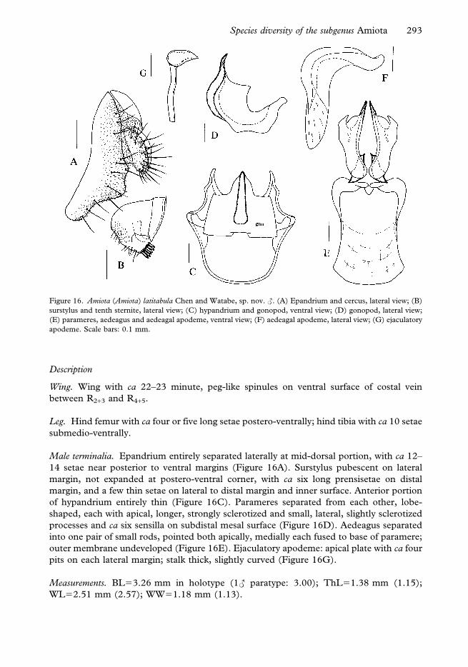
Description
Wing. Wing with ca 22–23 minute, peg-like spinules on ventral surface of costal vein
between R2+3 and R4+5.
Leg. Hind femur with ca four or five long setae postero-ventrally; hind tibia with ca 10 setae
submedio-ventrally.
Male terminalia. Epandrium entirely separated laterally at mid-dorsal portion, with ca 12–
14 setae near posterior to ventral margins (Figure 16A). Surstylus pubescent on lateral
margin, not expanded at postero-ventral corner, with ca six long prensisetae on distal
margin, and a few thin setae on lateral to distal margin and inner surface. Anterior portion
of hypandrium entirely thin (Figure 16C). Parameres separated from each other, lobe-
shaped, each with apical, longer, strongly sclerotized and small, lateral, slightly sclerotized
processes and ca six sensilla on subdistal mesal surface (Figure 16D). Aedeagus separated
into one pair of small rods, pointed both apically, medially each fused to base of paramere;
outer membrane undeveloped (Figure 16E). Ejaculatory apodeme: apical plate with ca four
pits on each lateral margin; stalk thick, slightly curved (Figure 16G).
Measurements. BL53.26 mm in holotype (1„ paratype: 3.00); ThL51.38 mm (1.15);
WL52.51 mm (2.57); WW51.18 mm (1.13).
Figure 16. Amiota (Amiota) latitabula Chen and Watabe, sp. nov. „. (A) Epandrium and cercus, lateral view; (B)
surstylus and tenth sternite, lateral view; (C) hypandrium and gonopod, ventral view; (D) gonopod, lateral view;
(E) parameres, aedeagus and aedeagal apodeme, ventral view; (F) aedeagal apodeme, lateral view; (G) ejaculatory
apodeme. Scale bars: 0.1 mm.
Species diversity of the subgenus Amiota 293

Indices. arb54–5/3–4 (4–5/3–5), avd50.60 (0.50), adf51.20 (1.20), flw51.65 (1.50), FW/
HW50.35, ch/o50.05 (0.06), prorb51.00 (1.00), rcorb50.70 (0.70), vb50.45 (0.40),
dcl50.60 (0.60), presctl50.65 (0.70), sctl51.10 (1.10), sterno50.90 (0.95), orbito51.60
(1.80), dcp50.40 (0.35), sctlp51.00 (0.95), C52.50 (2.31), 4c51.33 (1.36), 4v52.78
(2.58), 5x51.20 (1.20), ac54.00 (3.71), M50.67 (0.63), C3F50.60 (0.75).
HOLOTYPE: „, Yunnan: Mt Jizu, 17 August 2000, H.-W. Chen leg. (KIZ).
PARATYPES: 3„, Yunnan: Bamboo Temple, 31 July 2000, 15 July 2002, J.-J. Gao leg. (KIZ,
DBSU and SEHU).
Distribution. China (Yunnan).
Relationship. This species somewhat resembles A. (A.) cuii in the shapes of paramere and
aedeagus, but can be distinguished from it by the vertical lobe of gonopod with one large,
much-sclerotized process apico-medially.
Etymology. In reference to the broad, large vertical lobe of gonopod.
Amiota (Amiota) luguhuensis Chen and Watabe, sp. nov.
(Figure 17)
Diagnosis. Vertical lobe of gonopod slightly sclerotized apically, height longer than width
(Figure 17C).
Description
Wing. Wing with ca 21–22 minute, peg-like spinules on ventral surface of costal vein
between R2+3 and R4+5.
Leg. Hind femur with three long setae postero-ventrally; hind tibia with ca 10 setae
ventrally.
Male terminalia. Epandrium entirely separated laterally at mid-dorsal portion, with ca eight
setae near posterior to ventral margins (Figure 17A). Surstylus glabrous on medial mesal
surface, not expanded at postero-ventral corner, with five longer prensisetae on distal
margin, and several thin setae on distal margin to inner surface. Anterior portion of
hypandrium entirely thin (Figure 17C). Parameres separated from each other, lobe-shaped,
each with apical and lateral, strongly sclerotized, pointed processes and ca five sensilla on
medial mesal surface (Figure 17D). Aedeagus separated into one pair of small rods, each fused
to base of paramere; outer membrane undeveloped (Figure 17D, E). Ejaculatory apodeme:
apical plate with ca five pits on each lateral margin; stalk thick, curved (Figure 17F).
Measurements. BL52.70 mm; ThL51.13 mm; WL52.67 mm; WW51.20 mm.
Indices. arb54/4, avd50.30, adf51.20, flw51.30, FW/HW50.35, ch/o50.07,
prorb51.00, rcorb50.70, vb50.35, dcl50.65, presctl50.65, sctl51.10, sterno50.90,
orbito52.20, dcp50.25, sctlp51.10, C52.07, 4c51.53, 4v53.00, 5x51.25, ac53.63,
M50.79, C3F50.65.
294 H.-W. Chen et al.

HOLOTYPE: „, Yunnan: Lake Lugu, 25 July 2001, L.-P. He leg. (KIZ).
Distribution. China (Yunnan).
Relationship. This species resembles A. (A.) arcuata sp. nov. in the shape of paramere and
aedeagus, but can be distinguished from it by the vertical lobe of gonopod height longer
than width.
Etymology. In reference to the type locality.
Amiota (Amiota) nozawai Chen and Watabe, sp. nov.
(Figure 18)
Diagnosis. Vertical lobe of gonopod broadened, large, with three pointed processes; medial
process slender, less sclerotized; lateral ones much sclerotized (Figure 18C); paramere
apically with one pointed, sclerotized process and submedially with one bifurcated process
(Figure 18D).
Description
Wing. Wing with 22–23 minute, peg-like spinules on ventral surface of costal vein between
R2+3 and R4+5.
Leg. Hind femur with ca four to six long setae postero-ventrally; hind tibia with a row of ca
four to six setae ventrally.
Figure 17. Amiota (Amiota) luguhuensis Chen and Watabe, sp. nov. „ (see Figure 12 for further explanation).
Species diversity of the subgenus Amiota 295

Male terminalia. Epandrium entirely separated laterally at mid-dorsal portion, with 14–16
setae near posterior to ventral margins (Figure 18A). Surstylus pubescent on lateral margin,
not expanded at postero-ventral corner, with six long prensisetae on distal margin and a few
thin setae on lateral and ventral margins. Anterior portion of hypandrium entirely thin
(Figure 18C). Parameres separated from each other, lobe-shaped, each with apical, longer,
strongly sclerotized and small, lateral, slightly sclerotized processes and six sensilla on
subdistal mesal surface (Figure 18D). Aedeagus separated into one pair of small rods,
pointed apically, medially each fused to base of paramere; outer membrane undeveloped
(Figure 18E, F). Ejaculatory apodeme: apical plate with four pits on each lateral margin;
stalk thick, long (Figure 18G).
Measurements. BL52.82 mm in holotype (5„ paratypes: 2.63–2.97); ThL51.20 mm
(1.17–1.23); WL52.25 mm (2.15–2.38); WW51.00 mm (0.93–1.00).
Indices. arb54/3 (4/2–3), avd50.80 (0.70–0.85), adf51.20 (1.20–1.50), flw51.50 (1.40–
1.60), FW/HW50.35 (0.30–0.35), ch/o50.05 (0.05–0.06), prorb51.00 (0.95–1.00),
rcorb50.70 (0.60–0.75), vb50.35 (0.30–0.35), dcl50.55 (0.50–0.55), presctl50.60
(0.60–0.65), sctl51.10 (1.10–1.20), sterno50.90 (0.90–0.95), orbito52.20 (2.20–2.50),
dcp50.25 (0.20–0.25), sctlp51.00 (1.00–1.10), C52.00 (1.70–2.04), 4c51.50 (1.38–
1.59), 4v52.75 (2.43–2.92), 5x51.20 (1.18–1.38), ac54.28 (3.83–4.50), M50.79 (0.65–
0.72), C3F50.73 (0.59–0.77).
HOLOTYPE: „, Hunan: Mt Badagong, 3 August 2000, M. Nozawa leg. (KIZ).
Figure 18. Amiota (Amiota) nozawai Chen and Watabe, sp. nov. „ (see Figure 2 for further explanation).
296 H.-W. Chen et al.

PARATYPES: 9„, same data as the holotype except for 1–7 August 2000, M. Nozawa, Y.-G.
Hu and H. Takamori leg. (KIZ, BDNU and SEHU).
Distribution. China (Hunan).
Relationship. This species somewhat resembles A. (A.) jizushanensis sp. nov. in the shape of
male genitalia, but can be distinguished from it by the vertical lobe of gonopod with three
processes.
Etymology. Patronym of the collector Mr M. Nozawa.
Amiota (Amiota) paraspinata Chen and Watabe, sp. nov.
(Figure 19)
Diagnosis. Vertical lobe of gonopod sclerotized, with two acute, much-sclerotized processes
laterally (Figure 19C).
Description
Wing. Wing with ca 21–22 minute, peg-like spinules on ventral surface of costal vein
between R2+3 and R4+5.
Leg. Hind femur with five or six long setae postero-ventrally; hind tibia with 10 setae
ventrally.
Figure 19. Amiota (Amiota) paraspinata Chen and Watabe, sp. nov. „ (see Figure 12 for further explanation).
Species diversity of the subgenus Amiota 297

Male terminalia. Epandrium entirely separated laterally at mid-dorsal portion, with ca 17–
18 setae near posterior to ventral margins (Figure 19A). Surstylus slightly pubescent on
medial surface, not expanded at postero-ventral corner, with ca six longer prensisetae on
distal margin, and several thin setae on lateral margin and inner surface. Anterior portion of
hypandrium entirely thin (Figure 19C). Parameres separated from each other, lobe-shaped,
each with apical, strongly sclerotized, pointed and lateral, less-sclerotized processes and ca
six sensilla on medial mesal surface (Figure 19D). Aedeagus separated into one pair of
small rods, each fused to base of paramere; outer membrane undeveloped (Figure 19E, D).
Ejaculatory apodeme: apical plate with ca three pits on each lateral margin; stalk thick, long
(Figure 19F).
Measurements. BL52.82 mm in holotype (5„ paratypes: 2.63–3.10); ThL51.14 mm
(1.00–1.23); WL52.38 mm (2.18–2.45); WW51.05 mm (1.00–1.18).
Indices. arb55/4 (4–5/2–4), avd50.50 (0.50–0.56), adf51.50 (1.50–1.80), flw51.80
(1.60–1.90), FW/HW50.35 (0.35), ch/o50.05 (0.05), prorb51.00 (1.00), rcorb50.75
(0.60–0.80), vb50.35 (0.35–0.40), dcl50.55 (0.55), presctl50.60 (0.60–0.65), sctl51.10
(1.10–1.20), sterno50.90 (0.90–0.95), orbito51.80 (1.60–1.80), dcp50.23 (0.23–0.25),
sctlp51.00 (1.00), C51.93 (2.04–2.39), 4c51.50 (1.28–1.59), 4v52.56 (2.53–2.82),
5x51.10 (1.10–1.38), ac53.86 (2.83–4.50), M50.61 (0.45–0.72), C3F50.61 (0.59–0.67).
HOLOTYPE: „, Yunnan: Mt Jizu, 17 August 2000, H.-W. Chen leg (KIZ).
PARATYPES: 10„, same data as the holotype except for 17 and 18 August 2000, L.-P. He,
J.-G. Xiangyu, H. Takamori, H. Watabe and H.-W. Chen, leg. (KIZ, DBSU and SEHU);
20„, Yunnan: Bamboo Temple, 31 July, 4 August 2000, 5 May, 15 July 2002, J.-J. Gao
and H.-W. Chen leg. (KIZ, DBSU and SEHU).
Distribution. China (Yunnan).
Relationship. This species resembles A. (A.) spinata in having the vertical lobe of gonopod
with sclerotized, pointed process, but differs from it by the vertical lobe of gonopod with
two acute processes (with four acute processes in spinata).
Etymology. In reference to a close relationship to A. (A.) spinata.
Amiota (Amiota) shangrila Chen and Watabe, sp. nov.
(Figure 20)
Diagnosis. Vertical lobe of gonopod heavily sclerotized apically, basal two-thirds square-
shaped (Figure 20D); lateral process of paramere slender (Figure 20C).
Description
Wing. Wing with ca 21–22 minute, peg-like spinules on ventral surface of costal vein
between R2+3 and R4+5.
Leg. Hind femur with six long setae postero-ventrally; hind tibia with five setae ventrally.
298 H.-W. Chen et al.

Male terminalia. Epandrium nearly separated laterally at mid-dorsal portion, with ca 18–20
setae near posterior to ventral margins (Figure 20A). Surstylus glabrous on medial mesal
surface, not expanded at postero-ventral corner, with six longer prensisetae on distal
margin, and several thin setae on outer to inner surface (Figure 20B). Anterior portion of
hypandrium very thin (Figure 20C). Parameres separated from each other, lobe-shaped,
with ca three apically, much-sclerotized sensilla on medial surface (Figure 20D). Aedeagus
separated into one pair of small rods, each fused to base of paramere; outer membrane
undeveloped (Figure 20E). Ejaculatory apodeme: apical plate with ca four or five pits on
each lateral margin; stalk thick, long (Figure 20F).
Measurements. BL53.39 mm in holotype; ThL51.38 mm; WL52.83 mm; WW51.24 mm.
Indices. arb54/4–5,avd50.35,adf51.10, flw51.00,FW/HW50.35,ch/o50.07,prorb50.95,
rcorb50.80, vb50.40, dcl50.60, presctl50.80, sctl51.00, sterno50.95, orbito52.50,
dcp50.28,sctlp51.10,C52.23,4c51.77,4v53.53,5x51.09,ac53.33,M50.71,C3F50.63.
HOLOTYPE: „, Yunnan: Lake Lugu, 23 July 2001, J.-J. Gao leg. (KIZ).
Distribution. China (Yunnan).
Relationship. This species resembles A. (A.) ailaoshanensis sp. nov. in the general
morphology of male genitalia, especially paramere and aedeagus, but can be
distinguished from it by the vertical lobe of gonopod basal two-thirds quadrate (roundish
in ailaoshanensis).
Figure 20. Amiota (Amiota) shangrila Chen and Watabe, sp. nov. „ (see Figure 12 for further explanation).
Species diversity of the subgenus Amiota 299

Etymology. In reference to the type locality.
7. The rufescens species-group
Amiota (Amiota) rufescens species-group, Chen and Toda, 2001: 1547.
Diagnosis. Prensisetae on surstylus long, pointed apically.
Distribution. Oriental (southern China) and Palaearctic region.
Amiota (Amiota) magniflava Chen and Toda
Amiota (Amiota) magniflava Chen and Toda, 2001: 1547.
Specimen examined. Yunnan: 1„, Mt Ailo, 22 June 2001, H. Watabe leg.; 3„, Mt Wuliang,
2 July 2001, J.-J. Gao and L.-P. He leg.
Distribution. China (Hubei, Yunnan).
8. Ungrouped species
Amiota (Amiota) acuta Okada
Amiota (Amiota) acuta Okada, 1968: 306; Chen and Toda, 2001: 1549.
Specimen examined. Yunnan: 1„, Mt Jizu, 19 August 2000, H.-W. Chen leg.; 14„?,
Bamboo Temple, 15 July 2002, J.-J. Gao leg.
Distribution. China (Yunnan), Japan (Honshu).
Amiota (Amiota) dentata Okada
Amiota (Amiota) dentata Okada, 1971: 87; Maca and Lin, 1993: 2; Chen and Toda, 2001:
1550.
Specimens examined. Yunnan: 1„, Mt Jizu, 19 August 2000, H.-W. Chen leg.; 1„, Lake
Lugu, 25 July 2001, L.-P. He leg.
Distribution. China (Taiwan, Hubei, Yunnan), Japan (Hokkaido, Honshu).
Amiota (Amiota) furcata Okada
Amiota (Amiota) furcata Okada, 1971: 85 [Amiota (Amiota) alboguttata, forma furcata
Okada, 1960: 96 (part)]; Maca and Lin, 1993: 2; Chen and Toda, 2001: 1550.
Specimens examined. Hunan: 4„, Mt Badagong, 1–9 September 2000, Y.-G. Hu, M.
Nozawa and Takamori leg. Fujian: 5„, Mt Wuyi, 17–19 August 2001, ex tree trunks, M.
Nozawa, H. Watabe and H.-W. Chen leg. Guangdong: 2„, Nanling, 23 July 2002, H.
Takamori leg. Yunnan: 11„, Mt Jizu, 19 August 2000, L.-P. He, J.-G. Xiangyu, H.
Takamori H. Watabe and H.-W. Chen leg.; 4„, Lake Lugu, 25 July 2001, H. Watabe, J.-J.
Gao and L.-P. He leg.; 6„, Bamboo Temple, 15 July 2002, J.-J. Gao leg.
300 H.-W. Chen et al.

Distribution. China (Taiwan, Hubei, Hunan, Fujian, Sichuan, Yunnan), Japan (Hokkaido,
Honshu, Kyushu).
Amiota (Amiota) subfurcata Okada
Amiota (Amiota) subfurcata Okada, 1971: 85 [Amiota (Amiota) alboguttata, forma furcata
Okada, 1960: 96 (part)]; Maca and Lin, 1993: 2; Chen and Toda, 2001: 1551.
Amiota (Amiota) pacifica Sidorenko, 1989: 63 (synonymized by Sidorenko, 1992: 260).
Specimens examined. Fujian: 81„, Mt Wuyi, 13–19 August 2001, ex. tree trunks, H.
Watabe M. Nozawa and H.-W. Chen leg. Guangdong: 7„, Nanling, 23 July 2002, H.
Takamori leg. Guangxi: 1„, Nonggang, 21 July 1994, Y.-S. Cui leg. Guizhou: 3„, Longgong,
21–24 August 2000, ex. tree trunks, J.-J. Gao leg. Yunnan: Mt Jizu, 6„, 17–19 August 2000,
L.-P. He, J.-G. Xiangyu, H. Watabe, H. Takamori and H.-W. Chen leg.; Bamboo Temple,
1„, 15 July 2002, J.-J. Gao leg.; 4„, Yexianggu, 13 and 14 September 2002, B.-C. Wang and
H.-W. Chen leg.; 2„, Yixiang, 15 September 2002, H.-W. Chen leg.
Distribution. Russia, China (Jilin, Beijing, Zhejiang, Fujian, Taiwan, Guangdong, Guangxi,
Sichuan, Yunnan), Japan.
Amiota (Amiota) fuscata Chen and Zhang, sp. nov.
(Figure 21)
Diagnosis. All femora dark brown; paramere and aedeagus slender, much sclerotized and
pointed apically (Figure 21D, E).
Description
Wing. Wing with ca 21–22 minute, peg-like spinules on ventral surface of costal vein
between R2+3 and R4+5.
Male terminalia. Epandrium entirely separated into lateral lobes at mid-dorsal portion, with
ca 16–17 setae near posterior to ventral margins (Figure 21A). Surstylus glabrous, with
finger-like process at postero-ventral corner, 10 short prensisetae and several setae on distal
margin, and a few stout, spine-like setae on inner surface (Figure 21B). Anterior portion of
hypandrium slightly broadened at middle (Figure 21C). Paramere with ca three sensilla
subbasally. Vertical lobe of gonopod slightly sclerotized, round apically (Figure 21D).
Outer membrane of aedeagus slightly erected basally. Ejaculatory apodeme: apical plate
with ca five pits on each lateral margin; stalk thick (Figure 21F).
Measurements. BL53.33 mm in holotype (1„ paratype: 3.23); ThL51.30 mm (1.20);
WL52.70 mm (2.63); WW51.20 mm (1.15).
Indices. arb53–4/3 (4–5/3–4), avd50.33 (0.30), adf51.10 (1.10), flw51.20 (1.20), FW/
HW50.35 (0.35), ch/o50.07 (0.07), prorb50.80 (0.85), rcorb50.60 (0.65), vb50.35
(0.35), dcl50.60 (0.60), presctl50.65 (0.65), sctl51.10 (1.10), sterno51.00 (1.00),
orbito52.20 (2.20), dcp50.28 (0.25), sctlp51.10 (1.10), C52.00 (1.93), 4c51.88
(1.76), 4v53.18 (2.94), 5x51.00 (1.00), ac53.75 (3.75), M50.75 (0.71), C3F50.78
(0.75).
Species diversity of the subgenus Amiota 301

HOLOTYPE: „, Yunnan: Gaoligongshan, 15 August 2000, ex tree trunk, H.-W. Chen leg.
PARATYPES: 2„, Yunnan: Mt Wuliang, 1 July 2001, H. Watabe and J.-J. Gao leg. (KIZ and
SEHU).
Distribution. China (Yunnan).
Relationship. This species resembles A. (A.) albilabris in the coloration of all femora, but
differs from it by yellow mid and hind tibiae (dark brown in albilabris).
Etymology. In reference to the dark brown colour of the femora.
Amiota (Amiota) wangi Chen and Zhang, sp. nov.
(Figure 22)
Diagnosis. Vertical lobe of gonopod with one penta-furcated, sclerotized, pointed process;
aedeagus and parameres much sclerotized, apically pointed, basally fused to each other
(Figure 22C).
Figure 21. Amiota (Amiota) fuscata Chen and Zhang, sp. nov. „. (A) Epandrium and cercus, lateral view; (B)
surstylus and tenth sternite, lateral view; (C) hypandrium and gonopod, ventral view; (D, E) parameres, aedeagus
and aedeagal apodeme: (D) ventral view, (E) lateral view; (F) ejaculatory apodeme. Scale bars: 0.1 mm.
302 H.-W. Chen et al.

Description
Wing. Wing with 24–26 minute, peg-like spinules on ventral surface of costal vein between
R2+3 and R4+5.
Leg. Leg yellow; mid and hind femora slightly brownish.
Male terminalia. Epandrium nearly entirely separated laterally at mid-dorsal portion, with
ca 20 setae near posterior to ventral margins (Figure 22A). Surstylus with a few setae
medially, finger-like process at postero-ventral corner, ca eight prensisetae on distal margin,
and many setae on ventral margin and on outer mesal surface (Figure 22B). Anterior portion of
hypandrium slightly broadened submedially (Figure 22C, D). Ejaculatory apodeme: apical
plate with four pits on each lateral margin; stalk thick, long (Figure 22E, F).
Measurements. BL52.25 mm; ThL50.90 mm; WL51.80 mm; WW50.70 mm.
Indices. arb53–4/3, avd50.65, adf51.20, flw51.20, FW/HW50.35, ch/o50.07,
prorb50.95, rcorb50.70, vb50.30, dcl50.50, presctl50.55, sctl51.10, sterno50.95,
orbito52.50, dcp50.22, sctlp51.00, C51.43, 4c52.06, 4v52.82, 5x51.50, ac54.38,
M50.88, C3F50.71.
HOLOTYPE: „, Yunnan: Wangtianshu, 10 September 2002, ex tree trunk, B.-C. Wang leg.
(DBSU).
Distribution. China (Yunnan).
Figure 22. Amiota (Amiota) wangi Chen and Zhang, sp. nov. „ (see Figure 21 for further explanation).
Species diversity of the subgenus Amiota 303

Relationship. This species closely resembles A. (A.) acuta in the morphology of male
terminalia, but can be distinguished from it by the vertical lobe of gonopod with one penta-
furcated (vertical lobe of gonopod bifurcated in acuta).
Etymology. Patronym of the collector Mr B.-C. Wang.
Amiota (Amiota) wuyishanensis Chen and Zhang, sp. nov.
(Figure 23)
Diagnosis. Parameres slightly sclerotized, distally with two pointed processes (Figure 23C,
D), inter-laterally with one triangular process and one knot-like process having four
sensilla.
Description
Wing. Wing with 24–26 minute, peg-like spinules on ventral surface of costal vein between
R2+3 and R4+5.
Male terminalia. Epandrium separated laterally at mid-dorsal portion, with ca 23 setae near
posterior to ventral margins (Figure 23A). Surstylus pubescent, slightly expanded at
posteroventral corner, medially with three setae, with eight prensisetae on distal margin,
Figure 23. Amiota (Amiota) wuyishanensis Chen and Zhang, sp. nov. „. (A) Epandrium and cercus, lateral view; (B)
surstylus and tenth sternite, lateral view; (C) hypandrium and gonopod, ventral view; (D) parameres and aedeagal
apodeme, ventral view; (E) hypandrium, paramere, gonopod and aedeagal apodeme, lateral view; (F) ejaculatory
apodeme. Scale bars: 0.1 mm.
304 H.-W. Chen et al.

and many setae on ventral margin and outer mesal surface. Gonopods separated medially,
sclerotized medially, each with three finger-like, apical processes and two sclerotized
lateral processes (Figure 23C). Aedeagus fused to paramere (Figure 23D, E).
Ejaculatory apodeme: apical plate with four pits on each lateral margin; stalk thick, long
(Figure 23F).
Measurements. BL52.52 mm in holotype (2„ paratypes: 2.45–2.47); ThL51.00 mm
(1.00–1.10); WL51.92 mm (1.90–2.00); WW50.88 mm (0.84–0.90).
Indices. arb54/3–4 (4–5/3–4), avd51.00 (1.00), adf51.10 (1.10), flw51.80 (1.70–1.80),
FW/HW50.35 (0.35), ch/o50.05 (0.05), prorb51.00 (1.00), rcorb50.90 (0.80–0.95),
vb50.45 (0.45–0.50), dcl50.55 (0.55), presctl50.60 (0.60), sctl51.30 (1.30),
sterno50.95 (0.85–0.90), orbito52.00 (2.00–2.20), dcp50.40 (0.35–0.40), sctlp51.00
(1.00), C51.52 (1.38–1.60), 4c51.79 (1.67–2.00), 4v52.57 (2.40–2.85), 5x51.71 (1.38–
1.57), ac55.00 (5.00), M50.86 (0.73–0.85), C3F50.72 (0.69–0.74).
HOLOTYPE: „, Fujian: Mt Wuyi, 18 July 2001, ex tree trunk, H.-W. Chen leg. (KIZ).
PARATYPES: 2„, same data as the holotype (DBSU and SEHU). Yunnan: 2„, Yixiang, 15
September 2002, ex tree trunks, H.-W. Chen leg. (DBSU).
Distribution. China (Fujian, Yunnan).
Relationship. This species closely resembles A. (A.) dentata in the morphology of male
terminalia, but can be distinguished from it by the paramere with two processes (with one
pointed process in dentata).
Etymology. In reference to the type locality.
Discussion
Our data revealed that Yunnan province is the most abundant in species diversity of the
subgenus Amiota among the seven provinces of southern China examined in the present
study (Table I). Six of seven established species-groups of the subgenus Amiota (Chen and
Toda 1998a, 1998b, 2001) were found in Yunnan. This province has very diverse
geographical and biological environments. The northern parts are highlands running from
the Qinghai-Xizang Plateau with an elevation of more than 2000 m, and the southern parts
are lowlands with an elevation of less than 80 m. This topography brings a three-
dimensional differentiation of climate and diverse vegetation (Li and Xue 1997), e.g.
temperate deciduous forest mixed with conifer in the north-western district, evergreen
broad-leaved forest in the central district and tropical rain forest in the southern district.
About 14 000 species of flowering plant and nearly 20 000 known species of insect (Wang
1992) are reported in Yunnan Province. The distribution pattern of the species-groups of
Amiota in Yunnan well reflects the adaptation of the species-groups to various climatic and
biological environments. The basdeni and alboguttata species-groups that prefer cooler
habitats were found in northern Yunnan, whereas the sinuata species-group is restricted to
the tropical region of Yunnan (Xishuangbanna).
The subgenus Amiota is mainly distributed in the temperate regions of the Northern
Hemisphere (Chen and Toda 2001). The seven provinces of southern China where we
Species diversity of the subgenus Amiota 305

conducted the present drosophilid survey are situated at the northern borders of the
Oriental zoogeography region, between 18 and 30uN and between 97 and 120uE. Table II
shows the world geographic distribution patterns of the species-groups and ungrouped
species. As suggested from the distribution pattern of the species-groups in Yunnan, the
worldwide tendency of adaptation of each species-group to their favourable environment is
shown. Four species-groups, the basdeni, taurusata, alboguttata and rufescens groups, are
recorded in the Palearctic region of East Asia (isolated from the Oriental region by the
Yangtze River as a demarcation in rough), three species-groups (the basdeni, alboguttata and
rufescens groups) in Europe and two species-groups (the alboguttata and rufescens groups) in
North to Central America. Moreover, both the apodemata and sinuata species-groups are
distributed in South-East Asia, tropical districts of the Oriental region, and further the
sinuata species-group in northern parts of Australia. The Chinese part of the Oriental
region possesses a high degree of species diversity for the subgenus Amiota. All of the seven
species-groups so far established, together with many of ungrouped species, are found in
this region (Table II). The information on geographic distribution strongly suggests that an
early adaptive radiation of the subgenus Amiota might have occurred in southern China and
then spread to Palearctic, Nearctic or Australian regions.
Key to all the species of the subgenus Amiota from southern China
Male
1.– Face, postpronotal lobe and wing base each with distinctly milky white spot;
aedeagal apodeme broadened, dorso-ventrally flattened (subgenus Amiota) . . 2
2. Postocellar setae absent; fifth tergite laterally with coloured strips; lateral margin of
sixth tergite apically pointed, not reaching ventral margin of fifth tergite; aedeagus
single, basally fused to apodeme (apodemata species-group) . . . . . . .
. . . . . . . . . . . . . . . . apodemata Gupta and Panigrahy
– Postocellar setae present; fifth tergite laterally without coloured strips; lateral
margin of sixth tergite, reaching ventral margin of fifth tergite; aedeagus, if present,
basally not fused to apodeme . . . . . . . . . . . . . . . 3
Table II. Geographical distributions of 94 species of the subgenus Amiota in the world.
Europe
North–
Central
America
East Asia
South-
East Asia Australia Africa
All the
worldPalaearctic Oriental
Species-groups
apodemata 2 2 4
sinuata 5 5 1 10
nagatai 3 3
basdeni 1 6 10 14
taurusata 2 4 6
alboguttata 3 1 10 11 25
rufescens 1 1 1 1 4
Ungrouped spp. 5 7 7 8 1 1 3 28
Total 10 9 26 44 8 2 3 94
1.
306 H.-W. Chen et al.

3. Body yellow; hypandrium broadened, anteriorly connected to ventro-medial
portion of parameres by articulating plate; parameres basally to medially fused to
each other (sinuata species-group) . . . . . . . . . . . . . . 4
– Body black; hypandrium narrow, separated from paramere; parameres entirely
separated from each other . . . . . . . . . . . . . . . . 8
4. Prescutellar setae present; outer membrane of aedeagus with fine serration . . .
. . . . . . . . . . . . . . aculeata Chen and Aotsuka, sp. nov.
– Prescutellar setae absent . . . . . . . . . . . . . . . . . 5
5. Vertical lobe of gonopod basally extremely elongated, forming one pair of
strongly curved, rod-shaped processes with one pair of sickle-like projections
submedially . . . . . . . . . . subsinuata Chen and Aotsuka, sp. nov.
– Vertical lobe of gonopod basally without elongated process . . . . . . . 6
6. Epandrium postero-ventrally unpubescent; surstylus with stout spines on inner
surface; aedeagal apodeme as long as wide, basally much expanded laterad like
fan; paramere with only ordinary sensilla; ejaculatory apodeme with five pits on
each side of apical plate . . . . . . . . . . . pengi Chen and Toda
– Epandrium postero-ventrally pubescent; surstylus without stout spines on inner
surface; aedeagal apodeme longer than wide, basally not strongly expanded
laterad; paramere distally with some very minute sensilla in addition to ordinary
ones; ejaculatory apodeme with two or three pits on each side of apical plate . 7
7. Distal part of paramere as long as basal part, apically slightly narrowing in lateral
view . . . . . . . . . . . . . . . . . . . sinuata Okada
– Distal part of paramere distinct longer than basal part, apically broadened in
lateral view . . . . . . . . . xishuangbanna Chen and Aotsuka, sp. nov.
8. Ventral branches of arista as long as dorsal ones; wing with ca 14–15 distinct, peg-
like spinules on ventral surface of costal vein between R2+3 and R4+5 (nagatai
species-group) . . . . . . . . . . . . . . . . . . . . 9
– Ventral branches of arista shorter than dorsal ones; wing with more than 18 small,
peg-like spinules there . . . . . . . . . . . . . . . . . 10
9. Vertical lobe of gonopod apically with one pair of horn-like processes nagatai Okada
– Vertical lobe of gonopod apically round, without any processes. . . . . .
. . . . . . . . . . . . . . . . . . . . okinawana Okada
10. Surstylus with a few of aristate processes separated from or fused to each other on
mesal surface; vertical lobe of gonopod with two sclerotized, basally fused
M-shaped processes (basdeni species-group) . . . . . . . . . . 11
– Surstylus without aristate processes; vertical lobe of gonopod without M-shaped
process . . . . . . . . . . . . . . . . . . . . . 19
11. Paramere less sclerotized, pubescent . . . . . . . . . . . . 12
– Paramere sclerotized, glabrous . . . . . . . . . . . . . . 13
12. Aedeagus and paramere slender, rod-shaped; aedeagus slightly sclerotized;
paramere entirely pubescent . . . . . . . . . . . . palpifera Okada
– Aedeagus and paramere lobe-shaped, aedeagus heavily sclerotized, black;
paramere partially pubescent, submedially with a few tiny setulae . . . . .
. . . . . . . . . . . . . . . brevipartita Chen and Gao, sp. nov.
Species diversity of the subgenus Amiota 307

13. Aedeagus much sclerotized and black . . . . . . . . . . . . 14
– Aedeagus sclerotized, black only at apical portion in some species . . . . 16
14. Paramere apically round, basally articulated with aedeagus; aedeagus subapically
with short, additional process . . . . . . . . . . curvistyla Okada
– Paramere apically pointed, basally fused to aedeagus; aedeagus subapically lacking
process . . . . . . . . . . . . . . . . . . . . . 15
15. Paramere curved apically; aedeagus very small, entirely fused to base of
paramere . . . . . . . . . . . . . . . onchopyga Nishiharu
– Paramere slightly curved submedially; aedeagus fused to paramere only at
base . . . . . . . . . . . . curvispinata Chen and Gao, sp. nov.
16. Paramere broadened, with one sclerotized process on lateral side . . . . .
. . . . . . . . . . . . . . . . . huae Chen and Gao, sp. nov.
– Paramere rod-shaped, lacking process . . . . . . . . . . . . 17
17. Paramere and aedeagus apically pointed; aedeagus curved toward medial portion
of paramere . . . . . . . . . . . longispinata Chen and Gao, sp. nov.
– Paramere and aedeagus apically blunt; aedeagus curved toward subapical portion
of paramere . . . . . . . . . . . . . . . . . . . . 18
18. Paramere much sclerotized, basally curved in lateral view . . . . . . .
. . . . . . . . . . . . . . . . lipingae Chen and Gao, sp. nov.
– Paramere less sclerotized, straight . . . . . . . . macai Chen and Toda
19. Hind femur ventro-basally with nearly hyaline, small, lobe-like flap (taurusata
species-group) . . . . . . . . . . . . . . . . . . . 20
– Hind femur without any flap . . . . . . . . . . . . . . . 22
20. All femora dark brown; hind femur curved basally; vertical lobe of gonopod nearly
triangular . . . . . . . . . . femorata Chen and Takamori, sp. nov.
– Leg entirely yellow, hind femur straight; vertical lobe of gonopod nearly round 21
21. Parameres basally fused to aedeagus . asymmetrica Chen and Takamori, sp. nov.
– Parameres entirely separated from aedeagus yixiangna Chen and Takamori, sp. nov.
22. Hind tibia ventrally with a row of long setae; hind second to fifth tarsomeres
broadened: length of second tarsomere at least shorter than 1.5 times its width
(alboguttata species-group) . . . . . . . . . . . . . . . 23
– Hind tibia ventrally without long setae; hind second to fifth tarsomeres nearly
straight . . . . . . . . . . . . . . . . . . . . . 30
23. Vertical lobe of gonopod medio-apically with three processes . . . . . .
. . . . . . . . . . . . . . . nozawai Chen and Watabe, sp. nov.
Vertical lobe of gonopod apically with two processes . . . . . . . . 24
23.–Vertical lobe of gonopod apically with one process . . . . . . . . 26
24. Vertical lobe of gonopod and aedeagus less sclerotized . . . . . . . .
. . . . . . . . . . . . . jizushanensis Chen and Watabe, sp. nov.
– Vertical lobe of gonopod much sclerotized, black . . . . . . . . . 25
25. Vertical lobe of gonopod medially separated into lateral lobes . . . . . .
. . . . . . . . . . . . . . dehiscentia Chen and Watabe, sp. nov.
–
–
308 H.-W. Chen et al.

– Vertical lobe of gonopod medially not separated, apically with two pointed
processes . . . . . . . . . . paraspinata Chen and Watabe, sp. nov.
26. Vertical lobe of gonopod basally roundish . . . . . . . . . . . 27
– Vertical lobe of gonopod basally quadrate . . . . . . . . . . . 28
27. Vertical lobe of gonopod much sclerotized distally, broadened, large, width as long
as height. . . . . . . . . . . . arcuata Chen and Watabe, sp. nov.
– Vertical lobe of gonopod less-sclerotized apically, height longer than width . . . .
. . . . . . . . . . . . . . luguhuensis Chen and Watabe, sp. nov.
28. Apical process of vertical lobe of gonopod pointed in ventral view . . . . .
. . . . . . . . . . . . . . shangrila Chen and Watabe, sp. nov.
– Apical process of vertical lobe of gonopod roundish in ventral view . . . 29
29. Width of vertical lobe of gonopod longer than height; paramere basally fused to
median part of aedeagus . . . . . . latitabula Chen and Watabe, sp. nov.
– Width of vertical lobe of gonopod shorter than height; paramere articulated with
basal part of aedeagus . . . . . . ailaoshanensis Chen and Watabe, sp. nov.
30. Body large (more than 4.0 mm in length); prensisetae on surstylus long, apically
pointed (rufescens species-group); paramere lobe-like, less sclerotized; vertical lobe
of gonopod short . . . . . . . . . . . magniflava Chen and Toda
– Body small (less than 4.0 mm in length); prensisetae on surstylus short and
apically blunt (ungrouped species) . . . . . . . . . . . . . 31
31. All femora dark brown; paramere and aedeagus slender and much sclerotized . .
. . . . . . . . . . . . . . . fuscata Chen and Zhang, sp. nov.
– All femora yellow; aedeagus fused to parameres . . . . . . . . . 32
32. Fifth sternite triangularly notched on posterior margin, with dense, long
setae . . . . . . . . . . . . . . . . . . . . . . 33
– Fifth sternite quadrate, not notched postero-medially, without dense, long setae 34
33. Surstylus with 11–12 prensisetae; mid tibia medio-ventrally with five to seven
erected setae longer than others . . . . . . . . . . . furcata Okada
– Surstylus with six or seven prensisetae; mid tibia ventrally without any long,
erected seta . . . . . . . . . . . . . . . . subfurcata Okada
34. Epandrium entirely separated mid-dorsally; vertical lobe of gonopod medially
separated from each other . . . . . . . . . . . . . . . 35
– Epandrium deeply constricted mid-dorsally but not separated laterally; vertical
lobe of gonopod narrowly elongated apically, not separated medially . . . 36
35. Paramere much sclerotized, with one antero-apically pointed and basally quadrate
process . . . . . . . . . . . . . . . . . . dentata Okada
– Parameres less sclerotized, distally with one triangular pointed process and one
knot-like process . . . . . . . wuyishanensis Chen and Zhang, sp. nov.
36. Vertical lobe of gonopod less sclerotized, apically elongated narrowly and
bifurcated . . . . . . . . . . . . . . . . . . acuta Okada
– Vertical lobe of gonopod much sclerotized, apically pointed and pentafurcated . .
. . . . . . . . . . . . . . . . wangi Chen and Zhang, sp. nov.
Species diversity of the subgenus Amiota 309

Acknowledgements
We would like to express our sincere thanks to Dr J.-G. Xiangyu, Ms L.-P. He and Mr
B.-C. Wang (KIZ), Dr Y.-G. Hu (Institute of Low Temperature Science, Hokkaido
University, Japan), Mr H. Ito (Hokkaido University of Education) and Mr M. Nozawa
(Tokyo Metropolitan University), who helped us with field collections and provided us
with invaluable specimens. This work was supported by the Japan Society for the
Promotion of Science (Nos 12375002 and 01111), the National Natural Science
Foundation of China (Nos 39930100 and 30024004) and the Chinese Academy of
Sciences (No. KSCX2-1-05).
References
Chen HW, Aotsuka T. 2003. A survey of the genus Leucophenga (Diptera, Drosophilidae) in Iriomote-jima of
Japan, with descriptions of three new species. The Canadian Entomologist 135:1–16.
Chen HW, Toda MJ. 1998a. Amiota (Amiota) apodemata species-group, with descriptions of two new species from
Southeast Asia (Diptera, Drosophilidae). Entomological Science 1:271–275.
Chen HW, Toda MJ. 1998b. Amiota (Amiota) sinuata species-group, with descriptions of five new species from
Southeast Asia (Diptera, Drosophilidae). Entomological Science 1:409–416.
Chen HW, Toda MJ. 2001. A revision of the Asian and European species in the subgenus Amiota Loew (Diptera,
Drosophilidae) and establishment of species-groups based on phylogenetic analysis. Journal of Natural
History 35:1517–1563.
Gupta JP, Panigrahy KR. 1987. Some further additions to the list of Indian fauna of Drosophilidae. Proceedings of
Zoological Society, Calcutta, 36:57–66.
Li DZ, Xue JR. 1997. The biodiversity and conservation of bamboos in Yunnan, China. In: Chapman GP, editor.
The Bamboos. London: The Linnean Society of London and Academic Press. p 83–94.
Loew H. 1862. Diptera Americae septentrionalis indigena. Centuria secunda, Berliner Entomologische Zeitschrift,
6:185–232.
Maca J. 1980. European species of the subgenus Amiota s. str. (Diptera, Drosophilidae). Acta Entomological
Bohemoslovaca 77:328–346.
Maca J, Lin FJ. 1993. The Drosophilidae of Taiwan: genera Amiota (excluding subgenus Phortica) and
Leucophenga (Nankangomyia subg. nov.). Bulletin of the Institute Zoology, Academia Sinica 32:1–11.
Nishiharu S. 1979. Three new species of Drosophilidae from Japan (Diptera). Kontyu, Tokyo, 47:38–43.
Okada T. 1960. On the Japanese species of the genus Amiota Loew (Diptera, Drosophilidae). Mushi 34:89–102.
Okada T. 1968. Addition to the fauna of the family Drosophilidae of Japan and adjacent countries (Diptera,
Drosophilidae). I. Genera Stegana, Amiota, Leucophenga, and Microdrosophila, with discussion on the
homology of phallic organs. Kontyu, Tokyo 36:303–323.
Okada T. 1971. A revision and taxometric analysis of the genus Amiota Loew of Japan and adjacent countries
(Diptera, Drosophilidae). Kontyu, Tokyo 39:82–98.
Sidorenko VS. 1992. Family Drosophilidae. In, Insects of Khingansky Reserve, Part II. Vladivostok: Dal’nauka
Publishing House. p 259–264).
Takada H, Beppu K, Toda MJ. 1979. Drosophila survey of Hokkaido, XXXVI. New and unrecorded species of
Drosophilidae (Diptera). Journal of the Faculty of General Education, Sapporo University 14:105–129.
Toda MJ, Peng TX. 1992. Some species of the subfamily Steganinae (Diptera, Drosophilidae) from Guangdong
Province, southern China. Annales de la Societe Entomologique de France (N. S.) 28:201–213.
Toda MJ, Sidorenko VS, Watabe H, Sergey KK, Vinokurov NN. 1996. A revision of the Drosophilidae (Diptera)
in East Siberia and Russian Far East: taxonomy and biogeography. Zoological Science 13:455–477.
Wang YX. 1992. Present situation and crisis of animal diversity in Yunnan. In: Wu ZY, editor. Symposium of
Seminar on Biodiversity in Yunnan. Yunnan Science and Technology Press. p 101–104.
Wheeler MR. 1952. The Drosophilidae of the Neararctic region, exclusive of the genus Drosophila. The
University of Texas Publication, 5204:162–218.
Zhang WX, Chen HZ, Peng TX. 1997. The family Drosophilidae. In: Xue WQ, Chao CM, editors. Flies of
China. Shenyang, China: Liaoning Science and Technology Press. p 280–489.
310 H.-W. Chen et al.