Specific Arrest of Cardiogenesis in Cultured - CORE BIOLOGY 196, 237–247 (1998) ARTICLE NO....
11
DEVELOPMENTAL BIOLOGY 196, 237–247 (1998) ARTICLE NO. DB988862 Specific Arrest of Cardiogenesis in Cultured Embryonic Stem Cells Lacking Cripto-1 1 Chunhui Xu,* Giovanna Liguori,² Eileen D. Adamson,* ,2 and Maria G. Persico² *The Burnham Institute, La Jolla Cancer Research Center, 10901 North Torrey Pines Road, La Jolla, California 92037; and ²International Institute of Genetics and Biophysics, CNR, 80125 Napoli, Italy The molecular events of cardiac lineage specification and differentiation are largely unknown. Here we describe the involvement of a growth factor with an EGF-like domain, Cripto-1 (Cr-1), in cardiac differentiation. During embryonic development, Cr-1 is expressed in the mouse blastocyst, primitive streak, and later is restricted to the developing heart. To investigate the role of Cr-1, we have generated Cr-1-negative embryonic stem (ES) cell lines by homologous recombina- tion. The resulting double ‘‘knockout’’ ES cells have selectively lost the ability to form beating cardiac myocytes, a process that can be rescued by reintroducing Cr-1 gene back into the Cr(0/0) cells. Furthermore, the lack of functional Cr-1 is correlated with absence of expression of cardiac-specific myosin light and heavy chain genes during differentiation. Differentiation into other cell types including skeletal muscle is not disrupted. These results suggest that Cr-1 is essential for contractile cardiomyocyte formation in vitro. q 1998 Academic Press Key Words: homologous recombination; gene targeting; markers for ectoderm, mesoderm and endoderm; teratocarcino- mas; mutant phenotype; rescue. INTRODUCTION tively little is known about the early stage of vertebrate heart development. In this report, we describe the involve- ment of Cripto-1 (see below) in the early stages of differenti- The development of the heart is a complex process com- ation of cardiomyocytes. posed of two major stages: an early specification and differ- Cripto-1 (Cr-1), encoded by the teratocarcinoma-derived entiation of cardiomyocytes and later morphogenesis growth factor-1 (tdgf-1) gene (Liguori et al., 1996), is a (Olson and Srivastava, 1996). Murine cardiac progenitor growth factor with an epidermal growth factor-like motif cells derive from the anterior lateral plate mesoderm that (Ciccodicola et al., 1989). Although Cr-1 folds in the charac- arises from the primitive streak on the sixth day of gestation teristic EGF-like manner using its six cysteine residues, the (E6.5) (Olson and Srivastava, 1996). These cells differentiate, resulting polypeptide is unable to bind to members of the express cardiac muscle structural genes such as myosin EGF receptor family due to truncation of some sequences heavy chains and atrial and ventricular myosin light chains (Brandt et al., 1994). Cr-1 is a growth factor for mammary (Kubalak et al., 1994; O’Brien et al., 1993), and form the cells by interaction with an unidentified receptor to activate primitive cardiac tube at E7.5 – 8.0 (Lyons et al., 1990; Mor- intracellular components through ras/raf/MAPK pathway kin, 1993; Sassoon et al., 1988). The newly formed cardiac (Kannan et al., 1997). Cr-1 and the related Cryptic gene have tube undergoes extensive morphological changes before been assigned to a distinct family, with the proposed name, forming the mature heart. Through gene targeting tech- CFC (Shen et al., 1997). niques, many genes have been identified as being involved Both human and mouse Cr-1 are encoded by 6 exons (exon in the later processes of heart development. However, rela- 4 contains the EGF-like domain) producing (in mouse) a secreted protein of 171 amino acid residues (Liguori et al., 1996). Cr-1 is expressed at three main stages in develop- 1 The last two authors contributed equally. ment. The earliest expression of Cr-1 occurs in 4-day mouse 2 To whom correspondence and reprint requests should be ad- blastocysts in both trophoblast and inner cell mass cells dressed. Fax: (619) 646-3195. E-mail: [email protected] or eadam [email protected]. (Johnson et al., 1994). At day 6.5, Cr-1 is present in epiblast 237 0012-1606/98 $25.00 Copyright q 1998 by Academic Press All rights of reproduction in any form reserved.
Transcript of Specific Arrest of Cardiogenesis in Cultured - CORE BIOLOGY 196, 237–247 (1998) ARTICLE NO....

DEVELOPMENTAL BIOLOGY 196, 237–247 (1998)ARTICLE NO. DB988862
Specific Arrest of Cardiogenesis in CulturedEmbryonic Stem Cells Lacking Cripto-11
Chunhui Xu,* Giovanna Liguori,† Eileen D. Adamson,*,2
and Maria G. Persico†*The Burnham Institute, La Jolla Cancer Research Center, 10901 North Torrey Pines Road,La Jolla, California 92037; and †International Institute of Genetics and Biophysics,CNR, 80125 Napoli, Italy
The molecular events of cardiac lineage specification and differentiation are largely unknown. Here we describe theinvolvement of a growth factor with an EGF-like domain, Cripto-1 (Cr-1), in cardiac differentiation. During embryonicdevelopment, Cr-1 is expressed in the mouse blastocyst, primitive streak, and later is restricted to the developing heart.To investigate the role of Cr-1, we have generated Cr-1-negative embryonic stem (ES) cell lines by homologous recombina-tion. The resulting double ‘‘knockout’’ ES cells have selectively lost the ability to form beating cardiac myocytes, a processthat can be rescued by reintroducing Cr-1 gene back into the Cr(0/0) cells. Furthermore, the lack of functional Cr-1is correlated with absence of expression of cardiac-specific myosin light and heavy chain genes during differentiation.Differentiation into other cell types including skeletal muscle is not disrupted. These results suggest that Cr-1 is essentialfor contractile cardiomyocyte formation in vitro. q 1998 Academic Press
Key Words: homologous recombination; gene targeting; markers for ectoderm, mesoderm and endoderm; teratocarcino-mas; mutant phenotype; rescue.
INTRODUCTION tively little is known about the early stage of vertebrateheart development. In this report, we describe the involve-ment of Cripto-1 (see below) in the early stages of differenti-The development of the heart is a complex process com-ation of cardiomyocytes.posed of two major stages: an early specification and differ-
Cripto-1 (Cr-1), encoded by the teratocarcinoma-derivedentiation of cardiomyocytes and later morphogenesisgrowth factor-1 (tdgf-1) gene (Liguori et al., 1996), is a(Olson and Srivastava, 1996). Murine cardiac progenitorgrowth factor with an epidermal growth factor-like motifcells derive from the anterior lateral plate mesoderm that(Ciccodicola et al., 1989). Although Cr-1 folds in the charac-arises from the primitive streak on the sixth day of gestationteristic EGF-like manner using its six cysteine residues, the(E6.5) (Olson and Srivastava, 1996). These cells differentiate,resulting polypeptide is unable to bind to members of theexpress cardiac muscle structural genes such as myosinEGF receptor family due to truncation of some sequencesheavy chains and atrial and ventricular myosin light chains(Brandt et al., 1994). Cr-1 is a growth factor for mammary(Kubalak et al., 1994; O’Brien et al., 1993), and form thecells by interaction with an unidentified receptor to activateprimitive cardiac tube at E7.5–8.0 (Lyons et al., 1990; Mor-intracellular components through ras/raf/MAPK pathwaykin, 1993; Sassoon et al., 1988). The newly formed cardiac(Kannan et al., 1997). Cr-1 and the related Cryptic gene havetube undergoes extensive morphological changes beforebeen assigned to a distinct family, with the proposed name,forming the mature heart. Through gene targeting tech-CFC (Shen et al., 1997).niques, many genes have been identified as being involved
Both human and mouse Cr-1 are encoded by 6 exons (exonin the later processes of heart development. However, rela-4 contains the EGF-like domain) producing (in mouse) asecreted protein of 171 amino acid residues (Liguori et al.,1996). Cr-1 is expressed at three main stages in develop-1 The last two authors contributed equally.ment. The earliest expression of Cr-1 occurs in 4-day mouse2 To whom correspondence and reprint requests should be ad-blastocysts in both trophoblast and inner cell mass cellsdressed. Fax: (619) 646-3195. E-mail: [email protected] or eadam
[email protected]. (Johnson et al., 1994). At day 6.5, Cr-1 is present in epiblast
237
0012-1606/98 $25.00Copyright q 1998 by Academic PressAll rights of reproduction in any form reserved.
AID DB 8862 / 6x3b$$$121 03-26-98 23:23:55 dba

238 Xu et al.
pCR-KO2 and selected in medium containing hygromycin B (400cells and is strongly expressed at the primitive streak stagemg/ml) and gancyclovir (0.2 mM).in the forming mesoderm. Then, a pattern restricted to de-
Differentiation of ES cells. Embryoid bodies (EBs) were formedveloping heart structures occurs (Dono et al., 1993; Johnsonby ES cell aggregate culture in hanging drops (Wobus et al., 1991).et al., 1994) with Cr-1 specifically expressed in the myocar-The differentiation medium was ES culture medium containingdium of the developing heart tubes in 8.5-day-old embryos20% fetal calf serum but without LIF. Rhythmic beating of theand in the outflow region, conotruncus, of the heart at 9.5–EBs, indicating cardiac muscle differentiation, was monitored by
10 days gestation, when the heart develops into a functional daily inspection of the cultures using phase microscopy. Inductionchambered organ. After 10.5 days of development, no in of differentiation into skeletal muscle was performed by thesitu hybridization signals have been detected in the embryo method similar to that described by Weitzer (Weitzer et al., 1995).(Dono et al., 1993). This pattern of Cr-1 expression suggests Induction of differentiation of ES cells into neurons was performeda role in the process of gastrulation, wherein the ectoderm by a method similar to that described by Bain (Bain et al., 1995).
Screening for recombinant clones. Genomic DNA samplesgerm layer gives rise to the mesoderm tissues includingwere extracted from individual clones and examined for the pres-those producing the heart and somites. In addition, theence of a targeted allele by PCR analysis using Taq Extender PCRhighly restricted expression patterns of Cr-1 raise the possi-additive system (Stratagene, San Diego, CA) or Expand PCR Systembility that Cr-1 may play a role in regulating cardiac gene(Boehringer Mannheim, Indianapolis, IN) with following primerexpression. We used the ES3 model system to test this possi-pairs. The Cr-1-specific primers, 5*-ACCTGCTCTGTGTCTCCT-bility.GATTTCC-3* (p-1), which is outside (3*) of the targeting vector, and5*-CACTAGGTGACCAATCTGGTCCAAC-3* (p-2), a neo gene-specific primer, 5*-GCAGCCTCTGTTCCACATACACTTC-3* (p-3), and a hyg gene-specific primer, 5*-TCACGTTGCAAGACC-MATERIALS AND METHODSTGCCTGAAAC-3* (p-4). The PCR conditions were as follows:947C for 4 min; 10 cycles of 947C for 10 s, 657C for 30 s, and 687Cfor 4 min, and followed by 20 cycles of 947C for 10 s, 657C for 30Construction of targeting vectors. The targeting vectors were
constructed with the pPNT plasmid (Tybulewicz et al., 1991). In s, and 687C for 4 min with 20-s increments for each cycle and athe vector pCR-KO1, a 5-kb 5* homologous sequence containing final extension step at 727C for 10 min. Additional PCR reactionexon 1 and 2 was inserted in front of a gene cassette encoding using primers within intron 3: 5*-GTCCCTGATAGTCTCTGA-neomycin phosphotransferase (neo) gene under the control of the TATTC-3* (p-5) and 5*-GAAATGTAAGAAAAGTCATGGGG-3*phosphoglycerokinase (PGK) promoter. A 3.8-kb 3* homologous (p-6) or a set of primer in neo gene: 5*-GTCAAGAAGGCGATA-fragment containing exon 6 was cloned into the BamHI site down- GAAGGCGATGCG-3* (p-7) and 5*-GGTGGAGAGGCTATT-stream to the neo. The targeting vector pCR-KO2 was prepared CGGCTATGACTG-3* (p-8) was also performed. The PCR reactionby inserting the PGK–hygromycin (PGK–hyg) downstream of the for primer p-4/p-5 and p-6/p-7 was performed by denaturing thePGK-neo in pCR-KO1. Negative selection to avoid random inser- DNA at 947C for 2 min, followed by 35 cycles of amplification:tion was provided by the herpes simplex virus thymidine kinase 947C for 30 s, 557C for 30 s, 727C for 1.5 min, and a final extension(hsv–tk) gene. step at 727C for 6 min.
Embryonic stem cell culture, electroporation, and selection. RT–PCR Analysis. Total RNA (3 mg) was isolated from ES cellsThe ES cells were maintained in the undifferentiated state by cul- and EBs, treated with RNase-free DNaseI (Stratagene, San Diego,ture on mitomycin C-treated mouse embryonic fibroblasts (MEF) CA) and converted into cDNA using oligo(dT) as a primer. Thefeeder layers according the standard protocols. The medium used cDNA was amplified by PCR using primers within the Cr-1 deletedwas high glucose Dulbecco’s modified Eagle’s medium (DMEM) region, 5*-ATCGGTCTTTCCAGTTCGTGCCTTC-3* in exon 3containing 15% fetal bovine serum, 0.1 mM b-mercaptoethanol, 1 and 5*-ATCTGCACAGGGAACACTTCTTGGG-3* in exon 5. AmM sodium pyruvate, 11 nonessential amino acids, 2 mM gluta- set of primers for GAPDH (Hummler et al., 1996), 5*-CGTCT-mine, 100 units penicillin/ml, 0.1 mg of streptomycin, and 2% TCACCACCATGGAGA-3* and 5*-CGGCCATCACGCCACAG-leukemia inhibitory factor (LIF) (conditioned medium collected TTT-3*, was also added to the same reaction as controls. The cDNAfrom confluent cultures of Chinese hamster ovary cells transfected from Day 10 EBs was amplified by PCR using specific primers forwith the LIF expression plasmid provided by permission of the MLC2v (Miller-Hance et al., 1993); specific primers for ANF, 5*-Genetics Institute, Andover, MA). The fetal bovine serum was ob- CGGTGTCCAACACAGATCTG-3* and 5*-TCTCTCAGAGGT-tained from Tissue Culture Biologicals (Tulare, CA). GGGTTGAC-3*, which should give a 187-bp product; specific
For the generation of Cr-1 (//0) cells, 1 1 107 R1 ES cells were primers for desmin, 5*-TGATGAGGCAGATGAGGGAG-3* andelectroporated with 30 mg linearized pCR-KO1 plasmid DNA at 5*-TGAGAGCTGAGAAGGTCTGG-3*, which should give a 246-300 V, 250 mF. Recombination events were selected with G418 (380 bp product, and GAPDH (Hummler et al., 1996). The cDNA frommg/ml) and gancyclovir (0.2 mM). To disrupt the second allele, Cr- Day 6 EBs was amplified by PCR using brachyury-specific primers,1 (//0) E31 and E40 cells were electroporated with linearized vector 5*-ATCAAGGAAGGCTTTAGCAAATGGG-3* and 5*-GAACC-
TCGGATTCACATCGTGAGA-3*, which should give a 159-bpproduct; GATA4 specific primers, 5*-CACTATGGGCACAGCA-GCTCC-3* and 5*-TTGGAGCTGGCCTGCGATGTC-3*, which3 Abbreviations used: DMEM, Dulbecco’s modified Eagle’s me-should give a 143-bp product; Mef 2C-specific primers (Martindium; ES cells, embryonic stem cells; EB, embryoid body; KO,et al., 1993); Nkx2.5-specific primers (Biben and Harvey, 1997),knockout; LIF, leukemia inhibitory factor; MEF, mouse embryonic5*-TGCAGAAGGCAGTGGAGCTGGACAAGCC-3* and 5*-TTG-fibroblasts; MHC, myosin heavy chain; MLC, myosin light chains;CACTTGTAGCGACGGTTCTGGAACCAG-3*, which shouldPGK, phosphoglycerokinase; tk, thymidine kinase; SRF, serum re-
sponse factor. give a 220-bp product; Egr-1-specific primers, 5*-CCGAGCTCT-
Copyright q 1998 by Academic Press. All rights of reproduction in any form reserved.
AID DB 8862 / 6x3b$$$122 03-26-98 23:23:55 dba

239No Cardiogenesis in Cr-1 KO ES cells
TCACACAACAACTTTTGTC-3* and 5*-CCGAGATCTCCCAG- Teratocarcinoma. Cells (61 106) were injected into two subcu-taneous sites of 6-week-old 129SvJ mice. Five mice were used forCTCATCATCAAAC-3*, which should give a 355-bp product and
GAPDH primers. cDNA samples from Day 28 EB outgrowths were each cell line. The tumors were processed for histological analysis3 weeks after the injection. The identification of tissue types wastested for skeletal muscle differentiation by PCR using myogenin-
specific primers selected from sequences in GenBank: 5*-TGA- made by a board-certified pathologist, Dr. D. Mercola, M.D., Ph.D.,FACP.GGGAGAGCGCAGGCTCAAG-3* and 5*-TGCTGTCCACGAT-
GGACGTAAGG-3*, which should give a 361-bp product. cDNAsamples derived from Day 25 EB outgrowths were tested for neu-ronal differentiation by PCR amplification using light chain neuro- RESULTSfilament-specific primers, derived from GenBank: 5*-CTCCTA-CTTGATGTCTGCTCGC-3* and 5*-TCAGACTCATCCTTGGC- Targeting the Cripto-1 Gene in ES Cells byAGC-3*, which should give a 219-bp product.
Homologous RecombinationThe PCR reactions for Cr-1, GAPDH, desmin, brachyury,GATA4, MEF2C, Egr-1, myogenin, and neurofilament were per- To precisely define the function(s) of Cr-1, we have dis-formed by denaturing the DNA at 947C for 2 min, followed by 35 rupted both copies of the wild-type Cr-1 alleles sequentiallycycles of amplification: 947C for 30 s, 60–657C for 30 s, 727C for by homologous recombination in R1 (Nagy et al., 1993) ES1.5 min, and a final extension step at 727C for 6 min. The RT–
cells. The two targeting vectors, pCR-KO1 and pCR-KO2,PCR assays for MLC2V were performed by denaturing the DNAcaused the replacement of Cr-1 exons 3, 4, and 5 by PGK-at 957C for 5 min, followed by 35 cycles of amplification: 947C forneo and by PGK-neo -hyg, respectively, as determined by30 s, 707C for 30 s, 727C for 30 s, and a final extension step atPCR analysis of genomic DNA (Figs. 1a–1d) and Southern727C for 6 min. The RT–PCR assays for ANF were performed byblotting analysis (data not shown). To further confirm thedenaturing the DNA at 957C for 5 min, followed by 35 cycles of
amplification: 947C for 30 s, 557C for 30 s, 727C for 50 s, and a final Cr-1 (0/0) mutation, reverse transcriptase–polymeraseextension step at 727C for 6 min. The RT–PCR assays for Nkx2.5 chain reaction (RT–PCR) was performed to examinewere made from cDNA converted by the Nkx2.5-specific 3* primer whether Cr-1 mRNA was present in the clones isolated. Noand performed by denaturing the DNA at 947C for 4 min, followed Cr-1 cDNA could be amplified from homozygous mutantby 35 cycles of amplification: 947C for 30 s, 597C for 1 min, 727C cells while a 216-bp product was obtained from wild-typefor 1 min, and a final extension step at 727C for 10 min.
and Cr-1 (//0) clones (Fig. 1e). In the same reaction, a 300-Western blotting. ES cells were cultured as aggregates to formbp fragment of GAPDH was produced from all the samplesEBs that continuously produced many differentiated tissues during(Fig. 1e). These tests confirmed that the Cr-1 gene was com-the time frame (Keller, 1995). EBs derived from different cell linespletely inactivated in the R1-derived clones selected.during a time course of differentiation were dissolved in lysis buffer
(Laemmli, 1970), separated by SDS–PAGE, and then transferredto polyvinyline difluoride membrane (Immobilon, Millipore Corp.,
ES Cell Differentiation as Aggregates in HangingBedford, MA) using standard Western blotting methods (Burnette,Drops1981). The samples were detected for myosin heavy chain (MHC) by
a monoclonal antibody MF20 (Developmental Studies Hybridoma Mouse ES cells differentiate in vitro into a variety of cellBank, NICHD, NIH), for MLC2A by a rabbit antiserum (a kind gift
types including spontaneously contracting cardiac myo-from Dr. K. Chien, University of California at San Diego), for ErbB2cytes (Keller, 1995). Since Cr-1 is expressed in the devel-and ErbB3 by rabbit antibodies (Santa Cruz Biotechnology Inc., CA),oping heart during embryogenesis, we examined whetherand for AFP and laminin by rabbit antisera (Dziadek and Adamson,Cr-1 knockout had any effect on cardiomyocyte formation1978; Grover et al., 1983). The blots were then incubated with the
peroxidase-labeled secondary antibodies. Signals for binding of the by in vitro differentiation analysis. Comparisons were madeantibodies were detected by enhanced chemiluminescence system among several clones, including wild-type R1, Cr-1 (//0)(ECL Amersham Corp., Aylesbury, UK). For controls, the blots were E31, and E40, two independent Cr-1 (0/0) ES cell lines,reprobed with mouse monoclonal antibodies against b-actin or a- DE7 and DE39, obtained from transfection of Cr-1 (//0)actinin (Sigma, St. Louis, MO) using a method developed by (Kra- E31 cells, and two other independent Cr-1 (0/0) lines, DE14jewski et al., 1996).
and DE48, derived from Cr-1 (//0) E40 cells. DifferentiatingStable transfection for rescue. Cells were cotransfected by lipo-cultures were initiated by removing ES cells from the feederfectamine (Gibco-BRL, Gaithersburg, MD) and plasmid vectors con-layer and forming size-controlled aggregates in hanging droptaining Cr-1 sense or antisense cDNA driven by the SV40 promotercultures (Wobus et al., 1991). All cell lines tested gave rise totogether with a plasmid DNA containing puromycin resistance
gene driven by the PGK promoter, according to the manufacturer’s embryoid bodies (EBs) of similar sizes and gross morphologyrecommendation. One week after the selection with 1.6 mg/ml pu- (data not shown). After 7 days in culture, spontaneouslyromycin, resistant clones were pooled, expanded, and subjected to contracting EBs were observed in cells derived from wild-the differentiation assay. type R1 cells. The beating activity was also observed in EBs
To test for the reexpression of Cripto-1 transcripts in these cul- derived from Cr-1 (//0) E31 and E40 after 9 days in culture.tures, semiquantitative RT–PCR was performed. Each cDNA sam-
The duration of continuous spontaneous contractile activ-ple was 10-fold serially diluted and used as templates for the PCRity in cardiac myocytes from R1, E31, or E40 was 1 to 2amplification with specific primers for the Cr-1 gene, usingweeks. In contrast, no beating cardiac cells were observedGAPDH gene transcripts as loading controls. The DNA bands pro-in EBs derived from any of the four independent Cr-1duced after 35 cycles of PCR were matched to compare levels be-
tween cell lines. (0/0) ES clones tested (Table 1), even during culture for an
Copyright q 1998 by Academic Press. All rights of reproduction in any form reserved.
AID DB 8862 / 6x3b$$$122 03-26-98 23:23:55 dba

240 Xu et al.
FIG. 1. Generation of Cr-1 (0/0) cells. (a and b) Strategies for disrupting Cr-1 alleles. Exons are depicted by the open boxes and numbered.B represents BamHI. (c and d) Genotyping of cell lines. The primer p-3 and p-1 amplified a 4.2-kb product in Cr-1 (//0) clones and 4.2-kb and6.8-kb products in Cr-1 (0/0) cells (under neo). The p-1 and p-2 amplified a 4.8-kb product from Cr-1 (///) and (//0) cells (under wt). The p-4 and p-1 gave a 5.2-kb product only in the Cr-1 (0/0) cells (under hyg). The p-5 and p-6 amplified a 242-bp product from Cr-1 (///) or (//0)cells while the p-7 and p-8 amplified a 739-bp product from Cr-1 (//0) and (0/0) cells. (e) RT–PCR verification of Cr-1 mutations.
Copyright q 1998 by Academic Press. All rights of reproduction in any form reserved.
AID DB 8862 / 6x3b$$8862 03-26-98 23:23:55 dba
241No Cardiogenesis in Cr-1 KO ES cells
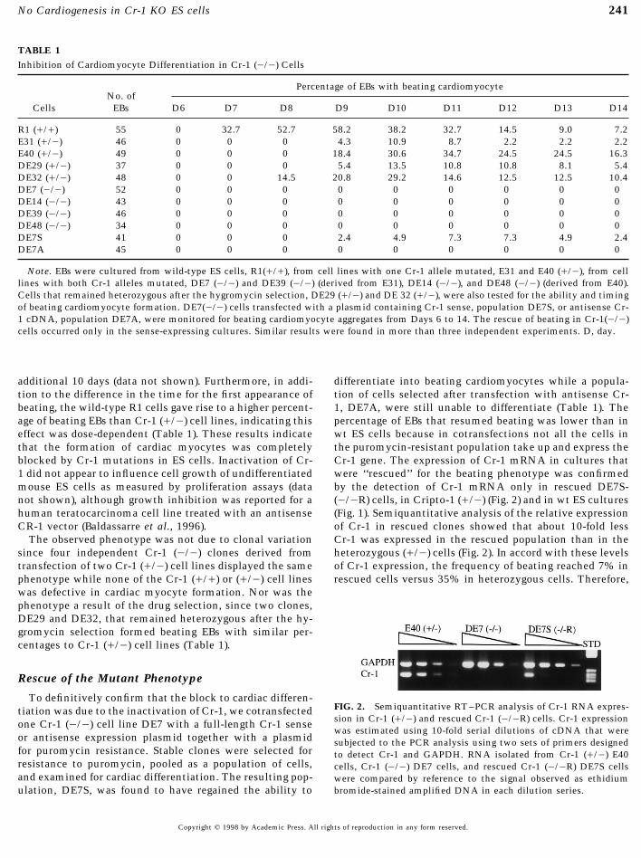
TABLE 1Inhibition of Cardiomyocyte Differentiation in Cr-1 (0/0) Cells
Percentage of EBs with beating cardiomyocyteNo. of
Cells EBs D6 D7 D8 D9 D10 D11 D12 D13 D14
R1 (///) 55 0 32.7 52.7 58.2 38.2 32.7 14.5 9.0 7.2E31 (//0) 46 0 0 0 4.3 10.9 8.7 2.2 2.2 2.2E40 (//0) 49 0 0 0 18.4 30.6 34.7 24.5 24.5 16.3DE29 (//0) 37 0 0 0 5.4 13.5 10.8 10.8 8.1 5.4DE32 (//0) 48 0 0 14.5 20.8 29.2 14.6 12.5 12.5 10.4DE7 (0/0) 52 0 0 0 0 0 0 0 0 0DE14 (0/0) 43 0 0 0 0 0 0 0 0 0DE39 (0/0) 46 0 0 0 0 0 0 0 0 0DE48 (0/0) 34 0 0 0 0 0 0 0 0 0DE7S 41 0 0 0 2.4 4.9 7.3 7.3 4.9 2.4DE7A 45 0 0 0 0 0 0 0 0 0
Note. EBs were cultured from wild-type ES cells, R1(///), from cell lines with one Cr-1 allele mutated, E31 and E40 (//0), from celllines with both Cr-1 alleles mutated, DE7 (0/0) and DE39 (0/0) (derived from E31), DE14 (0/0), and DE48 (0/0) (derived from E40).Cells that remained heterozygous after the hygromycin selection, DE29 (//0) and DE 32 (//0), were also tested for the ability and timingof beating cardiomyocyte formation. DE7(0/0) cells transfected with a plasmid containing Cr-1 sense, population DE7S, or antisense Cr-1 cDNA, population DE7A, were monitored for beating cardiomyocyte aggregates from Days 6 to 14. The rescue of beating in Cr-1(0/0)cells occurred only in the sense-expressing cultures. Similar results were found in more than three independent experiments. D, day.
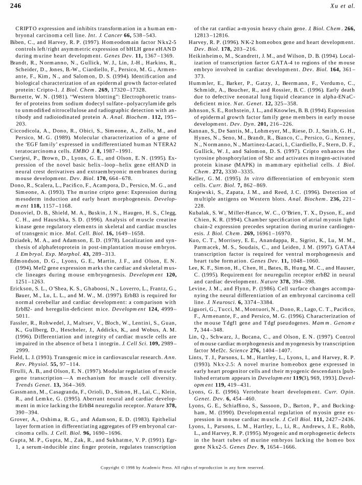
additional 10 days (data not shown). Furthermore, in addi- differentiate into beating cardiomyocytes while a popula-tion of cells selected after transfection with antisense Cr-tion to the difference in the time for the first appearance of
beating, the wild-type R1 cells gave rise to a higher percent- 1, DE7A, were still unable to differentiate (Table 1). Thepercentage of EBs that resumed beating was lower than inage of beating EBs than Cr-1 (//0) cell lines, indicating this
effect was dose-dependent (Table 1). These results indicate wt ES cells because in cotransfections not all the cells inthe puromycin-resistant population take up and express thethat the formation of cardiac myocytes was completely
blocked by Cr-1 mutations in ES cells. Inactivation of Cr- Cr-1 gene. The expression of Cr-1 mRNA in cultures thatwere ‘‘rescued’’ for the beating phenotype was confirmed1 did not appear to influence cell growth of undifferentiated
mouse ES cells as measured by proliferation assays (data by the detection of Cr-1 mRNA only in rescued DE7S-(0/0R) cells, in Cripto-1 (//0) (Fig. 2) and in wt ES culturesnot shown), although growth inhibition was reported for a
human teratocarcinoma cell line treated with an antisense (Fig. 1). Semiquantitative analysis of the relative expressionof Cr-1 in rescued clones showed that about 10-fold lessCR-1 vector (Baldassarre et al., 1996).
The observed phenotype was not due to clonal variation Cr-1 was expressed in the rescued population than in theheterozygous (//0) cells (Fig. 2). In accord with these levelssince four independent Cr-1 (0/0) clones derived from
transfection of two Cr-1 (//0) cell lines displayed the same of Cr-1 expression, the frequency of beating reached 7% inrescued cells versus 35% in heterozygous cells. Therefore,phenotype while none of the Cr-1 (///) or (//0) cell lines
was defective in cardiac myocyte formation. Nor was thephenotype a result of the drug selection, since two clones,DE29 and DE32, that remained heterozygous after the hy-gromycin selection formed beating EBs with similar per-centages to Cr-1 (//0) cell lines (Table 1).
Rescue of the Mutant Phenotype
To definitively confirm that the block to cardiac differen-FIG. 2. Semiquantitative RT–PCR analysis of Cr-1 RNA expres-tiation was due to the inactivation of Cr-1, we cotransfectedsion in Cr-1 (//0) and rescued Cr-1 (0/0R) cells. Cr-1 expressionone Cr-1 (0/0) cell line DE7 with a full-length Cr-1 sensewas estimated using 10-fold serial dilutions of cDNA that were
or antisense expression plasmid together with a plasmid subjected to the PCR analysis using two sets of primers designedfor puromycin resistance. Stable clones were selected for to detect Cr-1 and GAPDH. RNA isolated from Cr-1 (//0) E40resistance to puromycin, pooled as a population of cells, cells, Cr-1 (0/0) DE7 cells, and rescued Cr-1 (0/0R) DE7S cellsand examined for cardiac differentiation. The resulting pop- were compared by reference to the signal observed as ethidium
bromide-stained amplified DNA in each dilution series.ulation, DE7S, was found to have regained the ability to
Copyright q 1998 by Academic Press. All rights of reproduction in any form reserved.
AID DB 8862 / 6x3b$$$123 03-26-98 23:23:55 dba

242 Xu et al.
FIG. 3. (a) Western blot analysis of EBs. EBs derived from indicated cell lines at the specific day were detected for myosin heavy chain(MHC) by a monoclonal antibody MF20 (Developmental Studies Hybridoma Bank, NICHD, NIH), for MLC2A by a rabbit antiserum (akind gift from Dr. K. Chien, Dept. of Medicine, University of California at San Diego), for ErbB2 and ErbB3 by rabbit antibodies (SantaCruz Biotechnology Inc. CA), and for AFP and laminin by rabbit antisera. For a control, the blot was reprobed with antibody against b-actin or a-actinin. Signals for binding of the antibodies were detected by the enhanced chemiluminescence system (b) RT–PCR detectionof MLC 2V, ANF, and GAPDH in Day 10 EBs; brachyury, GATA4, Mef2C, Nkx2.5, Egr-1, and GAPDH in Day 6 EBs.
differentiation to cardiomyocytes is specifically restored by compared to that of wild-type EBs. In contrast, expressionof MHC in Cr-1 (0/0) EBs was undetectable. Similarly, ex-Cr-1 while differentiation is specifically blocked by the in-pression of MLC2A was observed only in the wild-type andactivation of the Cr-1 gene.Cr-1 (//0)-derived EBs but not in Cr-1 (0/0) EBs. Tran-scripts of cardiac-specific myosin light chain 2V (MLC2V)were undetectable in Cr-1 (0/0) EBs as examined by RT–Assays for Markers of Cardiac DifferentiationPCR (Fig. 3b, line 1). The expression of myosin polypeptides
To determine where Cr-1 might be acting during the pro- precedes contractile activity and so components upstreamcess of cardiac differentiation, we examined the expression of the myosin induction pathway must be affected. An earlyof the myosin heavy chain (MHC) and the cardiac-specific marker of muscle cell differentiation, desmin, the interme-myosin light chain 2A (MLC2A), two major contractile pro- diate filament protein, was present as transcripts in bothteins of cardiomyocytes, in 10-day old EBs from each cell wild-type and mutant cell cultures (data not shown), whiletype by Western blotting. As shown in Fig. 3a, the strongest transcripts of a later marker of heart differentiation, atrialsignal for MHC was detected in EBs derived from wild- natriuretic factor (ANF) were absent only in the mutant 10
day EB cultures (Fig. 3b, line 2).type R1 cells. Cr-1 (//0)-derived EBs gave weaker signals
Copyright q 1998 by Academic Press. All rights of reproduction in any form reserved.
AID DB 8862 / 6x3b$$$123 03-26-98 23:23:55 dba

243No Cardiogenesis in Cr-1 KO ES cells
As detected by RT–PCR, transcripts of precardiac meso- started strongly in the dense clusters of attached wild-typeR1 aggregates but was never observed in the Cr-1 (0/0)derm markers, transcriptional factors GATA4 (Edmondson
et al., 1994; Heikinheimo et al., 1994), were present in Cr- cells as described above, in agreement with the absence ofMLC2A in outgrowths of Cr (0/0) EBs (data not shown).1 (0/0) cell culture (Fig. 3b). The homeobox gene, Nkx2.5,
is related to tinman in Drosophila, where it is known to be Small myotubes started to form in both wild-type and mu-tant cell cultures after 2 weeks in culture and myotubesessential for heart formation and has been implicated in the
early stages of mouse cardiac myogenesis and morphogene- continued to grow into large skeletal muscle-like myotubes(Fig. 4a) that contained multiple nuclei and contracted fre-sis (Lints et al., 1993) in mice produced using knockout
technology (Biben and Harvey, 1997; Lyons et al., 1995). To quently on Days 20–25. The formation of skeletal musclewas further confirmed by expression of a skeletal-specificdetermine if Cr-1 acted upstream of the expression of this
gene, we analyzed differentiating EBs for Nkx2.5 mRNA by gene, myogenin, as detected by RT–PCR. Transcripts weredetected in RNA extracts of all differentiated ES cell linesRT–PCR. As shown in Fig. 3b, transcripts of Nkx2.5 were
detectable in Cr-1 (0/0) EBs. Egr-1, another transcription tested whether Cr-1 expressing or not (Fig. 5, top line).Differentiation into other mesoderm and into other cellfactor, which has been shown to be involved in stimulation
of cardiac MHC expression (Gupta et al., 1991), is expressed types was demonstrated by the generation of teratocarcino-mas, which form a wide range of recognizable differentiatedand is not affected by inactivation of Cr-1 (Fig. 3b). Expres-
sion of ErbB2 and ErbB3 in Cr-1 mutant EBs was not signifi- cell types. To examine differentiation in vivo, wild-type R1cells and Cr-1 (0/0) DE39 cells were injected (sc) into 129cantly different from wild-type EBs (Fig. 3a). This family of
genes are involved in later stages of cardiac development SvJ mice. After 3 weeks, tumors formed from both the mu-tant cells and wild-type cells and examination of hematoxy-because inactivation of ErbB2, ErbB3, ErbB4, and heregulin
in mice leads to inhibition of cardiac muscle trabeculae lin and eosin-stained sections indicated that the teratocarci-nomas contained many cell types. Dermis and squamousformation and causes midgestational death (Erickson et al.,
1997; Gassmann et al., 1995; Lee et al., 1995; Meyer and epidermis (Fig. 4c), cartilage (Fig. 4d), and epithelioid organs(Fig. 4e) were observed in teratocarcinomas derived fromBirchmeier, 1995).
The muscle-specific actin genes occur in four isoforms in both cell lines. Therefore, inactivation of Cr-1 did not affectdifferentiation of other mesoderm cell types in spite of thecardiac, skeletal, enteric, and vascular muscle cells. Their
ontogenesis has been described in ES cells and cardiac a- inability to differentiate into cardiomyocytes.To test if the mutant cells can differentiate into neurons,actin; transcripts appear on Day 5 of differentiation (Ng et
al., 1997). We used the same experimental approach to assay an ectoderm-derived cell type, the EBs were treated withtrans-retinoic acid which enhances neural differentiationfor the appearance of cardiac a-actin transcripts in ES and
differentiated EBs. In contrast to Ng et al., we found that (Levine and Flynn, 1986) and then plated on gelatin-coateddishes. One week after plating, cells with neuron-like mor-even undifferentiated ES cells express transcripts for a-car-
diac actin and continue to do so throughout the time course phology were found in the culture. Figure 4b shows neuriteoutgrowths in the Cr-1 (0/0) cell culture 2 weeks afterof differentiation. This was tested using the same condi-
tions for RT–PCR as described by Ng et al., as well as using plating. The axonal-like outgrowths expressed neurofila-ment transcripts because RT–PCR assays were positivedifferent primers under more stringent conditions (data not
shown). The same positive result was obtained for both Cr- (Fig. 5, lower panel) in differentiated cell cultures irrespec-tive of Cr-1 expression.1 (0/0) and Cr(///) genotypes, indicating that this gene is
poorly regulated in ES cells, and is not a good marker for To determine if inactivation of Cr-1 affects the differenti-ation of endoderm, the expression of a-fetoprotein (AFP), acardiac specific differentiation.marker for visceral endoderm cells, and laminin, a markerfor parietal endoderm cells among other tissues, was not
Assays for Markers of Other Differentiated Tissues significantly different in mutant EBs at Day 8 from thosein wild-type cells as determined by Western blotting analy-To test the possibility that Cr-1 disrupts early stages of
mesoderm differentiation, we examined the expression of sis (Fig. 3a). Thus, Cr-1 is not required for differentiation ofendoderm.an early mesoderm marker, brachyury (T) (Wilkinson et al.,
1990), since cardiomyocytes are mesoderm-derived. Asshown in Fig. 3b, brachyury transcripts were present in 6-day EBs derived from all cell lines tested, including wild- DISCUSSIONtype R1, two Cr-1 (//0) cell lines, and four Cr-1 (0/0) celllines as detected by RT–PCR. Therefore, inactivation of Cr- Functional and biochemical differentiation is readily
studied using ES cells because of their ability to differentiate1 did not affect expression of this early mesoderm marker.We then evaluated the role of Cr-1 on the generation of into a range of cell types in culture and into teratocarcino-
mas in adult hosts. The in vitro model of cardiogenesisother mesoderm-derived cell types such as skeletal muscleby culturing EBs on gelatin-coated dishes. Both wild-type using ES cells has been well characterized and the temporal
stages in cardiomyocyte differentiation and function haveand mutant EBs attached to the dishes within 1 day anddifferentiating cells grew outward. Cardiac contraction been mapped, although spatial signals and morphogenesis
Copyright q 1998 by Academic Press. All rights of reproduction in any form reserved.
AID DB 8862 / 6x3b$$$123 03-26-98 23:23:55 dba

244 Xu et al.
FIG. 4. In vitro and in vivo differentiation of Cr-1 (0/0) cells. (a) Differentiation of Cr-1 (0/0) cells into skeletal muscle. EBs derivedfrom the Cr-1 (0/0) cell line DE39 were formed by hanging drops and transferred to gelatin-coated dishes on Day 5. Strongly contractingmyotubes around the attached EBs were observed after culture for an additional 2 weeks. (b) Differentiation of Cr-1 (0/0) cells intoneurons. The EBs were also treated with 5 1 1007 M retinoic acid on Days 5–8 and transferred to gelatin-coated dishes after partialdissociation with trypsin–EDTA. Neurons were formed after plating. (c, d, e) Stained sections (hematoxylin and eosin) of teratocarcinomasderived from Cr-1 (0/0) ES cells. The tumors derived from Cr-1 (0/0) DE39 cells were fixed, embedded, and sectioned at 5 mm. Sectionsstained with hematoxylin and eosin contained readily recognizable dermis and squamous epidermis (c), cartilage (d), and epithelioid organs(e). Bar, 100 mm.
are lacking in this model (Fassler et al., 1996; Maltsev et cells to the cardiac lineage (reviewed in Baker and Lyons,1996). However, inactivation of these genes in mice did notal., 1993; Metzger et al., 1996; Miller-Hance et al., 1993;
Wobus et al., 1995). affect differentiation of cardiomyocytes, but affected laterevents in cardiogenesis (Lyons, 1996; Olson and Srivastava,Based on our results of in vitro and in vivo differentiation
of ES cells, we concluded that disruption of Cr-1 results in 1996). All of the transcription factors that we assayed thatwere shown to be involved in cardiac development werea specific defect in cardiac differentiation but there is no
detectable defect in differentiation of other mesodermal, transcribed in Cripto null ES cell cultures. Therefore Criptoacts independently of the expression of these genes. Otherendodermal and ectodermal cell types. These results em-
phasize a potentially important function for Cr-1 in cardio- than bone morphogenetic protein (BMP4) in chicks (Schul-theiss et al., 1997) and mouse (Winnier et al., 1995) andmyocyte differentiation at an early stage of cardiac develop-
ment, which is consistent with predictions based on the hepatocyte growth factor (HGF) in mouse (Rappolee et al.,1996) very little is known about how growth factors affectexpression pattern of Cr-1 during mouse embryogenesis.
Several transcription factor genes expressed in precardiac early cardiogenesis.The use of ES cells to model differentiation in vitro allowsmesoderm, such as Nkx2.5 (Biben and Harvey, 1997; Har-
vey, 1996), Mef2C (Lyons et al., 1995); Edmondson et al., the identification of induced gene activity in a temporalfashion but, except for a few good examples, such as neu-1994) (Lin et al., 1997), GATA4 (Heikinheimo et al., 1994;
Kuo et al., 1997; Molkentin et al., 1997), eHAND (Cserjesi ronal differentiation (Levine and Flynn, 1986), it is an inade-quate model for defining the events in a single pathwayet al., 1995), and dHAND (Srivastava et al., 1997) were pre-
dicted to be responsible for the commitment of mesodermal leading to defined cell types. This is because, without ma-
Copyright q 1998 by Academic Press. All rights of reproduction in any form reserved.
AID DB 8862 / 6x3b$$$124 03-26-98 23:23:55 dba

245No Cardiogenesis in Cr-1 KO ES cells
members and serum response factor (SRF), and it is thoughtthat specific combinations of factors modulate the type andactivities of the developing muscle cells (Firulli and Olson,1997). We describe here for the first time that inactivation ofthe growth factor gene, Cr-1, results specifically in defectiveearly differentiation of cardiomyocytes due to the absenceof contractile muscle proteins. This phenotype was specificto cardiogenesis because no other major tissue type wasabsent. The abrogation of the Cripto-1 gene was the causeof the phenotype because the reexpression of exogenousCripto-1 in the ‘‘knockout’’ cells reversed the mutant phe-
FIG. 5. RT–PCR analysis of myogenin and neurofilament expres- notype. We speculate that Cripto-1 may be a master switchsion in EB outgrowths. Cr-1 (///), Cr-1 (//0), and Cr-1 (0/0) cell gene that activates the myosin genes in the cardiomyocyteaggregates (EBs) were induced to differentiate into the structures via activation of the Ras/MAPK pathway (Kannan et al.,shown in Fig. 4a, skeletal muscle, or Fig. 4b, neurons (see Materials 1997) that could lead to activation of the SRF gene (Dono-and Methods). The cDNA samples from differentiated cultures
viel et al., 1996) and other type-specific combinatorial tran-were processed by PCR using primers specific for myogenin orscription factor genes.neurofilament. The predicted DNA bands were produced in all cul-
The unique specificity of Cripto-1 for cardiac cell differen-tures irrespective of Cr-1 expression.tiation is unusual because most factors activate both cardiacand skeletal muscle lineages. Possibly the EB provides thecorrect juxtaposition of primitive endoderm overlaying pre-cardiac mesoderm, a combination shown to be heart-induc-nipulation, multiple differentiation pathways occur simul-
taneously. Fortunately for this analysis, cardiomyocyte dif- tive in Xenopus (Nascone and Mercola, 1995). The result isthat Cr-1 is produced and this, in turn, induces unknownferentiation is one of the earliest events and these cells start
to beat on the seventh day of EB differentiation, reaching target genes which in combination lead to cardiomyogen-esis.an incidence of ú50% of total EBs. But even single EBs
contain mixtures of several cell types and these differ be- To devise strategies for cardiac muscle cell transplanta-tion in heart disease treatments (Field, 1993) it will be help-tween EBs and between cultures. Our finding that transcrip-
tion factors thought to be specific to cardiac lineage are ful to define how specific growth factors such as Cr-1 pro-mote cardiac lineages. Thus, these Cr-1 (0/0) and otherexpressed in this mixture of cell types does not allow assign-
ment of activities that are downstream or upstream of Cr- targeted ES cell lines will provide a good system for studyingspecification and differentiation of the cardiac lineage and1 in cardiomyogenesis because independent pathways are
mixed together. For instance, in our cultures there was al- may provide information for therapy programs aimed to-ward regeneration of myocardium.ways AFP-producing visceral endoderm, and these cells also
express GATA-4 (Heikinheimo et al., 1994). Primitive endo-derm and thyroid primordium as well as precardiomyocytesexpress Nkx2.5 (Lints et al., 1993). Mef2c is not specific ACKNOWLEDGMENTSto cardiac muscle since a low amount is also expressed inembryonic skeletal muscle and in neural crest cells (Ed- We thank the ES facility for gancyclovir, Dr. R. Oshima at thismondson et al., 1994). Therefore, we found that several Institute for the puromycin plasmid, and Drs. Carol MacLeod, Ce-
cilia Hertig, and Ken Chien (UCSD) for providing materials and‘‘cardiac-specifying’’ transcription factors are expressed inexcellent advice. Grant support from the Department of Defense,differentiating EBs that are unable to produce cardiac mus-Grant DAMD17-94-J-4286 to E.D.A.) and from the Italian Associa-cle. Indeed, transcripts for cardiac a-actin were detected intion for Cancer (AIRC) (to M.G.P.) is gratefully acknowledged. Thisundifferentiated ES cells. This may have been due to leakywork was done during the tenure of a research fellowship (97-71)regulation of this gene and/or due to the presence of a fewfrom the American Heart Association, California Affiliate (to C.X.).
remaining feeder cells. For example, NIH 3T3 cells (mouseembryo-derived fibroblasts) express high levels of Nkx2.5(Lints et al., 1993). In our studies, feeder cells were removed REFERENCESby two rounds of differential plating. Fibroblast feeder cellsin FBS-containing medium attach rapidly to plastic in con-
Bain, G., Kitchens, D., Yao, M., Huettner, J. E., and Gottlieb, D. I.trast to ES cells, which are then collected from the superna-(1995). Embryonic stem cells express neuronal properties in vitro.tant medium. It is therefore unlikely that feeder cell con-Dev. Biol. 168, 342–357.
tamination accounts for the presence of high levels of tran- Baker, R. K., and Lyons, G. E. (1996). Embryonic stem cells and inscripts for cardiac a-actin. vitro muscle development. Curr. Top. Dev. Biol. 33, 263–279.
The activation of muscle-specific genes, such as the mus- Baldassarre, G., Bianco, C., Tortora, G., Ruggiero, A., Moasser, M.,cle myosins and actins, is under the control of a large num- Dmitrovsky, E., Bianco, A. R., and Ciardiello, F. (1996). Transfec-
tion with a CRIPTO anti-sense plasmid suppresses endogenousber of transcription factors, usually including MEF2 family
Copyright q 1998 by Academic Press. All rights of reproduction in any form reserved.
AID DB 8862 / 6x3b$$$124 03-26-98 23:23:55 dba
246 Xu et al.
CRIPTO expression and inhibits transformation in a human em- of the rat cardiac a-myosin heavy chain gene. J. Biol. Chem. 266,12813–12816.bryonal carcinoma cell line. Int. J. Cancer 66, 538–543.
Biben, C., and Harvey, R. P. (1997). Homeodomain factor Nkx2-5 Harvey, R. P. (1996). NK-2 homeobox gene and heart development.Dev. Biol. 178, 203–216.controls left/right asymmetric expression of bHLH gene eHAND
during murine heart development. Genes Dev. 11, 1367–1369. Heikinheimo, M., Scandrett, J. M., and Wilson, D. B. (1994). Local-ization of transcription factor GATA-4 to regions of the mouseBrandt, R., Normanno, N., Gullick, W. J., Lin, J.-H., Harkins, R.,
Scheider, D., Jones, B.-W., Ciardiello, F., Persico, M. G., Armen- embryo involved in cardiac development. Dev. Biol. 164, 361–373.ante, F., Kim, N., and Salomon, D. S. (1994). Identification and
biological characterization of an epidermal growth factor-related Hummler, E., Barker, P., Gatzy, J., Beermann, F., Verdumo, C.,protein: Cripto-1. J. Biol. Chem. 269, 17320–17328. Schmidt, A., Boucher, R., and Rossier, B. C. (1996). Early death
due to defective neonatal lung liquid clearance in alpha-ENaC-Burnette, W. N. (1981). ‘‘Western blotting’’: Electrophoretic trans-fer of proteins from sodium dodecyl sulfate–polyacrylamide gels deficient mice. Nat. Genet. 12, 325–358.to unmodified nitrocellulose and radiographic detection with an- Johnson, S. E., Rothstein, J. L., and Knowles, B. B. (1994). Expressiontibody and radioiodinated protein A. Anal. Biochem. 112, 195– of epidermal growth factor family gene members in early mouse203. development. Dev. Dyn. 201, 216–226.
Ciccodicola, A., Dono, R., Obici, S., Simeone, A., Zollo, M., and Kannan, S., De Santis, M., Lohmeyer, M., Riese, D. J., Smith, G. H.,Persico, M. G. (1989). Molecular characterization of a gene of Hynes, N., Seno, M., Brandt, R., Bianco, C., Persico, G., Kenney,the ‘EGF family’ expressed in undifferentiated human NTERA2 N., Normanno, N., Martinez-Lacaci, I., Ciardiello, F., Stern, D. F.,teratocarcinoma cells. EMBO J. 8, 1987–1991. Gullick, W. J., and Salomon, D. S. (1997). Cripto enhances the
tyrosine phosphorylation of Shc and activates mitogen-activatedCserjesi, P., Brown, D., Lyons, G. E., and Olson, E. N. (1995). Ex-pression of the novel basic helix–loop–helix gene eHAND in protein kinase (MAPK) in mammary epithelial cells. J. Biol.
Chem. 272, 3330–3335.neural crest derivatives and extraembryonic membranes duringmouse development. Dev. Biol. 170, 664–678. Keller, G. M. (1995). In vitro differentiation of embryonic stem
cells. Curr. Biol. 7, 862–869.Dono, R., Scalera, L., Pacifico, F., Acampora, D., Persico, M. G., andSimeone, A. (1993). The murine cripto gene: Expression during Krajewski, S., Zapata, J. M., and Reed, J. C. (1996). Detection of
multiple antigens on Western blots. Anal. Biochem. 236, 221–mesoderm induction and early heart morphogenesis. Develop-ment 118, 1157–1168. 228.
Kubalak, S. W., Miller-Hance, W. C., O’Brien, T. X., Dyson, E., andDonoviel, D. B., Shield, M. A., Buskin, J. N., Haugen, H. S., Clegg,C. H., and Hauschka, S. D. (1996). Analysis of muscle creatine Chien, K. R. (1994). Chamber specification of atrial myosin light
chain-2 expression precedes septation during murine cardiogen-kinase gene regulatory elements in skeletal and cardiac musclesof transgenic mice. Mol. Cell. Biol. 16, 1649–1658. esis. J. Biol. Chem. 269, 16961–16970.
Kuo, C. T., Morrisey, E. E., Anandappa, R., Sigrist, K., Lu, M. M.,Dziadek, M. A., and Adamson, E. D. (1978). Localization and syn-thesis of alphafetoprotein in post-implantation mouse embryos. Parmacek, M. S., Soudais, C., and Leiden, J. M. (1997). GATA4
transcription factor is required for ventral morphogenesis andJ. Embryol. Exp. Morphol. 43, 289–313.heart tube formation. Genes Dev. 11, 1048–1060.Edmondson, D. G., Lyons, G. E., Martin, J. F., and Olson, E. N.
(1994). Mef2 gene expression marks the cardiac and skeletal mus- Lee, K. F., Simon, H., Chen, H., Bates, B., Hung, M. C., and Hauser,C. (1995). Requirement for neuregulin receptor erbB2 in neuralcle lineages during mouse embryogenesis. Development 120,
1251–1263. and cardiac development. Nature 378, 394–398.Levine, J. M., and Flynn, P. (1986). Cell surface changes accompa-Erickson, S. L., O’Shea, K. S., Ghaboosi, N., Loverro, L., Frantz, G.,
Bauer, M., Lu, L. L., and M. W., M. (1997). ErbB3 is required for nying the neural differentiation of an embryonal carcinoma cellline. J. Neurosci. 6, 3374–3384.normal cerebellar and cardiac development: a comparison with
ErbB2- and heregulin-deficient mice. Development 124, 4999– Liguori, G., Tucci, M., Montuori, N., Dono, R., Lago, C. T., Pacifico,5011. F., Armenante, F., and Persico, M. G. (1996). Characterization of
the mouse Tdgf1 gene and Tdgf pseudogenes. Mamm. GenomeFassler, R., Rohwedel, J., Maltsev, V., Bloch, W., Lentini, S., Guan,K., Gullberg, D., Hescheler, J., Addicks, K., and Wobus, A. M. 7, 344–348.(1996). Differentiation and integrity of cardiac muscle cells are Lin, Q., Schwarz, J., Bucana, C., and Olson, E. N. (1997). Controlimpaired in the absence of beta 1 integrin. J. Cell Sci. 109, 2989– of mouse cardiac morphogenesis and myogenesis by transcription2999. factor Mef2c. Science 276, 1404–1407.
Field, L. J. (1993). Transgenic mice in cardiovascular research. Ann. Lints, T. J., Parsons, L. M., Hartley, L., Lyons, I., and Harvey, R. P.Rev. Physiol. 55, 97–114. (1993). Nkx-2.5: A novel murine homeobox gene expressed in
early heart progenitor cells and their myogenic descendants [pub-Firulli, A. B., and Olson, E. N. (1997). Modular regulation of musclegene transcription—A mechanism for muscle cell diversity. lished erratum appears in Development 119(3), 969, 1993]. Devel-
opment 119, 419–431.Trends Genet. 13, 364–369.Gassmann, M., Casagranda, F., Orioli, D., Simon, H., Lai, C., Klein, Lyons, G. E. (1996). Vertebrate heart development. Curr. Opin.
Genet. Dev. 6, 454–460.R., and Lemke, G. (1995). Aberrant neural and cardiac develop-ment in mice lacking the ErbB4 neuregulin receptor. Nature 378, Lyons, G. E., Schiaffino, S., Sassoon, D., Barton, P., and Bucking-390–394. ham, M. (1990). Developmental regulation of myosin gene ex-
pression in mouse cardiac muscle. J. Cell Biol. 111, 2427–2436.Grover, A., Oshima, R. G., and Adamson, E. D. (1983). Epitheliallayer formation in differentiating aggregates of F9 embryonal car- Lyons, I., Parsons, L. M., Hartley, L., Li, R., Andrews, J. E., Robb,cinoma cells. J. Cell. Biol. 96, 1690–1696. L., and Harvey, R. P. (1995). Myogenic and morphogenetic defects
in the heart tubes of murine embryos lacking the homeo boxGupta, M. P., Gupta, M., Zak, R., and Sukhatme, V. P. (1991). Egr-1, a serum-inducible zinc finger protein, regulates transcription gene Nkx2-5. Genes Dev. 9, 1654–1666.
Copyright q 1998 by Academic Press. All rights of reproduction in any form reserved.
AID DB 8862 / 6x3b$$$125 03-26-98 23:23:55 dba

247No Cardiogenesis in Cr-1 KO ES cells
Maltsev, V. A., Rohwedel, J., Hescheler, J., and Wobus, A. M. (1993). factor and its receptor are expressed in cardiac myocytes duringearly cardiogenesis. Circ. Res. 78, 1028–1036.Embryonic stem cells differentiate in vitro into cardiomyocytes
Sassoon, D. A., Garner, I., and Buckingham, M. (1988). Transcriptsrepresenting sinusnodal, atrial and ventricular cell types. Mech.of alpha-cardiac and alpha-skeletal actins are early markers forDev. 44, 41–50.myogenesis in the mouse embryo. Development 104, 155–164.Martin, J. F., Schwarz, J. J., and Olson, E. N. (1993). Myocyte en-
Schultheiss, T. M., Burch, J. B., and Lassar, A. B. (1997). A role forhancer factor (MEF) 2C: A tissue-restricted member of the MEF-bone morphogenetic proteins in the induction of cardiac myogen-2 family of transcription factors. Proc. Natl. Acad. Sci. USA 90,esis. Genes Dev. 11, 451–462.5282–5286.
Shen, M. M., Wang, H., and Leder, P. (1997). A differential displayMetzger, J. M., Lin, W. I., and Samuelson, L. C. (1996). Vital stain-strategy identifies Cryptic, a novel EGF-related gene expresseding of cardiac myocytes during embryonic stem cell cardiogenesisin the axial and lateral mesoderm during mouse gastrulation.in vitro. Circ. Res. 78, 547–552.Development 124, 429–442.Meyer, D., and Birchmeier, C. (1995). Multiple essential functions
Srivastava, D., Thomas, T., Lin, Q., Kirby, M. L., Brown, D., andof neuregulin in development [see comments]. Nature 378, 386–Olson, E. N. (1997). Regulation of cardiac mesodermal and neural390.crest development by the Bhlh transcription factor, Dhand. Nat.Miller-Hance, W. C., LaCorbiere, M., Fuller, S. J., Evans, S. M., Ly-Genet. 16, 154–160.ons, G., Schmidt, C., Robbins, J., and Chien, K. R. (1993). In vitro
Tybulewicz, V. L., Crawford, C. E., Jackson, P. K., Bronson, R. T.,chamber specification during embryonic stem cell cardiogenesis.and Mulligan, R. C. (1991). Neonatal lethality and lymphopeniaExpression of the ventricular myosin light chain-2 gene is inde-in mice with a homozygous disruption of the c-abl proto-onco-
pendent of heart tube formation. J. Biol. Chem. 268, 25244–gene. Cell 65, 1153–1163.
25252.Weitzer, G., Milner, D. J., Kim, J. U., Bradley, A., and Capetanaki,
Molkentin, J. D., Lin, Q., Duncan, S. A., and Olson, E. N. (1997). Y. (1995). Cytoskeletal control of myogenesis: A desmin nullRequirement of the transcription factor Gata4 for heart tube for- mutation blocks the myogenic pathway during embryonic stemmation and ventral morphogenesis. Genes Dev. 11, 1061–1072. cell differentiation. Dev. Biol. 172, 422–439.
Morkin, E. (1993). Regulation of myosin heavy chain genes in the Wilkinson, D. G., Bhatt, S., and Herrmann, B. G. (1990). Expressionheart. Circulation 87, 1451–1460. pattern of the mouse T gene and its role in mesoderm formation.
Nagy, A., Rossant, J., Nagy, R., Abramow-Newerly, W., and Roder, Nature 343, 657–659.J. C. (1993). Derivation of completely cell culture-derived mice Winnier, G., Blessing, M., Labosky, P. A., and Hogan, B. L. (1995).from early-passage embryonic stem cells. Proc. Natl. Acad. Sci. Bone morphogenetic protein-4 is required for mesoderm forma-USA 90, 8424–8428. tion and patterning in the mouse. Genes Dev. 9, 2105–2116.
Nascone, N., and Mercola, M. (1995). An inductive role for the Wobus, A. M., Rohwedel, J., Maltsev, V., and Hescheler, J. (1995).endoderm in Xenopus cardiogenesis. Development 121, 515–523. Development of cardiomyocytes expressing cardiac-specific
Ng, W. A., Doetschman, T., Robbins, J., and Lessard, J. L. (1997). genes, action potentials, and ionic channels during embryonicMuscle isoactin expression during in vitro differentiation of mu- stem cell-derived cardiogenesis. Ann. NY Acad. Sci. 752, 460–
469.rine embryonic stem cells. Pediatr. Res. 41, 285–292.Wobus, A. M., Wallukat, G., and Hescheler, J. (1991). PluripotentO’Brien, T. X., Lee, K. J., and Chien, K. R. (1993). Positional speci-
mouse embryonic stem cells are able to differentiate into cardio-fication of ventricular myosin light chain 2 expression in themyocytes expressing chronotropic responses to adrenergic andprimitive murine heart tube. Proc. Natl. Acad. Sci. USA 90,cholinergic agents and Ca2/ channel blockers. Differentiation 48,5157–5161.173–182.Olson, E. N., and Srivastava, D. (1996). Molecular pathways con-
trolling heart development. Science 272, 671–676. Received for publication August 19, 1997Accepted January 25, 1998Rappolee, D. A., Iyer, A., and Patel, Y. (1996). Hepatocyte growth
Copyright q 1998 by Academic Press. All rights of reproduction in any form reserved.
AID DB 8862 / 6x3b$$$125 03-26-98 23:23:55 dba