SpecializedMechanosensoryNociceptorsMediating...
16
Article Specialized Mechanosensory Nociceptors Mediating Rapid Responses to Hair Pull Highlights d Identification of class of mechanoreceptors that make circumferential endings d Optogenetic activation of Circ-HTMRs evokes protective and avoidance behaviors d Circ-HTMRs are fast-conducting mechano-nociceptors that adapt slowly d Circ-HTMRs respond to single hairs and have spatially organized receptive fields Authors Nima Ghitani, Arnab Barik, Marcin Szczot, ..., Claire E. Le Pichon, Michael J. Krashes, Alexander T. Chesler Correspondence [email protected] In Brief Ghitani et al. identify a class of mechano- nociceptors that make circumferential endings around hair follicles. They demonstrate that these neurons respond to high-threshold mechanical stimuli, such as hair pulling, and that their activation evokes rapid protective and avoidance behaviors. Ghitani et al., 2017, Neuron 95, 944–954 August 16, 2017 Published by Elsevier Inc. http://dx.doi.org/10.1016/j.neuron.2017.07.024
Transcript of SpecializedMechanosensoryNociceptorsMediating...

Article
SpecializedMechanosens
ory NociceptorsMediatingRapid Responses to Hair PullHighlights
d Identification of class of mechanoreceptors that make
circumferential endings
d Optogenetic activation of Circ-HTMRs evokes protective and
avoidance behaviors
d Circ-HTMRs are fast-conducting mechano-nociceptors that
adapt slowly
d Circ-HTMRs respond to single hairs and have spatially
organized receptive fields
Ghitani et al., 2017, Neuron 95, 944–954August 16, 2017 Published by Elsevier Inc.http://dx.doi.org/10.1016/j.neuron.2017.07.024
Authors
Nima Ghitani, Arnab Barik,
Marcin Szczot, ..., Claire E. Le Pichon,
Michael J. Krashes,
Alexander T. Chesler
In Brief
Ghitani et al. identify a class of mechano-
nociceptors that make circumferential
endings around hair follicles. They
demonstrate that these neurons respond
to high-threshold mechanical stimuli,
such as hair pulling, and that their
activation evokes rapid protective and
avoidance behaviors.

Neuron
Article
Specialized Mechanosensory NociceptorsMediating Rapid Responses to Hair PullNima Ghitani,1,4 Arnab Barik,1,4 Marcin Szczot,1,4 James H. Thompson,1 Chia Li,2 Claire E. Le Pichon,3
Michael J. Krashes,2 and Alexander T. Chesler1,5,*1National Center for Complementary and Integrative Health (NCCIH)2National Institute of Diabetes and Digestive and Kidney Diseases (NIDDK)3National Institute of Child Health and Human Development (NICHD)
National Institutes of Health (NIH), Bethesda MD, USA4These authors contributed equally5Lead Contact*Correspondence: [email protected]
http://dx.doi.org/10.1016/j.neuron.2017.07.024
SUMMARY
The somatosensory system provides animals withthe ability to detect, distinguish, and respond todiverse thermal, mechanical, and irritating stimuli.While there has been progress in defining classesof neurons underlying temperature sensation andgentle touch, less is known about the neuronsspecific for mechanical pain. Here, we use in vivofunctional imaging to identify a class of cutaneoussensory neurons that are selectively activated byhigh-threshold mechanical stimulation (HTMRs). Weshow that their optogenetic excitation evokes rapidprotective and avoidance behaviors. Unlike othernociceptors, these HTMRs are fast-conductingAd-fibers with highly specialized circumferential end-ings wrapping the base of individual hair follicles.Notably, we find that Ad-HTMRs innervate uniquebut overlapping fields and can be activated by stimulias precise as the pulling of a single hair. Together, thedistinctive features of this class of Ad-HTMRs appearoptimized for accurate and rapid localization of me-chanical pain.
INTRODUCTION
Peripheral sensory neurons innervating the skin provide animals
with important details about their environment. For humans, the
activity of these neurons is responsible for our sense of touch
and evokes a range of emotional responses from pleasure to
pain. Our understanding of how sensory neurons detect diverse
stimuli is rooted in work from the late 19th century. In particular,
Maximilian von Frey is credited with formalizing the idea that
there are four sensations (touch, heat, cold, and pain), each de-
tected by morphologically distinct receptor types (Norrsell et al.,
1999). Von Frey posited that touch (including pressure and vibra-
tion) is detected by afferents with highly specialized endings
(such as corpuscles and hair-associated endings), while pain is
944 Neuron 95, 944–954, August 16, 2017 Published by Elsevier Inc.
sensed by afferents that make free nerve endings. This model
has proved to be remarkably prescient. The past two decades
have provided definitive links between specific classes of sen-
sory neurons, the detection of particular stimuli and distinct
afferent morphologies (Le Pichon and Chesler, 2014).
Perhaps the best example of sensory specificity is the activa-
tion of neurons expressing the heat-gated ion channel Tran-
sient Receptor Potential Cation Channel Subfamily V Member
1 (Trpv1) by capsaicin (Caterina et al., 1997), which is sufficient
to elicit the sensation of burning, whereas the ablation of these
neurons causes insensitivity to heat (Pogorzala et al., 2013).
Genetic labeling of Trpv1-expressing neurons showed, just as
predicted for ‘‘pain’’ sensation, that these neurons have free
nerve endings in the superficial layers of skin (Cavanaugh
et al., 2011). Similarly, molecular studies have also confirmed
that low-threshold mechanoreceptors (LTMRs) have special-
ized nerve endings with two distinct morphologies along the
shaft of hair follicles, lanceolate endings running parallel to
the hair shaft (Li et al., 2011), and circumferential endings wrap-
ping around each hair follicle (Bai et al., 2015; Bardoni
et al., 2014).
Despite these advances, decades of electrophysiological re-
cordings have demonstrated the existence of populations of
sensory neurons that remain poorly understood. These include
several types of broadly tuned, polymodal neurons and, notably,
classes of neurons that respond exclusively to high-threshold
mechanical stimuli (HTMRs) that are likely critically important
for pain (Bessou and Perl, 1969; Schmidt et al., 1995). Further-
more, molecules commonly used to define classes of sensory
neurons seem to target more than one subtype. For example,
neurons that are immuno-positive for Calcitonin gene-related
peptide (CGRP) have long been classified as peptidergic noci-
ceptors (Gibson et al., 1984). Ablation of neurons expressing
Calca, one of two genes that encode CGRP, has shown that
they are critical for thermosensation but not required for me-
chanical pain behaviors (McCoy et al., 2013). However, electrical
recording from CGRP neurons demonstrated that many can
respond to mechanical stimulation (Lawson et al., 2002; Weyer
et al., 2015). Anatomical studies have also suggested that
CGRP neurons are likely heterogeneous. In hairy skin, CGRP-
neurons have been shown to make free nerve endings typically
A
D E
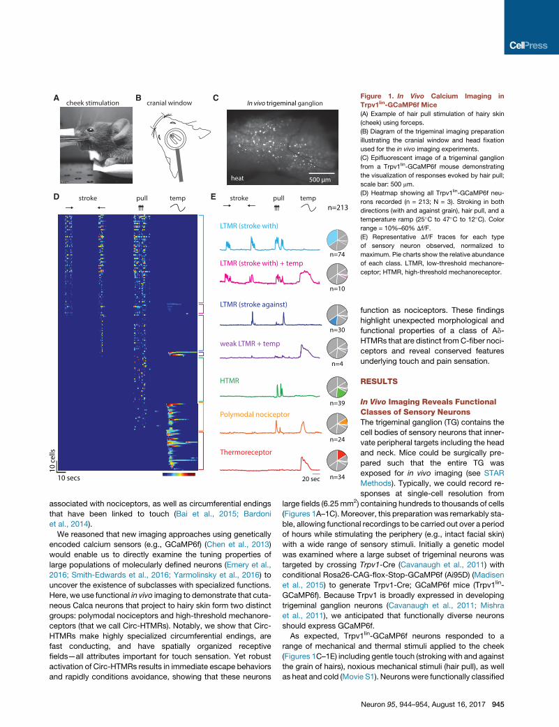
B C Figure 1. In Vivo Calcium Imaging in
Trpv1lin-GCaMP6f Mice
(A) Example of hair pull stimulation of hairy skin
(cheek) using forceps.
(B) Diagram of the trigeminal imaging preparation
illustrating the cranial window and head fixation
used for the in vivo imaging experiments.
(C) Epifluorescent image of a trigeminal ganglion
from a Trpv1lin-GCaMP6f mouse demonstrating
the visualization of responses evoked by hair pull;
scale bar: 500 mm.
(D) Heatmap showing all Trpv1lin-GCaMP6f neu-
rons recorded (n = 213; N = 3). Stroking in both
directions (with and against grain), hair pull, and a
temperature ramp (25�C to 47�C to 12�C). Colorrange = 10%–60% Df/F.
(E) Representative Df/F traces for each type
of sensory neuron observed, normalized to
maximum. Pie charts show the relative abundance
of each class. LTMR, low-threshold mechanore-
ceptor; HTMR, high-threshold mechanoreceptor.
associated with nociceptors, as well as circumferential endings
that have been linked to touch (Bai et al., 2015; Bardoni
et al., 2014).
We reasoned that new imaging approaches using genetically
encoded calcium sensors (e.g., GCaMP6f) (Chen et al., 2013)
would enable us to directly examine the tuning properties of
large populations of molecularly defined neurons (Emery et al.,
2016; Smith-Edwards et al., 2016; Yarmolinsky et al., 2016) to
uncover the existence of subclasses with specialized functions.
Here, we use functional in vivo imaging to demonstrate that cuta-
neous Calca neurons that project to hairy skin form two distinct
groups: polymodal nociceptors and high-threshold mechanore-
ceptors (that we call Circ-HTMRs). Notably, we show that Circ-
HTMRs make highly specialized circumferential endings, are
fast conducting, and have spatially organized receptive
fields—all attributes important for touch sensation. Yet robust
activation of Circ-HTMRs results in immediate escape behaviors
and rapidly conditions avoidance, showing that these neurons
function as nociceptors. These findings
highlight unexpected morphological and
functional properties of a class of Ad-
HTMRs that are distinct fromC-fiber noci-
ceptors and reveal conserved features
underlying touch and pain sensation.
RESULTS
In Vivo Imaging Reveals FunctionalClasses of Sensory NeuronsThe trigeminal ganglion (TG) contains the
cell bodies of sensory neurons that inner-
vate peripheral targets including the head
and neck. Mice could be surgically pre-
pared such that the entire TG was
exposed for in vivo imaging (see STAR
Methods). Typically, we could record re-
sponses at single-cell resolution from
large fields (6.25mm2) containing hundreds to thousands of cells
(Figures 1A–1C). Moreover, this preparation was remarkably sta-
ble, allowing functional recordings to be carried out over a period
of hours while stimulating the periphery (e.g., intact facial skin)
with a wide range of sensory stimuli. Initially a genetic model
was examined where a large subset of trigeminal neurons was
targeted by crossing Trpv1-Cre (Cavanaugh et al., 2011) with
conditional Rosa26-CAG-flox-Stop-GCaMP6f (Ai95D) (Madisen
et al., 2015) to generate Trpv1-Cre; GCaMP6f mice (Trpv1lin-
GCaMP6f). Because Trpv1 is broadly expressed in developing
trigeminal ganglion neurons (Cavanaugh et al., 2011; Mishra
et al., 2011), we anticipated that functionally diverse neurons
should express GCaMP6f.
As expected, Trpv1lin-GCaMP6f neurons responded to a
range of mechanical and thermal stimuli applied to the cheek
(Figures 1C–1E) including gentle touch (stroking with and against
the grain of hairs), noxious mechanical stimuli (hair pull), as well
as heat and cold (Movie S1). Neurons were functionally classified
Neuron 95, 944–954, August 16, 2017 945
ED
A B
heat/hair pullhair pullheat (>40C)
tdT Nefh
C
# of
neu
rons
# of
neu
rons
100 100
50 50
0 0
all heat responsive hair pull only
anti-tdT
hair pull
10-1515-20
20-2525-30
30-3535-40
40+10-15
15-2020-25
25-3030-35
35-4040+
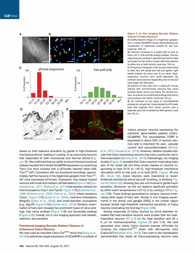
Figure 2. In Vivo Imaging Reveals Distinct
Classes of Calca Neurons
(A) Epifluorescent image of a trigeminal ganglion
from a Calca-GCaMP6f mouse demonstrating the
visualization of responses evoked by hair pull;
scale bar: 500 mm.
(B) Calcium increases in a single field of view to
heat (>40�C, left) and hair pulling (middle). The two
responses are overlaid in pseudocolor (red for heat
and green for hair pull) to reveal cells that respond
to either one or both stimuli; scale bar: 100 mm.
(C) Frequency histogram of all neurons responding
to heat (red, left panel) and hair pull (green, right
panel) ordered by soma size (5 mm bins). Heat-
responsive neurons have small diameters. By
contrast Calca neurons responding only to hair pull
have larger cell diameters.
(D) Section of hairy skin from a Calca-tdT mouse
stained with anti-tdTomato showing free nerve
endings (black arrow) just below the dermal sur-
face, as well as circumferential endings (red arrow)
surrounding a hair follicle. Scale bar: 50 mm.
(E) An example of two types of circumferential
endings at a single hair, those labeled by tdTomato
(red) that originate from Calca neurons and a
separate type that is labeled by Nefh (green); scale
bar: 25 mm.
based on their selective activation by gentle or high-threshold
mechanical stimuli, heating or cooling, or as polymodal neurons
that responded to both mechanical and thermal stimuli (Fig-
ure 1E). We confirmed that our ability to record diverse functional
classes resulted from broad GCaMP6f expression by examining
Trpv1-Cre mice crossed with a tdTomato reporter strain (Ai9;
Trpv1lin-tdT). Consistent with our functional recordings, approx-
imately half the neurons in the trigeminal ganglion from Trpv1lin-
tdT mice expressed tdTomato. Expression was biased toward
neuronswith small andmedium cell diameters (Figure S1A) (Cav-
anaugh et al., 2011; Mishra et al., 2011) that express markers for
thermoreceptors (Trpv1 and Trpm8, Figure S1D) (Caterina et al.,
1999; McKemy et al., 2002; Peier et al., 2002), irritant receptors
(Trpa1, Figure S1D) (Jordt et al., 2004), mechano-nociceptors
(MrgprD) (Zylka et al., 2005), and small-diameter nociceptors
(e.g., Npy2R, Figure S1D) (Usoskin et al., 2015). Similarly, exam-
ination of hairy skin revealed two prominent types of nerve end-
ings: free nerve endings (Figure S1B) and lanceolate endings
(Figure S1C). Overall, our in vivo imaging approach was reliable,
selective, and sensitive.
Functional Imaging Reveals Distinct Classes ofCutaneous Calca NeuronsWe next used an inducible Calca-CreERT2 driver line (Song et al.,
2012) to selectively target expression of GCaMP6f in a subset of
946 Neuron 95, 944–954, August 16, 2017
mature sensory neurons expressing the
calcitonin gene-related peptide (Calca-
GCaMP6f). The neuropeptide CGRP is
expressed in about 30% of sensory neu-
rons and is important for pain, vascular
control, and neuroinflammation (McCoy
et al., 2012; Russell et al., 2014). However, ablation studies indi-
cate that Calca-expressing neurons may have a particular role in
thermosensation (McCoy et al., 2013). Interestingly, our imaging
studies (Figure 2) revealed that Calca neurons innervating hairy
skin of the cheek fall into three broad classes of neurons re-
sponding to heat (31% to >40�C), high-threshold mechanical
stimulation (44% to hair pull), or to both (25%, Figures 2B and
2C, Movie S2). Calca neurons were insensitive to lower-
threshold mechanical stimuli (air puff, brushing, or stroking; Fig-
ure S2; Movie S2), showing they are not involved in gentle touch
sensation. Moreover, we did not observe significant activation
by either warm temperatures (<37�C) or by cooling (>13�C; Fig-ure S2B). These findings generalized to sensory neurons inner-
vating other types of hairy skin. Similar in vivo imaging experi-
ments in the dorsal root ganglia (DRG) in the lumbar region
showed similar high-threshold mechanical sensitivity of Calca
neurons innervating the hind leg (Figure S2C).
Sorting responses of Calca neurons by soma diameter re-
vealed that heat-sensitive neurons were smaller than the heat-
insensitive neurons (17 ± 2 mm for heat sensitive and 26 ±
6 mm for mechanosensitive; Figures 2C and S2A). We next
generated mice where Calca neurons express tdTomato by
crossing the Calca-CreERT2 strain with Ai9-reporter mice
(Calca-tdT) (Madisen et al., 2010). Two-color in situ hybridization
demonstrated that nearly all Calca-expressing neurons were
A
B
C
D E
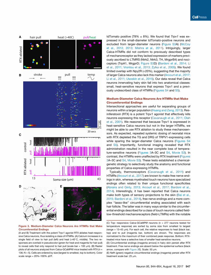
Figure 3. Medium-Diameter Calca Neurons Are HTMRs that Make
Circumferential Endings
(A and B) Treatment with the potent Trpv1 agonist RTX ablates heat-respon-
sive Calca neurons, thus isolating a class of HTMRs. (A) Calcium increases in a
single field of view to hair pull (left) and heat (>40�C, middle). The two re-
sponses are overlaid in pseudocolor (green for heat and magenta for hair pull)
to reveal cells that only respond to hair pull (scale bar = 100 mm). (B) Raster
plots of all neurons analyzed from Calca-GCaMP6f mice treated with RTX (n =
136; N = 5). Cells are ordered by size (largest to smallest, top to bottom). Color
scale range = 20%–100% Df/F.
tdTomato positive (78% ± 8%). We found that Trpv1 was ex-
pressed in the small-diameter tdTomato-positive neurons and
excluded from larger-diameter neurons (Figure S3A) (McCoy
et al., 2012, 2013; Mishra et al., 2011). Intriguingly, larger
Calca-HTMRs did not conform to previously described types
of mechanoreceptor as they lacked expression of markers previ-
ously ascribed to LTMRS (Ntrk2, Ntrk3, TH, MrgprB4) and noci-
ceptors (TrpA1, MrgprD; Figure S3D) (Bardoni et al., 2014; Li
et al., 2011; Vrontou et al., 2013; Zylka et al., 2005). We found
limited overlap with Npy2R (<20%), suggesting that the majority
of larger Calca neurons also lack this marker (Arcourt et al., 2017;
Li et al., 2011; Usoskin et al., 2015). Our data reveal that Calca
neurons innervating hairy skin fall into two anatomical classes:
small, heat-sensitive neurons that express Trpv1 and a previ-
ously undescribed class of HTMRs (Figures S4 and S5).
Medium-Diameter Calca Neurons Are HTMRs that MakeCircumferential EndingsIntersectional approaches are useful for separating groups of
neurons within a larger population (Huang and Zeng, 2013). Res-
iniferatoxin (RTX) is a potent Trpv1 agonist that effectively kills
neurons expressing this receptor (Cavanaugh et al., 2011; Olah
et al., 2001). We reasoned that because Trpv1 is expressed in
heat-sensitive Calca neurons but not in the larger HTMRs, we
might be able to use RTX ablation to study these mechanosen-
sors. As expected, repeated systemic dosing of neonatal mice
with RTX depleted the TG and DRG of TRPV1-expressing cells
while sparing the larger-diameter Calca neurons (Figures S4
and S5). Importantly, functional imaging revealed that RTX
administration resulted in the near complete loss of tempera-
ture-sensitive neurons (Figures 3A–3C and S4, Movie S3). In
contrast, the HTMRs were unaffected by RTX treatment (Figures
3A–3C and S4, Movie S3). These tests established a chemical-
genetic strategy to selectively study the anatomy and functional
properties of Calca-expressing HTMRs.
Typically, thermoreceptors (Cavanaugh et al., 2011) and
HTMRs (Arcourt et al., 2017) are known to make free nerve end-
ings in skin, whereas myelinated touch neurons have specialized
endings often related to their unique functional specificities
(Abraira and Ginty, 2013; Arcourt et al., 2017; Bardoni et al.,
2014). Interestingly, it has been reported that Calca neurons
make both types of sensory projections to the skin (Bai et al.,
2015; Bardoni et al., 2014), free nerve endings and a more com-
plex ‘‘lasso-like’’ circumferential ending associated with each
hair follicle. The latter was in many ways similar to the circumfer-
ential endings described for a class of touch neurons called field-
low-threshold mechanoreceptors (field-LTMRs) with the notable
(C) Top: responsive Calca-GCaMP6f neurons (n = 277 neurons tested for
temperature response) are sorted by soma size from smallest to largest
(range = 13–43 mm). For each cell, the relative responses to heat (black bar,
top) and to pull (magenta bar, bottom) are shown. The responses are
normalized to the maximum of their combined peak response. Bottom: RTX-
treated mice have a selective loss of smaller heat-sensitive neurons.
(D) Circumferential endings (magenta arrows) in hairy skin persist after RTX
treatment. Free nerve endings are absent below the epidermal surface (black
arrow, compare with Figure 2E). Scale: 50 mm.
(E) Nefh (green) negative circumferential endings (magenta) persist after RTX
treatment scale bar: 25 mm.
Neuron 95, 944–954, August 16, 2017 947

exception that Calca endings did not stain with NF200, a reliable
marker for A-fiber neurons (Bai et al., 2015). To study the sensory
terminals of Calca-HTMRs, we examined skin from Calca-
CreERT2 mice crossed with the Ai9-reporter strain (Calca-tdT)
(Madisen et al., 2010). Free nerve endings in superficial layers
of skin and circumferential endings associated with each hair
were tdT positive (Figures 2D and 2E). However, no reporter
expression was observed in skin cells includingMerkel cells (Fig-
ure S6A). Calca innervation in hairy skin from cheek, belly, and
back (innervated by the TG or DRG) was indistinguishable (Fig-
ures S6B and S7A). Furthermore, tdTomato afferents are
NF200 negative, demonstrating that these endings are distinct
from a recently identified class of HTMR (Figure S6C) (Arcourt
et al., 2017).
We treated Calca-tdT mice with RTX to ablate heat-sensitive
neurons. Notably, circumferential endings resisted RTX ablation
(Figures 3D and 3E; Figures S7A and S7B). In contrast, free nerve
endings, which were prominent in Calca-tdT mice, were elimi-
nated by RTX treatment (Figure 3D; Figure S7). Thus, we chose
to call these cells ‘‘Circ-HTMRs’’ to highlight that their response
properties are reminiscent of nociceptors but they have sensory
specializations normally associated with neurons involved in
touch. Notably, a recent study identified a population of fast-
conducting HTMRs using a different genetic strategy (Arcourt
et al., 2017). Unlike the Circ-HTMRs studied here, these neurons
had free nerve endings that were NF200 positive but also ap-
peared positive for CGRP. Importantly, Circ-HTMRs projecting
to the skin were never NF200 positive (Figure S6C), and free
nerve endings were not observed after RTX ablation (Figures
3D and 3E; Figure S7). Therefore, the Circ-HTMRs are distinct
from the neurons identified by Arcourt et al. (2017) and Calca-
CreER recombination does not label these neurons.
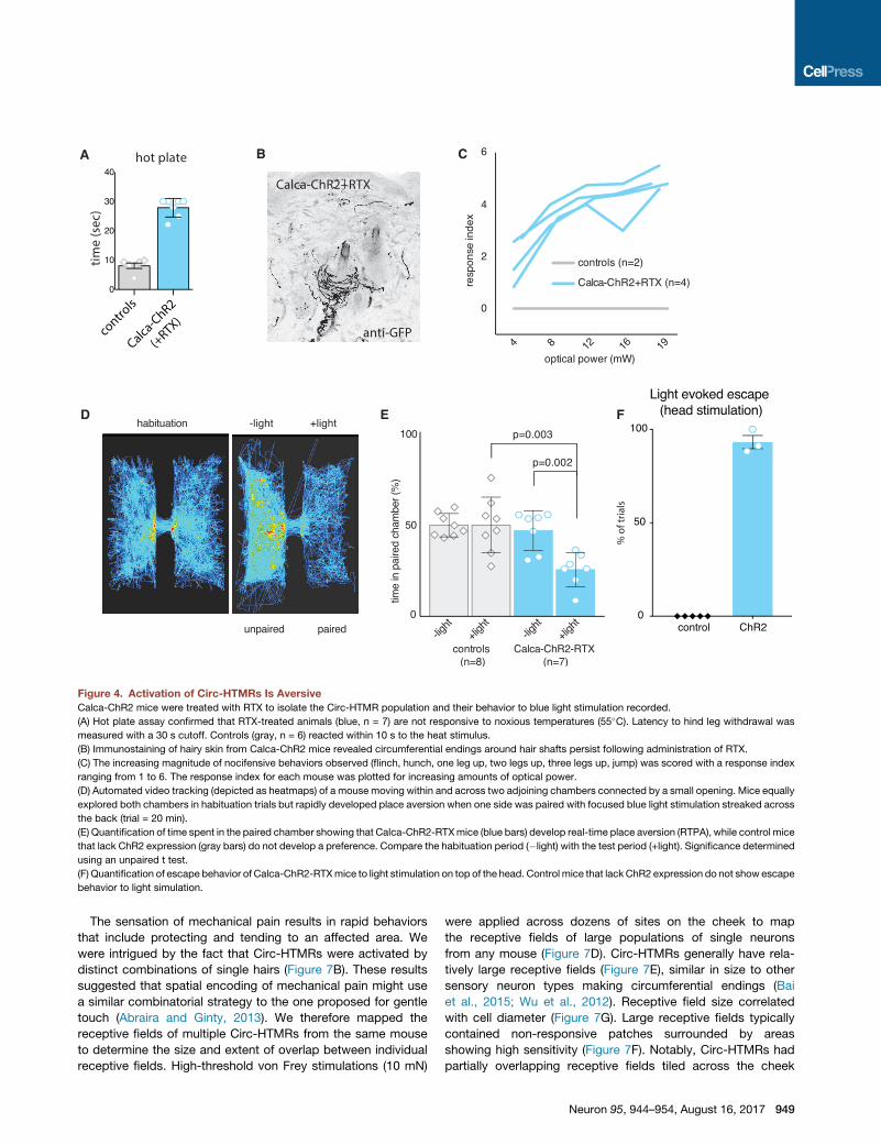
Optogenetic Activation of Circ-HTMRsReveals They AreNociceptorsA defining feature of nociceptors is that their activation is suffi-
cient to evoke protective reflexes and behaviors (Daou et al.,
2013). Therefore, to directly determine whether Circ-HTMRs
are nociceptors, we selectively expressed channelrhodopsin-2
(ChR2) in these neurons (Calca-creERT2 X Rosa26-CAG-ChR2)
and examined behavioral responses to optogenetic stimulation
in mice treated with RTX (Figures 4). As a control, we confirmed
that thesemice were insensitive to heat (Figure 4A), verifying that
they lacked Trpv1 nociceptors. Furthermore, examination of
hairy skin showed that ChR2 protein was found in circumferential
endings (Figure 4B). A flash of blue light on the hairy skin of freely
moving Calca-ChR2-RTX mice elicited a rapid withdrawal
response. Blue light evoked immediate flinching and jumping re-
sponses within milliseconds of stimulation that were never seen
in response to red light or in control mice. The magnitude of
behavioral responses was proportional to light intensity (Fig-
ure 4C). Repeated stimulation caused mice to develop nocifen-
sive/guarding postures (hunching). Notably, rapid escape
behaviors could be elicited by sweeping a spot of blue light
across their backs and heads, something not seen with red light
or in controls. This type of optical stimulation was sufficient to
cause rapid avoidance of a light-paired chamber, whether
mice were stimulated on their back (Figures 4D and 4E) or top
948 Neuron 95, 944–954, August 16, 2017
of the head (Figure 4F). Thus, selective activation of Circ-HTMRs
is strongly aversive, demonstrating the nociceptive phenotype of
these neurons.
Circ-HTMRs Are Ad-Mechano-NociceptorsWe noticed that the speed with which mice reacted to light stim-
ulation was remarkably fast. We observed the belly begin to
recoil from the fiber within �22 ms from the onset of light
(21.63 ± 1.4 SD, n = 5 animals repeated at least 20 times per an-
imal). The majority of classes of nociceptors are slowly con-
ducting C-fibers, but fast Ad-fibers have also been shown to
respond to high-threshold stimuli (Brown and Iggo, 1967;
Burgess and Perl, 1967; Leem et al., 1993). Interestingly, over
half a century ago Burgess and Perl (1967) described fast re-
sponding neurons from cats that responded to hair pull (Burgess
and Perl, 1967). We wondered whether the Circ-HTMRs were a
similar type of fast-conducting mechanical nociceptor. To test
this hypothesis, we measured conduction velocities (CVs) from
Circ-HTMRs in the DRG by performing extracellular sharp elec-
trode recordings of action potentials (APs) triggered by electrical
stimulation of hairy skin (Figures 5A–5D). These experiments
were simultaneously performed with calcium imaging to target
neurons responsive to the site of stimulation (Figure 5A). Interest-
ingly, we found that single action potentials could be reliably re-
ported by GCaMP6f signals with high fidelity (Figure 5B). We
found that small-diameter Calca neurons (19.8 ± 1.5 mm, n = 6)
had an average CV in the C-fiber range (0.71 ± 0.13 m/s), while
medium-diameter Calca neurons (34.8 ± 2.2 mm, n = 6) had an
average CV in the Ad range (means: 3.6 ± 0.47 m/s) (Figures
5C and 5D). Additionally, we measured CVs in the Ad range in
RTX-treated Calca-ChR2 mice from evoked compound action
potentials in the exposed sciatic nerve using optogenetic stimu-
lation of hairy skin (means: 5.87 ± 0.44 m/s, n = 5). Thus, Circ-
HTMRs have the response tuning (Figures 3A–3C), biophysical
properties (Figures 5C and 5D), and the specialized sensory end-
ings (Figures 3D and 3E) that might be expected for hair pull neu-
rons (Burgess and Perl, 1967).
Circ-HTMRs Have Sustained Responses to Single HairPull and Have Partially Overlapping Receptive FieldsTo further define the response properties of Circ-HTMRs, we
examined their responses to a panel of high-threshold mechan-
ical stimuli. Circ-HTMRs could be activated by pinching the skin
(Figures 6A and 6C) as well as by stiff punctate stimuli (von Frey
filaments > 4 mN; Figures 6B–6D). Notably, sustained mechani-
cal stimulation produced sustained responses (>10 s; Figures
6A–6C). The threshold for activation was measured by repeated
stimulation of the receptive field with von Frey filaments cali-
brated at different forces. Circ-HTMRs required forces in excess
of 4.0 mN for activation, similar to what we found for HTMRs
labeled in Trpv1lin-GCaMP6f mice. The threshold for activation
was much less sensitive than for LTMRs in that we found reliably
activated with forces as little as 0.7 mN (Figure 6D). Therefore,
Circ-HTMRs are slowly adapting mechano-nociceptors that
can be activated by multiple types of high-threshold stimuli.
Remarkably, pulling a single guard hair reliably activated a small
but reproducible group of Circ-HTMRs (<10 neurons/field of
view; Figures 7A–7C).
A
D E F
B C
Figure 4. Activation of Circ-HTMRs Is Aversive
Calca-ChR2 mice were treated with RTX to isolate the Circ-HTMR population and their behavior to blue light stimulation recorded.
(A) Hot plate assay confirmed that RTX-treated animals (blue, n = 7) are not responsive to noxious temperatures (55�C). Latency to hind leg withdrawal was
measured with a 30 s cutoff. Controls (gray, n = 6) reacted within 10 s to the heat stimulus.
(B) Immunostaining of hairy skin from Calca-ChR2 mice revealed circumferential endings around hair shafts persist following administration of RTX.
(C) The increasing magnitude of nocifensive behaviors observed (flinch, hunch, one leg up, two legs up, three legs up, jump) was scored with a response index
ranging from 1 to 6. The response index for each mouse was plotted for increasing amounts of optical power.
(D) Automated video tracking (depicted as heatmaps) of a mouse moving within and across two adjoining chambers connected by a small opening. Mice equally
explored both chambers in habituation trials but rapidly developed place aversion when one side was paired with focused blue light stimulation streaked across
the back (trial = 20 min).
(E) Quantification of time spent in the paired chamber showing that Calca-ChR2-RTXmice (blue bars) develop real-time place aversion (RTPA), while control mice
that lack ChR2 expression (gray bars) do not develop a preference. Compare the habituation period (�light) with the test period (+light). Significance determined
using an unpaired t test.
(F) Quantification of escape behavior of Calca-ChR2-RTXmice to light stimulation on top of the head. Control mice that lack ChR2 expression do not show escape
behavior to light simulation.
The sensation of mechanical pain results in rapid behaviors
that include protecting and tending to an affected area. We
were intrigued by the fact that Circ-HTMRs were activated by
distinct combinations of single hairs (Figure 7B). These results
suggested that spatial encoding of mechanical pain might use
a similar combinatorial strategy to the one proposed for gentle
touch (Abraira and Ginty, 2013). We therefore mapped the
receptive fields of multiple Circ-HTMRs from the same mouse
to determine the size and extent of overlap between individual
receptive fields. High-threshold von Frey stimulations (10 mN)
were applied across dozens of sites on the cheek to map
the receptive fields of large populations of single neurons
from any mouse (Figure 7D). Circ-HTMRs generally have rela-
tively large receptive fields (Figure 7E), similar in size to other
sensory neuron types making circumferential endings (Bai
et al., 2015; Wu et al., 2012). Receptive field size correlated
with cell diameter (Figure 7G). Large receptive fields typically
contained non-responsive patches surrounded by areas
showing high sensitivity (Figure 7F). Notably, Circ-HTMRs had
partially overlapping receptive fields tiled across the cheek
Neuron 95, 944–954, August 16, 2017 949

Figure 5. Extracellular Recordings of Action Potentials Revealed Circ-HTMRs Have Conduction Velocities in the Ad Range(A) Schematic drawing of the in vivo experimental setup, whereby peripherally evoked action potentials were recorded simultaneously by calcium imaging and by
an extracellular sharp electrode.
(B) Fluorescence images of medium-diameter (top left) and small-diameter (top right) Calca neurons in the DRG, overlaid with drawings of extracellular recording
pipettes (in color code). Calcium responses to electrical stimulation delivered to the hairy skin of the leg (bottom traces). Each transient was evoked by a single
action potential simultaneously measured with the recording pipette.
(C) Extracellular recordings of action potentials from the same neurons in (A) and (B) showing large difference in response latency (electrical stimulus indicated by
black line over traces). The boxed region shows an expanded timescale.
(D) Plots of conduction velocity (CV) measurements from sixmedium-diameter neurons with CVs in the A-d fiber range (means: 3.6 ± 0.47m/s, 34.8 ± 2.2 um), and
six small-diameter neurons with CVs in the C-fiber range (means: 0.71 ± 0.13 m/s, 19.8 ± 1.5 um).
(Figure 7D). Since having a single hair pulled evokes sharp, local-
ized, sustained pain, Circ-HTMR activation seems ideally suited
to encoding these features.
DISCUSSION
The past two decades have seen a number of advances in the
genetic and functional dissection of the neurons for temperature,
itch, and touch sensation (Le Pichon andChesler, 2014). To date,
most studies on nociception have focused on the unmyelinated
C-fibers. Meanwhile, our understanding of Ad-nociceptors re-
mained much less complete. Here we provide evidence that a
subset of cutaneous Calca neurons that lack TRPV1 expression
950 Neuron 95, 944–954, August 16, 2017
are Ad-HMTRs. We established a chemical-genetic intersec-
tional approach to allow us to ascertain their tuning properties,
morphology, and function.
Using in vivo functional imaging, we demonstrated that sensory
neurons expressing Calca and innervating hairy skin respond to
noxious mechanical and thermal stimuli. Previously, it was shown
that ablation of the Calca neurons resulted inmarked thermal def-
icitsbutnot significantmechanosensory impairment (McCoyetal.,
2013), perhaps because their studies focused on glabrous skin
that lacks Circ-HTMRs. Our finding that a large subset of Calca
neurons that innervate hairy skin is activated by high-threshold
mechanical stimuli highlights that, unlike thermosensation, me-
chanosensation is likely encoded by sensory populations with
A
B von Frey (19 mN)
5 sec
indi
vidu
al n
euro
ns
pinching
80% Δf/f
pinching
2 mV2 sec
D
0.7 1.6 4.0
6 1014vo
n Fr
ey fo
rce
(mN
)
prop
ortio
n re
spon
ding
10 sec
von Frey force (mN)
LTMR TRPV1
10210110010-1
0.5
1.0
0.0
von Frey force (mN)
HTMR TRPV1
10210110010-1
0.5
1.0
0.0
von Frey force (mN)
Calca
10210110010-1
0.5
1.0
0.0
2 sec
C
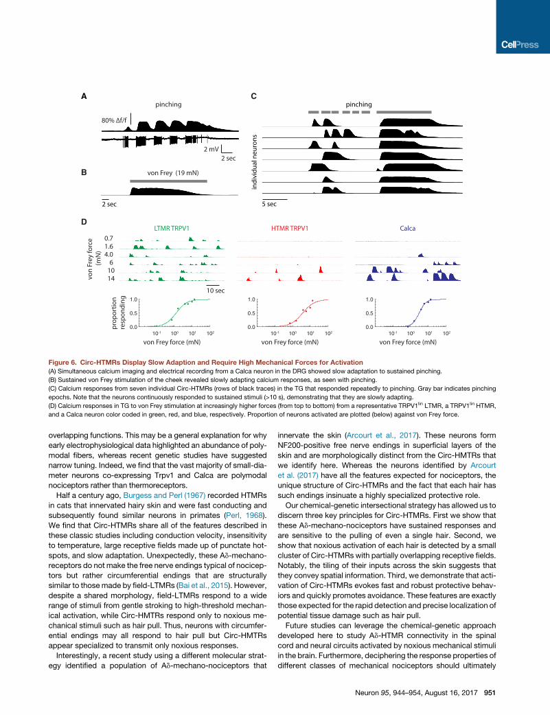
Figure 6. Circ-HTMRs Display Slow Adaption and Require High Mechanical Forces for Activation
(A) Simultaneous calcium imaging and electrical recording from a Calca neuron in the DRG showed slow adaptation to sustained pinching.
(B) Sustained von Frey stimulation of the cheek revealed slowly adapting calcium responses, as seen with pinching.
(C) Calcium responses from seven individual Circ-HTMRs (rows of black traces) in the TG that responded repeatedly to pinching. Gray bar indicates pinching
epochs. Note that the neurons continuously responded to sustained stimuli (>10 s), demonstrating that they are slowly adapting.
(D) Calcium responses in TG to von Frey stimulation at increasingly higher forces (from top to bottom) from a representative TRPV1lin LTMR, a TRPV1lin HTMR,
and a Calca neuron color coded in green, red, and blue, respectively. Proportion of neurons activated are plotted (below) against von Frey force.
overlapping functions. This may be a general explanation for why
early electrophysiological data highlighted an abundance of poly-
modal fibers, whereas recent genetic studies have suggested
narrow tuning. Indeed, we find that the vast majority of small-dia-
meter neurons co-expressing Trpv1 and Calca are polymodal
nociceptors rather than thermoreceptors.
Half a century ago, Burgess and Perl (1967) recorded HTMRs
in cats that innervated hairy skin and were fast conducting and
subsequently found similar neurons in primates (Perl, 1968).
We find that Circ-HTMRs share all of the features described in
these classic studies including conduction velocity, insensitivity
to temperature, large receptive fields made up of punctate hot-
spots, and slow adaptation. Unexpectedly, these Ad-mechano-
receptors do not make the free nerve endings typical of nocicep-
tors but rather circumferential endings that are structurally
similar to those made by field-LTMRs (Bai et al., 2015). However,
despite a shared morphology, field-LTMRs respond to a wide
range of stimuli from gentle stroking to high-threshold mechan-
ical activation, while Circ-HMTRs respond only to noxious me-
chanical stimuli such as hair pull. Thus, neurons with circumfer-
ential endings may all respond to hair pull but Circ-HMTRs
appear specialized to transmit only noxious responses.
Interestingly, a recent study using a different molecular strat-
egy identified a population of Ad-mechano-nociceptors that
innervate the skin (Arcourt et al., 2017). These neurons form
NF200-positive free nerve endings in superficial layers of the
skin and are morphologically distinct from the Circ-HMTRs that
we identify here. Whereas the neurons identified by Arcourt
et al. (2017) have all the features expected for nociceptors, the
unique structure of Circ-HTMRs and the fact that each hair has
such endings insinuate a highly specialized protective role.
Our chemical-genetic intersectional strategy has allowed us to
discern three key principles for Circ-HTMRs. First we show that
these Ad-mechano-nociceptors have sustained responses and
are sensitive to the pulling of even a single hair. Second, we
show that noxious activation of each hair is detected by a small
cluster of Circ-HTMRs with partially overlapping receptive fields.
Notably, the tiling of their inputs across the skin suggests that
they convey spatial information. Third, we demonstrate that acti-
vation of Circ-HTMRs evokes fast and robust protective behav-
iors and quickly promotes avoidance. These features are exactly
those expected for the rapid detection and precise localization of
potential tissue damage such as hair pull.
Future studies can leverage the chemical-genetic approach
developed here to study Ad-HTMR connectivity in the spinal
cord and neural circuits activated by noxious mechanical stimuli
in the brain. Furthermore, deciphering the response properties of
different classes of mechanical nociceptors should ultimately
Neuron 95, 944–954, August 16, 2017 951
A B C
0 20 40 600.1
1
10
100
cell diameter (um)
tree
leng
th (m
m)
1mm
single guard hair pull
freq
uenc
y
# of HTMRs responding1 105
0
5
10
D
10 seconds
hair#1 hair#2 hair#3 hair#2 hair#4 hair#5
indi
vidu
al n
euro
ns
E F
1mm
G
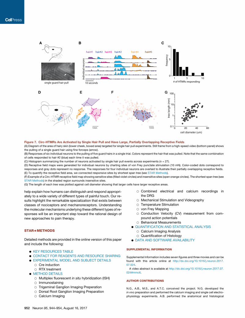
Figure 7. Circ-HTMRs Are Activated by Single Hair Pull and Have Large, Partially Overlapping Receptive Fields
(A) Diagram of the area of hairy skin (lower cheek, boxed area) targeted for single hair pull experiments. Still frame from a high-speed video (bottom panel) shows
the pulling of a single guard hair using fine forceps (arrow).
(B) Responses of six individual neurons to the pulling of five guard hairs in a single trial. Colors represent the hair that was pulled. Note that the same combination
of cells responded to hair #2 (blue) each time it was pulled.
(C) Histogram summarizing the number of neurons activated by single hair pull events across experiments (n = 27).
(D) Receptive field maps were generated for individual neurons by charting sites of von Frey punctate stimulation (10 mN). Color-coded dots correspond to
responses and gray dots represent no response. The responses for four individual neurons are overlaid to illustrate their partially overlapping receptive fields.
(E) To quantify the receptive field area, we connected responsive sites by shortest span tree (see STAR Methods).
(F) Example of a Circ-HTMR receptive field map showing sensitive sites (filled violet circles) and insensitive sites (open orange circles). The shortest span tree (see
STAR Methods) in the shaded region surrounds insensitive sites.
(G) The length of each tree was plotted against cell diameter showing that larger cells have larger receptive areas.
help explain how humans can distinguish and respond appropri-
ately to a wide variety of different types of painful touch. Our re-
sults highlight the remarkable specialization that exists between
classes of nociceptors and mechanoreceptors. Understanding
themolecular mechanisms underlying these different types of re-
sponses will be an important step toward the rational design of
new approaches to pain therapy.
STAR+METHODS
Detailed methods are provided in the online version of this paper
and include the following:
d KEY RESOURCES TABLE
d CONTACT FOR REAGENTS AND RESOURCE SHARING
d EXPERIMENTAL MODEL AND SUBJECT DETAILS
952
B Cre induction
B RTX treatment
d METHOD DETAILS
B Multiplex fluorescent in situ hybridization (ISH)
B Immunostaining
B Trigeminal Ganglion Imaging Preperation
B Dorsal Root Ganglion Imaging Preparation
B Calcium Imaging
Neuron 95, 944–954, August 16, 2017
B Combined electrical and calcium recordings in
the DRG
B Mechanical Stimulation and Videography
B Temperature Stimulation
B von Frey Mapping
B Conduction Velocity (CV) measurement from com-
pound action potentials
B Behavioral Measurements
d QUANTIFICATION AND STATISTICAL ANALYSIS
B Calcium Imaging Analysis
B Quantification of Histology
d DATA AND SOFTWARE AVAILABILITY
SUPPLEMENTAL INFORMATION
Supplemental Information includes seven figures and three movies and can be
found with this article online at http://dx.doi.org/10.1016/j.neuron.2017.
07.024.
A video abstract is available at http://dx.doi.org/10.1016/j.neuron.2017.07.
024#mmc6.
AUTHOR CONTRIBUTIONS
N.G., A.B., M.S., and A.T.C. conceived the project. N.G. developed the
in vivo preparation and performed the calcium imaging and single cell electro-
physiology experiments. A.B. performed the anatomical and histological
analyses. A.B. and C.E.L.P performed the in situ hybridization experiments.
M.S. wrote the software for calcium imaging analyses and performed the op-
togenetic electrophysiological recordings. N.G., J.H.T., C.L., and M.J.K. per-
formed the behavioral tests. N.G., A.B., M.S., and A.T.C. analyzed and inter-
preted data. A.T.C. wrote the manuscript, with input from all authors.
ACKNOWLEDGMENTS
We are indebted to Nick Ryba and Mark Hoon (NIDCR) for their invaluable
advice. Eileen Nguyen, Ruby Lam, Hanna Silberberg, and Latoya Hyson
(NCCIH) provided assistance. We thank Mark Stopfer (NICHD) for assistance
with single-cell electrical recordings. We thank David Ide, Bruce Pritchard,
Tom Talbot, Danny Trang, Phuoc Pham, Daniel Yochelson, and George Dold
at the NIH Section on Instrumentation for design and fabrication of custom-
built instruments used in this study. We thank Pao-Tien Chuang (UCSF) for
providing the Calca-CREERT2 mice and the GENIE project for the development
of GCaMP6. This work was supported by the Intramural Research Program of
the NIH, National Center for Complementary and Integrative Health (ATC).
Received: December 5, 2016
Revised: April 27, 2017
Accepted: July 21, 2017
Published: August 16, 2017
REFERENCES
Abraira, V.E., and Ginty, D.D. (2013). The sensory neurons of touch. Neuron 79,
618–639.
Arcourt, A., Gorham, L., Dhandapani, R., Prato, V., Taberner, F.J., Wende, H.,
Gangadharan, V., Birchmeier, C., Heppenstall, P.A., and Lechner, S.G. (2017).
Touch Receptor-Derived Sensory Information Alleviates Acute Pain Signaling
and Fine-Tunes Nociceptive Reflex Coordination. Neuron 93, 179–193.
Bai, L., Lehnert, B.P., Liu, J., Neubarth, N.L., Dickendesher, T.L., Nwe, P.H.,
Cassidy, C., Woodbury, C.J., and Ginty, D.D. (2015). Genetic Identification
of an Expansive Mechanoreceptor Sensitive to Skin Stroking. Cell 163,
1783–1795.
Bardoni, R., Tawfik, V.L., Wang, D., Francois, A., Solorzano, C., Shuster, S.A.,
Choudhury, P., Betelli, C., Cassidy, C., Smith, K., et al. (2014). Delta opioid re-
ceptors presynaptically regulate cutaneous mechanosensory neuron input to
the spinal cord dorsal horn. Neuron 81, 1312–1327.
Bessou, P., and Perl, E.R. (1969). Response of cutaneous sensory units with
unmyelinated fibers to noxious stimuli. J. Neurophysiol. 32, 1025–1043.
Brown, A.G., and Iggo, A. (1967). A quantitative study of cutaneous receptors
and afferent fibres in the cat and rabbit. J. Physiol. 193, 707–733.
Burgess, P.R., and Perl, E.R. (1967). Myelinated afferent fibres responding
specifically to noxious stimulation of the skin. J. Physiol. 190, 541–562.
Caterina, M.J., Schumacher, M.A., Tominaga, M., Rosen, T.A., Levine, J.D.,
and Julius, D. (1997). The capsaicin receptor: a heat-activated ion channel in
the pain pathway. Nature 389, 816–824.
Caterina, M.J., Rosen, T.A., Tominaga, M., Brake, A.J., and Julius, D. (1999). A
capsaicin-receptor homologue with a high threshold for noxious heat. Nature
398, 436–441.
Cavanaugh, D.J., Chesler, A.T., Braz, J.M., Shah, N.M., Julius, D., and
Basbaum, A.I. (2011). Restriction of transient receptor potential vanilloid-1 to
the peptidergic subset of primary afferent neurons follows its developmental
downregulation in nonpeptidergic neurons. J. Neurosci. 31, 10119–10127.
Chen, T.W., Wardill, T.J., Sun, Y., Pulver, S.R., Renninger, S.L., Baohan, A.,
Schreiter, E.R., Kerr, R.A., Orger, M.B., Jayaraman, V., et al. (2013).
Ultrasensitive fluorescent proteins for imaging neuronal activity. Nature 499,
295–300.
Daou, I., Tuttle, A.H., Longo, G., Wieskopf, J.S., Bonin, R.P., Ase, A.R., Wood,
J.N., De Koninck, Y., Ribeiro-da-Silva, A., Mogil, J.S., and Seguela, P. (2013).
Remote optogenetic activation and sensitization of pain pathways in freely
moving mice. J. Neurosci. 33, 18631–18640.
Emery, E.C., Luiz, A.P., Sikandar, S., Magnusdottir, R., Dong, X., and Wood,
J.N. (2016). In vivo characterization of distinct modality-specific subsets of so-
matosensory neurons using GCaMP. Sci. Adv. 2, e1600990.
Gibson, S.J., Polak, J.M., Bloom, S.R., Sabate, I.M., Mulderry, P.M., Ghatei,
M.A., McGregor, G.P., Morrison, J.F.B., Kelly, J.S., Evans, R.M., et al.
(1984). Calcitonin gene-related peptide immunoreactivity in the spinal cord
of man and of eight other species. J. Neurosci. 4, 3101–3111.
Huang, Z.J., and Zeng, H. (2013). Genetic approaches to neural circuits in the
mouse. Annu. Rev. Neurosci. 36, 183–215.
Jordt, S.E., Bautista, D.M., Chuang, H.H., McKemy, D.D., Zygmunt, P.M.,
Hogest€att, E.D., Meng, I.D., and Julius, D. (2004). Mustard oils and cannabi-
noids excite sensory nerve fibres through the TRP channel ANKTM1. Nature
427, 260–265.
Lawson, S.N., Crepps, B., and Perl, E.R. (2002). Calcitonin gene-related pep-
tide immunoreactivity and afferent receptive properties of dorsal root ganglion
neurones in guinea-pigs. J. Physiol. 540, 989–1002.
Le Pichon, C.E., and Chesler, A.T. (2014). The functional and anatomical
dissection of somatosensory subpopulations using mouse genetics. Front.
Neuroanat. 8, 21.
Leem, J.W., Willis, W.D., and Chung, J.M. (1993). Cutaneous sensory recep-
tors in the rat foot. J. Neurophysiol. 69, 1684–1699.
Li, L., Rutlin, M., Abraira, V.E., Cassidy, C., Kus, L., Gong, S., Jankowski, M.P.,
Luo, W., Heintz, N., Koerber, H.R., et al. (2011). The functional organization of
cutaneous low-threshold mechanosensory neurons. Cell 147, 1615–1627.
Madisen, L., Zwingman, T.A., Sunkin, S.M., Oh, S.W., Zariwala, H.A., Gu, H.,
Ng, L.L., Palmiter, R.D., Hawrylycz, M.J., Jones, A.R., et al. (2010). A robust
and high-throughput Cre reporting and characterization system for the whole
mouse brain. Nat. Neurosci. 13, 133–140.
Madisen, L., Garner, A.R., Shimaoka, D., Chuong, A.S., Klapoetke, N.C., Li, L.,
van der Bourg, A., Niino, Y., Egolf, L., Monetti, C., et al. (2015). Transgenicmice
for intersectional targeting of neural sensors and effectors with high specificity
and performance. Neuron 85, 942–958.
McCoy, E.S., Taylor-Blake, B., and Zylka, M.J. (2012). CGRPa-expressing
sensory neurons respond to stimuli that evoke sensations of pain and itch.
PLoS ONE 7, e36355.
McCoy, E.S., Taylor-Blake, B., Street, S.E., Pribisko, A.L., Zheng, J., and
Zylka, M.J. (2013). Peptidergic CGRPa primary sensory neurons encode
heat and itch and tonically suppress sensitivity to cold. Neuron 78, 138–151.
McKemy, D.D., Neuhausser, W.M., and Julius, D. (2002). Identification of a
cold receptor reveals a general role for TRP channels in thermosensation.
Nature 416, 52–58.
Mishra, S.K., Tisel, S.M., Orestes, P., Bhangoo, S.K., and Hoon, M.A. (2011).
TRPV1-lineage neurons are required for thermal sensation. EMBO J. 30,
582–593.
Norrsell, U., Finger, S., and Lajonchere, C. (1999). Cutaneous sensory spots
and the ‘‘law of specific nerve energies’’: history and development of ideas.
Brain Res. Bull. 48, 457–465.
Olah, Z., Szabo, T., Karai, L., Hough, C., Fields, R.D., Caudle, R.M., Blumberg,
P.M., and Iadarola, M.J. (2001). Ligand-induced dynamic membrane changes
and cell deletion conferred by vanilloid receptor 1. J. Biol. Chem. 276,
11021–11030.
Peier, A.M., Moqrich, A., Hergarden, A.C., Reeve, A.J., Andersson, D.A., Story,
G.M., Earley, T.J., Dragoni, I., McIntyre, P., Bevan, S., and Patapoutian, A.
(2002). A TRP channel that senses cold stimuli andmenthol. Cell 108, 705–715.
Perl, E.R. (1968). Myelinated afferent fibres innervating the primate skin and
their response to noxious stimuli. J. Physiol. 197, 593–615.
Pogorzala, L.A., Mishra, S.K., and Hoon, M.A. (2013). The cellular code for
mammalian thermosensation. J. Neurosci. 33, 5533–5541.
Russell, F.A., King, R., Smillie, S.J., Kodji, X., and Brain, S.D. (2014). Calcitonin
gene-related peptide: physiology and pathophysiology. Physiol. Rev. 94,
1099–1142.
Neuron 95, 944–954, August 16, 2017 953
Schmidt, R., Schmelz, M., Forster, C., Ringkamp, M., Torebjork, E., and
Handwerker, H. (1995). Novel classes of responsive and unresponsive C noci-
ceptors in human skin. J. Neurosci. 15, 333–341.
Smith-Edwards, K.M., DeBerry, J.J., Saloman, J.L., Davis, B.M., and
Woodbury, C.J. (2016). Profound alteration in cutaneous primary afferent ac-
tivity produced by inflammatory mediators. eLife 5, 5.
Song, H., Yao, E., Lin, C., Gacayan, R., Chen, M.H., and Chuang, P.T. (2012).
Functional characterization of pulmonary neuroendocrine cells in lung
development, injury, and tumorigenesis. Proc. Natl. Acad. Sci. USA 109,
17531–17536.
Usoskin, D., Furlan, A., Islam, S., Abdo, H., Lonnerberg, P., Lou, D., Hjerling-
Leffler, J., Haeggstrom, J., Kharchenko, O., Kharchenko, P.V., et al. (2015).
Unbiased classification of sensory neuron types by large-scale single-cell
RNA sequencing. Nat. Neurosci. 18, 145–153.
954 Neuron 95, 944–954, August 16, 2017
Vrontou, S., Wong, A.M., Rau, K.K., Koerber, H.R., and Anderson, D.J. (2013).
Genetic identification of C fibres that detect massage-like stroking of hairy skin
in vivo. Nature 493, 669–673.
Weyer, A.D., O’Hara, C.L., and Stucky, C.L. (2015). Amplified Mechanically
Gated Currents in Distinct Subsets of Myelinated Sensory Neurons
following In Vivo Inflammation of Skin and Muscle. J. Neurosci. 35,
9456–9462.
Wu, H., Williams, J., and Nathans, J. (2012). Morphologic diversity of cuta-
neous sensory afferents revealed by genetically directed sparse labeling.
eLife 1, e00181.
Yarmolinsky, D.A., Peng, Y., Pogorzala, L.A., Rutlin, M., Hoon, M.A., and
Zuker, C.S. (2016). Coding and Plasticity in the Mammalian Thermosensory
System. Neuron 92, 1079–1092.
Zylka, M.J., Rice, F.L., and Anderson, D.J. (2005). Topographically distinct
epidermal nociceptive circuits revealed by axonal tracers targeted to
Mrgprd. Neuron 45, 17–25.

STAR+METHODS
KEY RESOURCES TABLE
REAGENT or RESOURCE SOURCE IDENTIFIER
Antibodies
Chicken anti-NF200 Aves labs Cat# NFH
Rabbit anti-RFP Rockland Cat# 600-901-379; RRID: AB_10703148
AlexaFluor conjugate secondary
antibodies
Thermo Fisher Scientific https://www.thermofisher.com/us/en/home/brands/
molecular-probes/key-molecular-probes-products/
alexa-fluor/alexa-fluor-products.html
Chemicals, Peptides, and Recombinant Proteins
Paraformaldehyde Electron Microscopy Sciences Cat# 15714-S
OCT compound Thermo Fisher Scientific Cat# 23-730-571
Goat serum Rockland Cat# B304
Triton X-100 Sigma Cat# X100-100ML
ProLong gold mounting medium Thermo Fisher Scientific Cat# P36930
Resiniferatoxin (RTX) Sigma Cat# R8756-1G
Tamoxifen (TMX) Sigma Cat# T5648-1G
Thrombin Sigma Cat# T4648-10KU
Critical Commercial Assays
RNAscope multiplex fluorescent
in situ hybridization
Advanced Cell Diagnostics http://www.acdbio.com/
Experimental Models: Organisms/Strains
Mouse: Calca-cre: Calca-creERT2 Dr. Pao-Tien Chuang
Uuniversity of California,
San Francisco
N/A
Mouse: TRPV1-cre: B6.129S-
TRPV1tm1(cre)Bbm/J
Jackson Laboratories Stock# 017769; RRID: IMSR_JAX:017769
Mouse: Rosa-LSL-tdTomato: B6.129S-
Gt(ROSA)tm66.1(CAG-tdTomato)Hze/J
Jackson Laboratories Stock# 005975; RRID: IMSR_JAX:005975
Mouse: Rosa-LSL-GCaMP6f: B6J.Cg-
Gt(ROSA)26Sortm95.1(CAG-GCaMP6f)Hze/MwarJ
Jackson Laboratories Stock# 028865; RRID: IMSR_JAX:028865
Mouse: Rosa26-CAG-ChR2:
Ai32(RCL-ChR2(H134R)/EYFP)
Jackson Laboratories Stock# 012569; RRID: IMSR_JAX:012569
Software and Algorithms
Metamorph Molecular Devices https://www.moleculardevices.com/systems/
metamorph-research-imaging/metamorph-
microscopy-automation-and-image-analysis-software
FIJI ImageJ https://fiji.sc
MATLAB Mathworks Developed in house
Olympus Fluoview FV1000 Olympus http://www.olympus-lifesciences.com
Clampex 10 Molecular Devices https://www.moleculardevices.com/systems/conventional-
patch-clamp/pclamp-10-software
CleverSys CleverSys http://cleversysinc.com/CleverSysInc/
Other
Charged glass slides Daigger Scientific Cat# EF15978Z
Gelfoam dental sponges Thermo Fisher Scientific Cat# NC0490659
Rubber o-ring RT Dygert Cat# 1RJA3
Nair hair removal cream CVS Pharmacy N/A
Neuron 95, 944–954.e1–e4, August 16, 2017 e1

CONTACT FOR REAGENTS AND RESOURCE SHARING
Additional information and requests for reagents and other resources should be made to, and will be fulfilled by the Lead Contact,
Alexander Chesler ([email protected]).
EXPERIMENTAL MODEL AND SUBJECT DETAILS
All animal care and experimental procedures were performed in accordance with a protocol approved by the National Institute
for Neurological Diseases and Stroke Animal Care and Use Committee. TRPV1-Cre or B6.129-TRPV1tm1(cre)Bbm/J (Stock number
017769); Rosa-LSL-tdTomato or B6.129S-Gt(ROSA)tm66.1(CAG-tdTomato)Hze/J or Ai9 (Stock number 005975); Rosa-LSL-GCaMP6f
or B6J.Cg-Gt(ROSA)26Sortm95.1(CAG-GCaMP6f)Hze/MwarJ or Ai95D (Stock number 028865) strains were purchased from Jackson
Laboratories (Cavanaugh et al., 2011; Madisen et al., 2015; Madisen et al., 2010). Calca-creERT2 was generously provided by
Dr. Pao-Tien Chuang (Cardiovascular Research Institute, University of California, San Francisco, CA) (Song et al., 2012). Mice
of both sexes were used in all experiments. Genotyping was performed according to information provided by The Jackson Lab-
oratories and the Chuang lab (for Calca-Cre mice).
Cre inductionCre activity was induced by tamoxifen (TMX), administered twice by intraperitoneal injection at 100 mg/kg body weight at three
weeks of age and older. Injections were spaced by three days for recovery. Mice were used in experiments after at least one
week post tamoxifen injection.
RTX treatmentPups at 3-5 days old were injected intraperitoneally with 50 ml of 20 mMResiniferatoxin (RTX), and with double the dose on each suc-
cessive day to a final dose of 80 mM. Adult mice treated with RTX as pups were again re-dosed with RTX (final dose level) prior to use
in behavioral or imaging experiments.
METHOD DETAILS
Multiplex fluorescent in situ hybridization (ISH)Fresh TGs and DRGswere harvested frommice and rapidly frozen over dry ice. The tissues were embedded in OCT (Tissue-Tek) and
sectioned on a cryostat. 16 mm sections were mounted on charged glass slides (Daigger Scientific). Multiplex ISH was done with the
manual RNAscope assay (Advanced Cell Diagnostics).
ImmunostainingHair from the skin of the back of micewas removed using a depilatory cream (Nair), and cut into square pieces of approximately 5mm
x 5mm. The tissues were fixed in 4%PFA (ElectronMicroscopy Sciences) in PBS at 4�C for 3-5 days. After a PBSwash, tissues were
mounted in OCT medium (Tissue Tek), and sectioned at 80-90 mm on a cryostat (Leica). Skin sections were rinsed in PBS and incu-
bated in blocking buffer (5% goat serum; 0.5% Triton X-100) for 3 hours at room temperature. Sections were incubated in primary
antibodies (chicken anti-NF200, Aves labs; rabbit anti-RFP, Rockland) in blocking buffer at 4�C overnight. Sections were rinsed and
incubated overnight in AlexaFluor conjugated secondary antibodies (Thermofisher Scientific), washed in PBS, and mounted in
ProLong gold mounting media (Thermofisher Scientific). Z stack images were collected using a 40X oil objective on a laser scanning
confocal system (Olympus Fluoview FV1000), and processed using ImageJ/FIJI software (National Institutes of Health).
Trigeminal Ganglion Imaging PreperationMice of either sex at 4-8 weeks were anesthetized with inhalational Isoflurane/Oxygen mix and positioned in a custom built stereo-
tactic frame with heads fixed using ear and tooth bars. Continuous flow of isoflurane/Oxygen was provided through a nosepiece.
A hand warmer was placed under the mouse to stabilize body temperature. With the aid of a dissecting stereoscope (Leica), a small
strip of skin (2x10 mm) on top of the head was removed and the skull exposed. Connective tissue on the exposed skull was removed
using cotton tipped applicators. A round opening in the cranium (�7 mm in diameter, centered at Bregma) was made using a dental
drill (Ideal micro drill, Cellpoint scientific), and the dura was cut using surgical scissors. A glass Pasteur pipette, pulled at the tip to an
inner diameter of�500 mm, bent at a 70-90 angle, and under vacuum suction, was used to carefully aspirate the forebrain to expose
the left and right TGs. The cranium was repeatedly washed and bathed in HEPES buffer (in mM: 160 NaCl, 6 KCl, 13 Glucose,
13 HEPES, 2.5 CaCl2, 2.5 MgCl2, and pH adjusted to 7.2 with 10N NaOH). Excessive bleeding was controlled using Gelfoam dental
sponges (Pfizer). A rubber o-ring (9 mm diameter, RT Dygert) was glued over the opening on the skull using a cyanoacrylate based
adhesive. A custom-made stabilization bar securely mounted to the frame was attached to the o-ring and skull using dental cement.
e2 Neuron 95, 944–954.e1–e4, August 16, 2017
Dorsal Root Ganglion Imaging PreparationMice were positioned in a custom stereotactic frame under anesthesia as above. The hair on the back was shaved, and a sagittal
incision was made over the lumbar vertebrae. Muscle and connective tissues were carefully remove, avoiding damage to the
peripheral nerves. The vertebrae were stabilized at two sites using surgical clamps (Kopf). A dental drill was used to cut an opening
in the bone of the lumbar vertebrae covering a DRG on the left side. Forceps were used to carefully pull away bone cut by the dental
drill, to expose the DRG. HEPES buffer was used continuously to wash and hydrate the DRG. Excessive bleeding was controlled
using Gelfoam sponges, and with application of Trombin (Sigma).
Calcium ImagingThe surgical preparation was transferred on the stage of an epifluorescencemicroscope (MPERS, Olympus) equippedwith a 4X, 0.28
NA air objective. Illumination was provided with a 130 W halogen light source (U-HGLGPS, Olympus), using a standard green exci-
tation/emission filter cube. Imaging was performed using an Orca Flash 4.0 CMOS camera (Hamamatsu), in trials lasting 40 s, at
a 5 Hz frame rate, using Metamorph image acquisition software.
Combined electrical and calcium recordings in the DRGOur calcium imaging system with a 20X, 0.40 NA air objective was used to identify responsive neurons to mechanical and electrical
stimuli (30-70 V, 400 ms) delivered to the skin. Sharp micropipettes were fabricated from quartz glass capillaries using a P 2000 puller
(Sutter Instruments) and filled with 3M KCl, with a resistance of 80 MU. Pipettes were coupled to an Multiclamp700B amplifier head-
stage (Molecular Devices). A motorized micromanipulator (Sutter Instruments) was used to position pipettes to cells of interest under
visual guidance. Extracellular spikes and GCaMP6f signals were acquired simultaneously. Spikes were digitized at 10 KHz (Digidata
1550) and acquired using pClamp software (Molecular Devices). Synchronization of calcium imaging and spike recordings, and de-
livery of electrical stimulation to the skin was controlled by pClamp software and Digidata 1550. Conduction velocities were
measured for each neuron, by dividing distance (site of stimulation to DRG) by the latency to response.
Mechanical Stimulation and VideographyGentle stroking of the mouse cheek, with and against the grain of the hairs, was performedmanually using cotton tipped applicators.
Special care was taken to maintain each stroke consistent at �10-15 mN of applied force. Hair pull and skin pinch was performed
using forceps. Gentle air puff was made using a Picospritzer III (Parker), with a custom air nozzle positioned 2.5 cm from the cheek.
Video recordings were performed on all trials with mechanical stimulations using a CMOS video camera (acA2000-165um, Basler),
and a 0.6X telecentric lens (Edmunds Optics). For single hair pull experiments, a fine forceps and a 1X telecenric lens was used.
A trigger pulse generated by Digidata 1550 and controlled by Clampex 10 software (Molecular Devices) initiated video recordings
and calcium imaging synchronously.
Temperature StimulationTemperature ramps were applied to the mouse cheek using a custom-built apparatus, consisting of three M3 Lauda bath recircula-
tors (Lauda Brinkmann), set to cold, hot, and room temperatures, pumping water through a series of solenoid pinch valves
(NResearch Incorporated) which direct water through a copper thermode. Trigger pulses generated by the Digidata 1550 and Clam-
pex controlled shutting on and off of pinch valves, which produced the rise and fall of the temperature ramps. A digital thermometer
attached to the copper thermode measured the temperature ramps as an analog input to Digidata 1550 and was recorded using
Clampex.
von Frey MappingThe hairs on the mouse cheek were trimmed using a small hair clipper (Wahl), then removed completely using a hair removal
cream (Nair). A 10 mN von Frey filament was used to make localized mechanical stimulations, randomly distributed within the
confines of the depilated cheek, while simultaneously recording calcium signals and video of the stimulations. Mapping data
were analyzed using custom written MATLAB scripts (Mathworks). To compare relative sizes of mapped receptive fields we
calculated the minimum weight spanning tree connecting all responsive points, and total tree length was used as a measure
of receptive field extensity. To visualize responsive area boundaries, nonconvex polygon vertices encompassing all responsive
points were calculated.
Conduction Velocity (CV) measurement from compound action potentialsAdult mice were anesthetized with inhalational Isoflurane/Oxygen mix and positioned on the heating pad dorsal side up. Hind paws
were gently stretched and immobilized with adhesive tape. Hair on the lower part of the trunk and hind paws was shaved and depi-
lated to expose skin. A rectangular patch of the skin was excised to expose the gluteusmaximus and external obliquemuscles, which
were gently retracted to visualize a 6-8 mm stretch of the sciatic nerve. The sciatic nerve was continuously bathed in ACSF.
Compound action potentials (CAPs) were recorded with an ACSF-filled pipette suction electrode connected to a headstage of a
Multiclamp700B amplifier. The electrode was positioned over the nerve at a steep angle (�80�) and negative pressure was applied
to form a tight contact between pipette and nerve. CAPs were evoked by optogenetic stimulation of the intact thigh skin, using a
Neuron 95, 944–954.e1–e4, August 16, 2017 e3

blue fiber-coupled LED (Prizmatix) with the fiber tip positioned �1-2 mm from the skin. A 1 ms command pulse was used to drive
light stimulation with peak optical power of 37 mW. Stimulation was controlled with Clampex 10 software through Digidata 1550
acquisition system. For each site, stimulation was repeated > 90 times, data presented are averaged responses. 50 Hz electrical
interference was filtered offline. To calculate the CV, the distance between stimulation and recording sites was measured and
divided by response latency.
Behavioral MeasurementsFor behavioral testing with optogenetics, a strip of skin along the backs or bellies of RTX ablated Calca-ChR2 and age matched
Calca-GCaMP6f mice (as controls) was shaved and depilated a day before testing to allow blue light to efficiently reach the skin. Us-
ing a fiber coupled LED (Prizmatix), the fiber tip was manually positioned within 6-8 mm of the belly and the LED was switched on
while performing high speed (120 fps) video of the behavior. The mouse response to blue light stimulation at different power inten-
sities was scored as described in Figure 4. For the Real-Time Place Aversion (RTPA) assay, mice were placed in a cage with two
chambers separated by a wall with a round opening in the center, allowing mice to freely explore both chambers. Using a magnifying
lens and the Prizmatix blue LED, light was focused on the depilated backs of mice when they entered one chamber but not the other.
Mouse movement was video recorded and automatically tracked offline using CleverSys software.
QUANTIFICATION AND STATISTICAL ANALYSIS
Calcium Imaging AnalysisCalcium imaging stacks across multiple trials representing all applied stimuli were concatenated for each animal to generate a single
tiff stack using ImageJ. Tiff stacks were corrected for movement artifact using the Linear Stack Alignment plugin in ImageJ. Regions
of interest (ROIs) were selectedmanually in ImageJ using theCell MagicWand plugin. A total of 213 responsive Trpv1lin-GCaMP6f TG
(n = 213 neurons in 3 animals; see Figure 1), 430 responsive Calca-GCaMP6f TG neurons (n = 430 neurons from 15 animals; see Fig-
ure S2), 136Calca-GCaMP6f TG neurons from animals treatedwith RTX (n = 136 neurons from 5 animals; see Figure 3), and 52Calca-
GCaMP6f DRG neurons (n = 52 neurons form 3 animals, see Figure S2) were analyzed.
ROI signal analysis was performed using custom written MATLAB scripts (Mathworks). For Df/F0 determination F0 was calculated
as an average of 5 frames with lowest F from each stack of 200 frames. Custom written MATLAB scripts were used for neuropil sub-
traction: fluorescence signals of a donut shaped area around each ROI were subtracted from the cell response to remove possible
contribution of out of focus signal. Neurons were assigned as responsive to any given stimuli if dF/F0 > 20%. Cells were qualified as
mechano-, heat- or mechano-heat-responsive based on unbiased classification by a K-means clustering algorithm with assumed
cluster numbers equal to 3. Clustering centroids obtained from analysis of Calca-GCAMP6f results were used to classify responses
in Calca-GCAMP6f RTX-treated mice.
Quantification of HistologyCo-expression of Trpv1lin-tdT with sensory neuron markers was quantified for label overlap using ImageJ. For each experiment,
representative confocal images from tissue sections were quantified (n = 5 sections from 3 animals). Co-expression of tdTomato
with sensory neuronmarkers are quantified for label overlap using ImageJ (n = 5 sections from 3 animals). Quantification of hair shafts
with circumferential endingswas performed in ImageJ from confocal images (n = 10 skin preparations from 3 animals). To quantify the
lengths of free nerve endings, a line was traced using the free hand tool of ImageJ, and the absolute length wasmeasured (n = 10 skin
preparations from 3 animals).
DATA AND SOFTWARE AVAILABILITY
All data and custom made MATLAB codes for calcium imaging analysis are available upon request.
e4 Neuron 95, 944–954.e1–e4, August 16, 2017