Special Stains
15
SPECIAL STAINS AND H & E | 1 Chapter 1 | Introduction to Special Stains George L. Kumar, PhD, MBA and Gary W. Gill, CT(ASCP) Special stains are “special” because they are not routine. They are applied to tissue sections in addition to hematoxylin and eosin (H&E)-stained sections to answer questions that arise above and beyond those that can be answered by interpreting H&E-stained tissue morphology (Fig. 1). The term “special stains” is of uncertain provenance, but one can be certain that it began to be used after 1876 when H&E was introduced. Special stains (Fig. 2) can answer these questions: Is a certain class of molecules present or absent? Where are the molecules located in the preparation? How many of the molecules are present? Answering the last question requires sophisticated instrumentation and computation methods and, to our knowledge, this aspect of special stains is neither well-documented nor understood. In this article, we will describe some commonly used non- immunohistochemical stains. In the first part of the article, we will compare some key aspects between H&E and special stains, and certification of special stains by the “Biological Stain Commission”. In the second part of the article, we will delve into the technical details of special stains. H&E and Special Stains Comparing key aspects of H&E and special stains is instructive. Classification of Special Stains by the Biological Stain Commission The Biological Stain Commission (see Appendix) certifies biological stains. Among its objectives, the Biological Stain Commission strives to ensure the quality of dyes through independent testing according to appropriate rigorous chemical and performance criteria. The authors are unaware of comparable efforts elsewhere in the world to ensure the quality of dyes used as special stains and other applications. Sixty-four stains are on a certification basis with the Biological Stain Commission (Table 1). All but two, hematoxylin and orcein, are synthetic dyes. Twenty-nine of the 62 synthetic dyes were first used before 1909, and are highlighted in bold letters. Fifty-two dyes have Colour Index (C.I.) numbers; 12 do not. C.I. numbers are 5-digit numbers assigned by The Society of Dyers and Colourists in England to uniquely identify stains that are the same chemically, but have different names. These 5-digit numbers must be specified when purchasing dyes or publishing articles in which the dyes are cited to ensure using the same dye, even if identified by different names. Aspect H&E Special Stains Questions that can be answered Many One too many Primary interest Nucleus and cytoplasm Medical diagnosis (e.g., growth activity) Nucleus or cytoplasm Mostly in the diagnosis of infectious diseases and cancer based on chemical composition Basis of interpretation Morphology Morphology/Color Frequency of use Routine As needed Quantitative No No Controls needed No Yes Substrate specific No Yes
-
Upload
el-marie-salunga -
Category
Documents
-
view
106 -
download
1
description
Special Stains
Transcript of Special Stains

specialstainsandH&e | 1
Chapter 1 | Introduction to Special Stains
George L. Kumar, PhD, MBA and Gary W. Gill, CT(ASCP)
specialstainsare“special”becausetheyarenotroutine.theyare
applied to tissuesections inaddition tohematoxylinandeosin
(H&e)-stainedsectionstoanswerquestionsthatariseaboveand
beyondthosethatcanbeansweredbyinterpretingH&e-stained
tissuemorphology(Fig.1).theterm“specialstains”isofuncertain
provenance,butonecanbecertainthatitbegantobeusedafter
1876whenH&ewasintroduced.
specialstains(Fig.2)cananswerthesequestions:
isacertainclassofmoleculespresentorabsent?
Wherearethemoleculeslocatedinthepreparation?
Howmanyofthemoleculesarepresent?
answeringthelastquestionrequiressophisticatedinstrumentation
andcomputationmethodsand,toourknowledge,thisaspectof
specialstainsisneitherwell-documentednorunderstood.
in this article, we will describe some commonly used non-
immunohistochemicalstains.inthefirstpartofthearticle,wewill
comparesomekeyaspectsbetweenH&eandspecialstains,and
certificationofspecialstainsbythe“Biologicalstaincommission”.
inthesecondpartofthearticle,wewilldelveintothetechnical
detailsofspecialstains.
H&E and Special StainscomparingkeyaspectsofH&eandspecialstainsisinstructive.
Classification of Special Stains by the Biological Stain CommissiontheBiologicalstaincommission(seeappendix)certifiesbiological
stains.amongitsobjectives,theBiologicalstaincommissionstrives
toensurethequalityofdyesthroughindependenttestingaccordingto
appropriaterigorouschemicalandperformancecriteria.theauthors
areunawareofcomparableeffortselsewhereintheworldtoensure
thequalityofdyesusedasspecialstainsandotherapplications.
sixty-fourstainsareonacertificationbasiswiththeBiologicalstain
commission(table1).allbut two,hematoxylinandorcein,are
syntheticdyes.twenty-nineofthe62syntheticdyeswerefirstused
before1909,andarehighlightedinboldletters.
Fifty-twodyeshavecolourindex(c.i.)numbers;12donot.c.i.
numbersare5-digitnumbersassignedbythesocietyofdyersand
colouristsinenglandtouniquelyidentifystainsthatarethesame
chemically,buthavedifferentnames.these5-digitnumbersmust
bespecifiedwhenpurchasingdyesorpublishingarticlesinwhich
thedyesarecitedtoensureusingthesamedye,evenifidentified
bydifferentnames.
Aspect H&E Special Stains
Questions that can be answered Many One too many
Primary interest Nucleus and cytoplasm
Medical diagnosis (e.g., growth activity)
Nucleus or cytoplasm
Mostly in the diagnosis of infectious diseases
and cancer based on chemical composition
Basis of interpretation Morphology Morphology/Color
Frequency of use Routine As needed
Quantitative No No
Controls needed No Yes
Substrate specific No Yes

2|specialstainsandH&e specialstainsandH&e | 3
Introduction to Special StainsIntroduction to Special Stains
Figure 2. Special stained section of small
intestine. The special stain mucicarmine is
used for visualization of neutral epithelial
mucins in small intestine. The mucins are
stained rose to red, nuclei are blue/black
(Weigert’s iron hematoxylin), and other
tissue elements are yellow (metanil yellow
or tartrazine).
Figure 1. H&E-stained section of skin
with cutaneous blastomycosis. High
magnification (40x) view showing budding
yeast with the inflammatory infiltrate.
Hematoxylin stains the nuclei of cells blue
to bluish-purple, and eosin stains other
cellular elements in the tissues from pink
to red (Figure courtesy: Sunil Badve, MD,
FRCPath. Indiana University School of
Medicine, Indianapolis, IN, USA).
Acid fuchsin, C.I. 42685
Alcian blue 8 GX, C.I. 74240
Alizarin red S, C.I. 58005
Aniline blue WS, C.I. 42755
Auramine O, C.I. 41000
Azocarmine B
Azocarmine G, C.I. 50085
Azure A, C.I. 52005
Azure B, C.I. 52010
Azure C, C.I. 52002
Basic fuchsine, C.I. 42510
Bismarck brown Y, C.I. 21000
Brilliant cresyl blue, C.I. 51010
Brilliant green, C.I. 42040
Carmine, C.I. 75470
Chlorazol black E, C.I. 30235
Congo red, C.I. 22120
Cresyl violet
Crystal violet, C.I. 42555
Darrow red
Eosin B, C.I. 45400
Eosin Y, C.I. 45380
Erythrosin, C.I. 45430
Ethyl eosin, C.I. 45386
Ethyl green, C.I. 42590
Fast green F C F, C.I. 42053
Fluorescein Isothiocyanate
Giemsa Stain 1902,
modified in 1904.
Hematoxylin, C.I. 75290
(Bohmer 1865)
Indigo carmine, C.I. 73015
Janus green B, C.I. 11050
Jenner stain 1899
Light green SF, C.I. 42095
Malachite green, C.I. 42000
Martius yellow, C.I. 10315
Methyl orange, C.I. 13025
Methyl violet 2B, C.I. 42535
Methylene blue
Methylene blue, C.I. 52015
Methylene violet
(Bernthsen), C.I. 52041
Neutral red, C.I. 50040
Nigrosin, C.I. 50420
Nile blue A, C.I. 51180
Nuclear fast red, C.I. 60760
Oil Red O, C.I. 26125
Orange G, C.I. 16230
Orange II, C.I. 15510
Orcein
Pararosaniline, C.I. 42500
Phloxin B, C.I. 45410
Protargol S
Pyronine B, C.I. 45010
Pyronine Y, C.I. 45005
Resazurin
Rose Bengal, C.I. 45435
Safranine O, C.I. 50240
Sudan black B, C.I. 26150
Sudan III, C.I. 26100
Sudan IV, C.I. 26105
Tetrachrome stain (MacNeal)
Thionine, C.I. 52000
Toluidine blue, C.I. 52040
Weigert 1878
Wright stain (1908)
Table 1. 2009 biological stains certified by the Biological Stain Commission.

4|specialstainsandH&e specialstainsandH&e | 5
Introduction to Special Stains Introduction to Special Stains
Special Stainsspecialstainisatermthatismostlyusedinalaboratorysetting.
specialstainshavetwobroadareasofapplication:researchand
diagnostic.inresearch,specialstainsareusedasprobestoidentify
certainchemicalconstituentsinnormalandabnormalcells.the
informationsoobtainedisusedasabasisforfurtherstudyand
alsoasabaselineagainstwhichtheresultsofspecialstainingcan
becomparedindiagnosticapplications.Onthebasisofsucha
comparison,thesignificanceofthefindingscanbeinterpreted.
specialstainscanbeappliedtocellbiologyandhistology.some
usefulapplicationsare: (1) thedeterminationofdnaandRna
content,(2)themodeofactionofdrugs,hormonesorofpotentially
toxicfoodadditives,(3)metabolicbiochemistry,(4)biochemistryof
diseaseprocesses,(5)primarysitesofmanymetastatictumors,
(6) identification of non-pigmented metastatic melanomas,
(7)detectionofearlyinvadingtumors,(8)definitionofthemargins
ofsurgically resected tumors, (9) identificationofBarrbodies,
(10)stainingcells inways thatcanbeusedasabasis forcell
separationbyappropriateinstrumentation(e.g.,fluorescence),and
(11)identificationofmicro-organisms(e.g.,Cryptococcus neoformans,
Helicobacter pylori).seetable2.
thematerial,methodsandinterpretationofthesespecialstainscan
befoundinreferences5-7.Whenworkingwithspecialstains,keep
inmindthefollowingconsiderations:
specialstainingoftenrequirestheuseofunusualstainsand
reagentsthatareavailablefromonlyafewsources.Knowledge
ofsuchsourcesisessentialtoovercometechnicalbottlenecks.
Beawareofspecialstainsthatcontaincoloredandcolorless
impurities(e.g.,salts)asthesesubstancesmayinterferewith
thestaining.
special staining requires broad knowledge of the tissue or
cellstargeted.
Whenworkingwithspecialstains,careshouldbetakentocollect,
fixandpreparethespecimeninamannerthatwillmaintainthe
moleculeof interestwithincellsortissues.Forexample,it is
importanttoworkwithfrozensectionswhenattemptingtoidentify
enzymesandtoavoidfatsolventssuchasalcoholandxylene
whenattemptingtoidentifylipids.
Withcellsuspensions,itisessentialtodeterminebymicroscopy
whethercellsarepresent,andhowmanycellsaretobeusedwhen
makingtheslides.Usingthisqualitycontrolstepwillimprovethe
cellularpreparations.
controlpreparationsmustberuninparallelwithexperimental
preparations foroneormoreof the followingreasons: (1) to
determineifthespecialstainisworking,(2)toassessthedegree
ofnon-specificstaining,(3)todeterminewhetherareagentisstill
active,and(4)toserveasastandardinfractionalreductionof
stainingprocedures.ifapositivereactionisnotedwhenacontrol
isnotused,itcanstillbedeterminedthatthereactionisatleast
working(howwellorhowspecificallyisopentospeculation).
However,anegativereactionintheabsenceofacontrolmay
indicateeitherthatthesoughtconstituentisnotpresentorthe
reactionisnotworking.
controlslidesshouldbe: (1)sectionsof tissue/cellhigh ina
particularmolecule/constituent,(2)purifiedsamplesofaparticular
moleculeinsmears,(3)samplesofthesamespecimenpretreated
withsolventsorenzymes to remove thesoughtconstituent,
(4)samplesofthesamespecimenwiththeomittanceofessential
reagentsorsteps in thestainingprocedure,or (5) runasa
duplicatecellspreadinthesamemannerastheexperimentminus
oneessentialstep.
theamountofspecialstainswithinacellortissuerepresents
thedifferencebetweentheamounttakenupduringstainingand
theamountremovedbytherinsesfollowingstaining.toensure
theoptimalamount,theusermustemploythosematerialsand
methodsthatpromotestainuptakeduringandfollowingstaining
(e.g.,dyeconcentration,suitablesolvent,controloffavorablepH,
additionofsalts,ifnecessary,controlofionicconcentration,if
necessary,timeandtemperature).
tomaintaintherightamountandhueofthespecialstain,mount
thestainedspecimeninamediumthatdoesnotpromotebleaching
orleaching.
toensureoptimalimagequalityofthestainedspecimen,use
therightamountofmountingmedium,coverwithano.1cover
glass,anduseacleanmicroscopewiththeilluminationadjusted
accordingtothemethodofKöhler(seeappendix,page283).
Manual vs. Automated Special Staining Protocolsdependingonthefinancialsituationofthelaboratory,specimen
samplesize,andthenumberofpersonnelavailable,specialstain
protocolsareperformedeithermanuallyorbyusingautomated
systems.Manualstainingofslidesworkwellinaresearchsetting,
especially,whenthenumberofprocessedslidesarefewperday.
However,withincreasingnumbersofslidestobestained,themanual
methodbecomespronetoerrorresultingindecreasedflexibility
andproductivity.Withthemedicalcommunitydemandingfaster
turnaroundtimes,increasedflexibilityandproductivityaswellas
greater standardization,automated instrumentshave replaced
somemanualmethodsofstainingthusbecominganintegralpart
ofthelaboratory.automationcombinedwithspecializedsoftware
applicationsandconnectivityhavemademanyinstrumentscapable
of multiprogramming runs resulting in standardized protocols,
manageableworkschedules,enhancedworkflow,cost-effectiveness
andtheabilitytoadapttoregulatoryrequirements.
Special Stain Clinical Application Staining Specificity
For detecting micro-organisms and Helicobacter pylori
Acid-Fast Bacteria (AFB) (Ziehl-Neelsen Stain)
Detects nocardioform-actinomycete groups of bacteria, including Mycobacterium Spp (acid fast), Rhodococcus equi and Nocardia Spp (weakly acid fast) Fig. 3
Acid-fast bacilli retain a cationic dye that is extracted from all other types of bacteria and animal cells by acidified alcohol. The waxy wall (with mycolic acid) of mycobacteria retains the dye
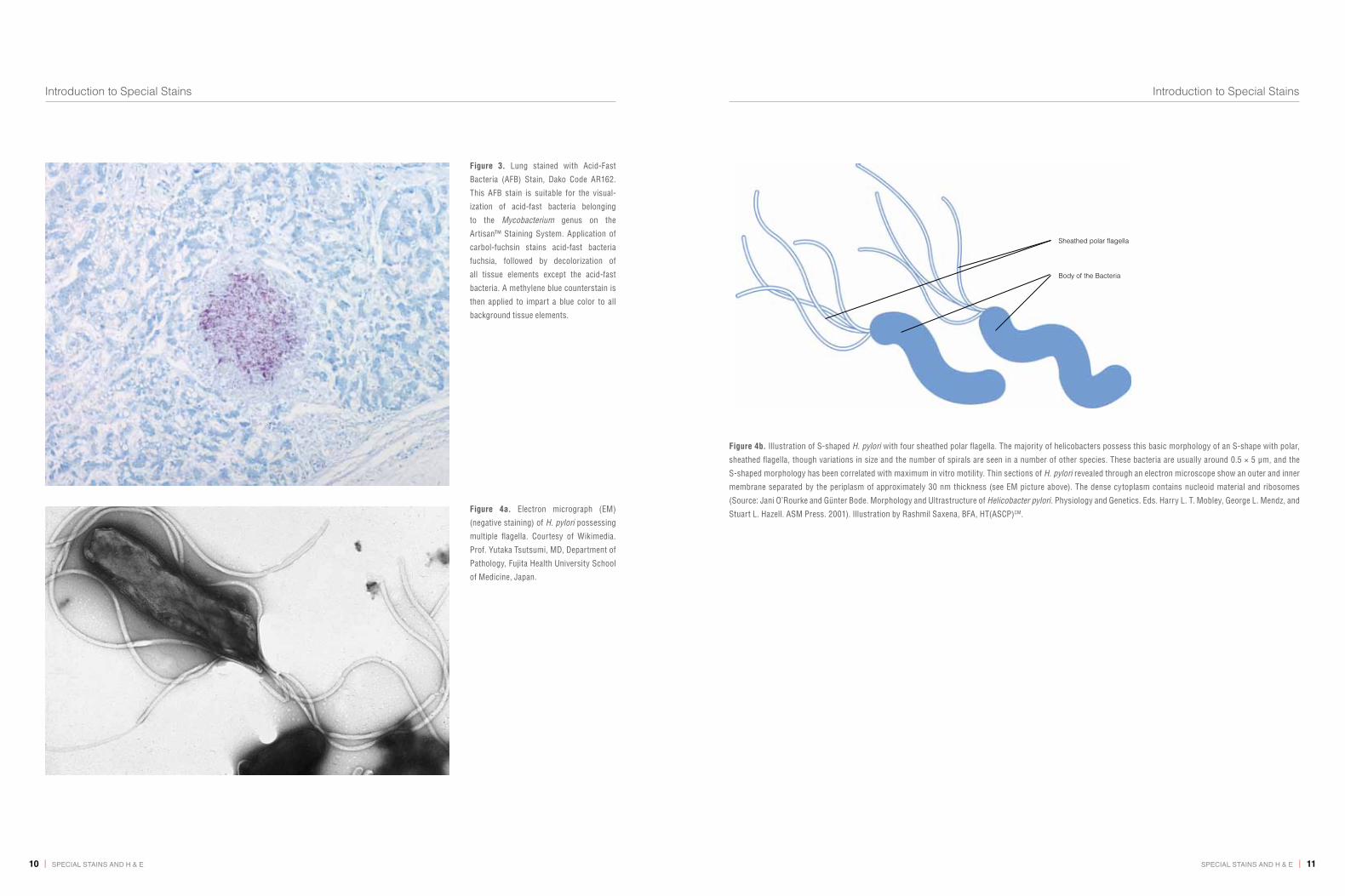
Alcian Yellow / Toluidine Blue (Leung) Stain Used for the detection of H. pylori See Fig. 4 for an electron micrograph and illustration of H. pylori
The yellow dye stains oxidized and sulphonated gastric mucus providing contrast for suspended Helicobacter organisms that are stained with toluidine blue
Dieterle’s Stain Identifies Borrelia burgdorferi, Legionella pneumophila, Treponema pallidum
Stains whole organisms
Diff-Quik Stain (Diff-Quik is the formerly trademarked name for a proprietary rapid Giemsa-like stain)
Detects H. pylori and some fungi(e.g., Pneumocystis jiroveci)
H. pylori and Pneumocystis jiroveci
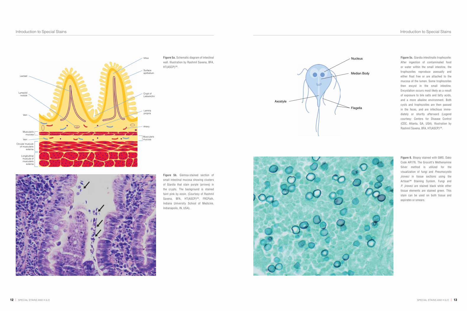
Giemsa Stain Used for staining H. pylori, Plasmodium vivax, Rickettsia prowazekii, Rickettsia rickettsii, Rickettsia tsutsugamushi, Trypanosoma cruzi, Giardia lamblia; Fig. 5a, b and c
Stains polyanions blue and polycations pink
Bacteria show up blue on account of their nucleic acids. Acidic capsules (e.g., Anthrax Bacilli, Cryptococcus) would be expected to be blue or purple
Gram Stain (Named after its inventor, the Danish scientist Hans Christian Gram, who developed the technique in 1884 to discriminate between two types of bacteria with similar clinical symptoms)
Used for the detection of Gram-positive (Clostridium botulinum, Clostridium tetani, Staphylococcus aureus and Corynebacterium diphtheriae) or Gram-negative bacteria (Salmonella, Shigella dysenteriae, Escherichia coli and Pseudomonas aeruginosa). Also used for the detection of Actinomyces Israeli, Legionella pneumophila, Neisseria gonorrhea, Neisseria meningitidis, Nocardia asteroides
Stains whole organisms
Table 2. Commonly used special stains.

6|specialstainsandH&e specialstainsandH&e | 7
Introduction to Special Stains Introduction to Special Stains
Special Stain Clinical Application Staining Specificity
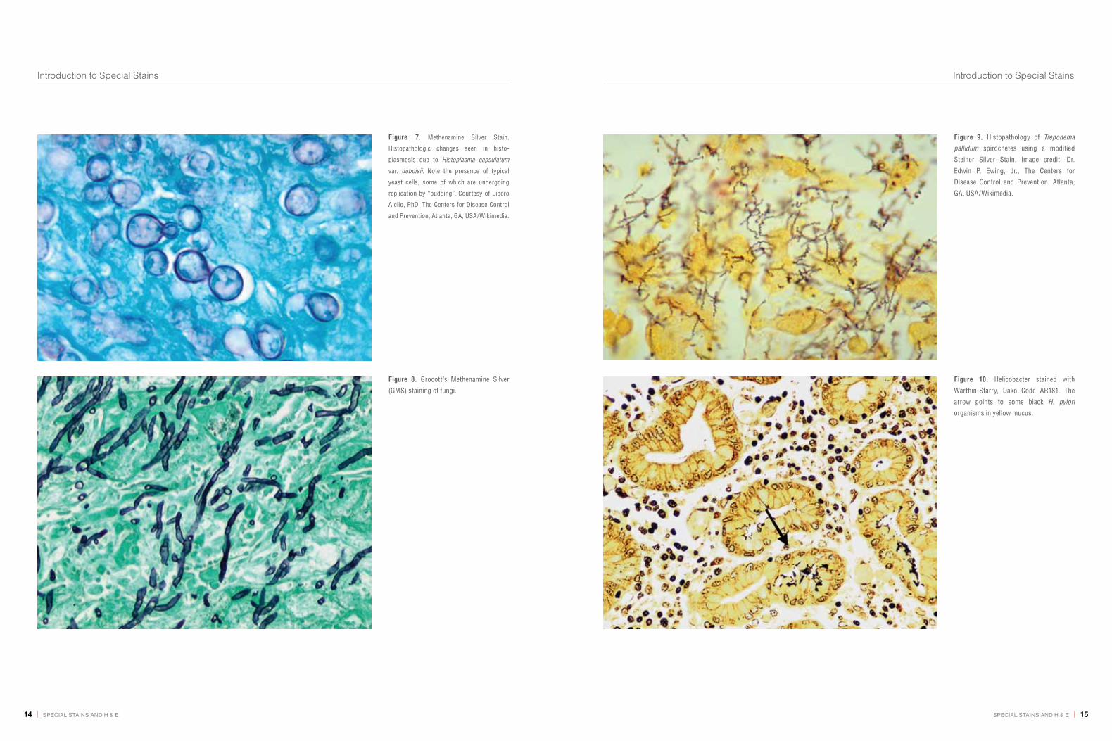
Grocott’s Methenamine Silver (GMS) Stain Useful in identifying a variety of pathogenic fungi, including
Aspergillus fumigatus, Blastomyces dermatitidis, Candida
albicans, Coccidioides immitis, Cryptococcus neoformans,
Histoplasma capsulatum, Nocardia asteroids, Pneumocystis
carinii, Pneumocystis Jiroveci (human) and Sporothrix
schenckii; Fig. 6-8
Polysaccharide components of the fungal cell wall
Mayer’s Mucicarmine Stain Detects encapsulated yeast-like fungus Cryptococcus
neofarmans
Polysaccharides on the capsule
Periodic Acid-Schiff (PAS) Stain Used for the identification of Aspergillus fumigatus,
Blastomyces dermatitidis, Candida albicans, Coccidioides
immitis, Cryptococcus neofarmans, Sporothrix schenckii
Polysaccharide components of the fungal cell wall
Sayeed’s Stain (Schiff’s reagent, 0.5% periodic acid,
Mayer’s hemalum)
Detects H. pylori H. pylori
Steiner & Steiner Staining Method Detects spirochetes and legionella, and pneumophila
bacteria, e.g., Borrelia burgdorferi, H. pylori, Legionella
pneumophila, Treponema pallidum; Fig. 9
Stains whole organisms
Warthin-Starry Stain (these are reduced silver
methods)
Identifies Alipia feles, Bartonella henselae, Borrelia burgdorferi,
H. pylori, Legionella pneumophila, Treponema pallidum; Fig. 10
Stains whole organisms
For demonstrating connective tissue, muscle, collagen, lipid and fibrin
Gomori’s One-Step Trichrome Stain Used for distinguishing collagen and smooth muscle fibers;
Fig.11
Collagen and smooth muscle fibers
Jones’ Basement Membrane
Periodic Schiff-Methenamine Silver (PASM) Stain
Used for the identification of basement membranes (of the
glomerulus in the kidney or in tissue samples); Fig. 12
Basement membranes
Masson’s Trichrome Stain (TRI) Used for distinguishing cells from surrounding connective
tissue which has several variants and is probably the
trichrome most used in histopathology. Black nuclei, red
cytoplasm (including muscle), blue or green collagen
(including fine fibers), cartilage and mucus; Fig. 13
Muscle, collagen fibers, fibrin and erythrocytes
Special Stain Clinical Application Staining Specificity
Russel-Movat Pentachrome Stain Used for simultaneous demonstration of muscle, elastic
fibers, collagen/reticular fibers, ground substance and
fibrinoid in tissues
Muscle, elastic fibers, collagen/reticular fibers
Oil Red O and Sudan Black B Stains Used for staining lipids in frozen sections and some
lipoproteins on paraffin sections
Lipids, including triglycerides (which necessarily are
neutral). Oil Red O stains only the most hydrophobic
lipids (triglycerides and cholesterol esters). Sudan
Black B stains these and also phospholipids and
sphingomyelins, which are less hydrophobic
Orcein Stain Used for staining elastic fibers Elastic fibers
Lendrum’s Method (Picro-Mallory Stain) Fibrin Fibrin
Phosphotungstic Acid-Hematoxylin (PTAH) Stain Used for demonstrating striated muscle fibers
Also used to stain abnormal neuroglia (reactive astrocytosis)
Muscle fibers, collagen
Silver methods for reticulum and
basement membranes
(e.g., Reticulin/ Nuclear Fast Red Stain)
Used for the identification of reticulin fibers in tissue
samples; Fig.14
Reticulin (collagen with high level of hexosylation,
including Type IV)
Verhoeff Stain
Van Gieson Stain
Used for the identification of elastic laminae and fibers
in tissues; Fig.15
The Verhoeven Stain is specific for elastic fibers.
The Van Gieson Stain is specific for collagen.
Verhoeff’s iron-hematoxylin stains elastin and
nuclei black. Van Gieson’s picro-fuchsine gives
yellow cytoplasm and red collagen fibers
For detecting nucleic acids
Ethyl Green-Pyronine Stain Used for differential demonstration of DNA and RNA A buffered mixture of the two dyes gives blue-green
DNA and red RNA (rRNA in cytoplasm, nucleoli)
Feulgen Stain Used for the identification of chromosomal material or
deoxyribonucleic acid (DNA in paraffin-embedded tissue
or cell specimens); Fig.16
Deoxyribonucleic acid (DNA)
Table 2. Commonly used special stains.

8|specialstainsandH&e specialstainsandH&e | 9
Introduction to Special Stains Introduction to Special Stains
Special Stain Clinical Application Staining Specificity
Neuropathology
Bielschowsky Silver Stain Used for diagnosing Alzheimer’s Disease to show neuritic
components of plaques and tangles
Neurofilament protein. Normal axons are also stained
Congo Red Used for the detection of amyloidal plaques in brain; Fig. 17 Extracellular amyloidal deposits
Cresyl Violet Stain Useful in identifying cell bodies of neurons in tissue
sections; Fig. 18
Nissl substance in neurons. The Cresyl Violet Stain
shows cell bodies of neurons by virtue of their abundant
rough ER and ribosomes (rRNA)
Phosphotungstic Acid-Hematoxylin
(PTAH) Stain
Used to stain abnormal neuroglia (reactive astrocytosis) Abnormal neuroglia (reactive astrocytosis)
For demonstrating myelin
Luxol Fast Blue (MBS) Stain Used for demonstrating myelin; Fig. 18 and 19 Myelin
Page’s Eriochrome Cyanine R Used for demonstrating myelin Myelin
Dermatopathology, hematology, pigment detection, minerals and bone
Alizarin Red S Stain Calcium detection in tissues Complexes with calcium
Chloroacetate Esterase (Leder) Stain Useful as a marker of neutrophils Histochemical detection of an enzyme of neutrophil
leukocytes
Hall’s Stain Used for the detection of bile pigment Bilirubin
Masson-Fontana Stain Used for the detection of melanin and some
neuroendocrine cells
Serotonin, melanin and other silver-reducing
(argentaffin) substances
Perls’ Prussian Blue Stain Demonstrates hemosiderin in bone marrow macrophages
and within erythroblasts
Hemosiderin (iron storage complex)
p-dimethylaminobenzylidenerhodanine Stain Used for the detection of copper in tissues Copper or copper-associated protein
Villanueva Osteochrome Bone Stain Gives uniform and reproducible results for mineralized
or undecalcified bone
Mineralized or undecalcified bone
Special Stain Clinical Application Staining Specificity
Miscellaneous and multipurpose stains
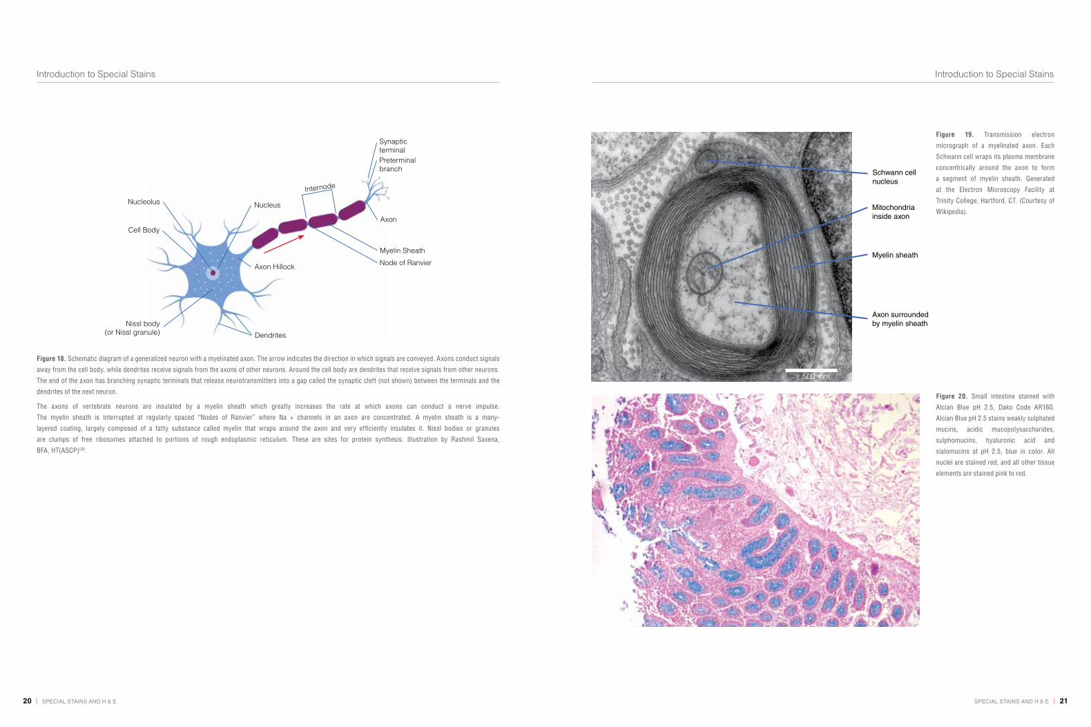
Alcian Blue Used in identifying mucins and glycosaminoglycans.
At pH 2.5, Alcian Blue stains sulphated and non-
sulphated acidic carbohydrates. At pH 1.0, only sulphated
carbohydrates are stained; Fig. 20 and 21
Mucins
Giemsa Stain Used in hematology, e.g., for the detection of
erythroidcolonies, binucleate normoblast, megaloblasts,
mast cells, etc. Giemsa is also used for chromosome
staining; Fig. 22a, 22b and 23
Specific for phosphate groups of DNA
Gomori’s Silver Stain Used for the detection of reticulin in bone marrow Reticulin
Mucicarmine Stain Detects mucins; Fig.1 Mucins
Periodic Acid-Schiff (PAS) Stain Used for staining structures containing a high proportion of
carbohydrate macromolecules (glycogen and glycoprotein),
basement membranes, collagen and primary cell types;
Fig. 24 and 25
Carbohydrate macromolecules by virtue of their content
of galactose, glucose, fucose and mannose
Periodic Acid-Silver Methenamine (PEM) Stain Used for the delineation of basement membranes Carbohydrate macromolecules by virtue of their content
of galactose, glucose, fucose and mannose
Table 2. Commonly used special stains.
10|specialstainsandH&e specialstainsandH&e | 11
Introduction to Special Stains Introduction to Special Stains
Figure 3. Lung stained with Acid-Fast
Bacteria (AFB) Stain, Dako Code AR162.
This AFB stain is suitable for the visual-
ization of acid-fast bacteria belonging
to the Mycobacterium genus on the
Artisan™ Staining System. Application of
carbol-fuchsin stains acid-fast bacteria
fuchsia, followed by decolorization of
all tissue elements except the acid-fast
bacteria. A methylene blue counterstain is
then applied to impart a blue color to all
background tissue elements.
Figure 4a. Electron micrograph (EM)
(negative staining) of H. pylori possessing
multiple flagella. Courtesy of Wikimedia.
Prof. Yutaka Tsutsumi, MD, Department of
Pathology, Fujita Health University School
of Medicine, Japan.
Figure 4b. Illustration of S-shaped H. pylori with four sheathed polar flagella. The majority of helicobacters possess this basic morphology of an S-shape with polar,
sheathed flagella, though variations in size and the number of spirals are seen in a number of other species. These bacteria are usually around 0.5 × 5 μm, and the
S-shaped morphology has been correlated with maximum in vitro motility. Thin sections of H. pylori revealed through an electron microscope show an outer and inner
membrane separated by the periplasm of approximately 30 nm thickness (see EM picture above). The dense cytoplasm contains nucleoid material and ribosomes
(Source: Jani O’Rourke and Günter Bode. Morphology and Ultrastructure of Helicobacter pylori. Physiology and Genetics. Eds. Harry L. T. Mobley, George L. Mendz, and
Stuart L. Hazell. ASM Press. 2001). Illustration by Rashmil Saxena, BFA, HT(ASCP)CM.
Sheathed polar flagella
Body of the Bacteria
12|specialstainsandH&e specialstainsandH&e | 13
Introduction to Special Stains Introduction to Special Stains
Figure 5a. Schematic diagram of intestinal
wall. Illustration by Rashmil Saxena, BFA,
HT(ASCP)CM.
Lactael
Vein
Lympoidnodule
Muscularismucosa
Vein
Circular musculeof muscularis
externa
Longitudinalmuscule ofmuscularis
externa
Villus
Surfaceepithelium
Crypt of Lieberkühn
Laminapropria
Artery
Muscularismucosa
Figure 5b. Giemsa-stained section of
small intestinal mucosa showing clusters
of Giardia that stain purple (arrows) in
the crypts. The background is stained
faint pink by eosin. (Courtesy of Rashmil
Saxena, BFA, HT(ASCP)CM, FRCPath,
Indiana University School of Medicine,
Indianapolis, IN, USA).
Figure 5c. Giardia intestinalis trophozoite:
After ingestion of contaminated food
or water within the small intestine, the
trophozoites reproduce asexually and
either float free or are attached to the
mucosa of the lumen. Some trophozoites
then encyst in the small intestine.
Encystation occurs most likely as a result
of exposure to bile salts and fatty acids,
and a more alkaline environment. Both
cysts and trophozoites are then passed
in the feces, and are infectious imme-
diately or shortly afterward (Legend
courtesy: Centers for Disease Control
(CDC, Atlanta, GA, USA). Illustration by
Rashmil Saxena, BFA, HT(ASCP)CM.
Nucleus
Median Body
Flagella
Axostyle
Figure 6. Biopsy stained with GMS, Dako
Code AR176. The Grocott’s Methenamine
Silver method is utilized for the
visualization of fungi and Pneumocystis
jiroveci in tissue sections using the
Artisan™ Staining System. Fungi and
P. jiroveci are stained black while other
tissue elements are stained green. This
stain can be used on both tissue and
aspirates or smears.
14|specialstainsandH&e specialstainsandH&e | 15
Introduction to Special Stains Introduction to Special Stains
Figure 7. Methenamine Silver Stain.
Histopathologic changes seen in histo-
plasmosis due to Histoplasma capsulatum
var. duboisii. Note the presence of typical
yeast cells, some of which are undergoing
replication by “budding”. Courtesy of Libero
Ajello, PhD, The Centers for Disease Control
and Prevention, Atlanta, GA, USA/Wikimedia.
Figure 8. Grocott’s Methenamine Silver
(GMS) staining of fungi.
Figure 9. Histopathology of Treponema
pallidum spirochetes using a modified
Steiner Silver Stain. Image credit: Dr.
Edwin P. Ewing, Jr., The Centers for
Disease Control and Prevention, Atlanta,
GA, USA/Wikimedia.
Figure 10. Helicobacter stained with
Warthin-Starry, Dako Code AR181. The
arrow points to some black H. pylori
organisms in yellow mucus.

16|specialstainsandH&e specialstainsandH&e | 17
Introduction to Special Stains Introduction to Special Stains
Figure 11. Liver section stained with
a modification of Gomori’s One-Step
Trichrome method that colors collagen
green rather than blue, Dako Code AR166.
Figure 12. Kidney stained with Jones’
Basement Membrane, Dako Code AR180.
The Jones’ Basement Membrane stain
is used for visualization of basement
membranes, specifically glomerular and
tubular basement membranes in renal
tissue. The Bowman’s capsule is stained
black, inner basement membrane - black
to gray, nuclei - red, collagen - rose, and
cytoplasm and other tissue are stained
pink. This stain has been optimized for use
on 2 µm thick tissue sections.
Figure 13. Biopsy stained with Masson’s
Trichrome, Dako Code AR173. This stain
is used to distinguish collagen from
muscle in tissue specimens using the
Artisan™ Staining System. The Trichrome
stain is often used to differentiate
between collagen and smooth muscle
and to identify an increase in collagenous
tissue. With the Masson’s Trichrome stain,
muscle is stained red, collagen - blue,
fibrin - pink, erythrocyte - red and nuclei
- blue/black.
Figure 14. Liver stained with Reticulin/
No Counterstain, Dako Code AR182. The
Reticulin/No Counterstain stain is used
for the visualization of reticulin fibers
in tissue sections using the Artisan™
Staining System.
18|specialstainsandH&e specialstainsandH&e | 19
Introduction to Special Stains Introduction to Special Stains
Figure 15a. Skin stained with Elastic
stain, Dako Code AR163. In this section
Verhoeff’s hematoxylin method has been
counterstained with Van Gieson’s picro-
fuchsine. The Elastic stain is based on
Verhoeff’s technique optimized for the
Artisan™ Staining System. Elastin fibers
and elastic lamina in histological specimens
are stained black, while remaining
tissue elements are stained as follows:
nuclei - blue/black, collagen - red, other
tissue elements - yellow.
Figure 15b. Schematic diagram of skin:
Cross-section. Dermis contains collagen
and elastin which give the skin its form,
shape and elasticity. Illustration by
Rashmil Saxena, BFA, HT(ASCP)CM.Epidermis
Meissner’scorpuscle
Dermis
Oil gland
Sweat gland
Hypodermis
Hair shaft
Arrector pili
Hair follicle
Hair root
Paciniancorpuscle
Vein
Artery
Figure 16. Breast tissue stained with
Feulgen, Dako Code AR174. The Feulgen
stain is used to demonstrate DNA in
tissue sections. RNA is not stained
by this procedure. The DNA is stained
magenta with Schiff’s reagent. The stained
DNA is contrasted against a light green
counterstain to allow better visualization
by light microscopy or image analysis.
Figure 17. Amyloid stained with Congo
Red, Dako Code AR161. The Congo
Red stain is used to detect amyloid, an
abnormal protein product that can be
found in various pathologic conditions.
This stain is based on Benhold’s and
demonstrates amyloid in pink to dark
salmon with light microscopy or the
characteristic “apple-green birefringence’’
with polarized light. Mayer’s hematoxylin
is used as a counterstain. The preferred
method for visualization of amyloid is
under polarized light.
20|specialstainsandH&e specialstainsandH&e | 21
Introduction to Special Stains Introduction to Special Stains
Figure 18. Schematic diagram of a generalized neuron with a myelinated axon. The arrow indicates the direction in which signals are conveyed. Axons conduct signals
away from the cell body, while dendrites receive signals from the axons of other neurons. Around the cell body are dendrites that receive signals from other neurons.
The end of the axon has branching synaptic terminals that release neurotransmitters into a gap called the synaptic cleft (not shown) between the terminals and the
dendrites of the next neuron.
The axons of vertebrate neurons are insulated by a myelin sheath which greatly increases the rate at which axons can conduct a nerve impulse.
The myelin sheath is interrupted at regularly spaced “Nodes of Ranvier” where Na + channels in an axon are concentrated. A myelin sheath is a many-
layered coating, largely composed of a fatty substance called myelin that wraps around the axon and very efficiently insulates it. Nissl bodies or granules
are clumps of free ribosomes attached to portions of rough endoplasmic reticulum. These are sites for protein synthesis. Illustration by Rashmil Saxena,
BFA, HT(ASCP)CM.
Nucleolus Nucleus
Internode
Node of Ranvier
Myelin Sheath
Axon
Synaptic terminal
Axon Hillock
Preterminalbranch
Cell Body
Nissl body(or Nissl granule) Dendrites
500 nm
Schwann cell nucleus
Mitochondriainside axon
Myelin sheath
Axon surrounded by myelin sheath
Figure 19. Transmission electron
micrograph of a myelinated axon. Each
Schwann cell wraps its plasma membrane
concentrically around the axon to form
a segment of myelin sheath. Generated
at the Electron Microscopy Facility at
Trinity College, Hartford, CT. (Courtesy of
Wikipedia).
Figure 20. Small intestine stained with
Alcian Blue pH 2.5, Dako Code AR160.
Alcian Blue pH 2.5 stains weakly sulphated
mucins, acidic mucopolysaccharides,
sulphomucins, hyaluronic acid and
sialomucins at pH 2.5, blue in color. All
nuclei are stained red, and all other tissue
elements are stained pink to red.

22|specialstainsandH&e specialstainsandH&e | 23
Introduction to Special Stains Introduction to Special Stains
Figure 22b. Giemsa staining. Atypical
mononuclear megakaryocyte in chronic
myeloid leukemia. (Figure from Dako
Education Guide, “The Illustrated Guide
to Bone Marrow Diagnosis,” 2nd Edition
(2009). Editors: Carlos Martin, MD, and
George L. Kumar, PhD).
Figure 21. Small intestine stained with Alcian Blue/PAS, Dako Code AR169. This stain is used for the demonstration of neutral and acidic mucosubstances on the
Artisan™ Staining System. Alcian Blue pH 2.5 imparts a blue color to the acidic mucins and other carboxylated or weakly sulphated acid mucosubstances. The
periodic acid-Schiff (PAS) reaction is then used to stain basement membranes, glycogen and neutral mucosubstances pink to red. Mixtures of neutral and acidic
mucosubstances will appear purple due to positive reactions with both Alcian Blue and PAS.
Figure 22a. Cell types seen in normal bone
marrow. Giemsa staining. (Figure from
Dako Education Guide, “The Illustrated
Guide to Bone Marrow Diagnosis,” 2nd
Edition (2009). Editors: Carlos Martin, MD,
and George L. Kumar, PhD).
Normoblast
Plasma cell
Megakaryocyte
Erythroblast
Eosinophil
Myelocyte
Neutrophil

24|specialstainsandH&e specialstainsandH&e | 25
Introduction to Special Stains Introduction to Special Stains
Figure 23. Spleen stained with Giemsa, Dako Code AR164. Cell types are stained as follows: mast-cell granules and basophils - purple, eosinophils - bright pink,
lymphocytes - blue.
Figure 24. Trachea stained with Alcian
Blue/PAS/Hematoxylin, Dako Code AR178.
This stain is used for the demonstration
of neutral and acidic mucosubstances
on the Artisan™ Staining System. Alcian
Blue pH 2.5 imparts a blue color to the
acidic mucins and other carboxylated or
weakly sulfated acid mucosubstances.
The periodic acid-Schiff (PAS) reaction is
then used to stain basement membranes,
glycogen and neutral mucosubstances
pink to red. Mixtures of neutral and acidic
mucosubstances will appear purple due to
positive reactions with both Alcian Blue
and PAS. A hematoxylin counterstain is
then applied to impart a blue/black color
to the nuclei.
Figure 25. Kidney stained with PAS, Dako
Code AR165.

26|specialstainsandH&e specialstainsandH&e | 27
Introduction to Special Stains Introduction to Special Stains
Conclusionspecialstainsbelongtoanassortedfamilyofstainsformicroscopic
visualizationandgeneralidentificationofcells,tissuesandmicro-
organisms. special stains remain an important tool for many
pathologistsandtechnologistsprovidingapowerfulcomplement
toimmunohistochemistry,flowcytometry,insituhybridizationand
otherdiagnostictechnologiesthatdefineapatient’smedicalprofile.
Withthemedicalcommunitydemandinggreaterstandardizationand
qualitycontrol,specialstainprotocolshavebecomeincreasingly
automatedresultinginhigherlevelsofproductivityandflexibility.
automationisnosubstituteforasolidunderstandingoftheprinciples
andpracticesofaqualitystaining.Weanticipatethatthistechnology
willcontinuetoevolveintheforeseeablefutureandexpectittoform
anintegralpartofpathologicdiagnosis.inanutshell,thisintroduction
wasintendedtoprovideguidancetohelpinterestedreadersacquire
proficiencyinselectingandperformingspecialstainsfasterthanthey
mighthaveotherwisedone.
Appendix Biological Stain Commissionthe Us-based Biological stain commission was an indirect
consequenceofWorldWari.duringtheGreatWartherewasa
blockadeofGermanproducts,includingdyes.By1920,thesupplyof
pre-wardyeswasalmostexhausted,foreignsupplieswereerratic,and
thedomesticdyeswerestilloftenunsatisfactory.asaconsequence,
severalconcernedgroupsandindividualscametogether,which
resultedintwokeyconferencesin1921onthestandardizationof
stains.Fromthisactivity,thecommissiononthestandardizationof
Biologicalstainsoriginated.By1923,thecommissionalreadyhad
aconstitutionthatisrecognizablytheforerunneroftheaimsofthe
presentcommission.inparallelwiththis,co-founderdr.HaroldJ.
conn,whilechairmanofthecommission,publishedthefirstedition
ofBiologicalstains in1925.thisbookhasbecomeastandard
sourceofreferenceintechnicalandresearchhistopathological
andbiologicallaboratoriesusingdyes.thebookhasbeenrevised
regularlywitha10thedition(2002)asthemostrecentversion.in
1944,thecommissiononthestandardizationofBiologicalstains
becametheBiologicalstaincommission.
theobjectivesoftheBiologicalstaincommissionare:1)toensure
anuninterruptedsupplyofdyesusedinbiologicalandmedical
applications, 2) to promote cooperation and dialogue among
manufacturers, vendors and users of dyes for histochemical
applications,3)toensurethequalityofdyesthroughindependent
testingaccordingtoappropriatelyrigorouschemicalandperformance
criteria,4)toeducateusersofbiologicalstainsaboutsourcesof
reliabledyesandhowtheymightbestbeused,and5)topublish
informationconcerningneworimprovedusesforbiologicaldyesand
relatedhistochemicaltechniques.
theseobjectivesaremetbywayof:1)analyzingdyecontentand
compositionofsamplessuppliedvoluntarilybydyemanufacturers
orvendors,2)testingtheperformanceofdyesamplesinrigorous,
standardizedproceduresknowntobediscerningtestsofthestaining
qualityofthedye,3)issuingcertificationlabelstobeattachedtothe
containersusedbycompaniesmarketingaccepteddyestoassure
consumersthatthesedyeshavemettheperformancecriteriaofthe
Biologicalstaincommission,4)conductingandsupportingresearch
onbiologicaldyesandhistochemicaltechniquesdependentondyes,
5)publishingbooksconcerningbiologicaldyesandhistochemical
techniques,andpublishingBiotechnic&Histochemistry,abimonthly
journalofmicrotechniqueandhistochemistry,and6)maintaining
anactivedialogueamongscientists,manufacturersandvendors
concernedwithbiologicalstains.
interestedreaderscanlearnmuchmoreabouttheBiologicalstain
commissionatitsWebsite:http://www.biologicalstaincommission.org/.
Anatomic Pathology Checklist by College of American PathologistsseveralthousandUsanatomicpathologylaboratoriesareinspected
bythecollegeofamericanpathologistsforaccreditationpurposes
requiredbytheclinicallaboratoryimprovementamendmentsof
1988(clia’88).theycanexpecttobeaskedthesetwoquestions
aboutspecialstains(seetextinbold,page27):

28|specialstainsandH&e specialstainsandH&e | 29
Introduction to Special Stains
Bibliography
1. Wissowzkya(1876).UeberdaseosinalsreagenzaufHämoglobinunddie Bildung von Blutgefässen und Blutkörperchen bei säugetier undHühnerembryonen.Archiv für mikroskopische Anatomie;13:479-496.
2. Horobin RW, Kiernan Ja, eds (2002). conn’s Biological stains:aHandbookofdyes,stainsandFluorochromesforUseinBiologyandMedicine.10thed.Oxford,UK:BiOsscientificpublishers.
3. thesocietyofdyersandcolouristsHomepage.accessedaugust27,2009at:http://www.sdc.org.uk/.
4. RotimiO,cairnsa,Grays,Moayyedip,dixonMF(2000).Histologicalidentification of Helicobacter pylori: comparison of staining methods.J Clin Pathol;53(10):756-759.
5. churukiancJ(2009).MethodoftheHistochemicalstains&diagnosticapplication, department of pathology and laboratory Medicine,University of Rochester, Rochester nY, second web edition (2009).accessedaugust27,2009at:http://www.urmc.rochester.edu/path/zqu/stainsManual/index.html.
6. carsonFl,Hladikc(2009).Histotechnology:aself-instructionaltext.3rded.chicago,il:ascppress;2009.
7. Wulffs,(ed.)(2004).educationGuide:specialstains.carpinteria,ca:daKO.
8. commission on laboratory accreditation: laboratory accreditationprogram. anatomic pathology checklist – Revised 06/15/2009.collegeofamericanpathologists,northfieldil.
9. Baker JR (1958). principles of Biological Microtechnique: a study ofFixationanddyeing.Bungay,suffolk:Methuen&co.,ltd.,1958.
10. Garrett RH, Grisham cM (2010). Biochemistry. 4th ed. Boston, Ma:cengagelearning.
11. Horobin RW, Bancroft Jd (1998). troubleshooting Histology stains.newYork:churchilllivingstone.
12. Horobin RW (1982). Histochemistry: an explanatory Outline ofHistochemistryandBiophysicalstaining.london:Butterworths;1982.
13. KiernanJa(2009).staining,Histochemistry,andHistotechnologyFaQ.accessedaugust21,2009at:http://publish.uwo.ca/~jkiernan/faqlist.htm.
14. JonesdB(1951).inflammationandrepairoftheglomerulus.Am J Path 27:991-1009.
AcknowledgmentsThe authors acknowledge with gratitude John A. Kiernan, PhD,
Alton D. Floyd, PhD, and Jamie Nowacek, BS, for their critical
reviews and helpful suggestions. We would also like to thank
Sunil Badve, MD and Rashmil Saxena, BFA, HT(ASCP)CM for
providing us H&E stained section of the skin and Giemsa stained
intestinal sample, respectively.
Chapter 2 General Oversight Stains for Histology and Histopathology
John A. Kiernan, MB, ChB, PhD, DSc
Fornormalordiseasedtissuesofhumansandothervertebrate
animals,aroutinelyusedstainingmethodforuseonparaffinorfrozen
(cryostat)sections4-7μmthickisexpectedtoprovideintenseblue,
purpleorblackcolorationthatislargelyconfinedtochromatininthe
nucleiofcells,togetherwithacontrastingandpalercolorsuchaspink
oryellowinthesurroundingcytoplasmandinextracellularstructures
(notablycollagenfibers).
Haemalum and EosinForabout130yearstheoversightnuclearstainoffirstchoicehas
beenhaemalum,whichmaybeoneofmanysolutionscontaining
hematein,aluminumionsandusuallyotheringredients.Haemalum
stainsnuclearchromatinblue.For thesecondcolor,called the
counterstain,reddyesaregenerallypreferred,especiallyeosin,
whichcanprovidearangeoforangehues.eosinalsochangesthe
colorofhaemalum-stainednuclearchromatinfrombluetowards
purple.sectionsstainedwithhaemalumandeosinarethefirstones
tobeexaminedbyhumanandveterinarypathologistsexamining
surgicalorpostmortemspecimens,andoftenaresufficientfor
diagnosis.Otherstainingmethodsareusedasrequired,especially
toshowcytoplasmicandextracellularcomponentsthatdonotstain
distinctlywitheitherhaemalumoreosin.
Haemalum
thecombinationofhaemalumandeosiniscalledH & E.thismethod
isalsocalledhematoxylin and eosinbecausehaemalumsolutions
aremadebydissolvinghematoxylininwateroralcohol,oxidizing
someofittohematein,andaddinganaluminumsalt.Hematoxylin
(colorlesswhenpure)andhematein(yellowinacid,red-violetin
neutralandalkalinesolutions,changingatpH5–6)probablyarenot
directlyinvolvedinthestainingofnucleibyhaemalum.
therearemanyformulationsofhaemalum.almostallcontainan
excessofaluminumionsoverhaemateinmoleculesandalsocontain
anorganicacidsuchasaceticorcitric;thepHisusuallyintherange
2.0–3.5.Othersubstances inhaemalumsolutionsmay include
anoxidizingagent,oftensodiumiodate,toacceleratehematein
generation,andanorganicliquidsuchasglycerolorethyleneglycol,
whichdoesnotaffectstainingpropertiesbutmayextendtheshelf-
lifebyretardingevaporationandprecipitationofinsolublematerials.
Mixtureswithahighaluminum:hemateinratiostainsectionsslowly,
withselectivecolorationofnuclearchromatintypicallybeingachieved
in5to15minutes.thisiscalledprogressive staining.solutions
withloweraluminum:hemateinratiosrapidlycolorallcomponents
of thetissue.selectivecolorationofnuclei is thenachievedby
differentiation(alsocalleddestaining)inadilutemineralacidsuch
as0.1MHcl(pH1.0).thisremovesdye-metalcomplexesfromthe
tissue,decomposesthecomplexesintohemateinandaluminum
ions,andacceleratesthefurtheroxidationofhemateintoother
compounds.aluminum-hemateincomplexesattachedtonuclear
chromatinaremoreresistanttodifferentiationthanthoseattached
toothersubstances.inpractice,differentiationisusuallydonewith
acid-alcohol,whichis70%or95%ethanolwith1%v/vconcentrated
Hcl.selectivenuclearstainingachievedbydifferentiationiscalled
regressive staining.
thechemicalcompositionsofsolutionscontaininghaemateinand
aluminumionshavebeeninvestigatedbyelectrophoresisandby
spectrophotometry.thesestudiesshowtheexistenceofcationic
hematein-aluminum(Hmal)complexes,principallythered[Hmal]2+
atpH2.6,whichistheacidityofpracticalstainingsolutions.asoluble
bluecomplex,[Hmal2]3+,existsatpH4.7andischangedathigher
pHtoinsolubleblue,presumablypolymeric,materialthatcanbere-
dissolvedbyacidification.theequilibriaaresummarizedinFigure1.
anioniccomplexes, including[Hm2al]−,havebeenshowntobe
involvedintextiledyeingbycomplexesofhemateinwithvarious
metalions,andtheirinvolvementinnuclearstainingbyhaemalum
hasbeenpostulated.
intissuesstainedbyhaemalum,progressivelyorregressively,cell
nucleiaredullbrownish-red.thecolorischangedtoblue(Figure2)
byrinsingintapwater(ifitspHisabove5)ordistilledwaterthat
hasbeenmadeslightlyalkaline,aswithafewdropsofammonium
hydroxide.this“blueing”convertstheredHm-alcomplexionsto
bluepolymersthatareinsolubleinwaterandorganicsolventsand
alsoareremarkablyresistanttofading.theblueproductresists
extractionbyweaklyacidiccounterstains,suchaseosinin0.5%
aceticacid,butitisremovedbysolutionsofstrongeracidssuchas