Specifi city in reactive oxidant signaling: think globally
9
THE JOURNAL OF CELL BIOLOGY JCB: MINI-REVIEW © The Rockefeller University Press $8.00 The Journal of Cell Biology, Vol. 174, No. 5, August 28, 2006 615–623 http://www.jcb.org/cgi/doi/10.1083/jcb.200605036 JCB 615 Introduction The now burgeoning field of oxidants in biological systems rose from obscurity with the cardinal discovery that the common red cell enzyme erythrocuprein was both ubiquitous and protec- tive, functioning as a superoxide dismutase (SOD; McCord and Fridovich, 1969). The resultant syllogism therefore depicted free radicals as both ubiquitous and dangerous, and oxidant stress was rapidly established as a common mechanism linking inflamma- tory, degenerative, and neoplastic processes in human disease. The propensity of oxidants to initiate chain reactions and select targets based on redox potential rather than cellular function further sug- gested a capriciousness of oxidative reactions that destroyed the delicate biochemical specificity required by various signaling machines. Only in the past decade has it become clear that most, if not all, multicellular organisms have evolved molecular strategies to intentionally produce these unruly chemicals for, of all things, signaling purposes, prompting the question “whence specificity?” The NADPH oxidases are evolutionarily ancient Although they are not the only source of oxidants, the NADPH oxidase (Nox) family members are the principal complexes that function solely to redox-couple NADPH and molecular oxygen to generate O 2 .− and, thence, H 2 O 2 . Thus, the examination of Nox biology reveals much about the cellular logic behind regu- lated oxidant production. The seven known human Noxs include Nox1–5 and Duox1–2, with Nox2 (gp91 phox ) being the founding member. As a family, these oxidases participate in a variety of adaptive functions, ranging from mitogenesis to immune cell signaling (Geiszt and Leto, 2004; Lambeth, 2004). Reflecting these varied biological roles, the Nox proteins have been impli- cated in several cell-fate pathways, such as the Ras mitogenic pathway (Irani et al., 1997), the MAP kinases (Gu et al., 2002; Xu et al., 2002), the JAK–STAT pathways (Schieffer et al., 2000), and NF-κB (Sulciner et al., 1996; Gu et al., 2003). Nox-dependent signaling has been a biologically success- ful device by all accounts, having appeared early and persisted throughout evolution on the aerobic earth. Orthologous Nox genes arose in concert with multicellular organization (Lalucque and Silar, 2003), and so are found as early as the slime mold Dictyostelium discoideum and the filamentous fungus Podospora anserina (Malagnac et al., 2004; Lardy et al., 2005). During starvation conditions, free Dictyostelial amoebae aggregate into a slug that behaves as a single organism, differentiating a dis- tinct organ, the spore-bearing fruiting body. Although single de- letions of any of the three nox genes or p22 phox fail to produce a phenotype in unicellular amoebae, starvation of these knockout mutants interrupts fruiting body morphogenesis (Lardy et al., 2005). Similarly, deletion of either of the two P. anserina Nox genes results in failed fruiting body differentiation. (Malagnac et al., 2004). Thus, Noxs control developmental signaling in the most primitive multicellular organisms, an ancestral function that foreshadowed their later involvement in basic mammalian cell fate pathways. One might fairly ask why the utilization of reactive oxidants has been so evolutionarily durable and how oxidants can manage to selectively relay a diverse array of sig- naling cassettes, especially because the different Noxs presum- ably produce the same oxidant species perceived by the cell as an oxidative threat. Physical organization of signaling elements is a common strategy for pathway specificity A general paradigm in cell signaling holds that information pro- ceeds through pathway-specific multimolecular complexes built Specificity in reactive oxidant signaling: think globally, act locally Lance S. Terada Department of Medicine, University of Texas Southwestern Medical Center, Dallas, TX 75390 Dallas VA Medical Center, Dallas, TX 75216 Although reactive oxidants have long been stigmatized as unwanted metabolic byproducts, the expression of oxi- dases specifically functioning to produce these same mol- ecules in a regulated fashion is surprisingly pervasive throughout metazoan and plant evolution. Although the involvement of oxidants in many signaling pathways is well documented, the cellular strategies for conferring pathway specificity to such reactive molecules have re- mained more recondite. Recent studies now suggest that cells may spatially restrict oxidant production to allow microdomain-specific signaling. Correspondence to [email protected] Abbreviations used in this paper: AMPK, AMP-activated protein kinase; BCR, B lymphocyte antigen receptor; GEF, guanine nucleotide exchange factor; PTP, protein tyrosine phosphatase; SOD, superoxide dismutase; VDAC, voltage- dependent anion channel. on April 4, 2019 jcb.rupress.org Downloaded from http://doi.org/10.1083/jcb.200605036 Published Online: 21 August, 2006 | Supp Info:
Transcript of Specifi city in reactive oxidant signaling: think globally
TH
EJ
OU
RN
AL
OF
CE
LL
BIO
LO
GY
JCB: MINI-REVIEW
© The Rockefeller University Press $8.00The Journal of Cell Biology, Vol. 174, No. 5, August 28, 2006 615–623http://www.jcb.org/cgi/doi/10.1083/jcb.200605036
JCB 615
IntroductionThe now burgeoning fi eld of oxidants in biological systems rose
from obscurity with the cardinal discovery that the common red
cell enzyme erythrocuprein was both ubiquitous and protec-
tive, functioning as a superoxide dismutase (SOD; McCord and
Fridovich, 1969). The resultant syllogism therefore depicted free
radicals as both ubiquitous and dangerous, and oxidant stress was
rapidly established as a common mechanism linking infl amma-
tory, degenerative, and neoplastic processes in human disease. The
propensity of oxidants to initiate chain reactions and select targets
based on redox potential rather than cellular function further sug-
gested a capriciousness of oxidative reactions that destroyed the
delicate biochemical specifi city required by various signaling
machines. Only in the past decade has it become clear that most, if
not all, multicellular organisms have evolved molecular strategies
to intentionally produce these unruly chemicals for, of all things,
signaling purposes, prompting the question “whence specifi city?”
The NADPH oxidases are evolutionarily ancientAlthough they are not the only source of oxidants, the NADPH
oxidase (Nox) family members are the principal complexes that
function solely to redox-couple NADPH and molecular oxygen
to generate O2.− and, thence, H2O2. Thus, the examination of
Nox biology reveals much about the cellular logic behind regu-
lated oxidant production. The seven known human Noxs include
Nox1–5 and Duox1–2, with Nox2 (gp91phox) being the founding
member. As a family, these oxidases participate in a variety of
adaptive functions, ranging from mitogenesis to immune cell
signaling (Geiszt and Leto, 2004; Lambeth, 2004). Refl ecting
these varied biological roles, the Nox proteins have been impli-
cated in several cell-fate pathways, such as the Ras mitogenic
pathway (Irani et al., 1997), the MAP kinases (Gu et al., 2002;
Xu et al., 2002), the JAK–STAT pathways (Schieffer et al.,
2000), and NF-κB (Sulciner et al., 1996; Gu et al., 2003).
Nox-dependent signaling has been a biologically success-
ful device by all accounts, having appeared early and persisted
throughout evolution on the aerobic earth. Orthologous Nox
genes arose in concert with multicellular organization (Lalucque
and Silar, 2003), and so are found as early as the slime mold
Dictyostelium discoideum and the fi lamentous fungus Podospora anserina (Malagnac et al., 2004; Lardy et al., 2005). During
starvation conditions, free Dictyostelial amoebae aggregate into
a slug that behaves as a single organism, differentiating a dis-
tinct organ, the spore-bearing fruiting body. Although single de-
letions of any of the three nox genes or p22phox fail to produce a
phenotype in unicellular amoebae, starvation of these knockout
mutants interrupts fruiting body morphogenesis (Lardy et al.,
2005). Similarly, deletion of either of the two P. anserina Nox
genes results in failed fruiting body differentiation. (Malagnac
et al., 2004). Thus, Noxs control developmental signaling in the
most primitive multicellular organisms, an ancestral function
that foreshadowed their later involvement in basic mammalian
cell fate pathways. One might fairly ask why the utilization of
reactive oxidants has been so evolutionarily durable and how
oxidants can manage to selectively relay a diverse array of sig-
naling cassettes, especially because the different Noxs presum-
ably produce the same oxidant species perceived by the cell as
an oxidative threat.
Physical organization of signaling elements is a common strategy for pathway specifi cityA general paradigm in cell signaling holds that information pro-
ceeds through pathway-specifi c multimolecular complexes built
Specifi city in reactive oxidant signaling: think globally, act locally
Lance S. Terada
Department of Medicine, University of Texas Southwestern Medical Center, Dallas, TX 75390Dallas VA Medical Center, Dallas, TX 75216
Although reactive oxidants have long been stigmatized as
unwanted metabolic byproducts, the expression of oxi-
dases specifi cally functioning to produce these same mol-
ecules in a regulated fashion is surprisingly pervasive
throughout metazoan and plant evolution. Although the
involvement of oxidants in many signaling pathways is
well documented, the cellular strategies for conferring
pathway specifi city to such reactive molecules have re-
mained more recondite. Recent studies now suggest that
cells may spatially restrict oxidant production to allow
microdomain-specifi c signaling.
Correspondence to [email protected]
Abbreviations used in this paper: AMPK, AMP-activated protein kinase; BCR, B lymphocyte antigen receptor; GEF, guanine nucleotide exchange factor; PTP, protein tyrosine phosphatase; SOD, superoxide dismutase; VDAC, voltage- dependent anion channel.
on April 4, 2019jcb.rupress.org Downloaded from http://doi.org/10.1083/jcb.200605036Published Online: 21 August, 2006 | Supp Info:
JCB • VOLUME 174 • NUMBER 5 • 2006 616
on colocalizing scaffolds as a means of maximizing effi ciency
and attaining specifi city beyond what would be allowed by us-
ing the limited number of signaling proteins as individual, freely
diffusible agents within a crowded cytosol. The logic behind
such quaternary spatial organization would fi t well with the use
of oxidants as locally active mediators if two conditions are met.
First, the source of oxidants should likewise be tightly regu-
lated, not only from an agonistic standpoint but also in terms of
strict subcellular localization. Second, a broad fi eld of anti-
oxidant activity must be present within the cytosol to confi ne
oxidative effects to within proximity of their origin, in essence,
optimizing spatial signal-to-noise ratios. The latter criterion has
long been established, as several antioxidant enzymes are,
in fact, largely cytosolic, such as Cu/Zn SOD and glutathione
peroxidase. Pathways that produce oxidants as signifi cant meta-
bolic byproducts tend to be sequestered within organelles,
whose defenses are correspondingly buttressed by higher con-
centrations of these or other antioxidant enzymes (such as cata-
lase in peroxisomes or MnSOD in mitochondria). Even the
exceptions to antioxidant distribution tend to prove the rule. For
instance, peroxiredoxin II, through its association with PDGFR,
suppresses PDGF signaling, whereas the less targeted catalase
and glutathione peroxidase have no effect (Choi et al., 2005).
What evidence exists that the former criterion, i.e., that oxidases
are focally activated, is also fulfi lled?
Nox subunits are directed to specifi c platforms
The cytoskeleton. Different cells, when imaged with
different oxidant-detection methods, display subcellular restric-
tion of oxidant activity around regions of cytoskeletal rearrange-
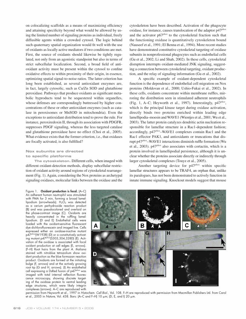
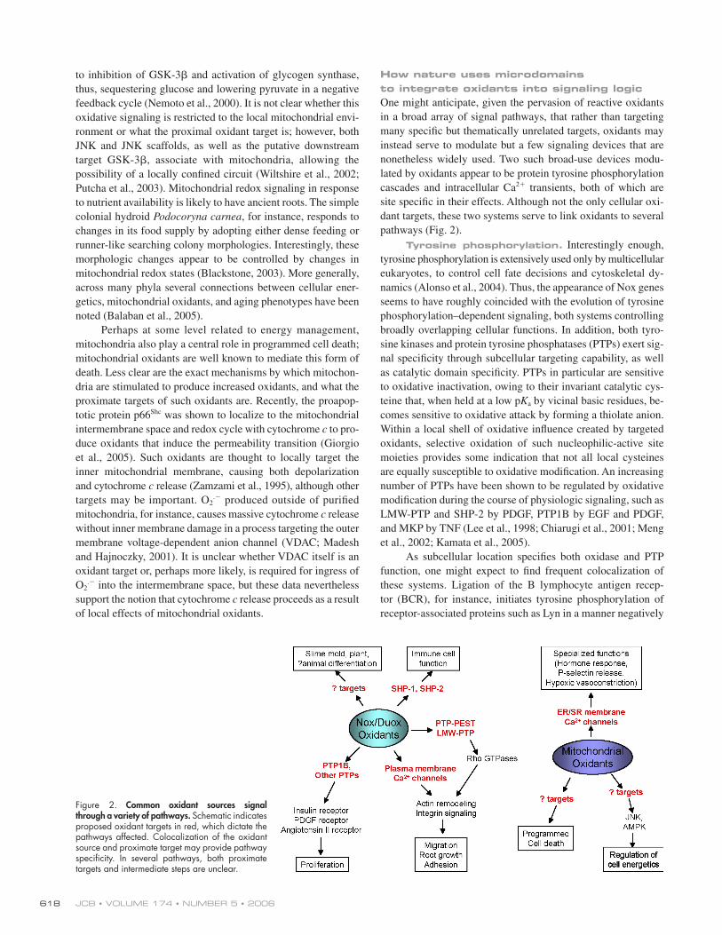
ment (Fig. 1).[ID]FIG1[/ID] Again, considering the Nox proteins as archetypal
signaling oxidases, molecular links between the oxidase and the
cytoskeleton have been described. Activation of the phagocyte
oxidase, for instance, causes translocation of the adaptor p47phox
and the activator p67phox to the cytoskeletal fraction such that
the functioning oxidase is quantitatively cytoskeleton bound
( Nauseef et al., 1991; El Benna et al., 1994). More recent studies
have demonstrated constitutive cytoskeletal targeting of oxidase
subunits in nonprofessional phagocytes such as endothelial cells
(Gu et al., 2002; Li and Shah, 2002). In these cells, cytoskeletal
disruption interrupts oxidant-mediated JNK signaling, suggest-
ing a connection between cytoskeletal targeting, oxidant produc-
tion, and the relay of signaling information (Gu et al., 2002).
A specifi c example of oxidant-dependent cytoskeletal
function is the dependence of endothelial cell migration on Nox
proteins (Moldovan et al., 2000; Ushio-Fukai et al., 2002). In
these cells, oxidants concentrate within membrane ruffl es, mir-
roring the distribution seen in stimulated adherent neutrophils
(Fig. 1, A–C; Heyworth et al., 1997). Interestingly, p47phox,
which is the principal kinase target during oxidase activation,
directly binds two proteins enriched within leading edge
lamellipodia–moesin and WAVE1 (Wientjes et al., 2001; Wu et al.,
2003). The latter protein catalyzes dendritic actin nucleation re-
sponsible for lamellar structure in a Rac1-dependent fashion;
accordingly, p47phox–WAVE1 complexes contain Rac1 and the
Rac1 effector PAK1, and antioxidants or truncations that dis-
rupt p47phox–WAVE1 interactions diminish ruffl e formation (Wu
et al., 2003). p47phox also associates with cortactin, which is a
protein involved in lamellipodial persistence, although it is un-
clear whether the proteins associate directly or indirectly through
larger cytoskeletal complexes (Touyz et al., 2005).
Another targeting device for p47phox within specifi c
lamellar structures appears to be TRAF4, an orphan that, unlike
its paralogues, has not been demonstrated to actively function in
innate immune signaling. Knockout models suggest that mouse
Figure 1. Oxidant production is focal. (A–C) An adherent human neutrophil was stimulated with PMA for 3 min, forming a broad lamel-lipodium (arrowheads). H2O2 was detected as a cerium perhydroxide reaction product (B) and was pseudocolored and overlaid on the phase-contrast image (C). Oxidants are heavily concentrated in the ruffl ing lamel-lipodium. (D and E) Endothelial cells were loaded with the oxidant-sensitive fl uorescent dye dichlorofl uorescein and imaged live. Cells expressed either an oxidase-inactive mutant p47phox(W193R) (D) or a constitutively activat-ing mutant p47phox(S303,304,328D) (E). Acti-vation of the oxidase is associated with focal oxidant production at cell edges (E, arrows). (F–H) Root hairs from the plant A. thaliana stained with nitroblue tetrazolium show oxi-dant production as the blue formazan reaction product. Oxidants are formed at the initiating bulge (F, arrows) and at the actively growing root tip (G and H, arrows). (I) An endothelial cell expressing a DsRed fusion of p47phox was imaged with total internal refl ection fl uores-cence microscopy, showing discrete target-ing of the oxidase protein to ventral leading edge structures, which were likely integrin complexes (arrows). A–C are reproduced with permission from Heyworth et al., 1997 in Histochem. Cell Biol., Vol. 108. F–H are reproduced with permission from Macmillan Publishers Ltd. from Carol et al., 2005 in Nature, Vol. 438. Bars: (A–C and F–H) 10 μm; (D, E, and I) 20 μm.
FOCAL OXIDANT SIGNALING • TERADA 617
TRAF4 and the Drosophila melanogaster orthologue dTRAF1
instead control ontogenic migration during respective dorsal
closure events (Liu et al., 1999; Regnier et al., 2002). In the
fl y, dTRAF1 operates within a Rho-GTPase/JNK cassette
during cell migration, and a parallel situation in human endo-
thelial cells may require a direct interaction between TRAF4
and p47phox. This interaction governs oxidant-dependent JNK
activation, and endothelial cell migration involves TRAF4-
dependent activation of the NADPH oxidase through the Rho-
GTPases and PAK1 (Xu et al., 2002; Wu et al., 2005). TRAF4
and p47phox target focal integrin complexes within the lamel-
lipodia of motile endothelial cells, tethered by the focal con-
tact scaffold Hic-5. Thus, TRAF4 appears to function in this
regard by focusing the activation of the oxidase to a specifi c
cytoskeletal structure.
Besides p47phox phosphorylation, Rac1 activation is also
required to activate many Noxs; thus, sites of Rac1 activation
may also be expected to specify the subcellular location of Nox-
dependent signaling complexes. Active Rac1, for instance, con-
centrates within ruffl ing lamellae, suggesting spatial coordination
of Rac’s cytoskeletal and prooxidant effects (Kraynov et al.,
2000). One potential mechanism for Rac1 targeting is through
the actin-binding scaffold IQGAP, which targets leading edge
actin structures and mediates cell migration (Mataraza et al.,
2003). IQGAP not only binds and, therefore, localizes the active
forms of Rac1 and Cdc42 but also associates with VEGFR2 and
Nox2 at leading edge structures, mediating VEGF-dependent
oxidant production (Ikeda et al., 2005).
Another tactic cells use to spatially restrict Rac1 function
is local exclusion of Rho-GDI. A striking example of how Rho-
GDI specifi es Nox activation sites was recently demonstrated in
the plant Arabidopsis thaliana. Focal cytoskeletal rearrange-
ments within the specialized trichoblast cell cause a single root
hair to extend from each cell. A mutation resulting in the root
hair–defective phenotype localizes to the gene for a plant Nox,
RHD2/AtrbohC (Foreman et al., 2003). Although wild-type
plants produced oxidants confi ned to the tip of extending root
hairs (Fig. 1, F–H), rhd2 mutants neither produced oxidants nor
formed root hairs. Conversely, diffuse exposure to exogenous
oxidants caused loss of spatial control, with the resultant forma-
tion of numerous aberrant root hairs. Two subsequent A. thaliana
mutants causing a similar phenotype of multiple aborted growth
bulges (supercentipede) were found to encode SCN1, which is
a Rho-GDI (Carol et al., 2005). Whereas wild-type plants dem-
onstrated a single focus of oxidant production at the growing
root hair tip, scn1 mutants displayed multiple foci of oxidants
corresponding to abnormal growth bulges. Therefore, the plant
Rho-GDI SCN1 functions to restrict oxidant production exclu-
sively to a single root tip.
A third method of localizing Rac1 activation is through
targeting of Rho guanine nucleotide exchange factors (GEFs)
with Rac1 activity. Recruitment of a Rac1 GEF is suggested by
the association of human Nox1 with the Rac GEF βPIX (Park
et al., 2004). Thus, βPIX, which is known to modulate EGFR
function, activates Rac1, causing EGF-dependent oxidant
production. In addition, Rap1a, which associates with the Nox2
complex, targets membrane protrusions and locally activates the
Rac GEFs Vav2 and Tiam1, and thus Rac1 itself, at the lame lli-
podial edge (Arthur et al., 2004).
Membrane rafts. Membrane rafts are known to facil-
itate the congregation of several signaling proteins, including Nox
subunits. In suspended myeloid cells, for example, the Nox2 cy-
tochrome subunits constitutively sequester in raft fractions, with
translocation of the soluble proteins p47phox and p67phox into rafts
after stimulation (Vilhardt and Van Deurs, 2004). Raft associa-
tion of the mitogenic Nox1 has also been noted in smooth mus-
cle cells (Hilenski et al., 2004), and angiotensin II stimulation,
which proceeds through Nox1, promotes Rac1 traffi cking into
rafts, whereas raft disruption blocks angiotensin II–dependent
oxidative signaling (Zuo et al., 2004). Similarly, rafts contain
the focal complex–associated TRAF4, and raft disruption blocks
TRAF4-dependent oxidative signaling (Wu et al., 2005).
The association of Nox proteins with raft microdomains
may explain, in part, why oxidant production by Noxs, which is
presumed to be directed outside the cell, can affect intracellular
targets. Plasma membrane rafts containing Rac1 are known to
be internalized in response to integrin signals (del Pozo et al.,
2004), and caveola-derived signaling endosomes, which are a
type of membrane-derived “signalosome,” continue to trans-
duce growth factor receptor signals after internalization. Indeed,
small caveolin-containing vesicles termed cavicles are thought
to be transported, possibly as microtubular cargo, between the
plasma membrane and pericentrosomal caveosomes (Mundy
et al., 2002). Although it is as yet unclear whether functioning
Nox complexes are transported within similar internalized
structures, Nox2, p47phox, p67phox, and p22phox clearly exist in
discrete, detergent-insoluble complexes within the cytosol of
endothelial cells in association with microtubules (Gu et al.,
2002; Li and Shah, 2002). More recently, IL-1β has been shown
to activate Nox2 within early endosomes containing IL-1R
(Li et al., 2006). The possible functioning of Nox complexes
within these or other intracellular membranous structures war-
rants further investigation.
Mitochondrial oxidants and mitochondrial signalingMitochondria have long been known to represent focal sources
of reactive oxidants, and more recently, have been appreciated
as important signaling organelles. Mitochondria, for instance,
regulate several facets of cellular energetics beyond ATP pro-
duction, at least some through local oxidant production. AMP-
activated protein kinase (AMPK), which is believed to serve as
an energy gauge, is activated by mitochondrial oxidants, perhaps
through mitochondrial c-Src (Zou et al., 2004). AMPK controls
several energy-related pathways, including the inhibition of
acetyl CoA carboxylase with suppression of fatty acid synthesis
and the activation of glycolysis and β-oxidation. Under hypoxic
conditions, mitochondrially derived oxidants cause activation
of AMPK; the compound metformin, which is commonly used
to treat diabetes, activates AMPK, again, through mitochondrial
oxidant production (Zou et al., 2004; Quintero et al., 2006).
Another mediator of cellular energetics is pyruvate, a water-
shed metabolite that drives mitochondrial respiration. Pyruvate-
induced mitochondrial oxidants appear to activate JNK, leading
JCB • VOLUME 174 • NUMBER 5 • 2006 618
to inhibition of GSK-3β and activation of glycogen synthase,
thus, sequestering glucose and lowering pyruvate in a negative
feedback cycle (Nemoto et al., 2000). It is not clear whether this
oxidative signaling is restricted to the local mitochondrial envi-
ronment or what the proximal oxidant target is; however, both
JNK and JNK scaffolds, as well as the putative downstream
target GSK-3β, associate with mitochondria, allowing the
possibility of a locally confi ned circuit (Wiltshire et al., 2002;
Putcha et al., 2003). Mitochondrial redox signaling in response
to nutrient availability is likely to have ancient roots. The simple
colonial hydroid Podocoryna carnea, for instance, responds to
changes in its food supply by adopting either dense feeding or
runner-like searching colony morphologies. Interestingly, these
morphologic changes appear to be controlled by changes in
mitochondrial redox states (Blackstone, 2003). More generally,
across many phyla several connections between cellular ener-
getics, mitochondrial oxidants, and aging phenotypes have been
noted (Balaban et al., 2005).
Perhaps at some level related to energy management,
mitochondria also play a central role in programmed cell death;
mitochondrial oxidants are well known to mediate this form of
death. Less clear are the exact mechanisms by which mitochon-
dria are stimulated to produce increased oxidants, and what the
proximate targets of such oxidants are. Recently, the proapop-
totic protein p66Shc was shown to localize to the mitochondrial
intermembrane space and redox cycle with cytochrome c to pro-
duce oxidants that induce the permeability transition (Giorgio
et al., 2005). Such oxidants are thought to locally target the
inner mitochondrial membrane, causing both depolarization
and cytochrome c release (Zamzami et al., 1995), although other
targets may be important. O2.− produced outside of purifi ed
mitochondria, for instance, causes massive cytochrome c release
without inner membrane damage in a process targeting the outer
membrane voltage-dependent anion channel (VDAC; Madesh
and Hajnoczky, 2001). It is unclear whether VDAC itself is an
oxidant target or, perhaps more likely, is required for ingress of
O2.− into the intermembrane space, but these data nevertheless
support the notion that cytochrome c release proceeds as a result
of local effects of mitochondrial oxidants.
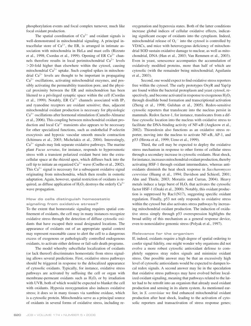
How nature uses microdomains to integrate oxidants into signaling logicOne might anticipate, given the pervasion of reactive oxidants
in a broad array of signal pathways, that rather than targeting
many specifi c but thematically unrelated targets, oxidants may
instead serve to modulate but a few signaling devices that are
nonetheless widely used. Two such broad-use devices modu-
lated by oxidants appear to be protein tyrosine phosphorylation
cascades and intracellular Ca2+ transients, both of which are
site specifi c in their effects. Although not the only cellular oxi-
dant targets, these two systems serve to link oxidants to several
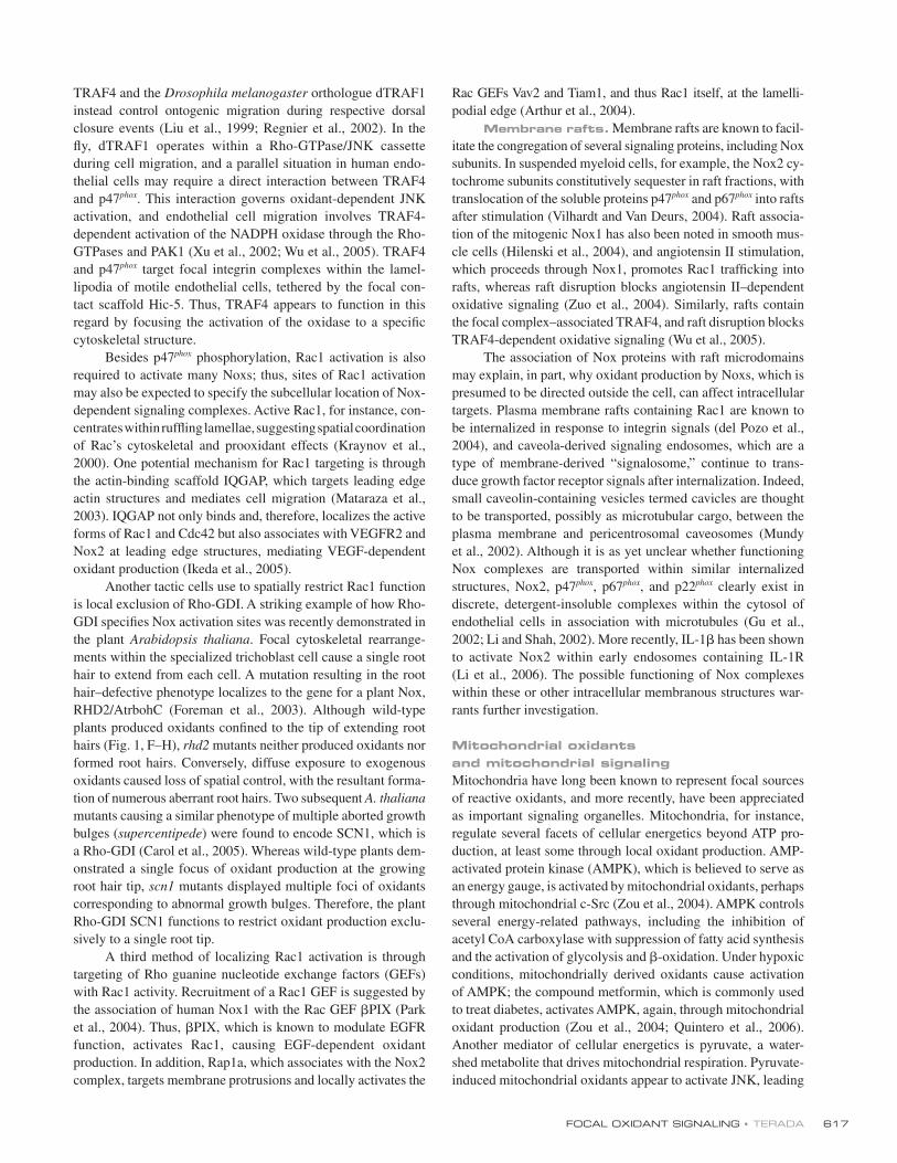
pathways (Fig. 2).
Tyrosine phosphorylation. Interestingly enough,
tyrosine phosphorylation is extensively used only by multicellular
eukaryotes, to control cell fate decisions and cytoskeletal dy-
namics (Alonso et al., 2004). Thus, the appearance of Nox genes
seems to have roughly coincided with the evolution of tyrosine
phosphorylation–dependent signaling, both systems controlling
broadly overlapping cellular functions. In addition, both tyro-
sine kinases and protein tyrosine phosphatases (PTPs) exert sig-
nal specifi city through subcellular targeting capability, as well
as catalytic domain specifi city. PTPs in particular are sensitive
to oxidative inactivation, owing to their invariant catalytic cys-
teine that, when held at a low pKa by vicinal basic residues, be-
comes sensitive to oxidative attack by forming a thiolate anion.
Within a local shell of oxidative infl uence created by targeted
oxidants, selective oxidation of such nucleophilic-active site
moieties provides some indication that not all local cysteines
are equally susceptible to oxidative modifi cation. An increasing
number of PTPs have been shown to be regulated by oxidative
modifi cation during the course of physiologic signaling, such as
LMW-PTP and SHP-2 by PDGF, PTP1B by EGF and PDGF,
and MKP by TNF (Lee et al., 1998; Chiarugi et al., 2001; Meng
et al., 2002; Kamata et al., 2005).
As subcellular location specifi es both oxidase and PTP
function, one might expect to fi nd frequent colocalization of
these systems. Ligation of the B lymphocyte antigen recep-
tor (BCR), for instance, initiates tyrosine phosphorylation of
receptor-associated proteins such as Lyn in a manner negatively
Figure 2. Common oxidant sources signal through a variety of pathways. Schematic indicates proposed oxidant targets in red, which dictate the pathways affected. Colocalization of the oxidant source and proximate target may provide pathway specifi city. In several pathways, both proximate targets and intermediate steps are unclear.
FOCAL OXIDANT SIGNALING • TERADA 619
regulated by the BCR-associated PTP SHP-1. Recently, BCR
signaling was found to depend on oxidants produced by the
Nox member Duox1 through oxidative inactivation of SHP-1
(Singh et al., 2005). Notably, only BCR-associated SHP-1, and
not SHP-1 isolated from BCR-depleted cytosol, sustained oxida-
tive inactivation, confi rming the local effect of oxidants. In a
similar fashion, T cell receptor cross-linking also induces an oxi-
dant burst that is necessary for downstream integrin activation.
This oxidant burst selectively inactivates SHP-2, which is
recruited directly to the T cell receptor adaptor complex, but
has no effect on SHP-1 (Kwon et al., 2005). SHP-2 is also
oxidatively inactivated by PDGF stimulation in a process that
requires its association with PDGFR, again indicating the im-
portance of spatial proximity between oxidant source and PTP
target (Meng et al., 2002).
In the context of cell migration, the phosphatase PTP-
PEST has been found to target peripheral focal integrin com-
plexes and thereby control lamellipodial dynamics. PTP-PEST
binds Hic-5 and diminishes Pyk2 and Src function; therefore,
TRAF4, by tethering p47phox to the Hic-5 complex, medi-
ates oxidative modifi cation of PTP-PEST, but not uninvolved
phosphatases such as MKP and SHP-2, suggesting spatial re-
striction of PTP inactivation (Wu et al., 2005). Rac1-induced
lamellipodial ruffl ing also requires oxidative inactivation of
LMW-PTP (Nimnual et al., 2003). This inactivation results in
phosphorylation (activation) of p190Rho-GAP, linking oxidant
production with RhoA inactivation within leading edge ruffl es.
A similar mechanism may facilitate tyrosine phosphorylation
of other integrin structures. Nox4 for instance, whose activity is
independent of p47phox, concentrates within focal adhesions in
smooth muscle cells (Hilenski et al., 2004). Transfected Nox4
also colocalizes with PTP1B in COS7 cells, and Nox4 enhances
insulin receptor tyrosine phosphorylation through PTP1B inac-
tivation, which is again consistent with spatial coordination be-
tween oxidant source and target (Mahadev et al., 2004; Martyn
et al., 2006).
Tyrosine phosphorylation events within mitochondria
control energetics and cell death, much like mitochondrial
oxidants. For instance, inactivation of Tim50, which is a mito-
chondrial inner membrane phosphatase, has been implicated
in apoptotic cytochrome c release (Guo et al., 2004). Another
phosphatase restricted to the mitochondrial inner membrane,
PTPMT1, controls ATP production and insulin secretion
(Pagliarini et al., 2005). Given that these PTPs are located in
close proximity to the mitochondrial respiratory chain, local
redox regulation would seem likely, although this specifi c rela-
tionship has not been well studied.
Intracellular Ca2+. A striking similarity exists be-
tween the proposed compartmentalization of oxidant signaling
and the spatial restriction of Ca2+ transients. Because of the
limited diffusion of free cytosolic Ca2+, both entry across the
plasma membrane and release from sarcolemmal stores can result
in focal Ca2+ accumulations such as puffs in Xenopus laevis
oocytes, sparks in cardiac myocytes, and quantum emission
domains in giant squid synapses. In the latter organ, Ca2+ con-
centrations of 300 μM confi ned within 0.5-μm regions have
been reported. This spatial control of Ca2+ transients to specifi c
microdomains is thought to be critical to the maintenance of
Ca2+ signal fi delity. In vascular smooth muscle, for example,
focal Ca2+ sparks cause relaxation, whereas diffuse increases in
intracellular Ca2+ result in contraction. It should be noted that
Ca2+ signals relay proliferative, cytoskeletal, and death signals,
similar to focal oxidants. Is there coordination between such
Ca2+-dependent signaling and oxidant production?
Many Nox proteins, including human Nox5 and DUOX1/2,
respond directly to Ca2+ through N-terminal EF-hand motifs,
providing one mechanism by which Ca2+ increases local oxi-
dant production. In plants, whose Nox proteins commonly
possess EF-hand domains, calmodulin signals have also been
shown to accentuate the Ca2+-dependent oxidant burst ( Harding
et al., 1997). Even Nox forms that lack EF-hand domains have
been shown to respond to Ca2+. The response of Nox2 to Ca2+,
which is required for maximal activation, is mediated by two
small proteins, MRP8 and MRP14, which are just large enough
to contain two EF-hands. In response to Ca2+ transients, these
proteins heterodimerize with each other, associate with Nox2,
and enhance oxidase activation synergistically with p47phox and
p67phox (Berthier et al., 2003). Possibly, therefore, some Nox
proteins have retained their Ca2+-response elements in trans
rather than cis.
Conversely, oxidants also trigger focal Ca2+ signals, in
part through direct activation of Ca2+ channels. Again in the
plant A. thaliana, root growth requires a high local concentra-
tion of cytosolic free Ca2+ that starts at the initiating bulge and
remains confi ned to the growing root tip, corresponding to the
localization of oxidants. Mutants defective in the plant Nox
RHD2 fail to establish Ca2+ gradients and, thus, lack normal
root hairs, whereas diffuse application of exogenous oxidants,
which create multiple aberrant root bulges, reactivates Ca2+
channels with resultant delocalized Ca2+ infl ux (Foreman et al.,
2003). In endothelial cells, H2O2 decreases the threshold of ino-
sitol 1,4,5 trisphosphate required to release intracellular Ca2+
stores, revealing an alternative mechanism for infl uencing
Ca2+-dependent signaling (Hu et al., 2000). The capacity of in-
tracellular Ca2+ and reactive oxidants to positively modulate
each other, in concept, allows the rapid establishment of local-
ized, positive feedback loops. After lymphocyte BCR ligation,
for instance, downstream Lyn phosphorylation is dependent on
both intracellular Ca2+ and Nox- dependent oxidant signaling,
which positively modulate each other in a monostable on–off
circuit (Singh et al., 2005).
Similar positive feedback loops also allow rapid asym-
metric amplifi cation of signals and early assignment of polarity
to cellular processes such as directed chemotactic migration.
At the leading edge of lamellar structures, these autoamplify-
ing loops involve local activation of Cdc42, PAK1, and Rac1
( DerMardirossian et al., 2004), and also appear to involve an
NADPH oxidase controlling focal complex dynamics (Wu
et al., 2005). Not surprisingly, then, Ca2+ transients restricted
to 2–3-μm foci within neuronal growth cones or fi broblast
pseudopodial extensions have recently been observed, which
are termed localized lamellipodial transients or localized fi -
broblast transients (Conklin et al., 2005). Both such Ca2+ tran-
sients increase with integrin activation and control tyrosine
JCB • VOLUME 174 • NUMBER 5 • 2006 620
phosphorylation events and focal complex turnover, much like
focal oxidant production.
The spatial coordination of Ca2+ and oxidant signals is
well-demonstrated in mitochondrial signaling. A principal in-
tracellular store of Ca2+, the ER, is arranged in intimate as-
sociation with mitochondria in HeLa and mast cells (Rizzuto
et al., 1998; Csordas et al., 1999). Opening of ER Ca2+ chan-
nels therefore results in local perimitochondrial Ca2+ levels
>20-fold higher than elsewhere within the cytosol, causing
mitochondrial Ca2+ uptake. Such coupled spikes in mitochon-
drial Ca2+ levels are thought to be important in propagating
Ca2+ oscillations, activating mitochondrial enzymes, and pos-
sibly activating the permeability transition pore, and the physi-
cal proximity between the ER and mitochondrion has been
likened to a privileged synaptic space within the cell (Csordas
et al., 1999). Notably, ER Ca2+ channels associated with IP3
and ryanodine receptors are oxidant sensitive; thus, adjacent
mitochondrial oxidant production is necessary for physiologic
Ca2+ oscillations after hormonal stimulation (Camello-Almaraz
et al., 2006). This coupling between mitochondrial oxidant pro-
duction and local Ca2+ transients also appears to be necessary
for other specialized functions, such as endothelial P-selectin
exocytosis and hypoxic vascular smooth muscle contraction
(Ichimura et al., 2003; Michelakis et al., 2004). Interestingly,
Ca2+ signals may link separate oxidative pathways. The marine
plant Fucus serratus, for instance, responds to hyperosmotic
stress with a transient polarized oxidant burst into the extra-
cellular space at the rhizoid apex, which diffuses back into the
cell tip to initiate an organized Ca2+ wave (Coelho et al., 2002).
This Ca2+ signal is necessary for a subsequent oxidative signal
originating from mitochondria, which then results in osmotic
adaptation. Again, however, spatial restriction of oxidants is re-
quired, as diffuse application of H2O2 destroys the orderly Ca2+
wave propagation.
How do cells distinguish homeostatic signaling from oxidative stress?To the extent that homeostatic signaling requires spatial con-
fi nement of oxidants, the cell may in many instances recognize
oxidative stress through the detection of diffuse cytosolic oxi-
dants that have escaped their usual designated locations. The
appearance of oxidants out of an appropriate spatial context
may represent reasonable cause to alert the cell to a dangerous
excess of exogenous or pathologically controlled endogenous
oxidants, to activate either defense or fail-safe death programs.
The model whereby subcellular localization of oxidants
(or lack thereof) discriminates homeostatic from stress signal-
ing allows several predictions. First, oxidative stress pathways
should be triggered in response to the delocalized appearance
of cytosolic oxidants. Typically, for instance, oxidative stress
pathways are activated by suffusing the cell or organ with
membrane-permeant oxidants such as H2O2 or by irradiation
with UVB, both of which would be expected to blanket the cell
with oxidants. Hypoxia reoxygenation also induces oxidative
stress; it does so in many tissues via xanthine oxidase, which
is a cytosolic protein. Mitochondria serve as a principal source
of oxidants in several forms of oxidative stress, including re-
oxygenation and hyperoxia states. Both of the latter conditions
increase global indices of cellular oxidative effects, indicat-
ing signifi cant escape of oxidants into the cytoplasm. Indeed,
mitochondrial release of O2.− into the cytosol is controlled by
VDACs, and mice with heterozygous defi ciency of mitochon-
drial SOD sustain oxidative damage to nuclear, as well as mito-
chondrial, DNA (Han et al., 2003; Van Remmen et al., 2003).
Even in yeast, senescence accompanies the accumulation of
oxidatively modifi ed proteins, more than half of which are
cytosolic (with the remainder being mitochondrial; Aguilaniu
et al., 2003).
Second, one would expect to fi nd oxidative stress reporters
free within the cytosol. The early prototypes OxyR and Yap1p
are found within the bacterial protoplasm and yeast cytosol, re-
spectively, and become activated in response to exogenous H2O2
through disulfi de bond formation and transcriptional activation
(Zheng et al., 1998; Gulshan et al., 2005). Redox-sensitive
cytosolic reporters that translocate into the nucleus persist in
mammals. Redox factor-1, for instance, translocates from a dif-
fuse cytosolic location into the nucleus with oxidative stress to
facilitate the DNA-binding activity of NF-κB (Angkeow et al.,
2002). Thioredoxin also functions as an oxidative stress re-
porter, moving into the nucleus to activate NF-κB, AP-1, and
p53 (Hirota et al., 1999; Ueno et al., 1999).
Third, the cell may be expected to deploy the oxidative
stress mechanism in response to other forms of cellular stress
through a secondary increase in cytosolic oxidants. Heat shock,
for instance, increases mitochondrial oxidant production, thereby
activating HSF-1 through oxidant intermediates, whereas anti-
oxidants diminish the heat shock response in Saccharomyces cerevisiae (Huang et al., 1994; Davidson and Schiestl, 2001;
Ahn and Thiele, 2003; Moraitis and Curran, 2004). Heavy
metals induce a large burst of H2O2 that activates the cytosolic
factor HSF-1 (Ozaki et al., 2000). Notably, this oxidant produc-
tion is suppressed by Rac1(N17), suggesting specifi c oxidant
regulation. Finally, p53 not only responds to oxidative stress
within the cytosol but also activates stress pathways by increas-
ing mitochondrial oxidant production. The induction of oxida-
tive stress simply through p53 overexpression highlights the
broad utility of this mechanism as a general response device,
even to nonoxidative genomic stress (Polyak et al., 1997).
Relevance for the organismIf, indeed, oxidants require a high degree of spatial ordering to
confer signal fi delity, one might wonder why organisms did not
evolve a more robust cytosolic antioxidant defense to com-
pletely suppress stray redox signals and minimize oxidant
stress. One possible answer may be that an excessively high
level of cytosolic antioxidants would be expected to dampen lo-
cal redox signals. A second answer may lie in the speculation
that oxidative stress pathways may have evolved before local-
ized oxidant signaling, meaning that pathways related to the lat-
ter had to be retrofi t into an organism that already used oxidant
production and sensing in its alarm system. As mentioned ear-
lier, in S. cerevisiae we fi nd enhanced mitochondrial oxidant
production after heat shock, leading to the activation of cyto-
solic reporters and transactivation of stress response genes;
FOCAL OXIDANT SIGNALING • TERADA 621
therefore, exuberant scavenging of cytosolic oxidants may gain-
say what, in this case, would be a protective stress response.
Both answers reveal the redox tightrope the cell is re-
quired to walk to spatially discriminate homeostatic from stress
signaling. This issue becomes particularly vexing in regard to
the mitochondrion, which, despite its ability to confi ne its oxi-
dative effects locally, can alternatively fl ood the cell with oxi-
dants and damage itself in the process, functioning as a principal
loudspeaker for sounding oxidant stress alarms. This scenario
highlights the exquisite control required for both oxidant
production and its escape into the cytosol. The consequence of
losing such control would appear to be inappropriate stress
responses, such as unscheduled cell cycle arrest or apoptosis, or
insensitivity to real stress with failure to activate these processes.
Not surprisingly, human states that refl ect these same cellular
signaling defects result in either degenerative or neoplastic
diseases that arise in the context of either excessive oxidative
stress or insensitivity to such stress.
The ubiquity of SOD in aerobic cells indeed refl ects the
dire consequences of poor oxidant regulation. The cellular strat-
egy of subsequently adopting these oxidants for signaling pur-
poses appears to have required the evolution of spatial control,
incorporation into other general signaling devices, and the pres-
ervation of a global oxidative distress pathway. When Emperor
Joseph II complained about the commissioned opera The Abduction from the Seraglio that there were “too many notes,
my dear Mozart,” Mozart is said to have responded: “(There
are) exactly the right number, your Majesty.” This comment
appears to apply to reactive oxidants as well, with the further
caveat that they should be in exactly the right places.
Dr. Terada is supported by the National Heart, Lung, and Blood Institute (grants R01-HL67256 and R01-HL61897) and the Veterans Administration.
Submitted: 4 May 2006Accepted: 24 July 2006
ReferencesAguilaniu, H., L. Gustafsson, M. Rigoulet, and T. Nystrom. 2003. Asymmetric
inheritance of oxidatively damaged proteins during cytokinesis. Science. 299:1751–1753.
Ahn, S.G., and D.J. Thiele. 2003. Redox regulation of mammalian heat shock factor 1 is essential for Hsp gene activation and protection from stress. Genes Dev. 17:516–528.
Alonso, A., J. Sasin, N. Bottini, I. Friedberg, A. Osterman, A. Godzik, T. Hunter, J. Dixon, and T. Mustelin. 2004. Protein tyrosine phosphatases in the human genome. Cell. 117:699–711.
Angkeow, P., S.S. Deshpande, B. Qi, Y.X. Liu, Y.C. Park, B.H. Jeon, M. Ozaki, and K. Irani. 2002. Redox factor-1: an extra-nuclear role in the regulation of endothelial oxidative stress and apoptosis. Cell Death Differ. 9:717–725.
Arthur, W.T., L.A. Quilliam, and J.A. Cooper. 2004. Rap1 promotes cell spread-ing by localizing Rac guanine nucleotide exchange factors. J. Cell Biol. 167:111–122.
Balaban, R.S., S. Nemoto, and T. Finkel. 2005. Mitochondria, oxidants, and aging. Cell. 120:483–495.
Berthier, S., M.H. Paclet, S. Lerouge, F. Roux, S. Vergnaud, A.W. Coleman, and F. Morel. 2003. Changing the conformation state of cytochrome b558 initiates NADPH oxidase activation: MRP8/MRP14 regulation. J. Biol. Chem. 278:25499–25508.
Blackstone, N.W. 2003. Redox signaling in the growth and development of colonial hydroids. J. Exp. Biol. 206:651–658.
Camello-Almaraz, M.C., M.J. Pozo, M.P. Murphy, and P.J. Camello. 2006. Mitochondrial production of oxidants is necessary for physiological calcium oscillations. J. Cell. Physiol. 206:487–494.
Carol, R.J., S. Takeda, P. Linstead, M.C. Durrant, H. Kakesova, P. Derbyshire, S. Drea, V. Zarsky, and L. Dolan. 2005. A RhoGDP dissociation inhibitor spatially regulates growth in root hair cells. Nature. 438:1013–1016.
Chiarugi, P., T. Fiaschi, M.L. Taddei, D. Talini, E. Giannoni, G. Raugei, and G. Ramponi. 2001. Two vicinal cysteines confer a peculiar redox regula-tion to low molecular weight protein tyrosine phosphatase in response to platelet-derived growth factor receptor stimulation. J. Biol. Chem. 276:33478–33487.
Choi, M.H., I.K. Lee, G.W. Kim, B.U. Kim, Y.H. Han, D.Y. Yu, H.S. Park, K.Y. Kim, J.S. Lee, C. Choi, et al. 2005. Regulation of PDGF signalling and vascular remodelling by peroxiredoxin II. Nature. 435:347–353.
Coelho, S.M., A.R. Taylor, K.P. Ryan, I. Sousa-Pinto, M.T. Brown, and C. Brownlee. 2002. Spatiotemporal patterning of reactive oxygen production and Ca(2+) wave propagation in fucus rhizoid cells. Plant Cell. 14:2369–2381.
Conklin, M.W., M.S. Lin, and N.C. Spitzer. 2005. Local calcium transients con-tribute to disappearance of pFAK, focal complex removal and deadhesion of neuronal growth cones and fi broblasts. Dev. Biol. 287:201–212.
Csordas, G., A.P. Thomas, and G. Hajnoczky. 1999. Quasi-synaptic calcium signal transmission between endoplasmic reticulum and mitochondria. EMBO J. 18:96–108.
Davidson, J.F., and R.H. Schiestl. 2001. Mitochondrial respiratory electron car-riers are involved in oxidative stress during heat stress in Saccharomyces cerevisiae. Mol. Cell. Biol. 21:8483–8489.
del Pozo, M.A., N.B. Alderson, W.B. Kiosses, H.H. Chiang, R.G. Anderson, and M.A. Schwartz. 2004. Integrins regulate Rac targeting by internalization of membrane domains. Science. 303:839–842.
DerMardirossian, C., A. Schnelzer, and G.M. Bokoch. 2004. Phosphorylation of RhoGDI by Pak1 mediates dissociation of Rac GTPase. Mol. Cell. 15:117–127.
El Benna, J., J.M. Ruedi, and B.M. Babior. 1994. Cytosolic guanine nucleotide-binding protein Rac2 operates in vivo as a component of the neutrophil respiratory burst oxidase. Transfer of Rac2 and the cytosolic oxidase components p47phox and p67phox to the submembranous actin cytoskel-eton during oxidase activation. J. Biol. Chem. 269:6729–6734.
Foreman, J., V. Demidchik, J.H. Bothwell, P. Mylona, H. Miedema, M.A. Torres, P. Linstead, S. Costa, C. Brownlee, J.D. Jones, et al. 2003. Reactive oxygen species produced by NADPH oxidase regulate plant cell growth. Nature. 422:442–446.
Geiszt, M., and T.L. Leto. 2004. The Nox family of NAD(P)H oxidases: host defense and beyond. J. Biol. Chem. 279:51715–51718.
Giorgio, M., E. Migliaccio, F. Orsini, D. Paolucci, M. Moroni, C. Contursi, G. Pelliccia, L. Luzi, S. Minucci, M. Marcaccio, et al. 2005. Electron trans-fer between cytochrome c and p66Shc generates reactive oxygen species that trigger mitochondrial apoptosis. Cell. 122:221–233.
Gu, Y., Y.C. Xu, R.F. Wu, R.F. Souza, F.E. Nwariaku, and L.S. Terada. 2002. TNF alpha activates c-jun amino terminal kinase through p47phox. Exp. Cell Res. 272:62–74.
Gu, Y., Y.C. Xu, R.F. Wu, F.E. Nwariaku, R.F. Souza, S.C. Flores, and L.S. Terada. 2003. p47phox Participates in Activation of RelA in Endothelial Cells. J. Biol. Chem. 278:17210–17217.
Gulshan, K., S.A. Rovinsky, S.T. Coleman, and W.S. Moye-Rowley. 2005. Oxidant-specifi c folding of Yap1p regulates both transcriptional activa-tion and nuclear localization. J. Biol. Chem. 280:40524–40533.
Guo, Y., N. Cheong, Z. Zhang, R. De Rose, Y. Deng, S.A. Farber, T. Fernandes-Alnemri, and E.S. Alnemri. 2004. Tim50, a component of the mitochon-drial translocator, regulates mitochondrial integrity and cell death. J. Biol. Chem. 279:24813–24825.
Han, D., F. Antunes, R. Canali, D. Rettori, and E. Cadenas. 2003. Voltage- dependent anion channels control the release of the superoxide anion from mitochondria to cytosol. J. Biol. Chem. 278:5557–5563.
Harding, S.A., S.H. Oh, and D.M. Roberts. 1997. Transgenic tobacco express-ing a foreign calmodulin gene shows an enhanced production of active oxygen species. EMBO J. 16:1137–1144.
Heyworth, P.G., J.M. Robinson, J. Ding, B.A. Ellis, and J.A. Badwey. 1997. Cofi lin undergoes rapid dephosphorylation in stimulated neutrophils and translocates to ruffl ed membranes enriched in products of the NADPH oxidase complex. Evidence for a novel cycle of phosphorylation and dephosphorylation. Histochem. Cell Biol. 108:221–233.
Hilenski, L.L., R.E. Clempus, M.T. Quinn, J.D. Lambeth, and K.K. Griendling. 2004. Distinct subcellular localizations of Nox1 and Nox4 in vascular smooth muscle cells. Arterioscler. Thromb. Vasc. Biol. 24:677–683.
Hirota, K., M. Murata, Y. Sachi, H. Nakamura, J. Takeuchi, K. Mori, and J. Yodoi. 1999. Distinct roles of thioredoxin in the cytoplasm and in the nucleus. A two-step mechanism of redox regulation of transcription fac-tor NF-kappaB. J. Biol. Chem. 274:27891–27897.
Hu, Q., G. Zheng, J.L. Zweier, S. Deshpande, K. Irani, and R.C. Ziegelstein. 2000. NADPH oxidase activation increases the sensitivity of intracellular
JCB • VOLUME 174 • NUMBER 5 • 2006 622
Ca2+ stores to inositol 1,4,5-trisphosphate in human endothelial cells. J. Biol. Chem. 275:15749–15757.
Huang, L.E., H. Zhang, S.W. Bae, and A.Y. Liu. 1994. Thiol reducing rea-gents inhibit the heat shock response. Involvement of a redox mecha-nism in the heat shock signal transduction pathway. J. Biol. Chem. 269:30718–30725.
Ichimura, H., K. Parthasarathi, S. Quadri, A.C. Issekutz, and J. Bhattacharya. 2003. Mechano-oxidative coupling by mitochondria induces proinfl amma-tory responses in lung venular capillaries. J. Clin. Invest. 111:691–699.
Ikeda, S., M. Yamaoka-Tojo, L. Hilenski, N.A. Patrushev, G.M. Anwar, M.T. Quinn, and M. Ushio-Fukai. 2005. IQGAP1 regulates reactive oxygen species-dependent endothelial cell migration through interacting with Nox2. Arterioscler. Thromb. Vasc. Biol. 25:2295–2300.
Irani, K., Y. Xia, J.L. Zweier, S.J. Sollott, C.J. Der, E.R. Fearon, M. Sundaresan, T. Finkel, and P.J. Goldschmidt-Clermont. 1997. Mitogenic signal-ing mediated by oxidants in Ras-transformed fi broblasts. Science. 275:1649–1652.
Kamata, H., S. Honda, S. Maeda, L. Chang, H. Hirata, and M. Karin. 2005. Reactive oxygen species promote TNFalpha-induced death and sus-tained JNK activation by inhibiting MAP kinase phosphatases. Cell. 120:649–661.
Kraynov, V.S., C. Chamberlain, G.M. Bokoch, M.A. Schwartz, S. Slabaugh, and K.M. Hahn. 2000. Localized Rac activation dynamics visualized in living cells. Science. 290:333–337.
Kwon, J., C.K. Qu, J.S. Maeng, R. Falahati, C. Lee, and M.S. Williams. 2005. Receptor-stimulated oxidation of SHP-2 promotes T-cell adhesion through SLP-76-ADAP. EMBO J. 24:2331–2341.
Lalucque, H., and P. Silar. 2003. NADPH oxidase: an enzyme for multicellularity? Trends Microbiol. 11:9–12.
Lambeth, J.D. 2004. NOX enzymes and the biology of reactive oxygen. Nat. Rev. Immunol. 4:181–189.
Lardy, B., M. Bof, L. Aubry, M.H. Paclet, F. Morel, M. Satre, and G. Klein. 2005. NADPH oxidase homologs are required for normal cell differentia-tion and morphogenesis in Dictyostelium discoideum. Biochim. Biophys. Acta. 1744:199–212.
Lee, S.R., K.S. Kwon, S.R. Kim, and S.G. Rhee. 1998. Reversible inactivation of protein-tyrosine phosphatase 1B in A431 cells stimulated with epidermal growth factor. J. Biol. Chem. 273:15366–15372.
Li, J.M., and A.M. Shah. 2002. Intracellular localization and preassembly of the NADPH oxidase complex in cultured endothelial cells. J. Biol. Chem. 277:19952–19960.
Li, Q., M.M. Harraz, W. Zhou, L.N. Zhang, W. Ding, Y. Zhang, T. Eggleston, C. Yeaman, B. Banfi , and J.F. Engelhardt. 2006. Nox2 and Rac1 regu-late H2O2-dependent recruitment of TRAF6 to endosomal interleukin-1 receptor complexes. Mol. Cell. Biol. 26:140–154.
Liu, H., Y.C. Su, E. Becker, J. Treisman, and E.Y. Skolnik. 1999. A Drosophila TNF-receptor-associated factor (TRAF) binds the ste20 kinase Misshapen and activates Jun kinase. Curr. Biol. 9:101–104.
Madesh, M., and G. Hajnoczky. 2001. VDAC-dependent permeabilization of the outer mitochondrial membrane by superoxide induces rapid and massive cytochrome c release. J. Cell Biol. 155:1003–1015.
Mahadev, K., H. Motoshima, X. Wu, J.M. Ruddy, R.S. Arnold, G. Cheng, J.D. Lambeth, and B.J. Goldstein. 2004. The NAD(P)H oxidase homolog Nox4 modulates insulin-stimulated generation of H2O2 and plays an in-tegral role in insulin signal transduction. Mol. Cell. Biol. 24:1844–1854.
Malagnac, F., H. Lalucque, G. Lepere, and P. Silar. 2004. Two NADPH oxidase isoforms are required for sexual reproduction and ascospore germina-tion in the fi lamentous fungus Podospora anserina. Fungal Genet. Biol. 41:982–997.
Martyn, K.D., L.M. Frederick, K. von Loehneysen, M.C. Dinauer, and U.G. Knaus. 2006. Functional analysis of Nox4 reveals unique characteristics compared to other NADPH oxidases. Cell. Signal. 18:69–82.
Mataraza, J.M., M.W. Briggs, Z. Li, A. Entwistle, A.J. Ridley, and D.B. Sacks. 2003. IQGAP1 promotes cell motility and invasion. J. Biol. Chem. 278:41237–41245.
McCord, J.M., and I. Fridovich. 1969. Superoxide dismutase: an enzymic func-tion for erythrocuprein (hemocuprein). J. Biol. Chem. 244:6049–6055.
Meng, T.C., T. Fukada, and N.K. Tonks. 2002. Reversible oxidation and inactiva-tion of protein tyrosine phosphatases in vivo. Mol. Cell. 9:387–399.
Michelakis, E.D., B. Thebaud, E.K. Weir, and S.L. Archer. 2004. Hypoxic pul-monary vasoconstriction: redox regulation of O2-sensitive K+ channels by a mitochondrial O2-sensor in resistance artery smooth muscle cells. J. Mol. Cell. Cardiol. 37:1119–1136.
Moldovan, L., N.I. Moldovan, R.H. Sohn, S.A. Parikh, and P.J. Goldschmidt-Clermont. 2000. Redox changes of cultured endothelial cells and actin dynamics. Circ. Res. 86:549–557.
Moraitis, C., and B.P. Curran. 2004. Reactive oxygen species may infl uence the heat shock response and stress tolerance in the yeast Saccharomyces cerevisiae. Yeast. 21:313–323.
Mundy, D.I., T. Machleidt, Y.S. Ying, R.G. Anderson, and G.S. Bloom. 2002. Dual control of caveolar membrane traffi c by microtubules and the actin cytoskeleton. J. Cell Sci. 115:4327–4339.
Nauseef, W.M., B.D. Volpp, S. McCormick, K.G. Leidal, and R.A. Clark. 1991. Assembly of the neutrophil respiratory burst oxidase. Protein kinase C promotes cytoskeletal and membrane association of cytosolic oxidase components. J. Biol. Chem. 266:5911–5917.
Nemoto, S., K. Takeda, Z.X. Yu, V.J. Ferrans, and T. Finkel. 2000. Role for mi-tochondrial oxidants as regulators of cellular metabolism. Mol. Cell. Biol. 20:7311–7318.
Nimnual, A.S., L.J. Taylor, and D. Bar-Sagi. 2003. Redox-dependent downregu-lation of Rho by Rac. Nat. Cell Biol. 5:236–241.
Ozaki, M., S.S. Deshpande, P. Angkeow, S. Suzuki, and K. Irani. 2000. Rac1 regulates stress-induced, redox-dependent heat shock factor activation. J. Biol. Chem. 275:35377–35383.
Pagliarini, D.J., S.E. Wiley, M.E. Kimple, J.R. Dixon, P. Kelly, C.A. Worby, P.J. Casey, and J.E. Dixon. 2005. Involvement of a mitochondrial phosphatase in the regulation of ATP production and insulin secretion in pancreatic beta cells. Mol. Cell. 19:197–207.
Park, H.S., S.H. Lee, D. Park, J.S. Lee, S.H. Ryu, W.J. Lee, S.G. Rhee, and Y.S. Bae. 2004. Sequential activation of phosphatidylinositol 3-kinase, beta Pix, Rac1, and Nox1 in growth factor-induced production of H2O2. Mol. Cell. Biol. 24:4384–4394.
Polyak, K., Y. Xia, J.L. Zweier, K.W. Kinzler, and B. Vogelstein. 1997. A model for p53-induced apoptosis. Nature. 389:300–305.
Putcha, G.V., S. Le, S. Frank, C.G. Besirli, K. Clark, B. Chu, S. Alix, R.J. Youle, A. LaMarche, A.C. Maroney, and E.M. Johnson Jr. 2003. JNK-mediated BIM phosphorylation potentiates BAX-dependent apoptosis. Neuron. 38:899–914.
Quintero, M., S.L. Colombo, A. Godfrey, and S. Moncada. 2006. Mitochondria as signaling organelles in the vascular endothelium. Proc. Natl. Acad. Sci. USA. 103:5379–5384.
Regnier, C.H., R. Masson, V. Kedinger, J. Textoris, I. Stoll, M.P. Chenard, A. Dierich, C. Tomasetto, and M.C. Rio. 2002. Impaired neural tube closure, axial skeleton malformations, and tracheal ring disruption in TRAF4- defi cient mice. Proc. Natl. Acad. Sci. USA. 99:5585–5590.
Rizzuto, R., P. Pinton, W. Carrington, F.S. Fay, K.E. Fogarty, L.M. Lifshitz, R.A. Tuft, and T. Pozzan. 1998. Close contacts with the endoplasmic reticulum as determinants of mitochondrial Ca2+ responses. Science. 280:1763–1766.
Schieffer, B., M. Luchtefeld, S. Braun, A. Hilfi ker, D. Hilfi ker-Kleiner, and H. Drexler. 2000. Role of NAD(P)H oxidase in angiotensin II-induced JAK/STAT signaling and cytokine induction. Circ. Res. 87:1195–1201.
Singh, D.K., D. Kumar, Z. Siddiqui, S.K. Basu, V. Kumar, and K.V. Rao. 2005. The strength of receptor signaling is centrally controlled through a coop-erative loop between Ca2+ and an oxidant signal. Cell. 121:281–293.
Sulciner, D.J., K. Irani, Z.X. Yu, V.J. Ferrans, P. Goldschmidt-Clermont, and T. Finkel. 1996. rac1 regulates a cytokine-stimulated, redox-dependent pathway necessary for NF-kappaB activation. Mol. Cell. Biol. 16:7115–7121.
Touyz, R.M., G. Yao, M.T. Quinn, P.J. Pagano, and E.L. Schiffrin. 2005. p47phox associates with the cytoskeleton through cortactin in human vascular smooth muscle cells: role in NAD(P)H oxidase regulation by angiotensin II. Arterioscler. Thromb. Vasc. Biol. 25:512–518.
Ueno, M., H. Masutani, R.J. Arai, A. Yamauchi, K. Hirota, T. Sakai, T. Inamoto, Y. Yamaoka, J. Yodoi, and T. Nikaido. 1999. Thioredoxin- dependent redox regulation of p53-mediated p21 activation. J. Biol. Chem. 274:35809–35815.
Ushio-Fukai, M., Y. Tang, T. Fukai, S.I. Dikalov, Y. Ma, M. Fujimoto, M.T. Quinn, P.J. Pagano, C. Johnson, and R.W. Alexander. 2002. Novel role of gp91(phox)-containing NAD(P)H oxidase in vascular endothelial growth factor-induced signaling and angiogenesis. Circ. Res. 91:1160–1167.
Van Remmen, H., Y. Ikeno, M. Hamilton, M. Pahlavani, N. Wolf, S.R. Thorpe, N.L. Alderson, J.W. Baynes, C.J. Epstein, T.T. Huang, et al. 2003. Life-long reduction in MnSOD activity results in increased DNA damage and higher incidence of cancer but does not accelerate aging. Physiol. Genomics. 16:29–37.
Vilhardt, F., and B. Van Deurs. 2004. The phagocyte NADPH oxidase depends on cholesterol-enriched membrane microdomains for assembly. EMBO J. 23:739–748.
Wientjes, F.B., E.P. Reeves, V. Soskic, H. Furthmayr, and A.W. Segal. 2001. The NADPH oxidase components p47(phox) and p40(phox) bind to moesin through their PX domain. Biochem. Biophys. Res. Commun. 289:382–388.
FOCAL OXIDANT SIGNALING • TERADA 623
Wiltshire, C., M. Matsushita, S. Tsukada, D.A. Gillespie, and G.H. May. 2002. A new c-Jun N-terminal kinase (JNK)-interacting protein, Sab (SH3BP5), associates with mitochondria. Biochem. J. 367:577–585.
Wu, R.F., Y. Gu, Y.C. Xu, F.E. Nwariaku, and L.S. Terada. 2003. Vascular endo-thelial growth factor causes translocation of p47phox to membrane ruffl es through WAVE1. J. Biol. Chem. 278:36830–36840.
Wu, R.F., Y.C. Xu, Z. Ma, F.E. Nwariaku, G.A. Sarosi Jr., and L.S. Terada. 2005. Subcellular targeting of oxidants during endothelial cell migration. J. Cell Biol. 171:893–904.
Xu, Y.C., R.F. Wu, Y. Gu, Y.S. Yang, M.C. Yang, F.E. Nwariaku, and L.S. Terada. 2002. Involvement of TRAF4 in oxidative activation of c-jun amino terminal kinase. J. Biol. Chem. 277:28051–28057.
Zamzami, N., P. Marchetti, M. Castedo, D. Decaudin, A. Macho, T. Hirsch, S.A. Susin, P.X. Petit, B. Mignotte, and G. Kroemer. 1995. Sequential reduction of mitochondrial transmembrane potential and generation of reactive oxygen species in early programmed cell death. J. Exp. Med. 182:367–377.
Zheng, M., F. Aslund, and G. Storz. 1998. Activation of the OxyR transcription factor by reversible disulfi de bond formation. Science. 279:1718–1721.
Zou, M.H., S.S. Kirkpatrick, B.J. Davis, J.S. Nelson, W.G. Wiles, U. Schlattner, D. Neumann, M. Brownlee, M.B. Freeman, and M.H. Goldman. 2004. Activation of the AMP-activated protein kinase by the anti-diabetic drug metformin in vivo. Role of mitochondrial reactive nitrogen species. J. Biol. Chem. 279:43940–43951.
Zuo, L., M. Ushio-Fukai, L.L. Hilenski, and R.W. Alexander. 2004. Microtubules regulate angiotensin II type 1 receptor and Rac1 localization in caveolae/lipid rafts: role in redox signaling. Arterioscler. Thromb. Vasc. Biol. 24:1223–1228.