Space in the brain: how the hippocampal formation...
18
rstb.royalsocietypublishing.org Review Cite this article: Hartley T, Lever C, Burgess N, O’Keefe J. 2014 Space in the brain: how the hippocampal formation supports spatial cognition. Phil. Trans. R. Soc. B 369: 20120510. http://dx.doi.org/10.1098/rstb.2012.0510 One contribution of 24 to a Theo Murphy Meeting Issue ‘Space in the brain: cells, circuits, codes and cognition’. Subject Areas: neuroscience, cognition, behaviour Keywords: hippocampus, entorhinal cortex, grid cells, place cells, head direction cells, boundary cells Author for correspondence: Tom Hartley e-mail: [email protected] Space in the brain: how the hippocampal formation supports spatial cognition Tom Hartley 1 , Colin Lever 2 , Neil Burgess 3 and John O’Keefe 4 1 Department of Psychology, University of York, York, UK 2 Department of Psychology, University of Durham, Durham, UK 3 Institute of Cognitive Neuroscience and Institute of Neurology, and 4 Sainsbury Wellcome Centre, University College London, London, UK Over the past four decades, research has revealed that cells in the hippocam- pal formation provide an exquisitely detailed representation of an animal’s current location and heading. These findings have provided the foundations for a growing understanding of the mechanisms of spatial cognition in mam- mals, including humans. We describe the key properties of the major categories of spatial cells: place cells, head direction cells, grid cells and boundary cells, each of which has a characteristic firing pattern that encodes spatial parameters relating to the animal’s current position and orientation. These properties also include the theta oscillation, which appears to play a functional role in the representation and processing of spatial information. Reviewing recent work, we identify some themes of current research and introduce approaches to computational modelling that have helped to bridge the different levels of description at which these mechanisms have been investigated. These range from the level of molecular biology and gen- etics to the behaviour and brain activity of entire organisms. We argue that the neuroscience of spatial cognition is emerging as an exceptionally integra- tive field which provides an ideal test-bed for theories linking neural coding, learning, memory and cognition. 1. Introduction How does an animal or human being know where it is, and how does it remem- ber distant goals, and then navigate efficiently towards them, while avoiding hazards and barriers? What kinds of representation underlie this kind of spatial ability? Over the past four decades, efforts to address such questions have pro- vided the foundations for a richly productive field connecting different levels of neuroscientific investigation, from membrane potentials and synaptic currents to individual neurons, from neuronal networks to complex behaviour and human cognition. In introducing the topic of ‘Space in the Brain’, we want to draw a distinc- tion between spatial frameworks tied to a particular body part, object or action and those that are fixed with respect to the outside world, independent of par- ticular actions and objects. The brain makes use of neural representations of both types. The first type of representation is crucial for behaviours such as catching a ball or picking a fruit from a tree. Behaviours such as navigating long distances over natural terrain or through a new city are also dependent on the second type of representation. This issue will focus primarily on the second type of spatial framework, which depends on a specialized system centred on the hippocampus, a phylogenetically ancient and well-preserved structure, which in humans is found deep in the medial temporal lobes. To understand what is so special about the hippocampal formation, it is first instructive to consider how spatial parameters are reflected in the firing of neur- ons in other parts of the brain. Throughout the brain, individual neurons are often found to have spatially restricted firing fields, which carry spatial infor- mation about the source of sensory information or destination of planned actions. Thus, a neuron in primary visual cortex might respond to a stimulus in a particular part of the visual field [1], a neuron in primary somatosensory & 2013 The Author(s) Published by the Royal Society. All rights reserved. on July 11, 2018 http://rstb.royalsocietypublishing.org/ Downloaded from
Transcript of Space in the brain: how the hippocampal formation...

on July 11, 2018http://rstb.royalsocietypublishing.org/Downloaded from
rstb.royalsocietypublishing.org
ReviewCite this article: Hartley T, Lever C, Burgess
N, O’Keefe J. 2014 Space in the brain: how the
hippocampal formation supports spatial
cognition. Phil. Trans. R. Soc. B 369: 20120510.
http://dx.doi.org/10.1098/rstb.2012.0510
One contribution of 24 to a Theo Murphy
Meeting Issue ‘Space in the brain: cells,
circuits, codes and cognition’.
Subject Areas:neuroscience, cognition, behaviour
Keywords:hippocampus, entorhinal cortex, grid cells,
place cells, head direction cells, boundary cells
Author for correspondence:Tom Hartley
e-mail: [email protected]
& 2013 The Author(s) Published by the Royal Society. All rights reserved.
Space in the brain: how the hippocampalformation supports spatial cognition
Tom Hartley1, Colin Lever2, Neil Burgess3 and John O’Keefe4
1Department of Psychology, University of York, York, UK2Department of Psychology, University of Durham, Durham, UK3Institute of Cognitive Neuroscience and Institute of Neurology, and 4Sainsbury Wellcome Centre, UniversityCollege London, London, UK
Over the past four decades, research has revealed that cells in the hippocam-
pal formation provide an exquisitely detailed representation of an animal’s
current location and heading. These findings have provided the foundations
for a growing understanding of the mechanisms of spatial cognition in mam-
mals, including humans. We describe the key properties of the major
categories of spatial cells: place cells, head direction cells, grid cells and
boundary cells, each of which has a characteristic firing pattern that encodes
spatial parameters relating to the animal’s current position and orientation.
These properties also include the theta oscillation, which appears to play a
functional role in the representation and processing of spatial information.
Reviewing recent work, we identify some themes of current research and
introduce approaches to computational modelling that have helped to
bridge the different levels of description at which these mechanisms have
been investigated. These range from the level of molecular biology and gen-
etics to the behaviour and brain activity of entire organisms. We argue that
the neuroscience of spatial cognition is emerging as an exceptionally integra-
tive field which provides an ideal test-bed for theories linking neural coding,
learning, memory and cognition.
1. IntroductionHow does an animal or human being know where it is, and how does it remem-
ber distant goals, and then navigate efficiently towards them, while avoiding
hazards and barriers? What kinds of representation underlie this kind of spatial
ability? Over the past four decades, efforts to address such questions have pro-
vided the foundations for a richly productive field connecting different levels of
neuroscientific investigation, from membrane potentials and synaptic currents
to individual neurons, from neuronal networks to complex behaviour and
human cognition.
In introducing the topic of ‘Space in the Brain’, we want to draw a distinc-
tion between spatial frameworks tied to a particular body part, object or action
and those that are fixed with respect to the outside world, independent of par-
ticular actions and objects. The brain makes use of neural representations of
both types. The first type of representation is crucial for behaviours such as
catching a ball or picking a fruit from a tree. Behaviours such as navigating
long distances over natural terrain or through a new city are also dependent
on the second type of representation. This issue will focus primarily on the
second type of spatial framework, which depends on a specialized system
centred on the hippocampus, a phylogenetically ancient and well-preserved
structure, which in humans is found deep in the medial temporal lobes.
To understand what is so special about the hippocampal formation, it is first
instructive to consider how spatial parameters are reflected in the firing of neur-
ons in other parts of the brain. Throughout the brain, individual neurons are
often found to have spatially restricted firing fields, which carry spatial infor-
mation about the source of sensory information or destination of planned
actions. Thus, a neuron in primary visual cortex might respond to a stimulus
in a particular part of the visual field [1], a neuron in primary somatosensory
rstb.royalsocietypublishing.orgPhil.Trans.R.Soc.B
369:20120510
2
on July 11, 2018http://rstb.royalsocietypublishing.org/Downloaded from
cortex might respond to a tactile stimulation of a particular
body part [2], and the firing of a motor neuron might help
to direct limb movements in a specific direction [3]. In each
case, neural activity reflects the spatial relationship between
a stimulus or response and a part of the body. Similar but
somewhat more abstract forms of spatial coding are found
beyond the primary sensory and motor cortices, notably in
parietal cortex where individual neurons’ receptive fields
may be fixed with respect to the hand, head or trunk, and
may respond to multiple sensory modalities [4–6]. Such
neural codes incorporate spatial information about stimuli
and responses in terms of various egocentric reference
frames (each anchored with respect to the body or part of
the body). They are well suited to mediating spatial behav-
iour in the immediate environment and to computing
transformations between visual and body-based reference
frames in the online control of action [7]. They carry spatial
information about stimuli and responses and can, in prin-
ciple, perform spatial computations linking one with the
other [8,9].
All of the above representations are ‘egocentric’ in terms of
their spatial reference frame. It is debateable whether they
represent space itself in an absolute sense, and when they do
represent locations in the world, those locations must be
updated as the various parts of the body, and the body
itself moves.
By contrast, and as we explain in more detail in §2, cells
in the hippocampal formation can represent an animal’s cur-
rent location or heading independently of individual sensory
cues and particular actions. Their firing fields are anchored to
the external environment (and thus termed ‘allocentric’ or
world-centred), rather than to individual objects, actions
or to the body. These cells appear to provide the basis for a
cognitive map: a representation of the environment and the
places and objects within it that is to some extent independ-
ent of bodily posture or orientation. As such it affords
long-term memory for the spatial relationships between
places, the routes between them, the resources, goals and
hazards they contain, in that it does not require continuous
updating as the animal goes about its daily life [10–13].
We briefly outline key aspects of the anatomy of
the hippocampal formation and the properties of its spatial
cells as characterized through in vivo extracellular unit record-
ing in freely behaving animals, mainly rodents. These cells
form the building blocks of spatial representation. Their fascin-
ating properties provide detailed quantitative constraints
on computational models which have been further supported
by advances in optogenetics, juxtacellular recording and
two-photon imaging in behaving animals, and human electro-
physiology and neuroimaging. These developments have
fuelled further discoveries, and we outline some of the
themes of current research and the new avenues which have
been opened up. The neuroscience of spatial cognition, we
will argue, is emerging as an exceptionally integrative field
which provides an ideal test-bed for theories linking neural
coding, learning, memory and cognition.
2. Anatomy and spatial cells of thehippocampal formation
In this section, we outline the anatomy of the hippocampal
formation and describe some of the spatial properties of the
neurons within it. Much of the evidence we refer to is
based on research in rodents, although as we explain later,
there is mounting evidence that the critical spatial properties
are maintained in other mammals, including humans. We
should also note that although our focus on the hippocampal
formation is justified by its central role in spatial cognition,
cells with related spatial properties, notably head direction
(HD) cells, are found in other brain regions.
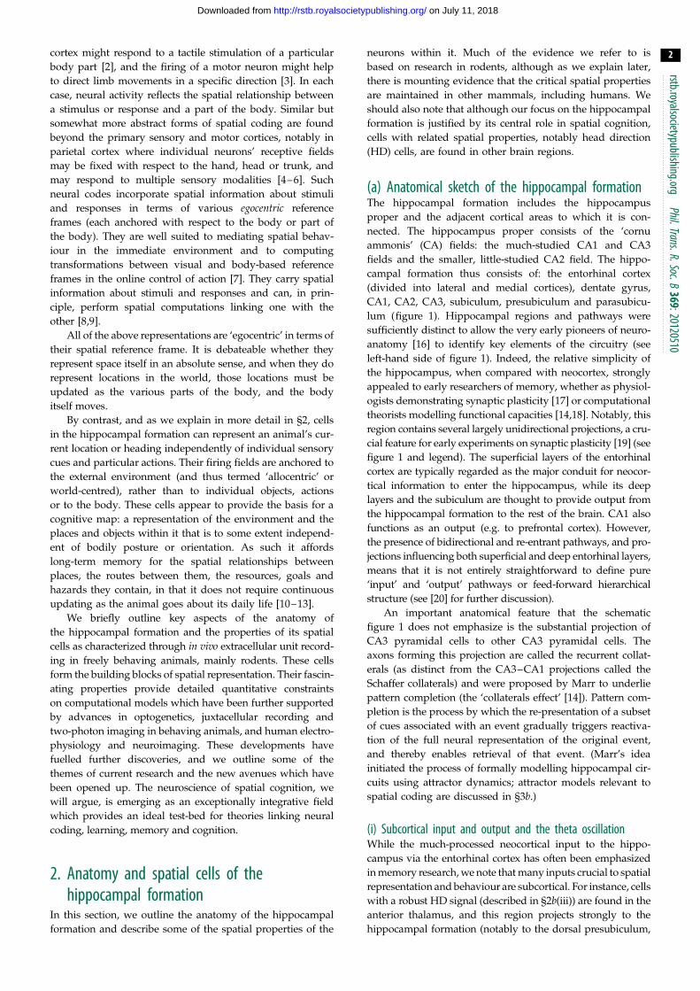
(a) Anatomical sketch of the hippocampal formationThe hippocampal formation includes the hippocampus
proper and the adjacent cortical areas to which it is con-
nected. The hippocampus proper consists of the ‘cornu
ammonis’ (CA) fields: the much-studied CA1 and CA3
fields and the smaller, little-studied CA2 field. The hippo-
campal formation thus consists of: the entorhinal cortex
(divided into lateral and medial cortices), dentate gyrus,
CA1, CA2, CA3, subiculum, presubiculum and parasubicu-
lum (figure 1). Hippocampal regions and pathways were
sufficiently distinct to allow the very early pioneers of neuro-
anatomy [16] to identify key elements of the circuitry (see
left-hand side of figure 1). Indeed, the relative simplicity of
the hippocampus, when compared with neocortex, strongly
appealed to early researchers of memory, whether as physiol-
ogists demonstrating synaptic plasticity [17] or computational
theorists modelling functional capacities [14,18]. Notably, this
region contains several largely unidirectional projections, a cru-
cial feature for early experiments on synaptic plasticity [19] (see
figure 1 and legend). The superficial layers of the entorhinal
cortex are typically regarded as the major conduit for neocor-
tical information to enter the hippocampus, while its deep
layers and the subiculum are thought to provide output from
the hippocampal formation to the rest of the brain. CA1 also
functions as an output (e.g. to prefrontal cortex). However,
the presence of bidirectional and re-entrant pathways, and pro-
jections influencing both superficial and deep entorhinal layers,
means that it is not entirely straightforward to define pure
‘input’ and ‘output’ pathways or feed-forward hierarchical
structure (see [20] for further discussion).
An important anatomical feature that the schematic
figure 1 does not emphasize is the substantial projection of
CA3 pyramidal cells to other CA3 pyramidal cells. The
axons forming this projection are called the recurrent collat-
erals (as distinct from the CA3–CA1 projections called the
Schaffer collaterals) and were proposed by Marr to underlie
pattern completion (the ‘collaterals effect’ [14]). Pattern com-
pletion is the process by which the re-presentation of a subset
of cues associated with an event gradually triggers reactiva-
tion of the full neural representation of the original event,
and thereby enables retrieval of that event. (Marr’s idea
initiated the process of formally modelling hippocampal cir-
cuits using attractor dynamics; attractor models relevant to
spatial coding are discussed in §3b.)
(i) Subcortical input and output and the theta oscillationWhile the much-processed neocortical input to the hippo-
campus via the entorhinal cortex has often been emphasized
in memory research, we note that many inputs crucial to spatial
representation and behaviour are subcortical. For instance, cells
with a robust HD signal (described in §2b(iii)) are found in the
anterior thalamus, and this region projects strongly to the
hippocampal formation (notably to the dorsal presubiculum,

CA3
CA2
CA1
Sb
MEC
LEC
PreSb ParaSb
DG
perirhinal Ctx
postrhinal Ctx
subcortical input
medial septum/diagonal band of Broca4–12 Hz (theta)
thalamusincluding HD signal
Figure 1. Schematic overview of major anatomical pathways in the hippocampal formation of the rat. Left-hand side of figure emphasizes gross morphology (ratbrain) of cell layers in hippocampus and dentate gyrus and long-established unidirectional projections. Classic trisynaptic pathway consists of projection from ento-rhinal cortex (LEC: lateral entorhinal cortex; MEC: medial entorhinal cortex) to dentate gyrus (DG), from DG to CA3, and from CA3 to CA1. Entorhinal input alsoconsists of direct monosynaptic LEC and MEC projections to CA3, to CA1, and to subiculum (Sb). CA1 projection to Sb and to LEC/MEC, and Sb projections to LEC/MEC,complete the circuit. Other circuits involve projections from subiculum to presubiculum (PreSb) and to parasubiculum (ParaSb), and projections from PreSb toMEC, and ParaSb to both LEC and MEC. Arrows indicate the direction of projection, and circles indicate cell bodies. For simplicity in this highly schematicfigure, omissions include the following: dendrites and dendritic location of axonal termination zones; commissural projections connecting left and right hemispheres;CA2-involving projections. Additional guidance. The term ‘hippocampal formation’ applies to regions contained within dashed box. Entorhinal pathways to DG, CA3,CA1 and Sb known as perforant pathway, DG to CA3 pathway as mossy fibre projection, CA3 to CA1 pathway as Schaffer collaterals. As well as projecting infeed-forward manner to CA1, the CA3 pyramidal cells project to other CA3 pyramidal cells; these recurrent collaterals were proposed by Marr to underlie patterncompletion (the ‘collaterals effect’ [14]). Postrhinal cortex is rat analogue of primate parahippocampal cortex (PHC), strongly implicated in visuospatial processing. Inrodents, term ‘postsubiculum’ (containing many HD cells) refers to dorsal portion of presubiculum. Two parallel pathways formed by projections from postrhinalcortex and presubiculum to MEC, and perirhinal cortex to LEC, are not fully illustrated. Inspired by [15].
rstb.royalsocietypublishing.orgPhil.Trans.R.Soc.B
369:20120510
3
on July 11, 2018http://rstb.royalsocietypublishing.org/Downloaded from
see [21] for review). Important subcortical outputs include
projections to the mammillary bodies and ventral striatum.
One perhaps under-emphasized subcortical projection is that
from the hippocampus and subiculum to the lateral septum.
Interestingly, there is a direct CA3–lateral septum projection.
This projection is part of a polysynaptic CA3–ventral tegmen-
tal area (VTA) pathway which supports associations between
reward and spatial contexts [22].
Importantly, all regions of the hippocampal formation
receive direct projections from the medial septum and diag-
onal band of Broca (hereafter, medial septum), and these
projections play a crucial role in generating and sculpting
the 4–12 Hz theta oscillation, a quasi-sinusoidal fluctuation
in the local field potential characteristically seen during loco-
motion [23]. This oscillatory medial septal input is a defining
feature of the hippocampal formation in rodents and the
theta rhythm has been hypothesized to play important
roles in processing novelty (see citations in [24]), in schedul-
ing memory encoding versus retrieval [25–27], and in
spatial representation [28,29].
Regarding spatial representation specifically, several
studies have shown that theta frequency and amplitude cor-
relate with running speed [30–35]. These correlations, and
the well-established relationship between theta phase of
firing and distance coding in both place cells and grid cells,
as further demonstrated by Jeewajee et al. in this issue [36],
have led to the suggestion that the theta oscillation plays a
role in coding for self-motion and can be used to estimate
spatial translation [11,37,38]. We consider the phenomenon
of phase precession in some detail in §2b(ii) and figure 3,
discussing the properties of place cells and grid cells. Intriguin-
gly, theta-modulated neurons have recently been discovered in
the anterior ventral thalamus and medial septum which
appear to combine the HD and theta self-motion signal,
using a theta frequency code for locomotion speed in a specific
allocentric direction. These have been called velocity-
controlled oscillators [39,40]. In general, the view that spatial
translation might be encoded using the theta oscillation
remains influential and controversial. In this special issue, sev-
eral authors explore this possibility [36,41,42], whereas others
take issue with it [43]. Jacobs in this issue [44] considers the
possibility that there is a navigation-related oscillation in
humans which is homologous to rodent theta, but which
occurs at a lower frequency [45,46].
We should emphasize that the hippocampal theta oscil-
lation has multiple generators and is dependent upon much
more than reciprocal anatomical connectivity with the
medial septum and other subcortical regions, important as
they are. Intrahippocampal connectivity and theta resonance
characteristics conferred by intrinsic membrane properties are
also crucial to normal functionality of hippocampal theta
[47,48]. Here, we note that the rich functionality conferred
by oscillatory systems in the hippocampal formation requires
a vast supportive network of inhibitory neurons. Somogyi
et al. in this issue [49] review important work on the classes
of interneurons (currently numbering ca 20 in CA1) that
support and control dominant hippocampal oscillations,
such as theta, gamma and ripples, emphasizing the way in

14.2
8.1
14.1
8.6
(a)
(b)
(c)
(d)
N
E
S
W
Figure 2. Four types of fundamental spatial cell. Figure shows one exampleof each type of fundamental spatial cell: (a) place cell; (b) HD cell; (c) gridcell; (d ) boundary cell. For each cell: left-hand column shows locational firingratemap (a,c,d) or directional firing polar plot (b), with peak firing rate inhertz shown top left of rate map/polar plot; right-hand column depictspath taken over whole trial (black line), on which are plotted the locationsat which spikes were recorded (green squares). In firing rate maps, one of fivecolours in locational bin indicates spatially smoothed firing rate in that bin(autoscaled to firing rate peak; dark blue, 0 – 20%; light blue, 20 – 40%;green, 40 – 60%; yellow, 60 – 80%; red, 80 – 100%). HD, grid and boundarycell recorded in 1 � 1 m ( place cell: 62 � 62 cm) square-walled box with50 cm-high walls. For boundary cell, 50 cm-long barrier inserted into boxelicits the second field along north side of barrier (as predicted by the bound-ary vector cell (BVC) model [53]) in addition to original field along southwall. Cells provided by Sarah Stewart and Colin Lever.
rstb.royalsocietypublishing.orgPhil.Trans.R.Soc.B
369:20120510
4
on July 11, 2018http://rstb.royalsocietypublishing.org/Downloaded from
which pyramidal cell function in CA1 (i.e. place cell function)
relies on a precise medley orchestrated by many spatio-
temporally specific patterns of inhibition. Different classes
of interneurons target different subcellular domains (e.g.
the axon hillock or the distal or proximal apical dendrites)
and/or provide inhibition at different phases of the theta
cycle (e.g. the trough or the peak). How does spatio-tem-
porally specific inhibition confer functionality? This will
take considerable research effort to unravel, but some clues
exist already. Mizuseki & Buzsaki in this issue [50] review
evidence implicating interneurons in phase precession,
including a study showing that silencing parvalbumin inter-
neurons, which provide perisomatic inhibition to CA1
pyramidal cells, results in narrowed theta phase variance of
pyramidal spikes, and thus disruption of the correlation
between spike phase and location. The scheduling of encod-
ing versus retrieval states by theta phase and acetylcholine in
CA1 and CA3 place cells [25,27,51,52] may also depend upon
spatio-temporally specific inhibition, in this case affecting the
balance between feed-forward inputs supporting encoding
versus recurrent inputs supporting retrieval.
(b) Spatial cellsIn this section, we briefly discuss all the major types of spatial
cells found in the hippocampal formation. Figure 2 provides
an introductory overview, showing one example of each of
the four fundamental spatial cells: a place cell (figure 2a),
an HD cell (figure 2b), a grid cell (figure 2c) and a boundary
cell (figure 2d ).
(i) Place cellsPlace cells were discovered in the hippocampus of the freely
behaving rat by O’Keefe & Dostrovsky [54]. They are principal
cells in the hippocampus proper and dentate gyrus. Place cells
characteristically fire at a low rate throughout most of the
environment, but each cell shows increased firing when
the animal is within a circumscribed region of the environment
(its ‘place field’). The spatial pattern of firing including
the place field(s) can be visualized using a firing rate map,
where the average firing rate at each location is represented
by the colour (see figure 2a, where the place field of the cell
in the specific environment shown occurs in a restricted
region towards the northern part of the east wall). Place cells
recorded at dorsal sites tend to have smaller fields while
those recorded at ventral sites are more broadly tuned
[55,56]. The pattern of firing across different trials in the same
environment can be very stable, but different patterns and
different subsets of cells are seen in sufficiently different, or
sufficiently experienced, environments (‘remapping’ [57–63]).
Different place cells recorded simultaneously in the same
environment have different place fields, such that at any
location only a small subset of place cells will be firing strongly.
By monitoring the firing rates of a small population of cells, the
animal’s current location can be reconstructed very accurately
[64,65]. Place fields can be remarkably robust to the removal
of one or more individual cues [66], though some cues are
more important than others (e.g. removing the bounding
walls of an environment reliably elicits remapping [61]).
In the open field, firing rates do not typically depend on the
animal’s orientation (HD or direction of travel [67]). Place
fields are seen on first exposure to an environment and do
not depend in a straightforward way on a particular task or
action. Although place cells are defined by the prominent
spatial correlates of their spiking activity, it should be
noted that their firing rates are sensitive to changes in other
environmental variables, such as odour and colour [68,69].
(ii) Phase precession and the theta oscillationTwo decades ago, O’Keefe & Recce [28] observed that the
spatial code expressed by place cells extended beyond the
rate-based code revealed by place field maps. Interestingly,
within the place field, spikes occurred at progressively earlier
phases of the local field potential (LFP) theta rhythm as the
animal progressed across the field (figure 3). The theta
phase precession phenomenon suggested that the locational
firing of place cells might be causally linked to theta, and
O’Keefe & Recce suggested that both phenomena might be

rstb.royalsocietypublishing.orgPhil.Trans.R.Soc.B
369:20120510
5
on July 11, 2018http://rstb.royalsocietypublishing.org/Downloaded from
understood in terms of interference between two velocity-
sensitive theta-band oscillations, one occurring at a slightly
higher frequency than the other, such as that of the LFP
and the neuron’s intrinsic oscillation. (We consider this
issue further in §3b(ii) in discussion of grid cell models.)
Importantly, theta phase coding of distance-through-field
observed in place cells, and also in grid cells [71], is cell-
specific. At the same time, as place cell X fires at a late
phase of pyramidal-layer theta as the rat enters cell X’s
place field, place cell Y will be firing at an intermediate
phase of pyramidal-layer theta in the middle of cell Y’s
field, and place cell Z will be firing at an early phase
of theta as the rat exits cell Y’s place field. Thus, place
cells firing at later, intermediate and early phases will have
firing fields, respectively, centred ahead of, centred on and
centred behind, the rat; the spatial order of firing fields on
the track will be present in the temporal order of firing
within each theta cycle. (While this effect is most clear on a
linear track [28,72], the same pattern can also be seen in ani-
mals foraging in open environments [73,74].) Considerable
information, then, exists in the potential of individual spatial
cells to fire at different phases of the theta cycle. In this issue,
Mizuseki & Buzsaki [50] argue for a more complex view of
oscillations beyond providing synchrony and consider how
theta increases information content by reducing redundancy,
i.e. by reducing synchrony of firing. They find that the spike
co-activation of principal cells (i.e. spike synchrony) in the
hippocampus and entorhinal cortex is reduced under theta
states (locomotion and rapid-eye-movement sleep), relative
to under slow wave sleep and immobility-associated states.
Rather, prominent synchrony is reserved for, and helps to
define, the ‘cell assembly’ level of organization which oper-
ates at a shorter period than the theta cycle. In this case,
the cell assembly refers to those co-actively spiking place
cells whose place fields occur at similar track locations.
(iii) Head direction cellsWhereas place cells’ activity represents where the animal is,
regardless of its orientation, HD cells, discovered by Ranck
and co-workers [75], provide a representation of allocentric
heading independent of location. HD cells are found in the
dorsal presubiculum [75] and entorhinal cortex [76], but
also, it should be noted, outside the hippocampal formation;
for example, in anterior dorsal thalamic nucleus and retro-
splenial cortex [77]. Each HD cell has a preferred direction
corresponding to a compass direction. It fires rapidly when-
ever the animal is facing in the preferred direction and only
weakly otherwise (see figure 2b, where the cell’s preferred
direction is east-south-east). The full range of directions
is represented such that at any time a subset of HD cells
will be firing. By monitoring the firing rates of a modest
number of cells recorded concurrently, the animal’s heading
can be reconstructed with great accuracy [78,79]. When polar-
izing cues are held constant, the preferred directions of HD
cells are stable, and the angular distance between two cells’
preferred directions remains constant. However, preferred
tunings of the entire system can be rotated by moving promin-
ent visual cues in an otherwise impoverished environment.
Changes that affect the directional tuning of the HD system
also affect the location of place fields. For example, in a
cylindrical environment, rotations of a single salient cue
that induce simultaneous rotation of HD cell tunings also
induce similar rotations of place field locations. These and
other data suggest that place cells rely on directional infor-
mation from the HD system. Consistent with this view,
lesioning the HD system disrupts the ability of visual cues
to control the orientation of place fields within a cylinder
[80]. Interestingly, the omnidirectionality of place cell firing
also depends upon intact HD input. In an open environment,
the direction that the rat travels through a given place field
(e.g. from east to west versus from west to east) generally
makes little difference to the cell’s firing rate [67,81]. How-
ever, after lesioning the HD system, place fields become
more directional [80]. In general, that spatial representations
in the hippocampal formation have an allocentric map-like
quality probably depends on intact HD function.
Several studies have examined the issue of sensory con-
trol over the HD system [77]. For instance, early studies
considered to what extent one cue set (e.g. ‘idiothetic’ cues
to self-motion derived from proprioceptive, motor and ves-
tibular sources) might typically predominate over another
(e.g. visual) [77,82,83]. Arguably, this question remains un-
resolved, though a common view is that self-motion cues
control moment-to-moment firing, with periodic and rapid
updating from distal cues at or beyond the boundaries of
explorable space [77,84–86]. One approach to this question
has been to explore influences on HD responses under cue con-
flict [77], showing that the influence of cues is not fixed by cue
type, but is considerably plastic according to circumstances.
For instance, Jeffery and colleagues had previously shown
interesting plasticity in the sensory control of place field orien-
tations (in effect a by-proxy study of the HD system): place cells
increasingly ‘distrust’ a prominent visual landmark if that
landmark is explicitly shown to be mobile with respect to
other cues [87]. In this issue, Knight et al. [88] set up a conflict
between visual cues and idiothetic-plus-background cues,
and found that HD cells followed the visual cue when conflicts
were small but ‘compromised’ between the two cue sets when
conflicts were large. These studies provide important insights
into the extent to which the HD system can adapt as different
directional cues become available.
(iv) Grid cellsIn 2005, the already very active field of spatial hippocampal
research was electrified by the discovery of a new class of
spatial cells, ‘grid cells’, by a group led by May-Britt and
Edvard Moser [89]. Grid cells were first identified in medial
entorhinal cortex (MEC) and have since been found in pre-
and parasubiculum [90]. Like place cells, they fire at specific
locations in the environment, but unlike place cells each grid
cell has multiple firing fields which tessellate the environ-
ment with a strikingly regular triangular pattern (figure 2c).
The grid field can be characterized in terms of three proper-
ties: scale (determined by the distance between adjacent firing
rate peaks), orientation (of grid axes relative to some reference
direction) and spatial phase (i.e. the two-dimensional offset of
the grid relative to an external reference point). Grid fields
from cells recorded from the same site can have widely differing
spatial phases, so that they may be offset with respect to one
another, even when they share the same scale and orientation.
As in place cells, the scale tuning of grid cells varies system-
atically along the dorsal–ventral axis of the hippocampal
formation, with fields recorded from cells in dorsal MEC being
smaller and closer together, whereas fields recorded from

(b)
(c)
200 ms
37.4 Hz0
(a) (d)
50 60 700
position (cm)
200
100
300
phas
e (°
)
running direction
Figure 3. Theta phase precession of place cell firing. (a) As a rat runs along a linear track, a place cell in the hippocampus fires as the animal moves through thefiring field (b). The firing rate code for location is also a temporal code (c): spikes (ticks) are fired at successively earlier phases of the theta rhythm of the local fieldpotential (blue trace), referred to as ‘theta phase precession’. The theta phase of firing correlates with the distance travelled through the place field (d ), even whenpooled over runs that might be fast or slow. Adapted from [70].
rstb.royalsocietypublishing.orgPhil.Trans.R.Soc.B
369:20120510
6
on July 11, 2018http://rstb.royalsocietypublishing.org/Downloaded from
ventral MEC are larger and more spread out [89,91]. This gradi-
ent parallels changes in the intrinsic temporal properties of the
cell membrane that in turn appear to be governed by properties
of a particular ion-channel (HCN1 [92,93]).
Initially, grid cells recorded from the same animal were
thought to show the same orientation, and grids recorded
from the same location in entorhinal cortex were thought to
have the same scale [89], but accumulating evidence began
to suggest that rather than forming a continuum, grid cells
might form discrete subsets marked by abrupt jumps in
scale [94]. The arrangement of grid scales has important con-
sequences for the ability of the grid system to encode very
large spatial scales [95,96], and this is explored further by
Towse et al. in this issue [97]. Recent evidence from studies
involving large numbers of grid cells recorded in the same
animal [98] shows that grids form modules with distinct
combinations of scale and orientation tunings. These are
anatomically overlapping while still showing an overall ten-
dency for scale to increase in the dorsal–ventral direction.
Although across the entire population the distribution of
orientations is far from uniformly distributed over their 608range, orientations of grids vary more between than within
modules, suggesting that different modules operate somewhat
independently. Moser et al. [43] discuss these findings in terms
of their implications for the underlying mechanisms of grid
formation, arguing that they point to the existence of dis-
tinct subpopulations with dissimilar patterns of inhibitory
interconnectivity, and that their independent responses to
environmental change may have a functional role in driv-
ing place cell remapping and the formation of distinctive
hippocampal codes for different environments.
The beautiful periodicity of grid fields has attracted wide-
spread attention, and many papers in this issue are concerned
with grid cells. Several explore how their characteristic firing
patterns might be derived and maintained, including that in
different environments [41,43,97,99]. What is the function of
grid cells? The consensus has developed rapidly since their
discovery in 2005 [89] that grid cells are involved in path inte-
gration. By path integration we mean the use of self-motion
signals to estimate travelled distances and directions, which
can in turn contribute to the maintenance of estimates of cur-
rent location. Although the original picture of grid cells (e.g.
that grid scale is invariant across environments) is being
revised [94,100], these revisions will probably not undermine
the widely held view that grid cells subserve path integration.
Arguably, the significant change of view is the increasing
appreciation that grid cells are not always crucial to place
cell function and cognitive mapping in all situations (as §3amakes clear). Rather, their importance to other spatial cells
and to behaviour may be restricted to those circumstances
when path integration is dominant and adaptive. Thus, in
this issue Poucet et al. [101] argue that the self-localizing prop-
erties of place cells will only be dependent upon grid cells
when external sensory information is unavailable or degraded,
as in the dark. These authors present an anatomically based
model whereby the ventral MEC specifically supports naviga-
tion in the light, whereas the dorsal MEC, rich in grid cells,
supports navigation in the dark. A synthesis may be that
place cells and spatial behaviour can rely either on self-
motion cues or external environmental cues but that precision,
stability and adaptability are maximized by combining signals
from both.
What, then, are the cues in the external environment that
the hippocampal formation uses in spatial mapping, and how
does it use them? One answer to this question is provided by
boundary cells.
(v) Boundary cellsEarly research on place cells often emphasized that the loca-
tional firing patterns were environment specific; that a cell
might fire in the north-west of a rectangle but in a circle,
fire at its centre or not at all [57]. However, it was later
observed, with manipulations affecting only environmental
geometry, that place cells typically fired at ‘corresponding’
locations in geometrically different environments—places
that tended to maintain their distance to the nearer walls of
each environment [61,102] (see also [103]). This suggested
that distances to the boundaries of the environment might
determine the spatial tunings of place cells, leading to the
prediction [53,102,104,105] that the input to the hippocampus
might include cells whose firing rates encoded preferred dis-
tances to environmental boundaries in specific allocentric
directions (in turn determined by the HD system). The
firing patterns of a given place cell under a variety of geome-
tric manipulations were well modelled as the thresholded
sum of a small number of the putative ‘boundary vector
cell’ (BVC) inputs [53]. On the basis of this computational
model, BVCs were predicted to have extended firing fields
parallel to the edges of the environment and to have addi-
tional fields where new barriers were inserted. For example,
a given BVC might fire whenever a wall or barrier is found
approximately 5 cm to the south of the rat; this cell would
be expected to fire along the southern perimeter of an

rstb.royalsocietypublishing.orgPhil.Trans.R.Soc.B
369:20120510
7
on July 11, 2018http://rstb.royalsocietypublishing.org/Downloaded from
enclosed environment and also along the northern side of a
barrier introduced into the same environment (figure 2d ).
Cells with such characteristics were subsequently discovered
in the subiculum [106,107], MEC [108,109] and presubiculum
and parasubiculum [90]. In other words, boundary cells are
found in all the regions of the hippocampal formation outside
the hippocampus proper.
BVCs were hypothesized to have a wide range of dis-
tance tunings such that a significant proportion would be
expected to fire at some remove from the environmental
boundary. However, initial evidence suggests the large
majority of boundary cells have firing fields very close to
the edges of the environment (i.e. they encode short bound-
ary vectors). Currently, the spatial properties of subicular
‘BVCs’ [106,107], entorhinal ‘boundary cells’ [109], entorhinal
and presubicular and parasubicular ‘border cells’ [108]
appear to show some overlap. It may be conservative to
regard them as belonging to a common functional category
which, to avoid any anatomical implication, one could label
as ‘boundary cells’.
In this issue, Stewart et al. [110] further investigate the issue
of what constitutes a boundary. They show that subicular
boundary cells respond to two major types of environmental
boundaries: vertical surfaces and drop edges. Both present
interruptions to the ground plane but generate very different
sensory perceptions. Stewart et al. show that a majority of
boundary cells treat walls and drop edges similarly. For
instance, a cell exhibiting an extra field in the location predicted
by the BVC model in response to an inserted vertical barrier,
as in figure 2d, will probably show the predicted extra field in
response to a newly created drop boundary. They conclude
that the cells they report are specialized to code environmental
boundaries and are well described by the BVC model. They also
report a subvariant of boundary cells (‘boundary-off cells’)
which clearly look like ‘inverse’ boundary cells (they fire every-where except where a short-range boundary cell might fire).
Taken together with interneuron-like boundary cells, these
results show that environmental boundaries can act in an
inhibitory manner, and may provide proof-of-concept for
some models of detour behaviour and place-input-dependent
grid cell formation [111].
The existence of boundary cells suggests that cues derived
from environmental geometry are among the more important
sources of external sensory information supporting cognitive
mapping in the hippocampal formation. That environmen-
tal boundaries influence place cell firing has been evident for
some time; increasingly, we are beginning to understand that
environmental boundaries influence grid cells too [94,111,112].
It is worth briefly mentioning, though it is beyond this review’s
scope to discuss in detail: (i) the hippocampal formation
seems particularly necessary for boundary-based, rather
than landmark-based, spatial learning in humans [113–115]
and (ii) boundary-based learning may follow different learn-
ing rules than landmark-based learning, although this is
controversial [113,114,116–118].
(vi) Other spatial cellsThe discovery of grid cells led to the development of sev-
eral computational models of grid formation [38,119,120]
described at different levels of complexity and detail, but
with the common property that grid fields result from the
summation of three sets of band-like inputs. The firing
fields of each set of inputs would resemble parallel bands
occurring throughout an environment (i.e. stripes) and the
orientation of each band would be separated by 608 from
the other two sets.
The periodic structure of MEC and parasubicular cells
was investigated by Krupic et al. [121] using two-dimensional
Fourier analysis to decompose the spatial firing patterns of
each cell into periodic components. Grid cells constituted
26% of the cells they recorded (characteristically showing
three periodic components of similar wavelength (scale)
orientated a 608 intervals), but a further 44% showed other
spatially periodic responses. These included a subset of
cells which could be described in terms of a single periodic
component (showing a firing field with parallel band-like fea-
tures, consistent with the models). Yet, across the population
recorded in an individual animal, cells’ tunings tended to be
clustered around a small set of orientations and scales
(common to both grid and periodic non-grid cells), sug-
gesting that input to each MEC/parasubicular cell might be
composed of a common, discrete set of band-like inputs.
Grid cells showed more stable periodic tunings than other
cells and, intriguingly, some cells that manifested a grid-
like field in one environment showed a less grid-like field
in another, again suggesting a link between properties of
the environment and the locus of grid cell firing. These obser-
vations raise the question as to whether grid cells form a
distinct class of spatially periodic cell, or whether they
might constitute an exceptionally ordered and stable extreme
within a continuum of cells which might combine periodic
inputs at different orientations. In this issue, Krupic et al.[111] draw on these data to present a model that addresses
the issue of environmental dependence of grid fields by
incorporating information about environmental boundaries.
(vii) Conjunctive cells, time cells and object cellsFor completeness, we should also mention a number of
classes of cells whose firing properties do not fall neatly
into the categories discussed above, but which may prove
to play an important role in the function of the hippocampal
formation (for example, in integrating different forms of
spatial and non-spatial information to form new memories).
First, we note that many of the spatial cells in the entorhinal
cortex show degrees of both locational (grid, boundary) and
directional (HD) information [76,122]. Indeed, in tasks that
constrain the animal’s movement to a set path (for example,
along a track) hippocampal place cells (normally insensitive
to direction) often become directional and fire only or princi-
pally in one direction [35,67]. Second, and perhaps relatedly,
in tasks involving repeated actions along a fixed path where
the action must be delayed in time with respect to an event or
location, a subset of hippocampal pyramidal cells (‘time
cells’) become attuned to specific delays or in some cases
jointly signal both time and location/distance along the
path [123,124].
(viii) ‘Spatial’ versus ‘non-spatial’ pathways?In the preceding discussion of spatial cells, we have not men-
tioned the lateral entorhinal cortex (LEC) which forms a
substantial input to the hippocampal formation. So, what is
its role in spatial cognition, if any? The HD signal arriving
via the dorsal presubiculum appears to underpin the spatial
processing of the entire hippocampal formation and it is

rstb.royalsocietypublishing.orgPhil.Trans.R.Soc.B
369:20120510
8
on July 11, 2018http://rstb.royalsocietypublishing.org/Downloaded from
notable that there are strong projections from the presubicu-
lum to the MEC but not to LEC. This alone could suggest
that the MEC is more involved in map-like spatial processing
than the LEC. Furthermore, MEC is preferentially recipro-
cally connected with the postrhinal/parahippocampal cortex
(PHC), strongly implicated in visuospatial processing, whereas
LEC is preferentially reciprocally connected with the perirhinal
cortex, strongly implicated in item/object processing. The
MEC and LEC projections to the dentate, CA fields and
subiculum, and the ensuing projections from those regions
in turn, form two broadly parallel pathways. As the first
approximation, these observations seem to imply is that the
MEC-related pathway is spatial, whereas the LEC-related
pathway is a ‘non-spatial’ or ‘what?’ pathway, perhaps.
The concept of spatial and non-spatial pathways may be a
useful shorthand, but it may also require some refinement. In
this issue, Knierim et al. [125] argue that the spatial MEC
versus non-spatial LEC dichotomy is too simple and that
the LEC can provide spatial information, but that this is
spatial in a different sense, using local frames of reference
in contrast to the global frame of reference used by MEC.
Interestingly, one function of the LEC is to code for the
remembered locations of objects [125–127].
(ix) Hippocampal replay and preplayBecause place cells fire so consistently at specific locations, it
is possible to reconstruct an animal’s location based on place
cell firing [64]. As the animal moves along a given trajectory,
place cells fire in a reliable order. Remarkably, these spatially
constrained trajectories can also be detected in brief bursts of
firing that occur when the animal is stationary and during
quiet wakefulness and sleep—a phenomenon termed
‘replay’ or ‘reactivation’ [128–131]. Replay events are syn-
chronized with electroencephalogram (EEG) features (‘sharp
waves’, ‘ripples’) and may run in either forward or in reverse
[132] directions. An individual ‘virtual’ replay trajectory (e.g.
place 1 through to place 10 from the beginning to end of a
track) takes one or two orders of magnitude less time than
that required to move through the actual trajectory in phys-
ical space (say a 2 m track). However, it should be noted
that such ‘compression’ of a longer trajectory is not unique
to sharp wave/ripple replay events. Longer trajectories are
also effectively seen in a single theta cycle (approx. 125 ms
duration in a rat) owing to the phenomenon of theta phase
precession (see discussion in §2b(ii)). A single theta cycle reca-
pitulates in a temporal sequence the spatial sequence of
locations centred behind, on, and then ahead of, the moving
animal. (These spatial sequences occur for space ‘nearby’ the
rat and will be shorter than whole-track trajectories.) Such
temporal compression phenomena render learning spatial
sequences more amenable to the associative rules underlying
long-term synaptic plasticity, such as pre-before-postsynaptic
neuron spiking within relatively short time windows (say up
to approx. 50 ms). Consonant with plasticity-enhancing tem-
poral firing sequences, the standard functional interpretation
of replay events is that they reinstantiate activity that occurred
during recent behaviour, and support a consolidation function
in learning and memory. Rehearsing routes offline might
aid the formation of behaviourally valuable spatial maps,
or aid in the transfer of rapidly acquired hippocampal spatial
memories to brain regions outside the hippocampus (systems
consolidation; see [133,134] for reviews).
A recent, and very welcome, development in the replay
literature has been to incorporate behavioural learning. In
this issue, for instance, Csicsvari & Dupret [135] review
their elegant work in a ‘cheeseboard’ task involving new
spatial goals each day, in which they show that sharp
wave/ripple replay of goal locations in sleep predicts sub-
sequent memory performance. Csicsvari & Dupret [135]
survey the replay literature as a whole to emphasize the
importance of replay events during waking behaviour as
well as sleep, and argue that replay events stabilize new cog-
nitive maps and adaptive spatial memories. Their review also
considers functions for replay that go beyond consolidation
in the simplest sense. For instance, a recent study has
shown that sharp wave/ripple reactivation sequences in the
awake rat often predict the future trajectory of the rat towards
a desired goal [136]. In this issue, Dragoi & Tonegawa [137]
discuss ‘preplay’, a related phenomenon [138,139], in which
cell assemblies fire in a constrained sequence in advance of
any relevant spatial experience. They argue that preplay
suggests a degree of preconfiguration within the hippocam-
pal spatial network and that patterns of place cell activity
during novel spatial experiences may involve selection from
among a set of pre-existing cell assemblies. These intriguing
findings are potentially challenging for a straightforward
experience-consolidation account of replay. However, they
are consistent with earlier observations that forward replay
events tend to precede the corresponding actions [140] and
with the idea that the hippocampus may be involved in plan-
ning future behaviour as well as in representing the present
and storing past experiences [141].
3. Current themes(a) The relationship between grid cells, head direction
cells, place cells and boundary cellsAn important theme of current work is to understand the
interactions between the various classes of spatial cell in the
hippocampal formation; where do these signals encoding
location, heading and environmental geometry arise, and
how are they combined? The standard view of the relation-
ship between grid cells and place cells has been that a
given place cell is formed by a linear summation of different
grid cells; many studies have modelled this relationship
[37,142,143]. A useful feature of the grid cell code is that,
despite its periodicity at the level of individual cells, a suit-
able combination of grids with different scales can provide
a highly specific code for a given location, because their
spatial phases only coincide rarely; linear summation of
such grids would thus lead to the highly circumscribed,
aperiodic firing characteristic of place cells.
At the time of their discovery, there were strong a priorigrounds to regard grid cells as key causal contributors to
the locational signal in hippocampus: grid cells were first
discovered in superficial layers of MEC which form the
major spatial input to the hippocampus (within which, of
the spatial cells described above, only place cells are encoun-
tered). Indeed, it was this well-established anatomical
relationship that had encouraged investigation of the spatial
properties of MEC cells [144]. However as noted by Witter
et al. [20], the traditional hierarchical view of the system is
giving way to a more sophisticated understanding as spatial

rstb.royalsocietypublishing.orgPhil.Trans.R.Soc.B
369:20120510
9
on July 11, 2018http://rstb.royalsocietypublishing.org/Downloaded from
cells with similar characteristics are discovered elsewhere in
the hippocampal formation.
Recent work ([145], reviewed in [144]) uses a new optogen-
etic technique to directly investigate the spatial functional
properties of cells immediately downstream of the hippo-
campus. Viral transduction is used to label entorhinal cells
afferent to place cells at a hippocampal injection site. The
infected entorhinal cells express channelrhodopsin, such
that they can be selectively activated by a specific wavelength
of laser light. Spatial properties of the light-activated ento-
rhinal cells are identified using conventional extracellular
recording techniques and their connectivity with hippocam-
pal target cells is inferred from the latency with which
postsynaptic spikes are observed in the hippocampus follow-
ing light stimulation of MEC. The results indicate that all
classes of spatial cells (i.e. grid, HD and boundary cells)
provide input to hippocampal neurons.
These findings confirm that, in adult animals, grid cells
provide an important input to hippocampus. However, it is
also clear that other spatial cells contribute to this pathway.
Thus, to the extent that grid cells do play a role in governing
the locational signal in the hippocampus proper, they may
not do so alone. Indeed, there is increasing evidence to
suggest that the other spatial inputs to the hippocampus
may be equally fundamental.
First, recent work ([146,147] reviewed by Wills et al.in the current issue [148]) has begun to address the
development of spatial properties in different classes of
spatial cell in the hippocampal formation. Wills et al. pro-
vide a wide-ranging review of the development of spatial
representation and behaviour in rodents, posing several
searching questions. For example, do hippocampal place
cells’ spatial properties depend on the pre-existence of grid
cells in the entorhinal cortex? In fact, the evidence indicates
that place cells are already established before stable grid
fields are apparent in MEC, suggesting that place cells must
receive other, earlier, spatial inputs, possibly from HD and
boundary cells.
Second, inactivation of the medial septum strongly dis-
rupts grid cell firing patterns, while the properties of place
cells and HD cells (and seemingly boundary cells) are rela-
tively unperturbed [149]. Interestingly, inactivation of the
hippocampus also makes grid cells lose their gridness [150].
To what extent this disruption is a specific consequence of
the removal of place cell locational signals remains to be
shown, but it is clearly evidence, if anything, in favour of
the dependence of grid cells upon place cell firing.
In this issue, Krupic et al. [111] propose a speculative model
of grid cell formation in which grid cell properties depend
upon place-like signalling and upon inhibition from envir-
onmental boundaries; in other words, grid cells represent a
form of place cell output. Another theoretical approach
[29,151] has been to assume that boundary cells determine
the location and shape of place fields in relation to environ-
mental boundaries [53], and that place cells then anchor grid
cells to the environment. In this model, therefore, the relation-
ship between environmental boundaries and grid cells is
indirect. An alternative view of boundary cells is that they
directly anchor grid cells to environmental boundaries
[108,112]. One of the reasons the boundary-to-grid relation-
ship is important is that pure path integration processes
rapidly accumulate error. External environmental boundaries
potentially provide a valuable error-correction mechanism.
(b) Modelling mechanisms of spatial representation(i) Attractor models of spatial representationThe spatial responses of individual neurons in the hippocam-
pal formation potentially provide the basis for a neural code
for variables such as location, heading and speed. Compu-
tational models are required to understand the way in which
these spatial codes might be derived from more fundamental
signals and how they interact with each other and with non-
spatial information to form spatial memories and to guide
complex spatial behaviour. The ordered anatomy of the hippo-
campus made it an early target for computational modelling
and there was already an extensive literature, pre-dating the
discovery of grid cells [89], addressing the mechanisms of
spatial representation and memory in the hippocampus. How-
ever, the discovery of detailed and ordered spatial code in the
grid cells of the MEC added important new impetus [152].
While it is beyond the scope of the current article to review
the earlier modelling work in detail, we outline some key con-
cepts (continuous attractors, ring attractors) derived from early
modelling approaches that are necessary to follow current
research in the area.
One of the most important insights originates with Marr
[14], who pointed out that the recurrent collateral connections
of the CA3 field provided an ideal substrate for an associative
memory system, capable of storing patterns of cortical input
and recovering such memories from degraded cues (pattern
completion). CA3 pyramidal cells not only project to CA1
(Schaffer collaterals, the feed-forward projection), but also
to themselves (the recurrent collaterals). Marr assumed
synaptic plasticity between members of the set of CA3 neur-
ons that fired together during a particular event (‘cells that
fire together, wire together’). Under this assumption, the
initial firing of only some members of that set (corresponding
to the presentation of partial cues) would subsequently tend
to trigger the firing of the other members of that set, such that
eventually all the cells corresponding to the original event fired
(corresponding to retrieval of the entire event). Thus, the net-
work will tend to evolve towards one of these stable stored
states, known as ‘attractors’. A special type of attractor is the
continuous attractor, in which the stable states are not discrete
but form a continuous manifold—that is, depending on its
input (or potentially its internal connectivity) the network
can move through a family of such stable states without
encountering any ‘barrier’—it is argued (e.g. in [37,78]) that
this type of representation is ideal for the representation of
continuous variables (such as location and direction).
To date, one of the most promising applications of
continuous attractors is in modelling the HD system [78].
Here, the periodicity of directional information, combined
with the stability of the relative HD tunings of different
cells under cue rotation are suggestive of a specific type of
continuous attractor mechanism, known as the ring attractor.
Neurons representing different headings can be visualized as
if organized into a ring in which the strength of connections
between any pair of neurons is a function of the angular
difference between their preferred directions. With appropri-
ately chosen symmetrical connections, short-range excitation
establishes a stable ‘bump’ of activity at a particular location
in the ring, representing the current heading. Because the
attractor is continuous, the bump can be moved smoothly
around the ring by asymmetric interactions whose strength
relative to the symmetric interactions reflects the angular

rstb.royalsocietypublishing.orgPhil.Trans.R.Soc.B
36
10
on July 11, 2018http://rstb.royalsocietypublishing.org/Downloaded from
velocity of the head. The ring attractor has become the domin-
ant model of the HD system and related periodic continuous
attractors are incorporated into a number of models of the
grid system.
In an empirical investigation in this issue, Knight et al.[88] set up a conflict between different sets of cues and
tested predictions derived from a ring attractor model against
those of an alternative Bayesian cue integration model. The
results were consistent with attractor models when conflicts
were small (‘follow visual cue’) and with Bayesian cue inte-
gration models when conflicts were large (‘follow weighted
average of the two cue sets’). Importantly, experience altered
the responses. In Knight et al. and the accompanying compu-
tational study [153], the authors suggest that the results can
be explained by incorporating short-term and long-term plas-
ticity effects into a ring attractor network. Interestingly, these
results in HD cells follow similar findings in the orientation of
the place cell representation [154].
9:20120510(ii) Models of grid field formationMuch of the recent theoretical work on spatial representation
has focused on the mechanism of grid field formation. Early
models fell into two distinct categories, operating at different
levels of description:
One type of model stressed the role of intercellular inter-
actions in establishing the spatial pattern of grid cell firing
and ensuring its subsequent stability by forming a contin-
uous attractor network (CAN). CAN models had originally
been proposed to explain place field firing [155,156]. In
these models, a ‘bump’ of activity, this time representing
the animal’s two-dimensional location, is shifted by an asym-
metric input determined by the animal’s running speed. With
the inclusion of periodic boundary conditions (analogous to
those seen in ring attractors) [155], such mechanisms might
account for the regularity and stability of grid fields [157,158].
With appropriately chosen lateral connections (forming a
Mexican-hat function; local connections excite neighbouring
cells while more distant connections are weakly inhibitory), a
topographically organized population would spontaneously
form a stable triangular grid-like pattern of activity across the
cortex (the mechanisms of this self-organizing process are
similar to those identified by Alan Turing in a seminal paper
on the chemical basis of morphogenesis in biology [159], so
this system is sometimes called a ‘Turing layer’). As in the ear-
lier place cell models, this grid-like pattern could then be
smoothly shifted across the cortical surface driven by velocity
modulated input, with the result that the firing of any given
neuron would peak at a grid of spatial locations [157]. One dif-
ficulty with this initial suggestion was that it suggests a cortical
patterning of activity (neighbouring cells share similar spatial
phases), which is not observed experimentally. However, this
does not rule out the possibility that a continuous attract-
or could be established using non-local lateral connectivity.
One proposal is that the appropriate non-local connectivity
could be established by early postnatal learning [37]. The
idea is that a ‘hard-wired’ Turing layer outside MEC provides
a training input to the MEC. This patterned input enforces a
periodic structure on the developing spatial representation in
MEC. Competitive interactions between MEC cells ensure
that different neurons come to represent different spatial
phases, and Hebbian learning between MEC neurons then
establishes long-range coupling between neurons sharing
similar inputs from the Turing layer as required for continuous
attractor dynamics.
While CAN models aim to explain the form of grid fields
in terms of grid cell interactions, another type of model
focuses on temporal properties of grid and place cell firing
and the relationship between the timing of action potentials
and the ongoing theta oscillation that dominates the hippo-
campal formation, especially during active motion. Several
experimental phenomena link theta to functional properties
of spatial cells in the hippocampal formation, including
spatial representation, learning and memory. The most sali-
ent is the theta phase precession phenomenon [28,36]. Since
its discovery in place cells, it had been speculated that spatial
localization of action potentials might result from interference
between theta oscillations in the local field potential and
intrinsic oscillations within each place cell. Because the
theta frequency is linearly dependent on running speed, the
resulting ‘beating’ interference could produce localized
fields. However, this beating activity would be expected to
result in spatially periodic fields, in contrast to the typically
aperiodic character of hippocampal place fields [160]. The
discovery of (periodic) grid cells, naturally reinvigorated
interest in oscillatory interference (OI); in order to account
for the regularity of grid cells two-dimensional fields, they
would require input from multiple velocity-controlled oscil-
lators, each modulated by movement in a particular grid
direction (i.e. at 608 intervals) [29].
The observation of grid cells had inspired two distinct
computational accounts, and in turn the models’ predictions
stimulated a burst of experimental research. In the past
5 years, remarkable technical advances have allowed
researchers to record membrane potentials from isolated
cells in behaving animals for the first time [161,162]. OI
models received some support from a pioneering investi-
gation of intracellular dynamics of place cells that showed
the predicted interference of intrinsic oscillations in the
theta band with those of the LFP [161]. However, results
from a similar study in grid cells were more challenging for
the OI account: grid fields were found to coincide with
slow, ramping depolarizations [163], which it was argued
were more consistent with the collective activity of a con-
tinuous attractor. So while spike timing in grid cells and
place cells alike appeared to depend critically on intracellular
theta, the signal driving the lattice-like location of grid fields
appeared to have a different origin. CAN models appeared
more consistent with the observed ramping depolarizations
but did not address spike timing phenomena. By contrast,
early OI models regarded theta phase precession as an essen-
tial element in the formation of grid fields, but did not
simulate intercellular interactions, while noting that they
might play a part in maintaining the stability of spatial
responses across the population as a whole [38].
In the current issue, several authors point out that
CAN and OI models are not incompatible and that a
hybrid model which incorporates both mechanisms would
address intercellular interactions and temporal properties
simultaneously [99] (see also [164]). Indeed, Blair et al. [41]
put forward one such model in which populations of inter-
connected theta cells form ring attractors within which the
phases of theta bursts are modulated by the velocity of move-
ment. The joint activity of these cells constitutes a synchrony
code for location and, when they converge on a grid cell, both
spatial periodicity and temporal properties are captured.

rstb.royalsocietypublishing.orgPhil.Trans.R.Soc.B
369:20120510
11
on July 11, 2018http://rstb.royalsocietypublishing.org/Downloaded from
More generally, recent studies investigating the part played
by intracellular dynamics and temporal properties in generat-
ing and constraining spatial signals have brought a new, finer
focus to models which are specified at increasingly detailed
level. Hasselmo [42] investigates the phenomenon of theta
phase skipping, wherein the activity of distinct subgroups
of grid cells (and interneurons) alternates between theta
cycles. The model provides a new network-level explanation
of periodic spatial firing and theta phase skipping based on
an OI-like mechanism and dependent upon cells’ intrinsic
resonance, connecting with earlier work linking resonance
to the spatial scale of grid fields [92]. Interestingly, the
model provides an interference based explanation for spatial
periodicity even in cells with low-frequency resonance (corres-
ponding to slow, subtheta-band-intrinsic oscillations as
reported in bats and humans [45,165,166]).
Continuous attractor and OI mechanisms provide some
insight into how cellular and subcellular interactions could
give rise to the remarkable regularity of grid fields, but they
do not explain how the observed range of grid cells might
come into existence and, in particular, how the distribution
of grid cell tunings (scales, orientations) might emerge in a
population of cells. Another strand of modelling work has
stressed self-organizing processes that could underlie the
development of network-level attractors and the emergence
of specific tunings of grid cells. Grossberg & Pilly in this
issue [167] review recent work which develops these themes,
again exploiting velocity-driven ring attractor mechanisms,
but focusing on self-organizing principles to provide an
account for the formation of grid fields based on band-like
inputs [38,121], the selection of particular grid scales and the
development of grid modules. Linking functional, anatomical
observations of grid cells to their computational properties,
Brecht et al. [168] address the origin of grid field periodicity
in a very different way by raising the intriguing hypothesis
that it may arise from an isomorphic hexagonal organization
of calbindin-positive patches in MEC.
To date, most modelling work has naturally been con-
cerned with mechanisms underlying the settled periodicity of
grid fields in fixed environments, but there is growing evidence
that grid fields, and in particular their scales, orientations and
angular symmetry are sensitive to geometric change [94],
environmental novelty [100] and behavioural factors [112].
Ultimately, grid cell models will need to accommodate these
findings. Towse et al. [97] investigate the idea that the expan-
sion in grid scale observed when animals encounter new
environments may be explained as an optimal response to
spatial uncertainty, minimizing the effects of spatial inconsis-
tency between the locations represented by different modules
of grid cells.
The grid cell phenomenon may have wider implications for
neuroscience. The strikingly periodic representation of non-
periodic variables (two-dimensional spatial location) is as yet
unparalleled elsewhere in the nervous system. This type of
code has some powerful and distinctive computational proper-
ties [95–97,169]. In several current models, periodicity is seen
as a robust property of a dynamic CAN, which may have
some functional value in forming a coherent and stable popu-
lation response in the face of diverse multi-modal inputs of
varying reliability. In particular, periodicity means that grid
cells can form a continuous and isotropic representation
of space while extending over an arbitrary interval. It seems
possible that such properties may have applications to other
continuous variables, and one wonders whether non-spatial
‘grids’ may be detected elsewhere in the brain.
(iii) Novel methods and increasing integrationUnderstanding of the detailed functional properties of the
hippocampal formation has benefitted greatly from recent
technical advances which have allowed manipulation and
measurement of cellular activity at an unprecedentedly micro-
scopic resolution. Recording neural activity in awake behaving
animals is necessary in order to expose the spatial correlates of
activity in the hippocampal formation, but until fairly recently
this was only possible using extracellular techniques where the
location of the electrodes can only be coarsely estimated
through (postmortem) histology. Now, however, whole cell
recording techniques can be used in vivo to isolate and target
identifiable cells with specific anatomical and electrophysio-
logical characteristics and can also be carried out in awake
behaving, and even freely moving, animals [170,171]. Drawing
on an approach first used to facilitate human neuroimaging
studies of spatial behaviour, virtual reality can be used to pre-
sent realistic visual–spatial cues as an experimental animal
runs on a rollerball [172,173]. This allows for complex spatial
behaviours (and, potentially, drastic environmental mani-
pulations) while maintaining the stability of the head in the
recording apparatus. Studies combining virtual reality with
intracellular electrophysiology are generating new insights
into the detailed anatomy and subcellular structure of spatial
representation in the hippocampal formation [161,163,164].
Such methods can, in principle, be combined with optical
imaging and microscopy to resolve correlates of subcellular
activity at the level of individual dendrites and synapses
[174]. This will allow characterization of physical growth
processes involved in learning and memory.
Another very important technical advance has been the
development of optogenetics. This exciting technique [175]
permits neurons to be selectively labelled, and then reversibly
activated or inactivated using laser application of specific
wavelengths of light. As noted earlier, the technique is
already being applied to the characterization of functional
connections within the hippocampal formation [144], but it
opens up many more possibilities for research into the
causal role of different parts of the hippocampal formation
in supporting specific spatial behaviours and memories.
Work by Tonegawa and co-workers [176] indicates that opto-
genetics may be used to selectively label and later reactivate
the sparse ensemble of hippocampal neurons involved in the
encoding of a specific fear memory, eliciting ‘freezing’ behav-
iour (normally specific to the spatial context in which it was
acquired) at a new environment. There is great potential to
build on this approach in understanding the basis for spatial
and episodic memory (or its putative analogue in animals,
‘episodic-like’ memory [177]). Our growing understanding
of spatial representations in the hippocampal formation
should make it possible to target and manipulate specific
spatial-context-specific memories and behaviours, and show
how different cell types, ensembles and brain structures are
causally involved in episodic encoding, storage and retrieval.
(iv) Spatial representation in non-rodent speciesMost of our current understanding of the neuronal represen-
tations of space in the hippocampal formation are based on
experiments in rats and mice. Cells with spatial properties

rstb.royalsocietypublishing.orgPhil.Trans.R.Soc.B
369:20120510
12
on July 11, 2018http://rstb.royalsocietypublishing.org/Downloaded from
corresponding to those of place cells, HD cells and grid cells
have now all been reported in primates [178–180]. However,
methods established in rodents for extracellular recording
during free movement around an enclosure or along a track
are not practical in primates. As a result, these studies tend
to produce sparser data which make the unambiguous charac-
terization of detailed spatial correlates of spiking activity and
intercellular interactions much more difficult. There may also
be significant differences in the forms of sensory information
available to different mammalian groups. For instance, pri-
mates can perform a lot of useful spatial exploration purely
visually, using eye movements as an alternative to locomotion.
This has implications for primate processing based upon
‘spatial view’ [180,181].
A more recent suggestion of a species difference concerns
theta, and thus, potentially, self-motion processing. In rats
and mice, active navigation is often associated with a strong
theta oscillation, a phenomenon linked to spatial represen-
tation through the phase precession effect and theoretically
important in a class of models of grid cell formation. However,
grid cells can be identified in bats in the absence of such oscil-
lations [182] (but see also [183]). It will be important to establish
cross-species similarities and differences in the relationship
between locomotion speed and theta. One way to achieve
this will be to compare theta oscillations across different species
in similar circumstances, for example, comparing theta power
during navigation in virtual reality in mice [173] and in
humans [44,166,184].
(v) Human spatial cognition: functions of the hippocampalformation and wider navigation network
One of the central motivations for animal research on the hippo-
campal formation is to shed light on its function in the human
brain. Although this issue focuses on the spatial functions of
the system, the hippocampus has a more general role in
memory. For example, atrophy in the hippocampal formation
is often an early feature in the progression of Alzheimer’s dis-
ease [185], and patients with damaged hippocampi often have
difficulty in forming new, enduring memories of personally
experienced events [186,187]. This general episodic amnesia
coexists with marked deficits in spatial orientation and naviga-
tion [188]. Consistent with the omnidirectional nature of place
cell firing in the open field in rats and humans [67,189] the hip-
pocampus particularly supports view-independent and
allocentric spatial memory [190,191]. Spatial function is also
dependent on those medial temporal and parietal regions
through which the hippocampus receives its input: damage to
the posterior part of the parahippocampal gyrus (known as
parahippocampal cortex or PHC) leading to topographical
disorientation, a specific impairment of spatial memory and
behaviour [192], while damage to the retrosplenial/medial
parietal region also leads to navigational problems [193–195].
Understanding the basis of spatial cognition in the intact
human brain has been advanced through functional neuro-
imaging. Techniques such as functional magnetic resonance
imaging (fMRI) have limited temporal and spatial resolution,
but do allow for the imaging of the whole brain, so that
the role of different structures can be investigated on the
millimetre scale.
Responses to static visual stimuli (spatial scenes including
images of rooms, buildings, landscapes and so on) have
proved revealing, with regions of the ventral, medial parietal
and lateral occipital neocortex found to be selectively respon-
sive to different forms of visual stimulus. An area of PHC
lying close to the posterior collateral sulcus (the ‘parahippo-
campal place area’ (PPA) [196]) is characteristically active in
the processing of static spatial scenes and, for example, in sec-
tions of a movie which depict places [197]. Scene selective
activity is also observed on the lateral surface of the occipital
cortex and on the medial surface in the region of retrosplenial
cortex (RSC), often extending into adjacent medial parietal,
medial temporal and posterior parietal cortex. Although
these regions were initially identified in studies of high-
level visual processing, early evidence indicated that they
were specifically involved in spatial processing. For example,
the PPA is sensitive to spatially organized scenes but not to
visually similar scenes in which the same elements have
been scrambled [196]. Interestingly, although the PPA is nor-
mally unresponsive to images of isolated objects, it is more
active for ‘landmark’ objects which have previously been
seen at navigationally significant locations [198].
Neuroimaging investigations of spatial cognition have also
explored more active spatial tasks, implicating the hippocam-
pal formation, consistent with the cell-level literature outlined
above. For example, activity during a wayfinding task, which
requires participants to find new accurate routes within a
familiar virtual environment, can be compared to the activity
seen during a task where participants follow a visible trail.
Here, activity in the hippocampus is associated with accurate
navigation, with more accurate navigators showing greater
hippocampal activity during a wayfinding task [199,200].
These studies characteristically also show navigation-related
activation encompassing posterior and medial regions (includ-
ing RSC) and PHC (including PPA). It has been argued that this
‘core network’, centred on the hippocampus, is not unique to
navigation but is also active during tasks that involve spatial
imagery (for example, imagining another person’s point of
view, retrieving episodic memories), and thus subserving a
more general ‘self-projection’ or ‘scene construction’ function
[12,201–205].
Early fMRI studies focused on univariate task compari-
sons, which reveal regions that are more active during one
task than another, and on correlations between behaviour
and voxel-level neural activity. More recent advanced tech-
niques, such as fMR-adaptation and multi-voxel pattern
analysis, now permit the computational properties of to be
probed in more detail. For example, one study showed that
scene categories (for example, beach and building) can be
decoded from patterns of activity in the PPA in response to
static images [206]. One promising avenue for current research
is to determine whether spatial parameters, such as heading
and location, can be decoded from the activity within different
parts of the core spatial network. Epstein & Vass [207] review
some recent work from the Epstein laboratory using these
techniques to investigate the complementary functional roles
of regions within and beyond the hippocampal formation.
For example, they show that the retrosplenial/medial pari-
etal region might encode information about locations and
directions within well-learned, familiar environments.
The non-invasive character of fMRI makes it an ideal
technique for investigating function in healthy human partici-
pants, but the spatial resolution of the technique is limited to
(at best) around 1 mm, making it difficult to directly investigate
the cell-level properties which have been so critical to the study
of spatial representation in animals. However, the organization

rstb.royalsocietypublishing.orgPhil.Trans.R.Soc.B
369:20120510
13
on July 11, 2018http://rstb.royalsocietypublishing.org/Downloaded from
of the grid cell system may make some of these properties
accessible at the macroscopic level, rather as molecular struc-
tures are revealed in analyses of macroscopic properties of
crystals. Preliminary evidence came from a study by Doeller
et al. [208]. They investigated the grid system in humans using
fMRI by analysing activity during a virtual spatial memory
task in which participants explored a circular arena locating
objects. In entorhinal cortex, the degree of activation was
modulated by the direction of movement, with peak activity
observed when participants moved parallel to six axes separated
by 608, similar to the axes of grid fields which appear to be
clustered throughout entorhinal cortex in rats [94,98]. An advan-
tage of fMRI over cellular electrophysiology is that it allows
activity to be imaged across the whole brain. Interestingly,
for instance, the sixfold directional signature suggestive of
grid system activity was seen beyond the hippocampal forma-
tion in a network typically associated with autobiographical
memory retrieval.
More direct evidence of spatial representation in the
human hippocampal formation comes from studies involving
intracranial electrophysiology. The implantation of depth or
surface electrodes is sometimes required prior to surgery
for the treatment of pharmacologically intractable epilepsy
in order to identify the origin of the seizure. When microelec-
trodes are implanted within the brain, it is possible to record
not only EEG but also the spiking activity of individual
neurons as participants undertake spatial tasks. Place respon-
sive cells with properties that resemble rats’ place cells have
been found in the human hippocampal formation [189].
A proportion of cells were also found to be modulated
according to the current goal in a virtual navigation task,
and some cells were sensitive to specific objects or views. In
a separate virtual navigation study [209], cells in entorhinal
cortex (termed ‘path cells’) showed spatially distributed
firing patterns which were modulated by the direction of
movement (clockwise or anticlockwise). Excitingly, Jacobs
et al. [165] have recently reported cells with grid-like firing
in humans during virtual navigation. While many character-
istics of spatial cells seen in animals have been observed in
humans, these studies also point up potential differences.
For instance, in this issue, Jacobs [44] argues that the domi-
nant oscillation in humans is at a lower frequency than that
in rodents. A similar view has also been argued by Watrous
et al. [166], who find that human hippocampal rhythmicity
is centred around ca 3 Hz while that of rats is centred
around ca 8 Hz.
If something akin to the rodent grid system exists in
humans, it will be interesting to see to what extent its prop-
erties (such as scale, orientation coherence and graded
topographical organization and modularity) are preserved.
Practical limitations to electrophysiology in humans may
make detailed population-level analyses impractical, but the
unique macroscopic properties of the grid system may
make them accessible to fMRI [208]. More generally, we
might expect human neuroimaging, with its larger scope, to
contribute further to the bigger picture, helping to reveal
the modular and topographical structure, macroscopic func-
tions and interactions of spatial systems within and beyond
the hippocampal formation. It seems likely that, as with
other classes of spatial cell [165,189], boundary cells may
exist in humans, but this remains to be demonstrated. Estab-
lishing the existence and properties of boundary, geometry
and distance-related processing in humans is a potential
target for future neuroimaging research [114,115], where it
may make contact with the growing literature which
addresses related processes from the standpoint of human
and computer vision [206,210–213].
Overall, results from human neuropsychology, neuroima-
ging and electrophysiology strongly suggest an evolutionary
continuity spanning mammalian species and implicating the
hippocampal formation and its cortical inputs in allocentric
spatial processing in rodents, primates and humans alike.
4. ConclusionThe study of spatial cognition and the function of the hippo-
campal formation are proving to be a particularly successful
example of integrative neuroscience. It is a field in which we
can point to substantive empirical and theoretical links at
every stage between the most elemental mechanisms and
their most macroscopic manifestations in behaviour and cog-
nition. What is the secret of this successful integration? First,
the critical central phenomena, the observation and character-
ization of the spatial cells of the hippocampal formation,
provide a well-established and theoretically grounded coup-
ling of neural firing to behaviour [54,75,89,107–109].
Second, the field benefits from a solid foundation in anatomy
[16,214]. Third, there is an extensive, solid literature connect-
ing anatomy to learning, memory and behaviour [17,215].
Fourth, spatial neuroscience has an unusually mature, quan-
titative and mechanistic theoretical basis in computational
modelling [14,37,38,78,85,95,120,155,216] which acts to con-
nect empirical phenomena at one level to those at another
and to generate new testable predictions. Perhaps most
important of all, in the analysis of the brain basis of complex
behaviour, the field of spatial cognition benefits from a focus
on functions which appear to be largely preserved across ver-
tebrate species including humans. Insights with their bases in
the fundamental neuroscience of the hippocampal formation
in animals have proved to generalize to the mechanisms of
human cognition in demanding and naturalistic tasks such
as navigation. These foundations have provided the theor-
etical and empirical basis for targeted and connected
investigations of spatial cognition ranging from the level of
molecular biology and genetics to the behaviour and brain
activity of entire organisms, including humans.
If anything, the future looks even more exciting than the
past: the combination of well-established techniques, such as
single unit recording with powerful new approaches in opt-
ical imaging, optogenetics, intracellular electrophysiology
and human neuroimaging, should lead to even stronger
links between molecular, anatomical, neural, systems, behav-
ioural and cognitive neuroscience. In turn, this process is
already driving new discoveries, invigorating the field with
fresh questions which will continue to contribute, we believe,
not just to a deeper understanding of spatial cognition, but
also towards integrated theories of the mechanisms of learn-
ing, memory and neural representation more generally.
Acknowledgements. We thank the Royal Society, the Wellcome Trust andGatsby Foundation for supporting the Theo Murphy meeting whichled to this special issue.
Funding statement. The authors acknowledge the support of the BBSRC,the Wellcome Trust, EU SpaceCog and MRC for funding researchdescribed here. Work leading to this issue was supported by aUniversity of York Anniversary Lectureship award to T.H.

14
on July 11, 2018http://rstb.royalsocietypublishing.org/Downloaded from
References
rstb.royalsocietypublishing.orgPhil.Trans.R.Soc.B
369:20120510
1. Hubel DH, Wiesel TN. 1962 Receptive fields,binocular interaction and functional architecture inthe cat’s visual cortex. J. Physiol. 160, 106 – 154.
2. Kaas JH, Nelson RJ, Sur M, Lin C-S, Merzenich MM.1979 Multiple representations of the body withinthe primary somatosensory cortex of primates.Science 204, 521 – 523. (doi:10.1126/science.107591)
3. Georgopoulos AP, Schwartz AB, Kettner RE. 1986Neuronal population coding of movement direction.Science 233, 1416 – 1419. (doi:10.1126/science.3749885)
4. Colby CL. 1998 Action-oriented spatial referenceframes in cortex. Neuron 20, 15 – 24. (doi:10.1016/S0896-6273(00)80429-8)
5. Andersen RA. 1997 Multimodal integration for therepresentation of space in the posterior parietalcortex. Phil. Trans. R. Soc. Lond. B 352, 1421 – 1428.(doi:10.1098/rstb.1997.0128)
6. Burgess N, Jeffery KJ, O’Keefe J. 1999 Intergratinghippocampal and parietal functions: a spatial pointof view. In The hippocampal and parietalfoundations of spatial cognition (eds N Burgess,KJ Jeffery, J O’Keefe), pp. 3 – 29. Oxford, UK: OxfordUniversity Press.
7. Milner AD, Goodale MA. 1995 The visual brain inaction. New York, NY: Oxford University Press.
8. Salinas E, Abbott L. 1996 A model of multiplicativeneural responses in parietal cortex. Proc. Natl Acad.Sci. USA 93, 11 956 – 11 961. (doi:10.1073/pnas.93.21.11956)
9. Pouget A, Deneve S, Duhamel J-R. 2002 Acomputational perspective on the neural basis ofmultisensory spatial representations. Nat. Rev.Neurosci. 3, 741 – 747. (doi:10.1038/nrn914)
10. Tolman EC. 1948 Cognitive maps in rats and men.Psychol. Rev. 55, 189 – 208. (doi:10.1037/h0061626)
11. O’Keefe J, Nadel L. 1978 The hippocampus as acognitive map. Oxford, UK: Oxford University Press.
12. Burgess N, Becker S, King JA, O’Keefe J. 2001Memory for events and their spatial context: modelsand experiments. Phil. Trans. R. Soc. Lond. B 356,1493 – 1503. (doi:10.1098/rstb.2001.0948)
13. Milner AD, Dijkerman HC, Carey DP. 1999 Visuospatialprocessing in a case of visual form agnosia. In Thehippocampal and parietal foundations of spatialcognition (eds N Burgess, KJ Jeffery, J O’Keefe),pp. 443 – 466. Oxford, UK: Oxford University Press.
14. Marr D. 1971 Simple memory: a theory forarchicortex. Phil. Trans. R. Soc. Lond. B 262, 23 – 81.(doi:10.1098/rstb.1971.0078)
15. Doherty A. 1999 MRC Centre for Synaptic Plasticity,University of Bristol available at http://www.bristol.ac.uk/synaptic/pathways/ (accessed 27 August2013).
16. Cajal SRY. 1955 Histologie du systeme nerveux del’homme & des vertebres. Consejo Superior deInvestigaciones Cientificas, Instituto Ramon y Cajal.
17. Bliss TV, Lomo T. 1973 Long-lasting potentiation ofsynaptic transmission in the dentate area of the
anaesthetized rabbit following stimulation of theperforant path. J. Physiol. 232, 331 – 356.
18. Gardner-Medwin AR. 1976 The recall of eventsthrough the learning of associations betweentheir parts. Proc. R. Soc. Lond. B 194, 375 – 402.(doi:10.1098/rspb.1976.0084)
19. Andersen P, Morris R, Amaral D, Bliss T, O’Keefe J.2006 The hippocampus book. Oxford, UK: OxfordUniversity Press.
20. Witter MP, Canto CB, Couey JJ, Koganezawa N,O’Reilly KC. 2014 Architecture of spatial circuits inthe hippocampal region. Phil. Trans. R. Soc. B 369,20120515. (doi:10.1098/rstb.2012.0515)
21. Taube JS. 1998 Head direction cells and theneuropsychological basis for a sense of direction.Prog. Neurobiol. 55, 225 – 256. (doi:10.1016/S0301-0082(98)00004-5)
22. Luo AH, Tahsili-Fahadan P, Wise RA, Lupica CR,Aston-Jones G. 2013 Linking context with reward: afunctional circuit from hippocampal CA3 to ventraltegmental area. Science 333, 353 – 357. (doi:10.1126/science.1204622)
23. Vanderwolf CH. 1969 Hippocampal electricalactivity and voluntary movement in the rat.Electroencephalogr. Clin. Neurophysiol. 26,407 – 418. (doi:10.1016/0013-4694(69)90092-3)
24. Jeewajee A, Lever C, Burton S, O’Keefe J, Burgess N.2008 Environmental novelty is signaled by reductionof the hippocampal theta frequency. Hippocampus18, 340 – 348. (doi:10.1002/hipo.20394)
25. Hasselmo ME, Bodelon C, Wyble BP. 2002A proposed function for hippocampal theta rhythm:separate phases of encoding and retrieval enhancereversal of prior learning. Neural Comput. 14,793 – 817. (doi:10.1162/089976602317318965)
26. Easton A, Douchamps V, Eacott M, Lever C. 2012A specific role for septohippocampal acetylcholine inmemory? Neuropsychologia 50, 3156 – 3168.(doi:10.1016/j.neuropsychologia.2012.07.022)
27. Douchamps V, Jeewajee A, Blundell P, Burgess N,Lever C. 2013 Evidence for encoding versus retrievalscheduling in the hippocampus by theta phase andacetylcholine. J. Neurosci. 33, 8689 – 8704. (doi:10.1523/JNEUROSCI.4483-12.2013)
28. O’Keefe J, Recce ML. 1993 Phase relationshipbetween hippocampal place units and the EEGtheta rhythm. Hippocampus 3, 317 – 330.(doi:10.1002/hipo.450030307)
29. Burgess N, O’Keefe J. 2011 Models of place and gridcell firing and theta rhythmicity. Curr. Opin.Neurobiol. 21, 734 – 744. (doi:10.1016/j.conb.2011.07.002)
30. Rivas J, Gaztelu J, Garcia-Austt E. 1996Changes in hippocampal cell discharge patternsand theta rhythm spectral properties as afunction of walking velocity in the guinea pig.Exp. Brain Res. 108, 113 – 118. (doi:10.1007/BF00242908)
31. Slawinska U, Kasicki S. 1998 The frequency of rat’shippocampal theta rhythm is related to the speed
of locomotion. Brain Res. 796, 327 – 331. (doi:10.1016/S0006-8993(98)00390-4)
32. Lever C, Jeewajee A, Burton S, O’Keefe J, Burgess N.2009 Hippocampal theta frequency, novelty, andbehavior. Hippocampus 19, 409 – 410. (doi:10.1002/hipo.20557)
33. Hinman JR, Penley SC, Long LL, Escabi MA, ChrobakJJ. 2011 Septotemporal variation in dynamics oftheta: speed and habituation. J. Neurophysiol. 105,2675 – 2686. (doi:10.1152/jn.00837.2010)
34. Wells CE, Amos DP, Jeewajee A, Douchamps V,Rodgers J, O’Keefe J, Burgess N, Lever C. 2013Novelty and anxiolytic drugs dissociate twocomponents of hippocampal theta in behaving rats.J. Neurosci. 33, 8650 – 8667. (doi:10.1523/JNEUROSCI.5040-12.2013)
35. McNaughton BL, Barnes CA, O’Keefe J. 1983 Thecontributions of position, direction, and velocity tosingle unit activity in the hippocampus of freely-moving rats. Exp. Brain. Res 52, 41 – 49. (doi:10.1007/BF00237147)
36. Jeewajee A, Barry C, Douchamps V, Manson D, LeverC, Burgess N. 2014 Theta phase precession of gridand place cell firing in open environments. Phil.Trans. R. Soc. B 369, 20120532. (doi:10.1098/rstb.2012.0532)
37. McNaughton BL, Battaglia FP, Jensen O, Moser EI,Moser M-B. 2006 Path integration and the neuralbasis of the ’cognitive map’. Nat. Rev. Neurosci. 7,663 – 678. (doi:10.1038/nrn1932)
38. Burgess N, Barry C, O’Keefe J. 2007 An oscillatoryinterference model of grid cell firing. Hippocampus17, 801 – 812. (doi:10.1002/hipo.20327)
39. Welday AC, Shlifer IG, Bloom ML, Zhang K, Blair HT.2011 Cosine directional tuning of theta cell burstfrequencies: evidence for spatial coding byoscillatory interference. J. Neurosci. 31, 16 157 –16 176. (doi:10.1523/JNEUROSCI.0712-11.2011)
40. Burgess N. 2008 Grid cells and theta as oscillatoryinterference: theory and predictions. Hippocampus18, 1157 – 1174. (doi:10.1002/hipo.20518)
41. Blair HT, Wu A, Cong J. 2014 Oscillatoryneurocomputing with ring attractors: anetwork architecture for mapping locations inspace onto patterns of neural synchrony. Phil.Trans. R. Soc. B 369, 20120526. (doi:10.1098/rstb.2012.0526)
42. Hasselmo ME. 2014 Neuronal rebound spiking,resonance frequency and theta cycle skipping maycontribute to grid cell firing in medial entorhinalcortex. Phil. Trans. R. Soc. B 369, 20120523.(doi:10.1098/rstb.2012.0523)
43. Moser EI, Moser M-B, Roudi Y. 2014 Networkmechanisms of grid cells. Phil. Trans. R. Soc. B 369,20120511. (doi:10.1098/rstb.2012.0511)
44. Jacobs J. 2014 Hippocampal theta oscillations areslower in humans than in rodents: implications formodels of spatial navigation and memory. Phil.Trans. R. Soc. B 369, 20130304. (doi:10.1098/rstb.2013.0304)

rstb.royalsocietypublishing.orgPhil.Trans.R.Soc.B
369:20120510
15
on July 11, 2018http://rstb.royalsocietypublishing.org/Downloaded from
45. Heys JG, MacLeod KM, Moss CF, Hasselmo ME. 2013Bat and rat neurons differ in theta-frequencyresonance despite similar coding of space. Science340, 363 – 367. (doi:10.1126/science.1233831)
46. Barry C, Doeller CF. 2013 3D mapping in the brain.Science 340, 279 – 280. (doi:10.1126/science.1237569)
47. Buzsaki G. 2002 Theta oscillations in thehippocampus. Neuron 33, 325 – 340. (doi:10.1016/S0896-6273(02)00586-X)
48. Buzsaki G. 2009 Rhythms of the brain. Oxford, UK:Oxford University Press.
49. Somogyi P, Katona L, Klausberger T, Lasztoczi B,Viney TJ. 2014 Temporal redistribution of inhibitionover neuronal subcellular domains underlies state-dependent rhythmic change of excitability in thehippocampus. Phil. Trans. R. Soc. B 369, 20120518.(doi:10.1098/rstb.2012.0518)
50. Mizuseki K, Buzsaki G. 2014 Theta oscillationsdecrease spike synchrony in the hippocampus andentorhinal cortex. Phil. Trans. R. Soc. B 369,20120530. (doi:10.1098/rstb.2012.0530)
51. Manns JR, Zilli EA, Ong KC, Hasselmo ME,Eichenbaum H. 2007 Hippocampal CA1 spikingduring encoding and retrieval: relation to thetaphase. Neurobiol. Learn. Mem. 87, 9 – 20.(doi:10.1016/j.nlm.2006.05.007)
52. Jezek K, Henriksen EJ, Treves A, Moser EI, MoserM-B. 2011 Theta-paced flickering between place-cellmaps in the hippocampus. Nature 478, 246 – 249.(doi:10.1038/nature10439)
53. Hartley T, Burgess N, Lever C, Cacucci F, O’Keefe J.2000 Modeling place fields in terms of the corticalinputs to the hippocampus. Hippocampus 10,369 – 379. (doi:10.1002/1098-1063(2000)10:4,369::AID-HIPO3.3.0.CO;2-0)
54. O’Keefe J, Dostrovsky J. 1971 The hippocampus as aspatial map. Preliminary evidence from unit activityin the freely-moving rat. Brain Res. 34, 171 – 175.(doi:10.1016/0006-8993(71)90358-1)
55. Jung MW, Wiener SI, McNaughton BL. 1994Comparison of spatial firing characteristics of unitsin dorsal and ventral hippocampus of the rat.J. Neurosci. 14, 7347 – 7356.
56. Kjelstrup KB, Solstad T, Brun VH, Hafting T, LeutgebS, Witter MP, Moser EI, Moser M-B. 2008 Finitescale of spatial representation in the hippocampus.Science 321, 140 – 143. (doi:10.1126/science.1157086)
57. Muller RU, Kubie JL. 1987 The effects of changes inthe environment on the spatial firing ofhippocampal complex-spike cells. J. Neurosci. 7,1951 – 1968.
58. Bostock E, Muller RU, Kubie JL. 1991 Experience-dependent modifications of hippocampal place cellfiring. Hippocampus 1, 193 – 205. (doi:10.1002/hipo.450010207)
59. Sharp PE. 1997 Subicular cells generate similarspatial firing patterns in two geometrically andvisually distinctive environments: comparisonwith hippocampal place cells. Behav. BrainRes. 85, 71 – 92. (doi:10.1016/S0166-4328(96)00165-9)
60. Kentros C, Hargreaves E, Hawkins RD, Kandel ER,Shapiro M, Muller RV. 1998 Abolition of long-termstability of new hippocampal place cell maps byNMDA receptor blockade. Science 280, 2121 – 2126.(doi:10.1126/science.280.5372.2121)
61. Lever C, Wills T, Cacucci F, Burgess N, O’Keefe J.2002 Long-term plasticity in the hippocampal placecell representation of environmental geometry.Nature 416, 90 – 94. (doi:10.1038/416090a)
62. Knierim JJ. 2002 Dynamic interactions between localsurface cues, distal landmarks, and intrinsic circuitryin hippocampal place cells. J. Neurosci. 22,6254 – 6264.
63. Leutgeb S, Leutgeb JK, Treves A, Moser MB, MoserEI. 2004 Distinct ensemble codes in hippocampalareas CA3 and CA1. Science 305, 1295 – 1298.(doi:10.1126/science.1100265)
64. Wilson MA, McNaughton BL. 1993 Dynamics of thehippocampal ensemble code for space. Science 261,1055 – 1058. (doi:10.1126/science.8351520)
65. Zhang K, Ginzburg I, McNaughton BL, Sejnowski TJ.1998 Interpreting neuronal population activity byreconstruction: unified framework with applicationto hippocampal place cells. J. Neurophysiol. 79,1017 – 1044.
66. O’Keefe J, Conway DH. 1978 Hippocampal placeunits in the freely moving rat: why they fire wherethey fire. Exp. Brain Res. 31, 573 – 590. (doi:10.1007/BF00239813)
67. Muller RU, Bostock E, Taube JS, Kubie JL. 1994 Onthe directional firing properties of hippocampalplace cells. J. Neurosci. 14, 7235 – 7251.
68. Anderson MI, Jeffery KJ. 2003 Heterogeneousmodulation of place cell firing by changes incontext. J. Neurosci. 23, 8827 – 8835.
69. Leutgeb S, Leutgeb JK, Barnes CA, Moser EI,McNaughton BL, Moser M-B. 2005 Independentcodes for spatial and episodic memory inhippocampal neuronal ensembles. Science 309,619 – 623. (doi:10.1126/science.1114037)
70. Huxter J, Burgess N, O’Keefe J. 2003 Nature 425,828 – 832.
71. Hafting T, Fyhn M, Bonnevie T, Moser M-B, Moser EI.2008 Hippocampus-independent phase precession inentorhinal grid cells. Nature 453, 1248 – 1252.(doi:10.1038/nature06957)
72. Dragoi G, Buzsaki G. 2006 Temporal encoding ofplace sequences by hippocampal cell assemblies.Neuron 50, 145 – 157. (doi:10.1016/j.neuron.2006.02.023)
73. Burgess N, Recce M, O’Keefe J. 1994 A model ofhippocampal function. Neural Netw. 7, 1065 – 1081.(doi:10.1016/S0893-6080(05)80159-5)
74. Skaggs WE, McNaughton BL, Wilson MA, Barnes CA.1996 Theta phase precession in hippocampalneuronal populations and the compression oftemporal sequences. Hippocampus 6, 149 – 172.(doi:10.1002/(SICI)1098-1063(1996)6:2,149::AID-HIPO6.3.0.CO;2-K)
75. Taube JS, Muller RU, Ranck Jr JB. 1990 Head-direction cells recorded from the postsubiculum infreely moving rats. I. Description and quantitativeanalysis. J. Neurosci. 10, 420 – 435.
76. Sargolini F, Fyhn M, Hafting T, McNaughton BL,Witter MP, Moser M-B, Moser EI. 2006 Conjunctiverepresentation of position, direction, and velocity inentorhinal cortex. Science 312, 758 – 762. (doi:10.1126/science.1125572)
77. Taube JS. 2007 The head direction signal: originsand sensory-motor integration. Annu. Rev. Neurosci.30, 181 – 207. (doi:10.1146/annurev.neuro.29.051605.112854)
78. Zhang K. 1996 Representation of spatial orientationby the intrinsic dynamics of the head-direction cellensemble: a theory. J. Neurosci. 16, 2112 – 2126.
79. Johnson A, Seeland K, Redish AD. 2005Reconstruction of the postsubiculum head directionsignal from neural ensembles. Hippocampus 15,86 – 96. (doi:10.1002/hipo.20033)
80. Calton JL, Stackman RW, Goodridge JP, Archey WB,Dudchenko PA, Taube JS. 2003 Hippocampal placecell instability after lesions of the head direction cellnetwork. J. Neurosci. 23, 9719 – 9731.
81. Burgess N, Cacucci F, Lever C, O’Keefe J. 2004Characterizing multiple independent behavioralcorrelates of cell firing in freely moving animals.Hippocampus 15, 149 – 153. (doi:10.1002/hipo.20058)
82. Knierim JJ, Kudrimoti HS, McNaughton BL. 1998Interactions between idiothetic cues and externallandmarks in the control of place cells and headdirection cells. J. Neurophysiol. 80, 425 – 446.
83. Jeffery KJ. 1998 Learning of landmark stability andinstability by hippocampal place cells.Neuropharmacology 37, 677 – 687. (doi:10.1016/S0028-3908(98)00053-7)
84. Zugaro MLB, Arleo A, Berthoz A, Wiener SI. 2003Rapid spatial reorientation and head direction cells.J. Neurosci. 23, 3478 – 3482.
85. McNaughton BL et al. 1996 Deciphering thehippocampal polyglot: the hippocampus as a pathintegration system. J. Exp. Biol. 199, 173 – 185.
86. Cressant A, Muller RU, Poucet B. 1997 Failure ofcentrally placed objects to control the firing fieldsof hippocampal place cells. J. Neurosci. 17,2531 – 2542.
87. Jeffery KJ, Donnett JG, Burgess N, O’Keefe J. 1997Directional control of hippocampal place fields. Exp.Brain Res. 117, 131 – 142. (doi:10.1007/s002210050206)
88. Knight R, Piette CE, Page H, Walters D, Marozzi E,Nardini M, Stringer S, Jeffery KJ. 2014 Weighted cueintegration in the rodent head direction system.Phil. Trans. R. Soc. B 369, 20120512. (doi:10.1098/rstb.2012.0512)
89. Hafting T, Fyhn M, Molden S, Moser MB, Moser EI.2005 Microstructure of a spatial map in theentorhinal cortex. Nature 436, 801 – 806. (doi:10.1038/nature03721)
90. Boccara CN, Sargolini F, Thoresen VH, Solstad T,Witter MP, Moser EI, Moser M-B. 2010 Grid cells inpre-and parasubiculum. Nat. Neurosci. 13,987 – 994. (doi:10.1038/nn.2602)
91. Fyhn M, Hafting T, Witter MP, Moser EI, Moser MB.2008 Grid cells in mice. Hippocampus 18,1230 – 1238. (doi:10.1002/hipo.20472)

rstb.royalsocietypublishing.orgPhil.Trans.R.Soc.B
369:20120510
16
on July 11, 2018http://rstb.royalsocietypublishing.org/Downloaded from
92. Giocomo LM, Hasselmo ME. 2008 Time constants ofh current in layer II stellate cells differ along thedorsal to ventral axis of medial entorhinal cortex.J. Neurosci. 28, 9414 – 9425. (doi:10.1523/JNEUROSCI.3196-08.2008)
93. Giocomo LM, Hussaini SA, Zheng F, Kandel ER,Moser M-B, Moser EI. 2011 Grid cells use HCN1channels for spatial scaling. Cell 147, 1159 – 1170.(doi:10.1016/j.cell.2011.08.051)
94. Barry C, Hayman R, Burgess N, Jeffery K. 2007Experience-dependent rescaling of entorhinal grids.Nat. Neurosci. 10, 682 – 684. (doi:10.1038/nn1905)
95. Fiete IR, Burak Y, Brookings T. 2008 What grid cellsconvey about rat location. J. Neurosci. 28, 6858 –6871. (doi:10.1523/JNEUROSCI.5684-07.2008)
96. Gorchetchnikov A, Grossberg S. 2007 Space, timeand learning in the hippocampus: how fine spatialand temporal scales are expanded into populationcodes for behavioral control. Neural Netw. 20,182 – 193. (doi:10.1016/j.neunet.2006.11.007)
97. Towse BW, Barry C, Bush D, Burgess N. 2014Optimal configurations of spatial scale for gridcell firing under noise and uncertainty. Phil.Trans. R. Soc. B 369, 20130290. (doi:10.1098/rstb.2013.0290)
98. Stensola H, Stensola T, Solstad T, Froland K, MoserM-B, Moser EI. 2012 The entorhinal grid map isdiscretized. Nature 492, 72 – 78. (doi:10.1038/nature11649)
99. Schmidt-Hieber C, Hausser M. 2014 How to build agrid cell. Phil. Trans. R. Soc. B 369, 20120520.(doi:10.1098/rstb.2012.0520)
100. Barry C, Ginzberg LL, O’Keefe J, Burgess N. 2012Grid cell firing patterns signal environmentalnovelty by expansion. Proc. Natl Acad. Sci. USA 109,17 687 – 17 692. (doi:10.1073/pnas.1209918109)
101. Poucet B, Sargolini F, Song EY, Hangya B, Fox S,Muller RU. 2014 Independence of landmark andself-motion-guided navigation: a different role forgrid cells. Phil. Trans. R. Soc. B 369, 20130370.(doi:10.1098/rstb.2013.0370)
102. O’Keefe J, Burgess N. 1996 Geometric determinantsof the place fields of hippocampal neurons. Nature381, 425 – 428. (doi:10.1038/381425a0)
103. Muller RU, Kubie JL, Ranck Jr JB. 1987 Spatial firingpatterns of hippocampal complex-spike cells in afixed environment. J. Neurosci. 7, 1935 – 1950.
104. Burgess N, Jackson A, Hartley T, O’Keefe J. 2000Predictions derived from modelling thehippocampal role in navigation. Biol. Cybern. 83,301 – 312. (doi:10.1007/s004220000172)
105. Lever C, Burgess N, Cacucci F, Hartley T, O’Keefe J.2002 What can the hippocampal representation ofenvironmental geometry tell us about Hebbianlearning? Biol. Cybern. 87, 356 – 372. (doi:10.1007/s00422-002-0360-z)
106. Barry C, Lever C, Hayman R, Hartley T, Burton S,O’Keefe J, Jeffery K, Burgess N. 2006 The boundaryvector cell model of place cell firing and spatialmemory. Rev. Neurosci. 17, 71 – 97. (doi:10.1515/REVNEURO.2006.17.1-2.71)
107. Lever C, Burton S, Jeewajee A, O’Keefe J, Burgess N.2009 Boundary vector cells in the subiculum
of the hippocampal formation. J. Neurosci.29, 9771 – 9777. (doi:10.1523/JNEUROSCI.1319-09.2009)
108. Solstad T, Boccara CN, Kropff E, Moser MB, Moser EI.2008 Representation of geometric borders in theentorhinal cortex. Science 322, 1865 – 1868.(doi:10.1126/science.1166466)
109. Savelli F, Yoganarasimha D, Knierim JJ. 2008Influence of boundary removal on the spatialrepresentations of the medial entorhinal cortex.Hippocampus 18, 1270 – 1282. (doi:10.1002/hipo.20511)
110. Stewart S, Jeewajee A, Wills TJ, Burgess N, Lever C.2014 Boundary coding in the rat subiculum. Phil.Trans. R. Soc. B 369, 20120514. (doi:10.1098/rstb.2012.0514)
111. Krupic J, Bauza M, Burton S, Lever C, O’Keefe J.2014 How environment geometry affects grid cellsymmetry and what we can learn from it. Phil.Trans. R. Soc. B 369, 20130188. (doi:10.1098/rstb.2013.0188)
112. Derdikman D, Whitlock JR, Tsao A, Fyhn M, HaftingT, Moser M-B, Moser EI. 2009 Fragmentation of gridcell maps in a multicompartment environment. Nat.Neurosci. 12, 1325 – 1332. (doi:10.1038/nn.2396)
113. Doeller CF, Burgess N. 2008 Distinct error-correctingand incidental learning of location relative tolandmarks and boundaries. Proc. Natl Acad. Sci. USA105, 5909 – 5914. (doi:10.1073/pnas.0711433105)
114. Doeller CF, King JA, Burgess N. 2008 Parallel striataland hippocampal systems for landmarks andboundaries in spatial memory. Proc. Natl Acad.Sci. USA 105, 5915 – 5920. (doi:10.1073/pnas.0801489105)
115. Bird CM, Capponi C, King JA, Doeller CF, Burgess N.2010 Establishing the boundaries: the hippocampalcontribution to imagining scenes. J. Neurosci. 30,11 688 – 11 695. (doi:10.1523/JNEUROSCI.0723-10.2010)
116. Horne MR, Iordanova MD, Pearce JM. 2010 Spatiallearning based on boundaries in rats ishippocampus-dependent and prone toovershadowing. Behav. Neurosci. 124, 623 – 632.(doi:10.1037/a0020824)
117. Lee SA, Spelke ES. 2010 Two systems of spatialrepresentation underlying navigation. Exp. BrainRes. 206, 179 – 188. (doi:10.1007/s00221-010-2349-5)
118. Cheng K, Newcombe NS. 2005 Is there a geometricmodule for spatial orientation? Squaring theory andevidence. Psychon. Bull. Rev. 12, 1 – 23. (doi:10.3758/BF03196346)
119. Mhatre H, Gorchetchnikov A, Grossberg S. 2012Grid cell hexagonal patterns formed by fast self-organized learning within entorhinal cortex.Hippocampus 22, 320 – 334. (doi:10.1002/hipo.20901)
120. Hasselmo ME, Brandon MP. 2012 A modelcombining oscillations and attractor dynamics forgeneration of grid cell firing. Front. Neural Circuits6, 30. (doi:10.3389/fncir.2012.00030)
121. Krupic J, Burgess N, O’Keefe J. 2012 Neuralrepresentations of location composed of spatially
periodic bands. Science 337, 853 – 857. (doi:10.1126/science.1222403)
122. Cacucci F, Lever C, Burgess N, O’Keefe J. 2000Topodirectional cells in the hippocampal formationof the rat. Eur. J. Neurosci. 12, 86.
123. MacDonald CJ, Lepage KQ, Eden UT, Eichenbaum H.2011 Hippocampal ‘time cells’ bridge the gap inmemory for discontiguous events. Neuron 71,737 – 749. (doi:10.1016/j.neuron.2011.07.012)
124. Kraus BJ, Robinson II RJ, White JA, Eichenbaum H,Hasselmo ME. 2013 Hippocampal ‘time cells’: timeversus path integration. Neuron 78, 1090 – 1101.(doi:10.1016/j.neuron.2013.04.015)
125. Knierim JJ, Neunuebel JP, Deshmukh SS. 2014Functional correlates of the lateral and medialentorhinal cortex: objects, path integration andlocal – global reference frames. Phil. Trans. R. Soc. B369, 20130369. (doi:10.1098/rstb.2013.0369)
126. Tsao A, Moser M-B, Moser EI. 2013 Traces ofexperience in the lateral entorhinal cortex. Curr.Biol. 23, 399 – 405. (doi:10.1016/j.cub.2013.01.036)
127. Deshmukh SS, Knierim JJ. 2011 Representation ofnon-spatial and spatial information in the lateralentorhinal cortex. Front. Behav. Neurosci. 5, 69.(doi:10.3389/fnbeh.2011.00069)
128. Wilson MA, McNaughton BL. 1994 Reactivation ofhippocampal ensemble memories during sleep.Science 265, 676 – 679. (doi:10.1126/science.8036517)
129. Skaggs WE, McNaughton BL. 1996 Replay ofneuronal firing sequences in rat hippocampusduring sleep following spatial experience. Science271, 1870 – 1873. (doi:10.1126/science.271.5257.1870)
130. Nadasdy Z, Hirase H, Czurko A, Csicsvari J, Buzsaki G.1999 Replay and time compression of recurring spikesequences in the hippocampus. J. Neurosci. 19,9497 – 9507.
131. Lee AK, Wilson MA. 2002 Memory of sequentialexperience in the hippocampus during slow wavesleep. Neuron 36, 1183 – 1194. (doi:10.1016/S0896-6273(02)01096-6)
132. Foster DJ, Wilson MA. 2006 Reverse replay ofbehavioural sequences in hippocampal place cellsduring the awake state. Nature 440, 680 – 683.(doi:10.1038/nature04587)
133. O’Neill J, Pleydell-Bouverie B, Dupret D, Csicsvari J.2010 Play it again: reactivation of wakingexperience and memory. Trends Neurosci. 33,220 – 229. (doi:10.1016/j.tins.2010.01.006)
134. Girardeau G, Zugaro M. 2011 Hippocampal ripplesand memory consolidation. Curr. Opin. Neurobiol.21, 452 – 459. (doi:10.1016/j.conb.2011.02.005)
135. Csicsvari J, Dupret D. 2014 Sharp wave/ripplenetwork oscillations and learning-associatedhippocampal maps. Phil. Trans. R. Soc. B 369,20120528. (doi:10.1098/rstb.2012.0528)
136. Pfeiffer BE, Foster DJ. 2013 Hippocampalplace-cell sequences depict future paths toremembered goals. Nature 497, 74 – 79. (doi:10.1038/nature12112)
137. Dragoi G, Tonegawa S. 2014 Selection ofpreconfigured cell assemblies for representation of

rstb.royalsocietypublishing.orgPhil.Trans.R.Soc.B
369:20120510
17
on July 11, 2018http://rstb.royalsocietypublishing.org/Downloaded from
novel spatial experiences. Phil. Trans. R. Soc. B 369,20120522. (doi:10.1098/rstb.2012.0522)
138. Dragoi G, Tonegawa S. 2011 Preplay of futureplace cell sequences by hippocampal cellularassemblies. Nature 469, 397 – 401. (doi:10.1038/nature09633)
139. Dragoi G, Tonegawa S. 2013 Distinct preplay ofmultiple novel spatial experiences in the rat. Proc.Natl Acad. Sci. USA 110, 9100 – 9105. (doi:10.1073/pnas.1306031110)
140. Diba K, Buzsaki G. 2007 Forward and reversehippocampal place-cell sequences during ripples.Nat. Neurosci. 10, 1241 – 1242. (doi:10.1038/nn1961)
141. Buckner RL. 2010 The role of the hippocampus inprediction and imagination. Annu Rev. Psychol. 61,27 – 34. (doi:10.1146/annurev.psych.60.110707.163508)
142. Solstad T, Moser EI, Einevoll GT. 2006 From gridcells to place cells: a mathematical model.Hippocampus 16, 1026 – 1031. (doi:10.1002/hipo.20244)
143. Rolls ET, Stringer SM, Elliot T. 2006 Entorhinalcortex grid cells can map to hippocampal placecells by competitive learning. Netw. Comput. NeuralSyst. 17, 447 – 465. (doi:10.1080/09548980601064846)
144. Zhang S-J, Ye J, Couey JJ, Witter M, Moser EI, MoserM-B. 2014 Functional connectivity of theentorhinal – hippocampal space circuit. Phil.Trans. R. Soc. B 369, 20120516. (doi:10.1098/rstb.2012.0516)
145. Zhang S-J, Ye J, Miao C, Tsao A, Cerniauskas I,Ledergerber D, Moser M-B, Moser EI. 2013Optogenetic dissection of entorhinal – hippocampalfunctional connectivity. Science 340, 1232627.(doi:10.1126/science.1232627)
146. Langston RF, Ainge JA, Couey JJ, Canto CB, BjerknesTL, Witter MP, Moser EI, Moser M-B. 2010Development of the spatial representation system inthe rat. Science 328, 1576 – 1580. (doi:10.1126/science.1188210)
147. Wills TJ, Cacucci F, Burgess N, O’Keefe J. 2010Development of the hippocampal cognitive map inpreweanling rats. Science 328, 1573 – 1576. (doi:10.1126/science.1188224)
148. Wills TJ, Muessig L, Cacucci F. 2014 Thedevelopment of spatial behaviour and thehippocampal neural representation of space. Phil.Trans. R. Soc. B 369, 20130409. (doi:10.1098/rstb.2013.0409)
149. Koenig J, Linder AN, Leutgeb JK, Leutgeb S. 2011The spatial periodicity of grid cells is not sustainedduring reduced theta oscillations. Science 332,592 – 595. (doi:10.1126/science.1201685)
150. Bonnevie T, Dunn B, Fyhn M, Hafting T, DerdikmanD, Kubie JL, Roudi Y, Moser EI, Moser M-B. 2013Grid cells require excitatory drive from thehippocampus. Nat. Neurosci. 16, 309 – 317.(doi:10.1038/nn.3311)
151. O’Keefe J, Burgess N. 2005 Dual phase and ratecoding in hippocampal place cells: theoreticalsignificance and relationship to entorhinal grid cells.
Hippocampus 15, 853 – 866. (doi:10.1002/hipo.20115)
152. Zilli EA. 2012 Models of grid cell spatial firingpublished 2005 – 2011. Front. Neural Circuits 6, 16.(doi:10.3389/fncir.2012.00016)
153. Page HJI, Walters DM, Knight R, Piette CE, Jeffery KJ,Stringer SM. 2014 A theoretical account of cue averagingin the rodent head direction system. Phil. Trans. R. Soc. B369, 20130283. (doi:10.1098/rstb.2013.0283)
154. Rotenberg A, Muller RU. 1997 Variable place-cellcoupling to a continuously viewed stimulus:evidence that the hippocampus acts as a perceptualsystem. Phil. Trans. R. Soc. Lond. B 352,1505 – 1513. (doi:10.1098/rstb.1997.0137)
155. Samsonovich A, McNaughton BL. 1997 Pathintegration and cognitive mapping in a continuousattractor neural network model. J. Neurosci. 17,5900 – 5920.
156. Conklin J, Eliasmith C. 2005 A controlled attractornetwork model of path integration in the rat.J. Comput. Neurosci. 18, 183 – 203. (doi:10.1007/s10827-005-6558-z)
157. Fuhs MC, Touretzky DS. 2006 A spin glass model ofpath integration in rat medial entorhinal cortex.J. Neurosci. 26, 4266 – 4276. (doi:10.1523/JNEUROSCI.4353-05.2006)
158. Guanella A, Verschure PF. 2006 A model of gridcells based on a path integration mechanism.In Artificial Neural Networks—ICANN 2006, Part I,LNCS4131 (eds S Kollias, A Stafylopatis, W Duch,E Oja), pp. 740 – 749. Berlin, Germany: Springer.
159. Turing AM. 1990 The chemical basis ofmorphogenesis. Bull. Math. Biol. 52, 153 – 197.(doi:10.1007/BF02459572)
160. Fenton AA, Kao H-Y, Neymotin SA, Olypher A,Vayntrub Y, Lytton WW, Ludvig N. 2008 Unmaskingthe CA1 ensemble place code by exposures to smalland large environments: more place cells andmultiple, irregularly arranged, and expanded placefields in the larger space. J. Neurosci. 28, 11 250 –11 262. (doi:10.1523/JNEUROSCI.2862-08.2008)
161. Harvey CD, Collman F, Dombeck DA, Tank DW. 2009Intracellular dynamics of hippocampal place cellsduring virtual navigation. Nature 461, 941 – 946.(doi:10.1038/nature08499)
162. Chorev E, Epsztein J, Houweling AR, Lee AK, BrechtM. 2009 Electrophysiological recordings frombehaving animals—going beyond spikes. Curr.Opin. Neurobiol. 19, 513 – 519. (doi:10.1016/j.conb.2009.08.005)
163. Domnisoru C, Kinkhabwala AA, Tank DW. 2013Membrane potential dynamics of grid cells. Nature495, 199 – 204. (doi:10.1038/nature11973)
164. Schmidt-Hieber C, Hausser M. 2013 Cellularmechanisms of spatial navigation in the medialentorhinal cortex. Nat. Neurosci. 16, 325 – 331.(doi:10.1038/nn.3340)
165. Jacobs J et al. 2013 Direct recordings of grid-likeneuronal activity in human spatial navigation. Nat.Neurosci. 16, 1188 – 1190. (doi:10.1038/nn.3466)
166. Watrous AJ, Lee DJ, Izadi A, Gurkoff GG, Shahlaie K,Ekstrom AD. 2013 A comparative study of humanand rat hippocampal low-frequency oscillations
during spatial navigation. Hippocampus 23, 656 –661. (doi:10.1002/hipo.22124)
167. Grossberg S, Pilly PK. 2014 Coordinated learning ofgrid cell and place cell spatial and temporalproperties: multiple scales, attention andoscillations. Phil. Trans. R. Soc. B 369, 20120524.(doi:10.1098/rstb.2012.0524)
168. Brecht M, Ray S, Burgalossi A, Tang Q, Schmidt H,Naumann R. 2014 An isomorphic mappinghypothesis of the grid representation. Phil.Trans. R. Soc. B 369, 20120521. (doi:10.1098/rstb.2012.0521)
169. Burak Y, Fiete IR. 2009 Accurate path integration incontinuous attractor network models of grid cells.PLoS Comput. Biol. 5, e1000291. (doi:10.1371/journal.pcbi.1000291)
170. Lapray D et al. 2012 Behavior-dependentspecialization of identified hippocampalinterneurons. Nat. Neurosci. 15, 1265 – 1271.(doi:10.1038/nn.3176)
171. Epsztein J, Brecht M, Lee AK. 2011 Intracellulardeterminants of hippocampal CA1 place and silentcell activity in a novel environment. Neuron 70,109 – 120. (doi:10.1016/j.neuron.2011.03.006)
172. Holscher C, Schnee A, Dahmen H, Setia L, Mallot HA.2005 Rats are able to navigate in virtualenvironments. J. Exp. Biol. 208, 561 – 569. (doi:10.1242/jeb.01371)
173. Chen G, King JA, Burgess N, O’Keefe J. 2013 Howvision and movement combine in the hippocampalplace code. Proc. Natl Acad. Sci. USA 110, 378 – 383.(doi:10.1073/pnas.1215834110)
174. Dombeck DA, Harvey CD, Tian L, Looger LL, TankDW. 2010 Functional imaging of hippocampal placecells at cellular resolution during virtual navigation.Nat. Neurosci. 13, 1433 – 1440. (doi:10.1038/nn.2648)
175. Boyden ES, Zhang F, Bamberg E, Nagel G,Deisseroth K. 2005 Millisecond-timescale,genetically targeted optical control of neuralactivity. Nat. Neurosci. 8, 1263 – 1268. (doi:10.1038/nn1525)
176. Ramirez S, Liu X, Lin P-A, Suh J, Pignatelli M,Redondo RL, Ryan TJ, Tonegawa S. 2013 Creating afalse memory in the hippocampus. Science 341,387 – 391. (doi:10.1126/science.1239073)
177. Eacott MJ, Easton A. 2010 Episodic memory inanimals: remembering which occasion.Neuropsychologia 48, 2273 – 2280. (doi:10.1016/j.neuropsychologia.2009.11.002)
178. Hori E, Tabuchi E, Matsumura N, Tamura R, Eifuku S,Endo S, Nishijo H, Ono T. 2003 Representation ofplace by monkey hippocampal neurons in real andvirtual translocation. Hippocampus 13, 190 – 196.(doi:10.1002/hipo.10062)
179. Robertson RG, Rolls ET, Georges-Francois P, PanzeriS. 1999 Head direction cells in the primate pre-subiculum. Hippocampus 9, 206 – 219. (doi:10.1002/(SICI)1098-1063(1999)9:3,206::AID-HIPO2.3.0.CO;2-H)
180. Killian NJ, Jutras MJ, Buffalo EA. 2012 A map ofvisual space in the primate entorhinal cortex. Nature491, 761 – 764. (doi:10.1038/nature11587)
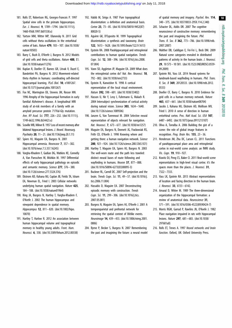
rstb.royalsocietypublishing.orgPhil.Trans.R.Soc.B
369:20120510
18
on July 11, 2018http://rstb.royalsocietypublishing.org/Downloaded from
181. Rolls ET, Robertson RG, Georges-Francois P. 1997Spatial view cells in the primate hippocampus.Eur. J. Neurosci. 9, 1789 – 1794. (doi:10.1111/j.1460-9568.1997.tb01538.x)
182. Yartsev MM, Witter MP, Ulanovsky N. 2011 Gridcells without theta oscillations in the entorhinalcortex of bats. Nature 479, 103 – 107. (doi:10.1038/nature10583)
183. Barry C, Bush D, O’Keefe J, Burgess N. 2012 Modelsof grid cells and theta oscillations. Nature 488, E1.(doi:10.1038/nature11276)
184. Kaplan R, Doeller CF, Barnes GR, Litvak V, Duzel E,Bandettini PA, Burgess N. 2012 Movement-relatedtheta rhythm in humans: coordinating self-directedhippocampal learning. PLoS Biol. 10, e1001267.(doi:10.1371/journal.pbio.1001267)
185. Fox NC, Warrington EK, Stevens JM, Rossor MN.1996 Atrophy of the hippocampal formation in earlyfamilial Alzheimer’s disease. A longitudinal MRIstudy of at-risk members of a family with anamyloid precursor protein 717Val-Gly mutation.Ann. NY Acad. Sci. 777, 226 – 232. (doi:10.1111/j.1749-6632.1996.tb34423.x)
186. Scoville WB, Milner B. 1957 Loss of recent memory afterbilateral hippocampal lesions. J. Neurol. Neurosurg.Psychiatry 20, 11 – 21. (doi:10.1136/jnnp.20.1.11)
187. Spiers HJ, Maguire EA, Burgess N. 2001Hippocampal amnesia. Neurocase 7, 357 – 382.(doi:10.1076/neur.7.5.357.16245)
188. Vargha-Khadem F, Gadian DG, Watkins KE, ConnellyA, Van Paesschen W, Mishkin M. 1997 Differentialeffects of early hippocampal pathology on episodicand semantic memory. Science 277, 376 – 380.(doi:10.1126/science.277.5324.376)
189. Ekstrom AD, Kahana MJ, Caplan JB, Fields TA, IshamEA, Newman EL, Fried I. 2003 Cellular networksunderlying human spatial navigation. Nature 425,184 – 188. (doi:10.1038/nature01964)
190. King JA, Burgess N, Hartley T, Vargha-Khadem F,O’Keefe J. 2002 The human hippocampus andviewpoint dependence in spatial memory.Hippocampus 12, 811 – 820. (doi:10.1002/hipo.10070)
191. Hartley T, Harlow R. 2012 An association betweenhuman hippocampal volume and topographicalmemory in healthy young adults. Front. Hum.Neurosci. 6, 338. (doi:10.3389/fnhum.2012.00338)
192. Habib M, Sirigu A. 1987 Pure topographicaldisorientation: a definition and anatomical basis.Cortex 23, 73 – 85. (doi:10.1016/S0010-9452(87)80020-5)
193. Aguirre GK, D’Esposito M. 1999 Topographicaldisorientation: a synthesis and taxonomy. Brain122, 1613 – 1628. (doi:10.1093/brain/122.9.1613)
194. Epstein RA. 2008 Parahippocampal and retrosplenialcontributions to human spatial navigation. TrendsCogn. Sci. 12, 388 – 396. (doi:10.1016/j.tics.2008.07.004)
195. Vann SD, Aggleton JP, Maguire EA. 2009 What doesthe retrosplenial cortex do? Nat. Rev. Neurosci. 10,792 – 802. (doi:10.1038/nrn2733)
196. Epstein R, Kanwisher N. 1998 A corticalrepresentation of the local visual environment.Nature 392, 598 – 601. (doi:10.1038/33402)
197. Hasson U, Nir Y, Levy I, Fuhrmann G, Malach R.2004 Intersubject synchronization of cortical activityduring natural vision. Science 303, 1634 – 1640.(doi:10.1126/science.1089506)
198. Janzen G, Van Turennout M. 2004 Selective neuralrepresentation of objects relevant for navigation.Nat. Neurosci. 7, 673 – 677. (doi:10.1038/nn1257)
199. Maguire EA, Burgess N, Donnett JG, Frackowiak RS,Frith CD, O’Keefe J. 1998 Knowing where andgetting there: a human navigation network. Science280, 921 – 924. (doi:10.1126/science.280.5365.921)
200. Hartley T, Maguire EA, Spiers HJ, Burgess N. 2003The well-worn route and the path less traveled:distinct neural bases of route following andwayfinding in humans. Neuron 37, 877 – 888.(doi:10.1016/S0896-6273(03)00095-3)
201. Buckner RL, Carroll DC. 2007 Self-projection and thebrain. Trends Cogn. Sci. 11, 49 – 57. (doi:10.1016/j.tics.2006.11.004)
202. Hassabis D, Maguire EA. 2007 Deconstructingepisodic memory with construction. TrendsCogn. Sci. 11, 299 – 306. (doi:10.1016/j.tics.2007.05.001)
203. Burgess N, Maguire EA, Spiers HJ, O’Keefe J. 2001 Atemporoparietal and prefrontal network forretrieving the spatial context of lifelike events.Neuroimage 14, 439 – 453. (doi:10.1006/nimg.2001.0806)
204. Byrne P, Becker S, Burgess N. 2007 Rememberingthe past and imagining the future: a neural model
of spatial memory and imagery. Psychol. Rev. 114,340 – 375. (doi:10.1037/0033-295X.114.2.340)
205. Schacter DL, Addis DR. 2007 The cognitiveneuroscience of constructive memory: rememberingthe past and imagining the future. Phil.Trans. R. Soc. B 362, 773 – 786. (doi:10.1098/rstb.2007.2087)
206. Walther DB, Caddigan E, Fei-Fei L, Beck DM. 2009Natural scene categories revealed in distributedpatterns of activity in the human brain. J. Neurosci.29, 10 573 – 10 581. (doi:10.1523/JNEUROSCI.0559-09.2009)
207. Epstein RA, Vass LK. 2014 Neural systems forlandmark-based wayfinding in humans. Phil. Trans.R. Soc. B 369, 20120533. (doi:10.1098/rstb.2012.0533)
208. Doeller CF, Barry C, Burgess N. 2010 Evidence forgrid cells in a human memory network. Nature463, 657 – 661. (doi:10.1038/nature08704)
209. Jacobs J, Kahana MJ, Ekstrom AD, Mollison MV,Fried I. 2010 A sense of direction in humanentorhinal cortex. Proc. Natl Acad. Sci. USA 107,6487 – 6492. (doi:10.1073/pnas.0911213107)
210. Oliva A, Torralba A. 2006 Building the gist of ascene: the role of global image features inrecognition. Prog. Brain Res. 155, 23 – 36.
211. Henderson JM, Zhu DC, Larson CL. 2011 Functionsof parahippocampal place area and retrosplenialcortex in real-world scene analysis: an fMRI study.Vis. Cogn. 19, 910 – 927.
212. Kravitz DJ, Peng CS, Baker CI. 2011 Real-world scenerepresentations in high-level visual cortex: it’s thespaces more than the places. J. Neurosci. 31,7322 – 7333.
213. Vass LK, Epstein RA. 2013 Abstract representationsof location and facing direction in the human brain.J. Neurosci. 33, 6133 – 6142.
214. Amaral D, Witter M. 1989 The three-dimensionalorganization of the hippocampal formation: areview of anatomical data. Neuroscience 31,571 – 591. (doi:10.1016/0306-4522(89)90424-7)
215. Morris RGM, Garrud P, Rawlins JN, O’Keefe J. 1982Place navigation impaired in rats with hippocampallesions. Nature 297, 681 – 683. (doi:10.1038/297681a0)
216. Rolls ET, Treves A. 1997 Neural networks and brainfunction. Oxford, UK: Oxford University Press.