SoxAX Binding Protein, a Novel Component of the Thiosulfate-Oxidizing Multienzyme System in
14
JOURNAL OF BACTERIOLOGY, Sept. 2008, p. 6097–6110 Vol. 190, No. 18 0021-9193/08/$08.000 doi:10.1128/JB.00634-08 Copyright © 2008, American Society for Microbiology. All Rights Reserved. SoxAX Binding Protein, a Novel Component of the Thiosulfate-Oxidizing Multienzyme System in the Green Sulfur Bacterium Chlorobium tepidum Takuro Ogawa, 1,2 Toshinari Furusawa, 1,2 Ryohei Nomura, 2 Daisuke Seo, 3 Naomi Hosoya-Matsuda, 2 Hidehiro Sakurai, 2,4 and Kazuhito Inoue 2 * Graduate School of Science, University of Tokyo, Bunkyo, Tokyo 113-0033, Japan 1 ; Department of Biological Sciences, Kanagawa University, Hiratsuka, Kanagawa 259-1293, Japan 2 ; Division of Material Science, Graduate School of Natural Science and Technology, Kanazawa University, Kakuma, Kanazawa, Ishikawa 920-1192, Japan 3 ; and Department of Biology, School of Education, Wasea University, Nishiwaseda, Shinjuku, Tokyo 169-8050, Japan 4 Received 7 May 2008/Accepted 11 July 2008 From the photosynthetic green sulfur bacterium Chlorobium tepidum (pro synon. Chlorobaculum tepi- dum), we have purified three factors indispensable for the thiosulfate-dependent reduction of the small, monoheme cytochrome c 554 . These are homologues of sulfur-oxidizing (Sox) system factors found in various thiosulfate-oxidizing bacteria. The first factor is SoxYZ that serves as the acceptor for the reaction intermediates. The second factor is monomeric SoxB that is proposed to catalyze the hydrolytic cleavage of sulfate from the SoxYZ-bound oxidized product of thiosulfate. The third factor is the trimeric cytochrome c 551 , composed of the monoheme cytochrome SoxA, the monoheme cytochrome SoxX, and the product of the hypothetical open reading frame CT1020. The last three components were expressed separately in Escherichia coli cells and purified to homogeneity. In the presence of the other two Sox factors, the recombinant SoxA and SoxX showed a low but discernible thiosulfate-dependent cytochrome c 554 reduction activity. The further addition of the recombinant CT1020 protein greatly increased the activity, and the total activity was as high as that of the native SoxAX-CT1020 protein complex. The recombinant CT1020 protein participated in the formation of a tight complex with SoxA and SoxX and will be referred to as SAXB (SoxAX binding protein). Homologues of the SAXB gene are found in many strains, comprising roughly about one-third of the thiosul- fate-oxidizing bacteria whose sox gene cluster sequences have been deposited so far and ranging over the Chlorobiaciae, Chromatiaceae, Hydrogenophilaceae, Oceanospirillaceae, etc. Each of the deduced SoxA and SoxX proteins of these bacteria constitute groups that are distinct from those found in bacteria that apparently lack SAXB gene homologues. Thiosulfate is one of the most-abundant forms of reduced sulfur in nature, and the ability to oxidize this compound is distributed over many microorganisms across different phyla (6, 7, 14, 19, 21, 43). It sometimes occurs that some members within a phylum can utilize thiosulfate while others cannot. This is the case with green sulfur bacteria; Chlorobium tepidum (pro synon. Chlorobaculum tepidum [29]) and Chlorobium limi- cola f. sp. thiosulfatophilum utilize thiosulfate, but Chlorobium limicola DSM 245 and Prosthecochloris estuarii do not (23, 24, 29, 30, 46). Almost all of the green sulfur bacteria, with one known exception of Chlorobium ferrooxidans, carry out anoxygenic photosynthesis with reduced sulfur compounds, such as sulfide and elemental sulfur, and some of them also use thiosulfate as the electron donor for the assimilation of various elements for growth (6, 7, 14, 23, 24, 43). Their reaction center (RC) is like photosystem I of oxygenic photosynthetic organisms, called type I or iron-sulfur type RC with ferredoxin and flavodoxin as immediate electron acceptors (27, 52, 56). The primary donor of the RC is a special pair of bacteriochlorophylls called P840, and its immediate electron donor is the RC-bound cytochrome (cyt) c 551 . In the moderately thermophilic C. tepidum, there seem to be multiple pathways for the reduction of the RC- bound cyt c 551 (the CT1639 protein, encoded by CT1639 of C. tepidum TLS). Itoh et al. (31) showed that a soluble mono- heme cyt, c 554 , of about 10 kDa (the CT0075 protein) donates electrons to the bound cyt c 551 rather than directly to oxidized P840. A similar small, monoheme cyt, c 555 , is an electron ac- ceptor in thiosulfate oxidation in C. limicola f. sp. thiosulfato- philum (39). C. tepidum seems to have an alternative electron transfer pathway, because mutant cells with the soluble cyt c 554 gene (CT0075) disrupted can grow phototrophically in a me- dium containing sulfide and thiosulfate, although at a lower rate than the wild type (59). The alternative pathway is pro- posed to be sulfide 3 membrane-bound sulfide-quinone re- ductase (57) 3 membrane-bound quinol oxidoreductase 3 RC-bound cyt c 551 3 P840. Two different biochemical pathways for thiosulfate oxidation are distinguishable among bacteria (22, 26, 36). In one type of pathway found in, e.g., Starkeya novella (34) and Allochroma- tium vinosum (28), thiosulfate is oxidized to sulfate through the cooperation of thiosulfate dehydrogenase, tetrathionate hy- drolase, and trithionate hydrolase with the formation of tetra- thionate as the intermediate and sulfate as the final product. In the other type of pathway, found in Paracoccus pantotrophus and some green sulfur bacteria, thiosulfate is oxidized by a * Corresponding author. Mailing address: Department of Biological Sciences, 15 Kanagawa University, Hiratsuka, Kanagawa 259-1293, Japan. Phone: 81 463-59-4111. Fax: 81 463-58-9684. E-mail: inoue-bio @kanagawa-u.ac.jp. Published ahead of print on 18 July 2008. 6097 on April 10, 2019 by guest http://jb.asm.org/ Downloaded from
Transcript of SoxAX Binding Protein, a Novel Component of the Thiosulfate-Oxidizing Multienzyme System in

JOURNAL OF BACTERIOLOGY, Sept. 2008, p. 6097–6110 Vol. 190, No. 180021-9193/08/$08.00�0 doi:10.1128/JB.00634-08Copyright © 2008, American Society for Microbiology. All Rights Reserved.
SoxAX Binding Protein, a Novel Component of the Thiosulfate-OxidizingMultienzyme System in the Green Sulfur Bacterium Chlorobium tepidum�
Takuro Ogawa,1,2 Toshinari Furusawa,1,2 Ryohei Nomura,2 Daisuke Seo,3 Naomi Hosoya-Matsuda,2Hidehiro Sakurai,2,4 and Kazuhito Inoue2*
Graduate School of Science, University of Tokyo, Bunkyo, Tokyo 113-0033, Japan1; Department of Biological Sciences,Kanagawa University, Hiratsuka, Kanagawa 259-1293, Japan2; Division of Material Science, Graduate School of
Natural Science and Technology, Kanazawa University, Kakuma, Kanazawa, Ishikawa 920-1192,Japan3; and Department of Biology, School of Education, Wasea University,
Nishiwaseda, Shinjuku, Tokyo 169-8050, Japan4
Received 7 May 2008/Accepted 11 July 2008
From the photosynthetic green sulfur bacterium Chlorobium tepidum (pro synon. Chlorobaculum tepi-dum), we have purified three factors indispensable for the thiosulfate-dependent reduction of the small,monoheme cytochrome c554. These are homologues of sulfur-oxidizing (Sox) system factors found invarious thiosulfate-oxidizing bacteria. The first factor is SoxYZ that serves as the acceptor for the reactionintermediates. The second factor is monomeric SoxB that is proposed to catalyze the hydrolytic cleavageof sulfate from the SoxYZ-bound oxidized product of thiosulfate. The third factor is the trimeric cytochromec551, composed of the monoheme cytochrome SoxA, the monoheme cytochrome SoxX, and the product of thehypothetical open reading frame CT1020. The last three components were expressed separately in Escherichiacoli cells and purified to homogeneity. In the presence of the other two Sox factors, the recombinant SoxA andSoxX showed a low but discernible thiosulfate-dependent cytochrome c554 reduction activity. The furtheraddition of the recombinant CT1020 protein greatly increased the activity, and the total activity was as highas that of the native SoxAX-CT1020 protein complex. The recombinant CT1020 protein participated in theformation of a tight complex with SoxA and SoxX and will be referred to as SAXB (SoxAX binding protein).Homologues of the SAXB gene are found in many strains, comprising roughly about one-third of the thiosul-fate-oxidizing bacteria whose sox gene cluster sequences have been deposited so far and ranging over theChlorobiaciae, Chromatiaceae, Hydrogenophilaceae, Oceanospirillaceae, etc. Each of the deduced SoxA and SoxXproteins of these bacteria constitute groups that are distinct from those found in bacteria that apparently lackSAXB gene homologues.
Thiosulfate is one of the most-abundant forms of reducedsulfur in nature, and the ability to oxidize this compound isdistributed over many microorganisms across different phyla(6, 7, 14, 19, 21, 43). It sometimes occurs that some memberswithin a phylum can utilize thiosulfate while others cannot.This is the case with green sulfur bacteria; Chlorobium tepidum(pro synon. Chlorobaculum tepidum [29]) and Chlorobium limi-cola f. sp. thiosulfatophilum utilize thiosulfate, but Chlorobiumlimicola DSM 245 and Prosthecochloris estuarii do not (23, 24,29, 30, 46).
Almost all of the green sulfur bacteria, with one knownexception of Chlorobium ferrooxidans, carry out anoxygenicphotosynthesis with reduced sulfur compounds, such as sulfideand elemental sulfur, and some of them also use thiosulfate asthe electron donor for the assimilation of various elements forgrowth (6, 7, 14, 23, 24, 43). Their reaction center (RC) is likephotosystem I of oxygenic photosynthetic organisms, calledtype I or iron-sulfur type RC with ferredoxin and flavodoxin asimmediate electron acceptors (27, 52, 56). The primary donorof the RC is a special pair of bacteriochlorophylls called P840,
and its immediate electron donor is the RC-bound cytochrome(cyt) c551. In the moderately thermophilic C. tepidum, thereseem to be multiple pathways for the reduction of the RC-bound cyt c551 (the CT1639 protein, encoded by CT1639 of C.tepidum TLS). Itoh et al. (31) showed that a soluble mono-heme cyt, c554, of about 10 kDa (the CT0075 protein) donateselectrons to the bound cyt c551 rather than directly to oxidizedP840. A similar small, monoheme cyt, c555, is an electron ac-ceptor in thiosulfate oxidation in C. limicola f. sp. thiosulfato-philum (39). C. tepidum seems to have an alternative electrontransfer pathway, because mutant cells with the soluble cyt c554
gene (CT0075) disrupted can grow phototrophically in a me-dium containing sulfide and thiosulfate, although at a lowerrate than the wild type (59). The alternative pathway is pro-posed to be sulfide 3 membrane-bound sulfide-quinone re-ductase (57) 3 membrane-bound quinol oxidoreductase 3RC-bound cyt c551 3 P840.
Two different biochemical pathways for thiosulfate oxidationare distinguishable among bacteria (22, 26, 36). In one type ofpathway found in, e.g., Starkeya novella (34) and Allochroma-tium vinosum (28), thiosulfate is oxidized to sulfate through thecooperation of thiosulfate dehydrogenase, tetrathionate hy-drolase, and trithionate hydrolase with the formation of tetra-thionate as the intermediate and sulfate as the final product. Inthe other type of pathway, found in Paracoccus pantotrophusand some green sulfur bacteria, thiosulfate is oxidized by a
* Corresponding author. Mailing address: Department of BiologicalSciences, 15 Kanagawa University, Hiratsuka, Kanagawa 259-1293,Japan. Phone: 81 463-59-4111. Fax: 81 463-58-9684. E-mail: [email protected].
� Published ahead of print on 18 July 2008.
6097
on April 10, 2019 by guest
http://jb.asm.org/
Dow
nloaded from

sulfur oxidizing system (Sox), which is also called thiosulfate-oxidizing multienzyme system (TOMES) (36). The Sox (orTOMES) pathway consists of several proteins and does notresult in the formation of tetrathionate (22). Some bacteria,e.g., S. novella (34) and A. vinosum, have both pathways (14,26, 28).
The biochemical pathway of thiosulfate oxidation by the Soxsystem has been intensively studied in facultative lithotrophicbacteria, such as Paracoccus pantotrophus (22) and Paracoccusversutus (formerly Thiobacillus versutus) (36). In these bacteria,Sox proteins are localized in the periplasm, and SoxYZ,SoxAX, and SoxB are essential components of thiosulfate ox-idation, with a small, soluble, monoheme c-type cyt of about 10kDa as the electron acceptor. The SoxYZ complex contains noprosthetic group and serves as the acceptor for the reactionintermediates (47). SoxX is a monoheme cyt c, and SoxA iseither a mono- or diheme cyt c depending on the bacterialspecies (1, 19, 32). SoxA and SoxX occur as a heterodimericcomplex in P. pantotrophus (20), Rhodovulum sulfidophilum(1), and S. novella (32). The initial oxidative reaction of thepathway is proposed to be the oxidative formation of a disul-fide linkage between the sulfane sulfur of thiosulfate and thecysteinyl-SH of SoxY (SoxY-SH) to yield -(S)-SSO3
�, accom-panied by the reduction of the small, monoheme cyt c cata-lyzed by SoxAX as follows (22): (SoxY-SH) � �SSO3
� � 2 cytcox3 (SoxY-S)-SSO3
� � 2 cyt cred � H�. SoxB contains twomanganese atoms (8, 17) and is thought to catalyze the hydro-lysis of (SoxY-S)-SSO3
� to yield sulfate and (SoxY-S)-SH.The fate of the -(S)-SH on SoxY seems to be different with
microorganisms. In bacteria such as P. pantotrophus that haveSoxCD, it is further oxidized to (-S)-SO3
� on SoxY, which isagain hydrolyzed by SoxB to regenerate (SoxY-SH), accompa-nied by the reduction of the small, monoheme cyt c (20, 22, 51).Complete oxidation of a thiosulfate generates eight electronsand two sulfate molecules.
In bacteria such as Thiothrix strains and green sulfur bacteriathat have no SoxCD, (SoxY-S)-SH presumably reacts with new�SSO3
� to yield SoxY that binds one more sulfur atom (sul-fane sulfur, or S0), accompanied by the donation of two elec-trons to external cyt c, and this reaction cycle is repeatedseveral times, yielding polysulfide groups on SoxY (54).
From the direct sequencing of the sox genes, as well as fromthe results of current whole-genome-sequencing projects,genes predicted to be involved in inorganic sulfur metabolismhave been identified in a variety of microorganisms, includingmany of the genes encoding Sox proteins (soxA, soxB, soxF,soxX, soxY, and soxW) that occur in a cluster as in P. pantotro-phus (19, 21, 22). In the green sulfur bacterium C. limicola f.thiosulfatophilum, Verte et al. (60) reported that sox genesoccur in a similar cluster. The entire genomic sequence of C.tepidum was elucidated for the first time, and its comparison tosequences of other green sulfur bacteria revealed a fairly largenumber of homologous genes involved in sulfur metabolism,including sox genes as found in other bacteria (16, 25). Withthe increasing availability of genomic sequences from othergreen sulfur bacterial strains, genes possibly involved in sulfurmetabolism may be compiled and compared (23, 24). The soxgene clusters were found in green sulfur bacteria that utilizethiosulfate, but generally not in those that do not utilize it,although some exceptions to the latter may exist (23, 24).
Moreover, soxC and soxD have not been found in green sulfurbacteria. When fed with thiosulfate, thiosulfate-utilizing greensulfur bacteria form elemental sulfur globules as intermediatesin the periplasmic space which are subsequently oxidized tosulfate (7, 9, 10, 11). The pathways for transport of elementalsulfur (or sulfane sulfur equivalent) and its subsequent metab-olism in green sulfur bacteria are unknown. In the metabolicpathway, the involvement of a dissimilatory sulfur reductasethat resides in cytoplasm is proposed (16, 23, 24, 25, 45). Thepossible pathway for transport of elemental sulfur (sulfanesulfur equivalent) or persulfide (23, 24) across the plasmamembrane has been discussed based on genomic sequenceanalysis (9, 10, 23, 24). Recently, Chan et al. (11) generated amutant from C. tepidum in which the region between CT0868and CT0876 was replaced by a transposon insertion and sub-sequently found that the mutant was completely defective forgrowth on thiosulfate as the sole electron donor, suggestingthat the protein(s) encoded by one or some of the genes in thisregion might be involved in the transport of sulfur (or a sulfanesulfur equivalent) across the plasma membrane.
The results of biochemical studies revealed the presence ofseveral components involved in thiosulfate oxidation in greensulfur bacteria. A small, monoheme cyt c called Chlorobium cytc555, a homolog of conventional cyt c in mitochondria, wasisolated from C. limicola f. sp. thiosulfatophilum and found tobe an electron acceptor of the thiosulfate-oxidizing enzymesystem (38, 39, 44). In C. tepidum, cyt c554 (a homolog of C.limicola cyt c555) seems to mediate electron transport betweenthe thiosulfate oxidation system and RC-bound cyt c551 asdescribed above (31). Meyer et al. (44) isolated the multihemecyt c551 from C. limicola f. sp. thiosulfatophilum, which appearsto correspond to a homologue of SoxAX found in variousthiosulfate-oxidizing bacteria. Kusai and Yamanaka (39) iso-lated thiosulfate-cyt c (multiheme cyt c551) reductase from thesame strain that binds no flavin or heme, with a molecular massof 80 kDa as estimated by sodium dodecyl sulfate-polyacryl-amide gel electrophoresis (SDS-PAGE) analysis in the pres-ence of mercaptoethanol. Brief summaries of the results ofbiochemical studies of thiosulfate oxidation in green sulfurbacteria have been presented in the literature (43, 62). Al-though the results of genome sequence analyses indicate thepresence of many sox genes in green sulfur bacteria that arehomologous to those found in other groups of bacteria (16, 23,24, 25), biochemical characterization of the products encodedby sox genes in green sulfur bacteria is largely lacking.
We have purified three components necessary for thiosulfateoxidation in C. tepidum and characterized some of their bio-chemical properties. We found that the SoxAX complex bindsa novel colorless protein factor, encoded by the hypotheticalopen reading frame CT1020. The gene product of CT1020,which will be referred to as SAXB (SoxAX binding protein),stimulates the thiosulfate oxidation activity of the Sox system inthis bacterium.
MATERIALS AND METHODS
Bacterial strains and growth conditions. C. tepidum strain TLS (obtained fromM. T. Madigan) was grown for 12 to 17 h at 40°C, essentially according to themethods described in reference 61.
Purification of thiosulfate-oxidizing proteins. The cells were harvested bycentrifugation (8,000 � g) at 4°C. The collected cells were resuspended in 50 mM
6098 OGAWA ET AL. J. BACTERIOL.
on April 10, 2019 by guest
http://jb.asm.org/
Dow
nloaded from
Tris-HCl (pH 7.8), 100 mM NaCl, 10 mM EDTA, 5 mM sodium ascorbate, 5 mMdithiothreithol, 1 mM 6-amino-n-caproic acid, 1 mM phenylmethylsulfonyl flu-oride and 1 mM p-aminobenzamidine·2HCl. The cell pellets were sonicatedbriefly and then disrupted with a French pressure cell at 140 MPa. The cell lysatewas centrifuged at 20,000 � g for 20 min, and unbroken cells were removed asprecipitates. The supernatant was further centrifuged at 160,000 � g for 2 h, andthe resultant supernatant was fractionated by ammonium sulfate, yielding thio-sulfate-oxidizing activity as the precipitate between 40% and 80% ammoniumsulfate saturation. Precipitated proteins were collected by centrifugation, and thepellet was resuspended in 20 mM Tris-HCl (pH 7.8) and dialyzed against thesame buffer with several changes. The dialyzed preparation was applied to aDEAE-Toyopearl 650 M column (2.5 by 25 cm; Tosoh) equilibrated with 20 mMTris-HCl (pH 7.8), and the flowthrough fractions that contained thiosulfate-oxidizing activity with cyt c554 as the electron acceptor were collected. The bufferof the flowthrough fractions was changed to 10 mM 2-morpholinoethanesulfonicacid (MES)-NaOH buffer (pH 6.0) by ultrafiltration (YM-3; Millipore) andapplied to a Hitrap SP column (two 5-ml columns in tandem; GE Healthcare).Protein was eluted with 100 ml of a linear gradient of 0 to 250 mM NaCl. Thefractions (5 ml each) that had a thiosulfate-dependent cyt c554 reduction activitywere combined (total volume, 20 ml) and desalted by ultrafiltration (YM-3;Millipore), and the concentrate was applied to a Hitrap Q column (two 5-mlcolumns in tandem). Protein was eluted with 100 ml of a linear gradient of 0 to300 mM NaCl in 10 mM MES-NaOH buffer (pH 6.0), and fractions (5 ml each)were collected.
At this stage, thiosulfate-oxidizing activity in the presence of externally addedcyt c554 was not detectable in any of the single fractions. Because it has oftenbeen shown that multiple factors are required for thiosulfate oxidation in otherbacteria (20, 22, 41, 42, 51), we tried to reconstitute the activity and found thatthe combination of three fractions (tentatively referred to as fractions I, II, andIII) restored the activity. We subsequently purified the active components sep-arately. Fraction I (eluted from the Hitrap Q column at about 20 mM NaCl) wasdesalted by ultrafiltration (YM-3; Millipore) with the buffer changed to 10 mMMES-NaOH (pH 6.0) and applied to a MonoS column (bed volume, 1 ml; GEHealthcare) equilibrated with the same buffer. Proteins were eluted with 20 mlof a linear gradient of 0 to 100 mM NaCl, and purified SoxYZ was eluted atabout 20 mM NaCl. The fractions eluted at about 60 mM NaCl contained acomplex of SoxYZ and SoxB, but we did not study the latter fractions in detail.Fraction II (eluted from the Hitrap Q column at about 80 mM NaCl) wasdesalted by ultrafiltration with the buffer changed to 10 mM Tris-HCl (pH 8.7)and applied to a MonoQ column (bed volume, 1 ml; GE Healthcare) equili-brated with the same buffer. SoxB was eluted at about 60 mM NaCl with a 30-mllinear gradient of 0 to 300 mM NaCl yielding purified SoxB. Fraction III (elutedfrom the Hitrap Q column at about 100 mM NaCl) was desalted by ultrafiltrationwith the buffer changed to 10 mM Tris-HCl (pH 8.7) and applied to a MonoQcolumn (bed volume, 1 ml; GE Healthcare) equilibrated with 10 mM Tris-HCl(pH 8.7). Protein was eluted at about 80 mM NaCl with a 30-ml linear gradientof 0 to 300 mM NaCl, yielding purified SoxAX-CT1020 protein.
Purification of cyt c554. Cyt c554 was purified to homogeneity from cell extractsas described previously (31).
Expression of rSoxA, rSoxX, and rCT1020 in Escherichia coli cells. All mo-lecular manipulations were carried out according to standard DNA techniques(53), and E. coli strains were grown in Luria-Bertani medium at 37°C. The finalconcentrations of antibiotics, when used, were 30 �g ml�1 for chloramphenicoland 100 �g ml�1 for ampicillin. Recombinant SoxA (rSoxA) and recombinantSoxX (rSoxX) were produced separately in E. coli strain BL21(DE3) harboring,in addition to the plasmid pEC86 (2), either pET23c::soxA or pET23c::soxX.Recombinant CT1020 protein (rCT1020) was produced in E. coli strainBL21(DE3) harboring the plasmid pET23c::CT1020. Expression of the recom-binant genes was induced by adding 0.5 mM isopropyl-�-D-thiogalacto-pyrano-side to the culture, followed by incubation for 16 to 18 h.
More precisely, these genes were amplified by PCR with KOD Dash (Toyobo)using the pair of oligonucleotides soxAfw (5�-CATATGAAAAAAACAATTCAGCGGGG-3�) and soxArv (5�-GAATTCTTATTTTCTTGATGCCGGG-3�) forsoxA, soxXfw (5�-GAATTCGTGGCGCGTGGTTTT-3�) and soxXrv (5�-AAGCTTTCAGAGCGTGTAGAGATAATCGAC-3�) for soxX, and ct1020fw (5�-CATATGAAAAAAGTGTTATCGCTCT-3�) and ct1020rv (5�-AAGCTTTCAGTTTTTAGGAATCATC-3�) for CT1020. These PCR-generated fragments wereligated into pCR2.1 to produce the plasmids pCR2.1::soxA, pCR2.1::soxX, andpCR2.1::CT1020, respectively. pCR2.1::soxA was digested with NdeI and EcoRI,and the released insert was ligated into NdeI/EcoRI-digested pET23c to producethe plasmid pET23c::soxA. pCR2.1::soxX was digested with EcoRI and HindIII,and the released insert was ligated into EcoRI/HindIII-digested pET23c toproduce the plasmid pET23c::soxX. pCR2.1::CT1020 was digested with NdeI and
HindIII, and the released insert was ligated into NdeI/HindIII-digested pET23cto produce the plasmid pET23c::CT1020. These plasmids were checked forcorrect sequences by automated DNA sequencing using an ABI Prism 310(Applied Biosystems).
Preparation of periplasmic extracts from overexpressing E. coli cells andpurification of rSoxA, rSoxX, and rCT1020. Periplasmic fractions were isolatedby osmotic shock treatment of E. coli cells according to the method specified byQiagen (13). Briefly, cells were harvested by centrifugation (10,000 � g for 10min), and the pellets from 4 liters of cultures were washed once with 30 mMTris-HCl (pH 7.8) and resuspended in 10 ml of 30 mM Tris-HCl (pH 7.8), 1 mMEDTA·2Na, and 20% sucrose. The cell suspension was stirred at room temper-ature for 10 min and centrifuged at 10,000 � g, 4°C for 10 min, and thesupernatant was retained. The pellets were resuspended in 30 mM Tris-HCl (pH7.8), and after being stirred for 10 min in an ice bath, the cells were againseparated by centrifugation as described above. The combined supernatants wereclarified by centrifugation at 184,000 � g for 1 h and used for purification of therespective recombinant proteins.
Briefly, purification of rSoxX was carried out as follows: a DEAE-Toyopearl650 M column (10 by 2.5 cm; Tosoh) with a linear gradient of 0 to 300 mM NaClin 20 mM Tris-HCl (pH 7.8), brought to 2 M ammonium sulfate, was used, andthe supernatant, after the precipitate was removed, was applied to a HiTrap PHEcolumn (bed volume, 5 ml; GE Healthcare), equilibrated with 2 M ammoniumsulfate in the same buffer, and eluted by using a decreasing linear gradient ofammonium sulfate from 2 to 0 M; concentration and desalting were performedby ultrafiltration (Amicon ultra; Millipore) and HiTrapQ column chromatogra-phy with elution on a linear gradient of 0 to 200 mM NaCl in the same buffer.
Purification of rCT1020 and rSoxA was carried out as follows: the activefractions were eluted in the flowthrough fractions from a DEAE-Toyopearl 650M column (10 by 2.5 cm; Tosoh) equilibrated with 20 mM Tris-HCl (pH 7.8),brought to 2 M ammonium sulfate, and the supernatant, after the precipitate wasremoved, was applied to a HiTrap PHE column (GE Healthcare), equilibratedwith 2 M ammonium sulfate, and eluted by using a decreasing linear gradient ofammonium sulfate from 2 to 0 M in the same buffer; concentration and desaltingwere performed by ultrafiltration (Amicon Ultra; Millipore).
Enzyme assays. Thiosulfate-dependent cyt c554 reduction activity was mea-sured by using a spectrophotometer (UV2500PC; Shimadzu) at 25°C in a volumeof 0.1 ml reaction mixture. The standard reaction mixture contained 20 mMMES-NaOH (pH 6.0), 50 �M cyt c554, 2 mM sodium thiosulfate, and 0.5 �Mpurified thiosulfate-oxidizing components (SoxYZ, SoxB, and SoxAX-CT1020protein) unless otherwise indicated. Cyt c reduction rates were calculated byusing the redox difference ε554 � 23.8 mM�1 � cm�1 (55). The kinetic constantsof enzymatic reactions were obtained by linear regression analyses.
Redox titrations. Redox titrations were carried out at 25°C under nitrogenatmosphere with a potentiometer (HM26S; Toa) equipped with a redox elec-trode (PTS-5011C; Toa). The solutions contained 5 �M of proteins, 10 �M2,3,5,6-tetramethyl-1,4-phenylenediamine, 10 �M duroquinone, and 10 �M1-methoxy-5-methylphenazinium methyl sulfate, in either 100 mM potassiumphosphate (pH 7.0) or 100 mM glycine-NaOH (pH 10). Reductive titrations werestarted at a high Eh by successive additions of 50 mM sodium dithionite and werefollowed by oxidative titrations (for pH 7.0 only) using successive additions of 10mM potassium ferricyanide.
Analytical methods. SDS-PAGE analysis was performed (40), and proteinswere stained with Coomassie brilliant blue R 250. Heme was stained with 3,3�-dimethoxy benzidine dihydrochloride (DMBZ) (18).
N-terminal amino acid sequences were determined by the Edman degradationmethod using a Procise cLC 494 sequencer (Applied Biosystems).
The molecular masses of SoxAX-CT1020 protein were determined by matrix-assisted laser ionization (MALDI)–time-of-flight mass spectrometry using a Shi-madzu AXIMA-CFR mass spectrometer with sinapinic acid as the matrix.
Protein was quantified by the method of Bradford (5) unless otherwise indi-cated.
The total heme content of SoxAX-CT1020 protein, rSoxA, and rSoxX weredetermined by using the alkaline pyridine hemochrome method (4). The proteinconcentrations of SoxA and SoxX were calculated from the heme concentrationby assuming that each contains one heme per subunit and the SoxAX-CT1020protein complex a total of two hemes per molecule.
The molecular masses of proteins were estimated by gel permeation chroma-tography using either (i) a fast protein liquid chromatography system (AKTA;GE Healthcare) equipped with a Superdex 200 10- by 300-mm GL column (GEHealthcare) with a flow rate of 1 ml � min�1 or (ii) an LC10-AD high-perfor-mance liquid chromatography (Shimadzu) TSK-Gel G3000PWXL 7.8- by300-mm column with a flow rate of 0.5 ml � min�1 using a low-molecular-weight
VOL. 190, 2008 THIOSULFATE OXIDATION IN GREEN SULFUR BACTERIA 6099
on April 10, 2019 by guest
http://jb.asm.org/
Dow
nloaded from

gel filtration kit (GE Healthcare) as the standard. The buffer was 50 mM Tris-HCl (pH 7.8) containing 150 mM NaCl.
RESULTS
Proteins essential for thiosulfate oxidation and their poly-peptide composition. We have purified the three components,namely, SoxYZ, SoxAX-CT1020 protein, and SoxB, to homo-geneity (Fig. 1) as essential proteins for thiosulfate oxidationin vitro with the small, monoheme cyt c554 as the electronacceptor.
The active protein purified from fraction I was colorless,with apparent molecular masses of about 40 and 26 kDa on gelpermeation chromatography (10- by 300-mm Superdex 200)analysis (data not shown). With SDS-PAGE analysis, thepolypeptide composition was the same, composed of two kindsof polypeptide of about 13 kDa and 9 kDa (Fig. 1b, lane 1).The N-terminal amino acid sequence of the 13-kDa polypep-tide was SWNEKAFSAS, which agrees with the one deducedfrom the soxY gene (CT1017) in the C. tepidum genomic da-tabase (http://www.tigr.org) that begins with Ser32, indicatingthat a signal peptide targeting the periplasmic space is cleavedbetween 31Ala and 32Ser. The N-terminal amino acid se-quence of the 9-kDa polypeptide was determined to be MKIKAVVQNN, which agrees with the one deduced from the soxZgene (CT1018) that begins with the initial methionine withoutcleavage of a signal peptide. When the purified preparationcontaining both the 26- and 40-kDa proteins was incubatedwith 1 mM dithiothreitol for 30 min at room temperature, theresults of the subsequent chromatography revealed that thepreparation contained only 26-kDa proteins. The same resultswere obtained when the preparation was incubated with only 1mM thiosulfate, suggesting that the 40-kDa protein wasformed during purification by the oxidation of the product-binding cysteine on SoxY between the two SoxYZ complexes.The SoxYZ activity was essentially unchanged by prior incu-bation of the preparation with either dithiothreitol or thiosul-fate. SoxY and SoxZ form a tight complex that is difficult toseparate into active components by ion-exchange chromatog-
raphy, hydrophobic interaction chromatography, or gel perme-ation chromatography.
The active protein purified from fraction II had an apparentmolecular mass of 63 kDa by gel permeation chromatographyanalysis (7.8- by 300-mm TSK-Gel G3000PWXL) (data notshown) and about 60 kDa by SDS-PAGE analysis (Fig. 1a, lane2). The N-terminal amino acid sequence of TKASSDLYDF,which agrees with the one deduced from the soxB gene(CT1021) that begins with 60Thr, indicates that a signal pep-tide targeting the periplasmic space is cleaved between 59Alaand 60Thr. Assuming that SoxB contains two Mn per mole-cule, the molecular mass of SoxB is calculated to be 61,655 Da,indicating that the purified SoxB exists as a monomer.
The active protein purified from fraction III was reddishbrown in color, with an apparent molecular mass of 42 kDa bygel permeation chromatography (10- by 300-mm Superdex200) analysis (data not shown), and composed of three kinds ofpolypeptide of about 30 kDa, 10 kDa, and 9 kDa by SDS-PAGE analysis (Fig. 1b, lane 2). The 30-kDa and the 10-kDabands were found to bind heme from DMBZ staining, but the9-kDa band did not (Fig. 1b, lane 3). The N-terminal aminoacid sequence of the 30-kDa band was EVNYQALVDADV,which agrees with the one deduced from the soxA gene(CT1019) that begins with 28Glu, indicating that a signal pep-tide targeting the periplasmic space is cleaved between 27Alaand 28Glu. The N-terminal amino acid sequence of the 10-kDaband was AAPAAVDSSV, which agrees with the one deducedfrom soxX gene (CT1016) that begins with 47Ala, indicatingthat a signal peptide targeting the periplasmic space is cleavedbetween 46Ala and 47Ala. The N-terminal amino acid se-quence of the 9-kDa band was EPAPAAPAAS, which agreeswith the one deduced from the hypothetical open readingframe CT1020 of previously unknown function that begins with22Glu, indicating that a signal peptide targeting the periplas-mic space is cleaved between 21Ala and 22Glu. The MALDI–time-of-flight MS measurements of the SoxAX-CT1020 pro-tein complex yielded masses in agreement with the aboveconclusions: 29,911 Da (SoxA, calculated mass of 29,934 Dafor a monoheme form; heme, 618 Da; and persulfide, 32 Da [3,12, 15, 35]), 11,128 Da (SoxX, calculated mass of 11,130 Da fora monoheme form), and 9,376 Da (CT1020 protein, calculatedmass of 9,373 Da) were the major peaks (data not shown).
Expression of rSoxA, rSoxX, and rCT1020 in E. coli cells.The SoxAX-CT1020 protein formed a tight complex, making itdifficult to prepare each component as a separate fractionretaining activity. We expressed each component separately inE. coli cells and purified rSoxA, rSoxX, and rCT1020 (Table 1and Fig. 1b). The dithionite-reduced form of the nativeSoxAX-CT1020 protein exhibited absorption peaks at 551,522, and 417 nm (Fig. 2a). The dithionite-reduced absorptionspectrum of rSoxAX-rCT1020 (Fig. 2b) was essentially identi-cal with that of the native SoxAX-CT1020 protein. rSoxA hasabsorption peaks at 551, 524, and 418 nm and rSoxX at 551,522, and 416 nm when reduced with dithionite, both typical forcyt c (Fig. 2c and d).
The absorption changes of the redox titration curves of na-tive SoxAX-CT1020 protein, rSoxA, and rSoxX are shown inFig. 3. At pH 10.0 (Fig. 3a), the midpoint redox potential ofrSoxX was �135 mV and that of rSoxA less than �550 mV.SoxAX-CT1020 protein exhibited two midpoint potentials,
FIG. 1. SDS-PAGE profiles of purified Sox proteins and purifiedrecombinant proteins. (a) Lanes: 1, SoxYZ; 2, SoxB; 3, SoxAX-CT1020 protein. (b) Lanes: 1, SoxYZ; 2 and 3, SoxAX-CT1020 pro-tein; 4 and 5, rCT1020; 6 and 7, rSoxX; 8 and 9, rSoxA. Lanes 1, 2, 4,6, and 8 were stained with Coomassie brilliant blue, and lanes 3, 5, 7,and 9 were stained with DMBZ.
6100 OGAWA ET AL. J. BACTERIOL.
on April 10, 2019 by guest
http://jb.asm.org/
Dow
nloaded from

with the higher one at �138 mV and the lower one less than�550 mV. From these results, the former is assigned to theheme on SoxX and the latter to the one on SoxA. At pH 7.0,only reduction of one heme with a midpoint potential of �161mV was observed, and the reduction-oxidation cycle was com-pletely reversible (Fig. 3b).
By SDS-PAGE analysis, the apparent molecular masses ofpurified rSoxA (30 kDa) and rSoxX (10 kDa) were indistin-guishable from those of the corresponding subunits of thenative C. tepidum SoxAX-CT1020 protein complex (Fig. 1b).
However, by SDS-PAGE analysis, rCT1020 showed two bands,with the major band smaller than the band found in the nativeSoxAX-CT1020 protein complex and the minor band indistin-guishable from the band in the native complex (Fig. 1b, lane 4).
FIG. 2. Redox absorption spectra of the SoxAX-CT1020 proteinand its components. (a) SoxAX-CT1020 protein. (b) Purified complexobtained by gel permeation chromatography from the mixture contain-ing rSoxA, rSoxX, and rCT1020. (c) Purified rSoxX. (d) PurifiedrSoxA. The concentration of each protein was 5 �M based on hemedetermination. Dithionite-reduced spectra (solid lines) were obtainedby the addition of sodium dithionite. Oxidized spectra (dotted lines)are those of the preparations as obtained.
FIG. 3. Redox titrations of SoxAX-CT1020 protein, rSoxA, andrSoxX. The absorbance was normalized based on heme determination.(a) Titrations at pH 10.0. Open squares, SoxAX-CT1020 protein; opentriangles, rSoxX; open circles, rSoxA. (b) Titrations at pH 7.0. Opensquares, SoxAX-CT1020 protein; open triangles, rSoxX. When Eh wasstabilized, typically 2 to 3 min after the addition of dithionite orferricyanide, the visible spectrum was recorded, and the differentialabsorption changes of A551 minus A565 were plotted. In panel b, theresults of both reductive and oxidative titrations are plotted in thesame figure. The solid line is fitted to the Nernst’s n � 1 equation withEm values of �161 mV (SoxAX-CT1020 protein, pH 7.0), �153 mV(rSoxX, pH 7.0), �138 mV (SoxAX-CT1020 protein, pH 10), and�135 mV (rSoxX, pH 10), respectively.
TABLE 1. Bacterial strains and plasmids used in this study
Strain or plasmid Relevant characteristics Reference or source
E. coli strainsXL-1 Blue MR �(mcrA)183 �(mcrCB-hsdSMR-mrr)173 recA1 endA1 gyrA96 thi-1 supE44 relA1 � lac StratageneBL21(DE3) F� ompT hsdSB(rB
� mB�) gal dcm (DE3) Novagen
PlasmidspCR2.1 Apr Kmr lacZ InvitrogenpET23c Apr T7 promoter NovagenpEC86 Cmr ccmABCDEFGH in pACYC184 2pCR2.1::soxA 870-bp soxA PCR fragment in pCR2.1 This studypCR2.1::soxX 447-bp soxX PCR fragment in pCR2.1 This studypCR2.1::CT1020 336-bp CT1020 PCR fragment in pCR2.1 This studypET23c::soxA 863-bp NdeI/HindIII fragment from pCR2.1::soxA in NdeI/HindIII of pET23c This studypET23c::soxX 441-bp EcoRI/HindIII fragment from pCR2.1::soxX in EcoRI/HindIII of pET23c This studypET23c::CT1020 329-bp NdeI/HindIII fragment from pCR2.1::CT1020 in NdeI/HindIII of pET23c This study
VOL. 190, 2008 THIOSULFATE OXIDATION IN GREEN SULFUR BACTERIA 6101
on April 10, 2019 by guest
http://jb.asm.org/
Dow
nloaded from

The N-terminal amino acid sequences of rSoxA and rSoxXwere EVNYQALVDA and AAPAAVDSSV, respectively, incomplete agreement with those of SoxA and SoxX in the pu-rified native C. tepidum SoxAX-CT1020 protein complex. Theresults of MALDI mass spectrometry showed a single masspeak of 11,112 Da for rSoxX and a major peak of 29,918 Da forrSoxA. The entire profiles of the MALDI mass spectrogramsin the regions of these polypeptides were very similar to thoseof the native C. tepidum SoxAX-CT1020 protein complex (datanot shown). The N-terminal amino acid sequences of therCT1020 preparation were found to be composed of a mixtureof two populations, with the major one starting at 27Ala andthe minor one at 23Glu, the latter being in agreement withthe native CT1020 protein in the purified C. tepidum com-plex. The MALDI mass spectrogram of rCT1020 showedtwo mass peaks of 8,923 Da (major) and 9,390 Da (minor)(data not shown), in agreement with assignments of startingpoints at 27Ala (8,908 Da) and 23Glu (9,373 kDa), respec-tively. The rCT1020 preparation used in this study is amixture of these two populations.
Thiosulfate-dependent cyt c554 reduction kinetics and theeffects of CT1020 protein. The combination of SoxAX-CT1020protein, SoxYZ, and SoxB was absolutely necessary for thethiosulfate-dependent reduction of the small, monoheme cytc554. The omission of any one of the components results in noactivity (Table 2), as found in other sulfur-oxidizing bacteria(20, 38, 41, 42, 51).
The relationship between the initial rate of cyt c554 reductionand the thiosulfate concentration (in the range of 0 to 2 mM)can be fitted to Michaelis-Menten-type kinetics with a Km
value for thiosulfate of 0.17 mM (Fig. 4a and Table 2). Athigher thiosulfate concentrations, inhibition of reactions wasnoted (data not shown), as with the Sox system of A. vinosum(28). The optimum pH for the reaction was found at about pH6.0 to 6.5 (data not shown). In this assay system, 2.1 mol of cytc554 was reduced per mole of thiosulfate oxidized and thereaction proceeded almost linearly for at least two minutes,during which time about 8 mol of thiosulfate was oxidized permole of SoxYZ (data not shown). These results indicate thatthe product of sulfane sulfur from thiosulfate is zero-valencesulfur, possibly remaining as polysulfane bound on SoxY oras the elemental sulfur subsequently formed (54). The pro-file of the temperature-activity curve of the complete system
was similar to those of ordinary enzyme reactions, with theoptimum temperature at about 55 to 60°C. The activitiesdetermined at 2 mM thiosulfate for 1 min were 6.7 (25°C),17.2 (35°C), 24.6 (40°C), 29.7 (45°C), 33.3 (50°C), 36.4(55°C), 36.9 (60°C), and 28.7 (65°C) �mol cyt c554
reduced � �mol Sox protein�1 � min�1.The functions of purified rSoxA, rSoxX, and rCT1020 were
examined in an assay system containing SoxYZ, SoxB, and cytc554 from C. tepidum, but not SoxAX-CT1020 protein (thebasic assay system). The addition of any one of the singlecomponents rSoxA, rSoxX, or rCT1020 to the basic assay sys-tem could not support thiosulfate oxidation. The same was truewith the two-component mixtures of rSoxA plus rCT1020 andrSoxX plus rCT1020 protein (data not shown). However, themixture of rSoxA and rSoxX, each at 0.5 �M, showed a low butdiscernible cyt c554 reduction activity when added to the basic
FIG. 4. Thiosulfate-dependent cyt c554 reduction activities. (a) Ef-fects of thiosulfate concentration on cyt c554 reduction rates. Thereaction mixture contained 0.5 �M SoxYZ, 0.5 �M SoxB, 0.5 �MSoxAX-CT1020 protein, 50 �M cyt c554, 20 mM MES-NaOH (pH 6.0),and the indicated concentrations of thiosulfate. (b) As above, exceptthat in place of 0.5 �M SoxAX-CT1020 protein (the basic assay mix-ture), the following were added: open triangles, 0.5 �M each of rSoxA,rSoxX, and rCT1020; open squares, 0.5 �M each of rSoxA and rSoxX.An enlargement of the kinetics of the last combination is shown in theinset. Cyt c554 reduction was monitored by the absorbance change at554 nm. (c) Effects of rSoxA and rSoxX concentrations in the absenceof rCT1020. The basic assay mixtures contained 1.0 �M rSoxA andindicated concentrations of rSoxX (open squares), 1.0 �M rSoxX andindicated concentrations of rSoxA (open circles), or equal concentra-tions of both rSoxA and rSoxX as indicated (open triangles). (d)Effects of rCT1020 concentration. The basic assay mixture contained0.5 �M rSoxA, 0.5 �M rSoxX, and the indicated concentrations ofrCT1020.
TABLE 2. Kinetics parameters in thiosulfate-dependent cyt c554reduction activitiesa
ComponentV (�mol cyt c554
reduction � �molSox protein�1 � min�1)
Km (mM)
SoxYZ, SoxB, and SoxAX-CT1020protein
7.9 0.2 0.17 0.02
SoxYZ, SoxB, and rSoxAX-rCT1020 7.8 0.1 0.18 0.02SoxYZ, SoxB, rSoxA, and rSoxX 0.24 0.08 0.16 0.02SoxYZ and SoxB NDSoxYZ and SoxAX-CT1020 protein NDSoxB and SoxAX-CT1020 protein ND
a Thiosulfate-dependent cyt c554 reduction activities and Km values weredetermined from the �S�0/v-versus-�S�0 plot in the thiosulfate concentrationrange of 0 to 2 mM. The values are the means standard deviations of theresults of three independent measurements similar to the ones whose results areshown in Fig. 4a and b. ND, not detectable ( 0.01 �mol cyt c554 reduced � �molSox protein�1 � min�1).
6102 OGAWA ET AL. J. BACTERIOL.
on April 10, 2019 by guest
http://jb.asm.org/
Dow
nloaded from
assay system (Fig. 4b and Table 2). When the rSoxX concen-tration was fixed at 1.0 �M, the rate of thiosulfate-dependentcyt c554 reduction initially increased with an increase in rSoxAconcentration but apparently was saturated at about 1.0 �Mof rSoxA (Fig. 4c). Similar results were obtained when therSoxX concentration was changed in the presence of a fixedconcentration of rSoxA of 1.0 �M. When rSoxA and rSoxXwere added simultaneously at the same concentration, thereaction rate increased with increasing concentrations ofrSoxA and rSoxX and with no indication of saturation in theconcentration range shown in Fig. 4c. Furthermore, thesimultaneous addition of increasing concentrations of rSoxAand rSoxX exceeded the maximum rates attained when theconcentration of one of the components was fixed at 1 �M(Fig. 4c).
When 0.5 �M rCT1020 was added to the basic assay systemcontaining 0.5 �M rSoxA and 0.5 �M rSoxX, the activity in-creased dramatically (Fig. 4d). The activity initially increasedwith increasing rCT1020 concentrations and, at around 0.5�M, reached a plateau at a level about 30-fold higher than inthe presence of rSoxA and rSoxX without rCT1020. The spe-cific activity and Km for thiosulfate of the assay system con-taining a mixture of rSoxA, rSoxX, and rCT1020 are shown inTable 2. The maximum activity of the assay mixture containingthe three recombinant proteins was comparable to that of themixture containing the native SoxAX-CT1020 protein com-plex, indicating that the recombinant proteins were overex-pressed in E. coli cells in their active forms. The temperature-activity curve of the basic assay system containing all threerecombinant proteins was similar to that containing nativeSoxAX-CT1020 protein until the temperature reached 55°C.The activity of the basic assay system containing the recom-binant proteins was slightly lower than that containing thenative SoxAX-CT1020 protein after 60°C, declining slightlyfaster than the latter with further increases in temperature(data not shown). Although the maximum activity of thesystem containing rSoxA and rSoxX was much lower thanthat of the complete system containing all three recombi-nant proteins, the Km value for thiosulfate was almost un-changed. The kinetics values obtained by various assays aresummarized in Table 2.
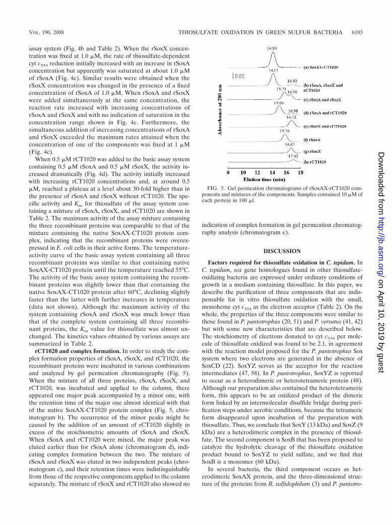
rCT1020 and complex formation. In order to study the com-plex formation properties of rSoxA, rSoxX, and rCT1020, therecombinant proteins were incubated in various combinationsand analyzed by gel permeation chromatography (Fig. 5).When the mixture of all three proteins, rSoxA, rSoxX, andrCT1020, was incubated and applied to the column, thereappeared one major peak accompanied by a minor one, withthe retention time of the major one almost identical with thatof the native SoxAX-CT1020 protein complex (Fig. 5, chro-matogram b). The occurrence of the minor peaks might becaused by the addition of an amount of rCT1020 slightly inexcess of the stoichiometric amounts of rSoxA and rSoxX.When rSoxA and rCT1020 were mixed, the major peak waseluted earlier than for rSoxA alone (chromatogram d), indi-cating complex formation between the two. The mixture ofrSoxA and rSoxX was eluted in two independent peaks (chro-matogram c), and their retention times were indistinguishablefrom those of the respective components applied to the columnseparately. The mixture of rSoxX and rCT1020 also showed no
indication of complex formation in gel permeation chromatog-raphy analysis (chromatogram e).
DISCUSSION
Factors required for thiosulfate oxidation in C. tepidum. InC. tepidum, sox gene homologues found in other thiosulfate-oxidizing bacteria are expressed under ordinary conditions ofgrowth in a medium containing thiosulfate. In this paper, wedescribe the purification of three components that are indis-pensable for in vitro thiosulfate oxidation with the small,monoheme cyt c554 as the electron acceptor (Table 2). On thewhole, the properties of the three components were similar tothose found in P. pantotrophus (20, 51) and P. versutus (41, 42)but with some new characteristics that are described below.The stoichiometry of electrons donated to cyt c554 per mole-cule of thiosulfate oxidized was found to be 2.1, in agreementwith the reaction model proposed for the P. pantotrophus Soxsystem where two electrons are generated in the absence ofSoxCD (22). SoxYZ serves as the acceptor for the reactionintermediates (47, 58). In P. pantotrophus, SoxYZ is reportedto occur as a heterodimeric or heterotetrameric protein (48).Although our preparation also contained the heterotetramericform, this appears to be an oxidized product of the dimericform linked by an intermolecular disulfide bridge during puri-fication steps under aerobic conditions, because the tetramericform disappeared upon incubation of the preparation withthiosulfate. Thus, we conclude that SoxY (13 kDa) and SoxZ (9kDa) are a heterodimeric complex in the presence of thiosul-fate. The second component is SoxB that has been proposed tocatalyze the hydrolytic cleavage of the thiosulfate oxidationproduct bound to SoxYZ to yield sulfate, and we find thatSoxB is a monomer (60 kDa).
In several bacteria, the third component occurs as het-erodimeric SoxAX protein, and the three-dimensional struc-ture of the proteins from R. sulfidophilum (3) and P. pantotro-
FIG. 5. Gel permeation chromatograms of rSoxAX-rCT1020 com-ponents and mixtures of the components. Samples contained 10 �M ofeach protein in 100 �l.
VOL. 190, 2008 THIOSULFATE OXIDATION IN GREEN SULFUR BACTERIA 6103
on April 10, 2019 by guest
http://jb.asm.org/
Dow
nloaded from
phus (15) have been determined by X-ray crystallography.SoxX from various bacteria has been reported to be a mono-heme protein, while SoxA is either a diheme protein in P.pantotrophus and R. sulfidophilum or a monoheme protein inStarkeya novella (32). For this study, we prepared a complexcontaining SoxAX and found that the complex binds theCT1020-encoded protein of about 9 kDa as the third compo-nent and that the complex exists as a heterotrimer (Fig. 1 andFig. 5).
Biochemical characterization of the CT1020 protein (SAXB).Rother and Friedrich (50) report having succeeded in coex-pressing soxA and soxX of P. pantotrophus in E. coli cells andhaving subsequently obtained the SoxAX complex in an activeform. They also overexpressed soxX, but the protein alone hadno detectable activity. We have succeeded in separately ex-pressing soxA, soxX, and CT1020 of C. tepidum in E. coli cells(Fig. 1b) and subsequently obtained each protein in an activepurified form (Fig. 4 and Table 2). rCT1020 is a colorlessprotein that does not appear to bind any prosthetic group orheavy metals. The mixture of rSoxA and rSoxX had low butdefinite thiosulfate-oxidizing activity in the presence of SoxYZand SoxB, and the activity was greatly accelerated by the ad-dition of rCT1020 (Fig. 4d). When rCT1020, rSoxA, and rSoxXwere mixed in various combinations, complex formation wasdemonstrated by the results of gel permeation chromatographyfor the combinations of rCT1020 and rSoxA and rCT1020,rSoxA, and rSoxX, but not rCT1020 and rSoxX or rSoxA andrSoxX (Fig. 5). These results indicate that the CT1020 proteinstrengthens the association of SoxA and SoxX by binding toSoxA and, possibly, also to SoxX. Accordingly, the CT1020protein will be referred to as SAXB (SoxAX binding protein).In addition to the structural role proposed above, SAXB mighthave a role in accelerating the catalytic activity by inducing aconformational change in SoxA and/or SoxX.
In the enzyme kinetics studies, when either rSoxA or rSoxXwas present at a fixed concentration, the reaction rate wassaturated when the concentration of the other component wasincreased (Fig. 4c). When the concentrations of both compo-nents were increased simultaneously, the oxidizing activity in-creased with the increase in the concentrations and exceededthe saturation rates attained when the concentration of eitherrSoxA or rSoxX was fixed. These results indicate that rSoxAand rSoxX do not work by the catalysis mechanism of randomcollision of the two components, because in such a mechanism,saturation of the rate (Fig. 4c) would not be observed in bothinstances when the concentration of each component wasfixed. The results of the kinetics experiments suggest that as-sociation of the two components is required for the catalyticreaction, although a complex formation between the two com-ponents was not directly demonstrated by the results of gelpermeation chromatography (Fig. 5). The association betweenthe two components does not appear to be great enough towithstand gel permeation chromatography in the absence ofSAXB.
Redox potentials of hemes of SoxAX-SAXB. The midpointredox potential at pH 10.0 of rSoxX was �135 mV, but that ofrSoxA was unusually low, with a value of less than �550 mV.Previously, the midpoint redox potentials of all the hemes ofSoxAX, irrespective of whether they were the diheme type (S.novella [33] and C. limicola f. sp. thiosulfatophilum [44]) or
triheme type (P. pantotrophus [49] and R. sulfidophilum [3])were reported to be in the range of about �135 to 200 mV.More recently, it was reported that one of the hemes has anunusually low midpoint redox potential of �432 mV in thetriheme SoxAX complex from P. pantotrophus (49) and �479mV in the diheme protein from Starkeya novella (33), respec-tively. Our results show that one of the hemes in the dihemeSoxAX-SAXB complex from C. tepidum has a very low redoxpotential and that rSoxA contains this low-potential heme(Fig. 3a).
Distribution of genes homologous to the SAXB gene. Asearch of the GenBank database reveals that homologues ofthe SAXB gene are found in a fairly large number of strains,comprising roughly one-third of the thiosulfate-oxidizing bac-teria whose sox gene cluster sequences have been deposited sofar and ranging over many genera, including members of theChlorobiaceae, Chromatiaceae, Hydrogenophilaceae, Oceano-spirillaceae, etc. In the Chlorobium limicola f. sp. thiosulfato-philum sox cluster, Verte et al. (60) noted the presence of anopen reading frame, a homologue of CT1020 (the SAXBgene), but they considered it not to be related to thiosulfateoxidation because the homologue was absent in the sox clustersof several bacteria, including P. pantotrophus (20). Hensen etal. (28) noted the presence of genes homologous to the SAXBgene immediately downstream of soxA in several thiosulfate-oxidizing bacteria, including Allochromatium vinosum. Frig-aard and Bryant (23, 24) noted that the cluster of sox genes inC. tepidum TLS, CT1015-soxXYZA-CT1020-soxBW, is con-served in the genomes of three other thiosulfate-utilizing greensulfur bacterial strains in addition to one (Chlorobium chloro-chromatii CaD3) that has not been reported to grow on thio-sulfate. They designated CT1020 as soxK and speculated as toits possible involvement in regenerating SoxYZ by mobilizingbound polysulfane. We have demonstrated here that the geneencodes SAXB that is required for tight binding of SoxA andSoxX. The gene is apparently absent in many other thiosulfate-oxidizing bacteria, including P. pantotrophus, S. novella, R. sul-fidophilum, etc. Amino acid sequence comparison of the de-duced mature SAXB homologues from various sourcesindicates sequence identities of from 20 to 79% to that of C.tepidum SAXB (SoxK). Some conserved amino acid sequencemotifs were identified (see Fig. 8a), implying a conserved func-tional identity.
The SAXB gene homologue is localized to an sox genecluster immediately downstream of soxA in all the bacteria thatare found to contain SAXB gene (soxK) homologues thus far,except for Thiobacillus denitrificans and Thiomicrospira cruno-gena XCL-2 (Fig. 6). T. denitrificans has two sox gene clustersbut only one putative SAXB gene homologue, which is foundat about 1.3 Mbp and about 0.87 Mbp away from soxA1 andsoxA2, respectively. We have failed to find an SAXB genehomologue in the genome of T. crunogena. In green sulfurbacteria, the SAXB gene (soxK) homologue is invariably foundimmediately upstream of soxB, as noted in references 23 and24. It is also found immediately upstream of soxB in some ofthe bacteria that belong to the other groups, but not in others.Interestingly, Bradyrhizobium japonicum USAD 110 containstwo sox gene clusters, in one of which the SAXB gene (soxK)is present and in the other of which it is absent. Moreover, thepredicted SoxAXs encoded by the two gene clusters belong to
6104 OGAWA ET AL. J. BACTERIOL.
on April 10, 2019 by guest
http://jb.asm.org/
Dow
nloaded from

different groups of proteins which are correlated with the pres-ence and absence of the SAXB gene (soxK) (see below).
Some amino acid sequence characteristics of SoxA andSoxX in bacteria in both the absence and presence of SAXBhomologues. The results of phylogenetic-tree analysis of bac-teria with deduced SAXB (SoxK), SoxA, and SoxX proteinsare shown in Fig. 7. The green sulfur bacteria with deducedSAXB (SoxK) proteins form a cluster away from other bacteria(Fig. 7a).
The SoxXs of various bacteria invariably contain one heme,and the SoxAs either one or two heme groups (1, 19, 32). Forexample, the SoxAs from P. pantotrophus and R. sulfidophilumeach have two c-type hemes and two conserved heme-bindingmotifs (CXXCH) (1, 20). By contrast, the SoxAs from S. no-vella and C. limicola f. sp. thiosulfatophilum each have only oneheme-binding motif, due to a difference in amino acid se-quence located within the N-terminal side (32, 37). From DNAsequence data, the predicted SoxA protein from C. tepidumbelongs to the monoheme type. From an amino acid sequencehomology search using the MEGA 4 program based on theneighbor-joining method, we found that SoxA sequences maybe classified into three groups (Fig. 7b): group 1 (diheme),represented by the SoxAs of Paracoccus and Rhodovulum bac-teria; group 2 (monoheme, type A), represented by the SoxAsof S. novella and R. palustris; and group 3 (monoheme, type B),represented by the SoxAs of members of the Chlorobiacieae(32). SoxXs may also be classified into three groups (Fig. 7c)that completely correspond to the grouping of SoxAs withrespect to strains, with the exception of SoxX from Sulfurimo-nas denitrificans, which constitutes an outlier in our analysisdespite the fact that its SoxA appears to belong to group 2 (Fig.7b). SoxAs and SoxXs from those bacteria that have SAXBinvariably belong to group 3. However, in the genome of Thio-microspira crunogena, whose SoxA and SoxX belong to group 3in the above analysis, we could not find a gene encoding anSAXB (SoxK) homologue using a BLAST search with anySAXB (SoxK) homologue protein as the query sequence. Asstated above, Thiobacillus denitrificans contains two sox geneclusters with the SAXB gene (soxK) located a distance awayfrom the clusters, and both of the predicted SoxAXs belong tothe groups that are correlated with the presence of the SAXBgene (soxK).
Although predicted SoxAs show considerable overall aminoacid sequence identity across strains, each group of SoxAs andSoxXs has its own characteristics (Fig. 7 and 8). Comparedwith group 1 and group 3 predicted SoxA sequences, the group2 predicted SoxA sequence has a shorter N terminus by about20 to 30 amino acids and insertions of 9 and 7 residues in themiddle and in the vicinity of the C terminus, respectively. Eachof the predicted sequences is assumed to have a signal peptide,and there may be some uncertainty in the sequence of themature protein on the N-terminal side. We have found that C.tepidum SoxA begins with the 28th glutamic acid sequence, andit seems that each group 2 predicted mature SoxA is actuallyshorter than the group 3 protein by about 20 to 30 amino acidson the N-terminal side. The group 1 predicted SoxA sequenceand its group 3 counterpart seem to be rather similar to eachother, with only minor differences of short insertions/deletionsof less than 6 residues. (Fig. 8b).
Although all of the predicted SoxX sequences show consid-
FIG. 6. Map of the sox gene cluster. SAXB gene (soxK) homologsare indicated in black. Other hypothetical sox-related genes are shownin light gray with soxA vertically and soxB obliquely striped. Thesources used were (organism, GenBank nucleotide sequence accessionnumber): Acidiphilium cryptum JF-5, CP000697; Allochromatium vino-sum, DQ441405; Bradyrhizobium japonicum USDA 110, BA000040;Bradyrhizobium sp. strain ORS278, CU234118; Chlorobium chlorochro-matii CaD3, CP000108; Chlorobium limicola f. sp. thiosulfatophilum,AY074395; Chlorobium tepidum TLS, AE006470; “Candidatus Ruthiamagnifica” strain Cm, CP000488; “Candidatus Vesicomyosocius oku-tanii” HA, AP009247; Nitrobacter hamburgensis X14, CP000319;Oceanospirillum sp. strain MED92, AAOW00000000; Paracoccus pan-totrophus GB17, X79242 (EMBL nucleotide sequence database acces-sion number); Pelodictyon phaeoclathratiforme BU-1, AAIK00000000;Prosthecochloris vibrioformis DSM 265, CP000607; Rhodopseudomonaspalustris CGA009, BX571963; Rhodovulum sulfidophilum, AY005800;Starkeya novella DSMZ 506T, AF139113; Thiobacillus denitrificansATCC 25259, CP000116; and Thiomicrospira crunogena XCL-2,CP000109.
VOL. 190, 2008 THIOSULFATE OXIDATION IN GREEN SULFUR BACTERIA 6105
on April 10, 2019 by guest
http://jb.asm.org/
Dow
nloaded from

FIG. 7. Unrooted phylogenetic tree of bacteria predicted to contain SAXB (a), SoxA (b), and SoxX (c). Phylogenetic relationships weregenerated using Molecular Evolutionary Genetic Analysis (MEGA) software version 4.0. The sequences used were [organism, protein (GenBank
6106 OGAWA ET AL. J. BACTERIOL.
on April 10, 2019 by guest
http://jb.asm.org/
Dow
nloaded from

erable amino acid sequence identity with each other, the over-all profile across the groups shows greater sequence diversitythan that predicted for the SoxA sequences. The predictedgroup 2 SoxX has a notable N-terminal extension of about 40to 90 amino acids as compared with the group 1 and group 3SoxX sequences (Fig. 8c). Due to the presence of a periplasmictargeting signal sequence on the N-terminal end, there may besome ambiguity in the prediction of mature proteins fromDNA sequences. In a comparison of SoxX proteins whoseN-terminal sequences have been biochemically determined,the S. novella group 2 SoxX has a longer N terminus than theSoxXs from the following: group 1, 47 residues (P. pantotro-phus) and 45 residues (R. sulfidophilum), and group 3, 86residues (C. tepidum). Compared with the group 1 SoxX, group2 and group 3 SoxX proteins have a significant 20-amino-aciddeletion (from residue 122 to 141 of R. sulfidophilum SoxX) (3)(Fig. 8c). The group 2 SoxX has a 12-residue insertion betweenresidues 60 and 61 of R. sulfidophilum. The group 3 SoxX hasno such insertion. The strings of 20-amino-acid sequences ofgroup 1 SoxXs corresponding to the deleted sequence in group3 and group 2 SoxXs were compared with SAXBs from severalgroup 3 bacteria by using CLUSTALW, and the results of theamino acid sequence alignment indicate that there exists sig-nificant homology between them (Fig. 8a). From the previouslyreported X-ray crystallographic results for the three-dimen-sional structure of the group 1 SoxAX complexes of R. sulfi-dophilum (3) and P. pantotrophus (15), the 20-amino-acid se-quence, which is absent in group 3 and group 2 SoxXs,constitutes a loop that seems to be important for complexformation with SoxA (3). From the results of the above-de-scribed sequence analyses (Fig. 8) and the gel permeationchromatography results (Fig. 5), we conclude that SAXB con-tributes to the tight complex formation between group 3 SoxAand SoxX. Group 2 bacteria do not seem to have the SAXBgene, although their SoxX protein has a 20-residue deletionsimilar to that in group 3 SoxX. In group 2 bacteria, it may be
that the 12-residue insertion described above or the long N-terminal extension in SoxX makes SAXB unnecessary for theassociation between SoxA and SoxX.
Several research groups have reported the preparation of cytc551s from group 3 bacteria that were essential for thiosulfateoxidation, including a 45- to 60-kDa cyt c551 from Chlorobiumlimicola f. sp. thiosulfatophilum strain NCIB 8346 (44); a di-meric, 30-kDa cyt c551 from Chlorobium limicola strain Tassa-jara (37); and a heterodimeric, 40-kDa cyt c551 from A. vino-sum (28). Thus far, the presence of SAXB has not beenreported for any of these strains, and the presence or absenceof SAXB in the SoxAX complex among group 3 bacteria willbe interesting to study in the future.
SoxB and thiosulfate-oxidizing enzyme. From C. tepidum,we have prepared SoxB as a monomer of about 60 kDa andSoxYZ either as a heterodimer or a tetramer of about 26- and40-kDa proteins (data not shown). Kusai and Yamanaka (39)purified a protein they called thiosulfate-oxidizing enzyme thatcatalyzed the thiosulfate-dependent reduction of cyt c551
(SoxAX homologue), but the requirement of SoxYZ for catal-ysis was not described. The enzyme was reported to be acolorless protein of 80 kDa in molecular mass as estimated bySDS-PAGE analysis in the presence of 0.5% mercaptoethanol.We found that SoxB and SoxAX-SAXB are not sufficient forthiosulfate-dependent cyt c554 reduction and that the partici-pation of SoxYZ is also necessary (Table 2). We purifiedSoxYZ and SoxB as separate proteins. However, as describedin Materials and Methods, C. tepidum cell extracts contained acomplex composed of SoxYZ and SoxB, and the partially pu-rified complex tended to dissociate into SoxB and SoxYZ inthe subsequent gel permeation chromatography in the pres-ence of a high salt concentration of 150 mM NaCl in 50 mMTris-HCl (pH 7.8). From the above observations, it is temptingto speculate that the “thiosulfate oxidase” preparation of Kusaiand Yamanaka (39) could be a complex of SoxYZ and SoxB,although they estimated the molecular mass of the “thiosulfate
accession number)]: Acidiphilium cryptum JF-5, SoxA (YP_001236154), SoxX (YP_001236151), and SAXB (YP_001236155); Allochromatiumvinosum, SoxA (ABE01361), SoxX (ABE01360), and SAXB (ABE01362); Anaeromyxobacter dehalogenans 2CP-1, SoxA (ZP_02323507) and SoxX(ZP_02323506); Aquifex aeolicus VF5, SoxA (NP_214239) and SoxX (NP_214238); Bradyrhizobium japonicum USDA 110, SoxA1 (NP_770154),SoxA2 (NP_767651), SoxX1 (NP_770151), SoxX2 (NP_767654), and SAXB (NP_767650); Bradyrhizobium sp. strain ORS278, SoxA1(YP_001205214), SoxA2 (YP_001208659), SoxX1 (YP_001205211), SoxX2 (YP_001208656), and SAXB (YP_001208656); Chlorobium chlorochro-matii CaD3, SoxA (YP_380216), SoxX (YP_380213), and SAXB (YP_380217); Chlorobium limicola f. sp. thiosulfatophilum, SoxA (AAL68886),SoxX (AAL68883), and SAXB (AAL68887); Chlorobium tepidum TLS, SoxA (NP_661911), SoxX (NP_661908), and SAXB (NP_661912,);Comamonas testosteroni KF-1, SoxA (ZP_01521176) and SoxX (ZP_01521177); Candidatus Ruthia magnifica” strain Cm, SoxA (YP_903997),SoxX (YP_904000), and SAXB (YP_903996); “Candidatus Vesicomyosocius okutanii” HA, SoxA (YP_001219566), SoxX (YP_001219569), andSAXB (YP_001219565); Herminiimonas arsenicoxydans, SoxA (YP_001099497) and SoxX (YP_001099498); Hydrogenophilus thermoluteolus, SoxA(BAF34123) and SoxX (BAF34124); Magnetospirillum gryphiswaldense MSR-1, SoxA (CAM76243) and SoxX (CAM76246); Marine gammapro-teobacterium strain HTCC2143, SoxA (ZP_01615028) and SoxX (ZP_01615032); Nitrobacter hamburgensis X14, SoxA (YP_578861) and SoxX(YP_578864); Oceanibulbus indolifex HEL-45, SoxA (ZP_02154073) and SoxX (ZP_02154077); Oceanospirillum sp. strain MED92, SoxA(ZP_01167150), SoxX (ZP_01167154), and SAXB (ZP_01167149); Paracoccus pantotrophus GB17, SoxA (CAA55827) and SoxX (CAB94379);Pelodictyon phaeoclathratiforme BU-1, SoxA (ZP_00588640), SoxX (ZP_00588637), and SAXB (ZP_00588641); Prosthecochloris vibrioformis DSM265, SoxA (YP_001129671), SoxX (YP_001129674), and SAXB (YP_001129670); Ralstonia eutropha H16, SoxA (YP_727992) and SoxX(YP_727991); Ralstonia pickettii 12J, SoxA (ZP_01661484) and SoxX (ZP_01661485); Ralstonia solanacearum GMI1000, SoxA (NP_521375) andSoxX (NP_521374); Ralstonia solanacearum UW551, SoxA (ZP_00944483) and SoxX (ZP_00944484); Rhodopseudomonas palustris CGA009, SoxA(NP_949804) and SoxX (NP_949805); Rhodovulum sulfidophilum, SoxA (AAF99434) and SoxX (AAF99431); Roseobacter sp. strain MED193,SoxA (ZP_01055913) and SoxX (ZP_01055916); Roseobacter denitrificans OCh 114, SoxA (YP_681833) and SoxX (YP_681830); Roseovariusnubinhibens ISM, SoxA (ZP_00961295) and SoxX (ZP_00961298); Starkeya novella DSMZ 506T, SoxA (AAR98727) and SoxX (AAR98728);Sulfitobacter sp. strain EE-36, SoxA (ZP_00956138) and SoxX (ZP_00956135); Sulfurimonas denitrificans DSM 1251, SoxA (YP_392779) and SoxX(YP_392776); Thiobacillus denitrificans ATCC 25259, SoxA1 (YP_314322), SoxA2 (YP_314676), SoxX1 (YP_314325), SoxX2 (YP_314675), andSAXB (YP_315511); Thiomicrospira crunogena XCL-2, SoxA (YP_390871) and SoxX (YP_390874); and Xanthobacter autotrophicus Py2, SoxA(YP_001416427) and SoxX (YP_001416428).
VOL. 190, 2008 THIOSULFATE OXIDATION IN GREEN SULFUR BACTERIA 6107
on April 10, 2019 by guest
http://jb.asm.org/
Dow
nloaded from

oxidase” as 80 kDa by SDS-PAGE analysis in the presence of2-mercaptoethanol.
In addition to the well-recognized soxXYZAB-encoded pro-teins in other bacteria, the CT1020-encoded protein (SAXB
[SoxK]) is also required for efficient in vitro oxidation of thio-sulfate in C. tepidum. SAXB gene (soxK) homologues are dis-tributed over fairly large groups of thiosulfate-oxidizing bacte-ria, and the SoxX and SoxA proteins of such bacteria
FIG. 8. Proteins and amino acid sequence alignments of component of SoxAX-SAXB. The alignment was performed using the programCLUSTALW, version 1.83. Amino acid sequence alignments of predicted SAXB (SoxK) (a), SoxA (b), and SoxX (c) proteins of some bacteriarepresentative of those in Fig. 7 are shown. (a) Amino acid sequence alignments of predicted SAXB (SoxK) proteins and the sequences of loopextension regions of type I SoxXs which are absent in group III SoxXs are compared. In panel c, the sequences of this loop extension region areunderlined. (b and c) The sequences in squares have been shown by X-ray crystallography to be important for the formation of complexes betweenSoxA and SoxX in R. sulfidophilum and P. pantotrophus (3, 15). Asterisks show identical residues; colons show conserved substitutions; dots showsemiconserved substitutions.
6108 OGAWA ET AL. J. BACTERIOL.
on April 10, 2019 by guest
http://jb.asm.org/
Dow
nloaded from

constitute a group that is distinct from those of bacteria that donot contain SAXB gene (soxK) homologues.
ACKNOWLEDGMENTS
We thank Linda Thony-Meyer for the plasmid pEC86.This work was supported in part by the Global COE program (in-
tegrative life science based on the study of biosignaling mechanisms),MEXT, Japan. This work was also supported by a high-tech researchcenter project, MEXT, Japan.
REFERENCES
1. Appia-Ayme, C., P. J. Little, Y. Matsumoto, A. P. Leech, and B. C. Berks.2001. Cytochrome complex essential for photosynthetic oxidation of boththiosulfate and sulfide in Rhodovulum sulfidophilum. J. Bacteriol. 183:6107–6118.
2. Arslan, E., H. Schulz, R. Zufferey, P. Kunzler, and L. Thony-Meyer. 1998.Overproduction of the Bradyrhizobium japonicum c-type cytochrome sub-units of the cbb3 oxidase in Escherichia coli. Biochem. Biophys. Res. Com-mun. 251:744–747.
3. Bamford, V. A., S. Bruno, T. Rasmussen, C. Appia-Ayme, M. R. Cheesman,B. C. Berks, and A. M. Hemmings. 2002. Structural basis for the oxidation ofthiosulfate by a sulfur cycle enzyme. EMBO J. 21:5599–5610.
4. Berry, E. A., and B. L. Trumpower. 1987. Simultaneous determination ofhemes a, b, and c from pyridine hemochrome spectra. Anal. Biochem. 161:1–15.
5. Bradford, M. M. 1976. A rapid and sensitive method for the quantitation ofmicrogram quantities of protein utilizing the principle of protein-dye bind-ing. Anal. Biochem. 72:248–254.
6. Brune, D. C. 1989. Sulfur oxidation by phototrophic bacteria. Biochim.Biophys. Acta 975:189–221.
7. Brune, D. C. 1995. Sulfur compounds as photosynthetic electron donors, p.847–870. In R. E. Blankenship, M. T. Madigan, and C. E. Bauer (ed.),Anoxygenic photosynthetic bacteria. Kluwer, Amsterdam, The Netherlands.
8. Cammack, R., A. Chapman, W.-P. Lu, A. Karagouni, and D. P. Kelly. 1989.Evidence that protein B of the thiosulphate-oxidizing system of Thiobacillusversutus contains a binuclear manganese cluster. FEBS Lett. 253:239–243.
9. Chan, L.-K., R. Morgan-Kiss, and T. E. Hanson. 2008. Genetic and pro-teomic studies of sulfur oxidation in Chlorobium tepidum (syn. Chlorobacu-lum tepidum), p. 357–371. In R. Hell, C. Dahl, D. B. Knaff, and T. Leustek(ed.), Sulfur metabolism in phototrophic organisms. Springer, Dordrecht,The Netherlands.
10. Chan, L.-K., R. Morgan-Kiss, and T. E. Hanson. 2008. Sulfur oxidation inChlorobium tepidum (syn. Chlorobaculum tepidum): genetic and proteomicanalyses, p. 117–126. In C. Dahl and C. G. Friedrich (ed.), Microbial sulfurmetabolism. Springer, Berlin, Germany.
11. Chan, L.-K., T. S. Weber, R. M. Morgan-Kiss, and T. E. Hanson. 2008. Agenomic region required for phototrophic thiosulfate oxidation in the greensulfur bacterium Chlorobium tepidum (syn. Chlorobaculum tepidum). Micro-biology 154:818–829.
12. Cheesman, M. R., P. J. Little, and B. C. Berks. 2001. Novel heme ligation ina c-type cytochrome involved in thiosulfate oxidation: EPR and MCD ofSoxAX from Rhodovulum sulfidophilum. Biochemistry 40:10562–10569.
13. Crowe, J., and K. Henco. 1992. The QIAexpressionist, 2nd ed. Qiagen, Inc.,Chatsworth, CA.
14. Dahl, C. 2008. Inorganic sulfur compounds as electron donors in purplesulfur bacteria, p. 289–317. In R. Hell, C. Dahl, D. B. Knaff, and T. Leustek(ed.), Sulfur metabolism in phototrophic organisms. Springer, Dordrecht,The Netherlands.
15. Dambe, T., A. Quentmeier, D. Rother, C. Friedrich, and A. J. Scheidig. 2005.Structure of the cytochrome complex SoxXA of Paracoccus pantotrophus, aheme enzyme initiating chemotrophic sulfur oxidation. J. Struct. Biol. 152:229–234.
16. Eisen, J. A., K. E. Nelson, I. T. Paulsen, J. F. Heidelberg, M. Wu, R. J.Dodson, R. Deboy, M. L. Gwinn, W. C. Nelson, D. H. Haft, E. K. Hickey, J. D.Peterson, A. S. Durkin, J. L. Kolonay, F. Yang, I. Holt, L. A. Umayam, T.Mason, M. Brenner, T. P. Shea, D. Parksey, W. C. Nierman, T. V. Feld-blyum, C. L. Hansen, M. B. Craven, D. Radune, J. Vamathevan, H. Khouri,O. White, T. M. Gruber, K. A. Ketchum, J. C. Venter, H. Tettelin, D. A.Bryant, and C. M. Fraser. 2002. The complete genome sequence of Chlo-robium tepidum TLS, a photosynthetic, anaerobic, green-sulfur bacterium.Proc. Natl. Acad. Sci. USA 99:9509–9514.
17. Epel, B., K.-O. Schafer, A. Quentmeier, C. Friedrich, and W. Lubitz. 2005.Multifrequency EPR analysis of the dimanganese cluster of the putativesulfate thiohydrolase SoxB of Paracoccus pantotrophus. J. Biol. Inorg. Chem.10:636–642.
18. Francis, R. T., Jr., and R. R. Becker. 1984. Specific indication of hemopro-teins in polyacrylamide gels using a double-staining process. Anal. Biochem.136:509–514.
19. Friedrich, C. G., F. Bardischewsky, D. Rother, A. Quentmeier, and J. Fi-scher. 2005. Prokaryotic sulfur oxidation. Curr. Opin. Microbiol. 8:253–259.
20. Friedrich, C. G., A. Quentmeier, F. Bardischewsky, D. Rother, R. Kraft, S.Kostka, and H. Prinz. 2000. Novel genes for lithotrophic sulfur oxidation ofParacoccus pantotrophus GB17. J. Bacteriol. 182:4677–4687.
21. Friedrich, C. G., A. Quentmeier, F. Bardischewsky, D. Rother, G. Orawski,P. Hellwig, and J. Fischer. 2008. Redox control of chemotrophic sulfuroxidation of Paracoccus pantotrophus, p. 139–150. In C. Dahl and C. G.Friedrich (ed.), Microbial sulfur metabolism. Springer, Berlin, Germany.
22. Friedrich, C. G., D. Rother, F. Bardischewsky, A. Quentmeier, and J. Fi-scher. 2001. Oxidation of reduced inorganic sulfur compounds by bacteria:emergence of a common mechanism? Appl. Environ. Microbiol. 67:2873–2882.
23. Frigaard, N.-U., and D. A. Bryant. 2008. Genomic and evolutionary perspec-tives on sulfur metabolism in green sulfur bacteria, p. 60–76. In C. Dahl andC. G. Friedrich (ed.), Microbial sulfur metabolism. Springer, Berlin, Ger-many.
24. Frigaard, N.-U., and D. A. Bryant. 2008. Genomic insights into the sulfurmetabolism of phototrophic green sulfur bacteria, p. 337–355. In R. Hell, C.Dahl, D. B. Knaff, and T. Leustek (ed.), Sulfur metabolism in phototrophicorganisms. Springer, Dordrecht, The Netherlands.
25. Frigaard, N.-U., A. G. M. Chew, H. Li, J. A. Maresca, and D. A. Bryant. 2003.Chlorobium tepidum: insight into the structure, physiology, and metabolismof a green sulfur bacterium derived from the complete genome sequence.Photosynth. Res. 78:93–117.
26. Grimm, F., F. Bettina, and C. Dahl. 2008. Thiosulfate and sulfur oxidation inpurple sulfur bacteria, p. 101–116. In C. Dahl and C. G. Friedrich (ed.),Microbial sulfur metabolism. Springer, Berlin, Germany.
27. Hauska, G., T. Schoedl, H. Remigy, and G. Tsiotis. 2001. The reaction centerof green sulfur bacteria. Biophys. Biochim. Acta 1507:260–277.
28. Hensen, D., D. Sperling, H. G. Truper, D. C. Brune, and C. Dahl. 2006.Thiosulphate oxidation in the phototrophic sulphur bacterium Allochroma-tium vinosum. Mol. Microbiol. 62:794–810.
29. Imhoff, J. F. 2003. Phylogenetic taxonomy of the family Chlorobiaceae on thebasis of 16S rRNA and fmo (Fenna-Matthews-Olson protein) gene se-quences. Int. J. Syst. Evol. Microbiol. 53:941–951.
30. Imhoff, J. F. 2008. Systematics of anoxygenic phototrophic bacteria, p. 269–287. In R. Hell, C. Dahl, D. B. Knaff, and T. Leustek (ed.), Sulfur metabo-lism in phototrophic organisms. Springer, Dordrecht, The Netherlands.
31. Itoh, M., D. Seo, H. Sakurai, and P. Setif. 2002. Kinetics of electron transferbetween soluble cytochrome c554 and purified reaction center complex fromthe green sulfur bacterium Chlorobium tepidum. Photosynth. Res. 71:125–135.
32. Kappler, U., K.-F. Aguey-Zinsou, G. R. Hanson, P. V. Bernhardt, and A. G.McEwan. 2004. Cytochrome c551 from Starkeya novella. Characterization,spectroscopic properties, and phylogeny of a diheme protein of the SoxAXfamily. J. Biol. Chem. 279:6252–6260.
33. Kappler, U., P. V. Bernhardt, J. Kilmartin, M. J. Riley, J. Teschner, K. J.McKenzie, and G. R. Hanson. SoxAX cytochromes: a new type of hemecopper protein involved in bacterial energy generation from sulfur com-pounds. J. Biol. Chem., in press.
34. Kappler, U., C. G. Friedrich, H. G. Truper, and C. Dahl. 2001. Evidence fortwo pathways of thiosulfate oxidation in Starkeya novella (formerly Thioba-cillus novellus). Arch. Microbiol. 175:102–111.
35. Kappler, U., G. R. Hanson, A. Jones, and A. G. McEwan. 2005. A recombi-nant diheme SoxAX cytochrome: implications for the relationship betweenEPR signals and modified heme-ligands. FEBS Lett. 579:2491–2498.
36. Kelly, D. P., J. K. Shergill, W.-P. Lu, and A. P. Wood. 1997. Oxidativemetabolism of inorganic sulfur compounds by bacteria. Antonie van Leeu-wenhoek 71:95–107.
37. Klarskov, K., F. Verte, G. V. Driessche, T. E. Meyer, M. A. Cusanovich, andJ. V. Beeumen. 1998. The primary structure cytochrome c551 from the pho-tosynthetic green sulfur bacterium Chlorobium limicola, strain Tassajara,reveals a novel c-type cytochrome. Biochemistry 37:10555–10562.
38. Kusai, A., and T. Yamanaka. 1973. A novel function of cytochrome c (555,Chlorobium thiosulfatophilum) in oxidation of thiosulphate. Biochem. Bio-phys. Res. Commun. 51:107–112.
39. Kusai, K., and T. Yamanaka. 1973. The oxidation mechanisms of thiosul-phate and sulphide in Chlorobium thiosulphatophilum: roles of cytochromec551 and cytochrome c553. Biochim. Biophys. Acta 325:304–314.
40. Laemmli, U. K. 1970. Cleavage of structural proteins during the assembly ofthe head of bacteriophage T4. Nature 227:680–685.
41. Lu, W.-P., B. E. P. Swoboda, and D. P. Kelly. 1985. Properties of thethiosulphate-oxidizing multi-enzyme system from Thiobacillus versutus. Bio-chim. Biophys. Acta 828:116–122.
42. Lu, W.-P., and D. P. Kelly. 1983. Purification and some properties of twoprincipal enzymes of the thiosulphate-oxidizing multi-enzyme system fromThiobacillus A2. J. Gen. Microbiol. 129:3549–3564.
43. Meyer, B., J. F. Imhoff, and J. Kuever. 2007. Molecular analysis of thedistribution and phylogeny of the soxB gene among sulfur-oxidizing bacteria:evolution of the Sox sulfur oxidation enzyme system. Environ. Microbiol.9:2957–2977.
44. Meyer, T. E., R. G. Bartsch, M. A. Cusanovich, and J. H. Mathewson. 1968.
VOL. 190, 2008 THIOSULFATE OXIDATION IN GREEN SULFUR BACTERIA 6109
on April 10, 2019 by guest
http://jb.asm.org/
Dow
nloaded from
The cytochromes of Chlorobium thiosulfatophilum. Biochim. Biophys. Acta153:854–861.
45. Meyer, T. E., and M. A. Cusanovich. 2003. Discovery and characterization ofelectron transfer proteins in the photosynthetic bacteria. Photosynth. Res.76:111–126.
46. Petri, R., L. Podgotsek, and J. F. Imhoff. 2001. Phylogeny and distribution ofthe soxB gene among thiosulfate-oxidizing bacteria. FEMS Microbiol. Lett.197:171–178.
47. Quentmeier, A., and C. G. Friedrich. 2001. The cysteine residue of the SoxYprotein as the active site of protein-bound sulfur oxidation of Paracoccuspantotrophus GB17. FEBS Lett. 503:168–172.
48. Quentmeier, A., P. Hellwig, F. Bardischewsky, G. Grelle, R. Kraft, and C. G.Friedrich. 2003. Sulfur oxidation in Paracoccus pantotrophus: interaction ofthe sulfur-binding protein SoxYZ with the dimanganese SoxB protein. Bio-chem. Biophys. Res. Commun. 312:1011–1018.
49. Reijerse, E., M. Sommerhalter, P. Hellwig, A. Quentmeier, D. Rother, C.Laurich, E. Bothe, W. Lubitz, and C. G. Friedrich. 2007. The unusual redoxcenters of SoxXA, a novel c-type heme-enzyme essential for chemotrophicsulfur-oxidation of Paracoccus pantotrophus. Biochemistry 46:7804–7810.
50. Rother, D., and C. G. Friedrich. 2002. The cytochrome complex SoxXA ofParacoccus pantotrophus is produced in Escherichia coli and functional in thereconstituted sulfur-oxidizing enzyme system. Biochim. Biophys. Acta 1598:65–73.
51. Rother, D., H.-J. Henrich, A. Quentmeier, F. Bardischewsky, and C. G.Friedrich. 2001. Novel genes of the sox gene cluster, mutagenesis of theflavoprotein SoxF, and evidence for a general sulfur-oxidizing system inParacoccus pantotrophus GB17. J. Bacteriol. 183:4499–4508.
52. Sakurai, H., N. Kusumoto, and K. Inoue. 1996. Function of the reactioncenter of green sulfur bacteria. Photochem. Photobiol. 64:5–13.
53. Sambrook, J., E. F. Fritsch, and T. Maniatis. 1989. Molecular cloning: alaboratory manual, 2nd ed. Cold Spring Harbor Laboratory, Cold SpringHarbor, NY.
54. Sauve, V., S. Bruno, B. C. Berks, and A. M. Hemmings. 2007. The SoxYZcomplex carries sulfur cycle intermediates on a peptide swinging arm. J. Biol.Chem. 282:23194–23204.
55. Selvaraj, F., D. Devine, W. Zhou, D. C. Brune, M. T. Lince, and R. E.Blankenship. 1998. Purification and properties of cytochrome c553 from thegreen sulfur bacterium Chlorobium tepidum, p. 1593–1596. In G. Garab (ed.),Photosynthesis: mechanisms and effects, vol. III. Kluwer Academic Publish-ers, Dordrecht, The Netherlands.
56. Seo, D., A. Tomioka, N. Kusumoto, M. Kamo, I. Enami, and H. Sakurai.2001. Purification of ferredoxins and their reaction with purified reactioncenter complex from the green sulfur bacterium Chlorobium tepidum. Bio-chim. Biophys. Acta 1503:377–384.
57. Shahak, Y., B. Arieli, E. Padan, and G. Hauska. 1992. Sulfide quininereductase (SQR) activity in Chlorobium. FEBS Lett. 299:127–130.
58. Stout, J., L. D. Smet, B. Vergauwen, S. Savvides, and J. van Beeumen. 2008.Structural insights into component SoxY of the thiosulfate-oxidizing mul-tienzyme system of Chlorobaculum thiosulfatophilim, p. 127–138. In C. Dahland C. G. Friedrich (ed.), Microbial sulfur metabolism. Springer, Berlin,Germany.
59. Tsukatani, Y., R. Miyamoto, S. Itoh, and H. Oh-oka. 2006. Soluble cyto-chrome c554, CycA, is not essential for photosynthetic electron transfer inChlorobium tepidum. FEBS Lett. 580:2191–2194.
60. Verte, F., V. Kostanjevecki, L. D. Smet, T. E. Meyer, M. A. Cusanovich, andJ. J. Van Beeumen. 2002. Identification of a thiosulfate utilization genecluster from the green phototrophic bacterium Chlorobium limicola. Bio-chemistry 41:2932–2945.
61. Wahlund, T. M., C. R. Woese, R. W. Castenholz, and M. T. Madigan. 1991.A thermophilic green sulfur bacterium from New Zealand hot springs, Chlo-robium tepidum sp. nov. Arch. Microbiol. 156:81–90.
62. Yamanaka, T. 1996. Mechanisms of oxidation of inorganic electron donors inautotrophic bacteria. Plant Cell Physiol. 37:569–574.
6110 OGAWA ET AL. J. BACTERIOL.
on April 10, 2019 by guest
http://jb.asm.org/
Dow
nloaded from