
Sound in medicine
53
medicine Sound in Sound is a major method of communication. General properties of sound: 1- A sound wave is a mechanical disturbance in a gas, liquid, or solid, that travels outward from the source with some definite velocity. 2- A sound wave is longitudinal wave in which the pressure changes occur in the same direction the wave travels. The vibrations cause local increases and decreases in pressure relative to atmospheric pressure. These pressure increases called compressions and decreases called rarefactions as shown in next (fig.). By: Dr. Enas S. AL-Mizban
Transcript of Sound in medicine

medicineSound in
Sound is a major method of communication.
General properties of sound:
1- A sound wave is a mechanical disturbance in a gas, liquid, or
solid, that travels outward from the source with some definite
velocity.
2- A sound wave is longitudinal wave in which the pressure
changes occur in the same direction the wave travels. The vibrations
cause local increases and decreases in pressure relative to
atmospheric pressure. These pressure increases called compressions
and decreases called rarefactions as shown in next (fig.).
By: Dr. Enas S. AL-Mizban

Sound in medicine
3- the relationship between the frequency of vibration (f) of the sound wave , the wave length (λ) and the velocity (v) of the sound wave is: v = λf
EX: Find the velocity for a sound wave with a frequency of 1000Hz in air at 200C ,if the wave length is (0.344m)?
Solu.// v = λf = 0.344×103 = 344m/sec

medicineSound in The decibel: The common unit of sound pressure or
intensity (dB).
The audible sound range : is usually defined as 20 Hz to 20,000Hz (20KHz) . Few people can hear over this entire range. Older people lose the ability to hear the frequencies above 10 KHz.
Infrasound : Refers to sound frequencies below the normal hearing range or less than 20Hz . It is produced by natural phenomena like earthquake waves and atmospheric pressure changes.
Ultrasound ; Is the frequency range above 20KHz.
Ultrasound is used clinically in a number of specialties

Sound in medicine
In Ultrasonic imaging of the body.
It used by obstetricians to examine the unborn child.
It often gives more information than an X-ray and it is less hazardous for the fetus.

Sound in medicine The Intensity of a Sound Wave Energy is carried by the wave as potential and kinetic
energy ,the intensity I of a sound wave is the energy
passing through 1m2/sec or watts per square meter, where
I is measured in bel or decibel (dB)
1bel = 10 dB
For hearing test, It is convenient to use a reference sound
intensity (or sound pressure) to which other sound
intensities can be compared.
The reference sound intensity Io is 10-16 w\cm2 .

Sound in medicine The Intensity of a Sound Wave
The most intense sound that ear can tolerate
without pain is about 120 dB .
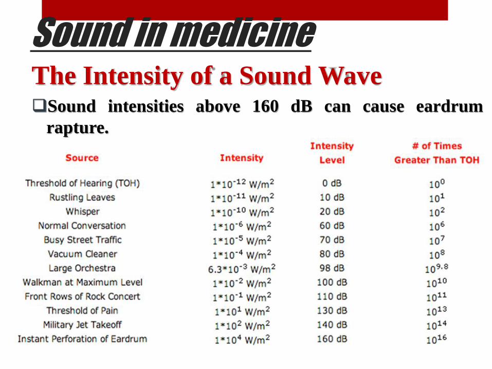
Sound in medicine The Intensity of a Sound Wave Sound intensities above 160 dB can cause eardrum
rapture.

Sound in medicine The Acoustic Impedance (Z) The acoustic impedance (Z) for the sound wave can be
calculated from the equation: Z= ρν, (see next table).
Where ρ : is the density of medium where the sound wave
transfer through it (Kg/m3).
v: is the velocity of sound wave in the medium (m/sec).
Substance ρ (kg/m3) ν (m/sec) Z(kg/m2.sec)
Air 1.29 3.31×10² 430
Water 1×10³ 14.8×10² 1.48×10⁶
Brain 1.02×10³ 15.3×10² 1.56×10⁶
Muscle 1.04×10³ 15.8×10² 1.64×10⁶
Bone 1.9×10³ 40.4×10² 7.68×10⁶

Sound in medicine Sound/Ultrasound Wave Reflection, Transmission and Absorption.
When sound wave hits the body, part of the wave is reflected and part is transmitted in to the body. The ratio of the reflected amplitude (R) to the incident amplitude (A0) depend on the impedances of the two media Z1 and Z2
Incident wave (A0) Transmitted wave (T)
Reflected wave (R)
Medium1(Air) / Z1 Medium2 (Body) / Z2

Sound in medicine Attenuation of Ultrasound Wave
When a sound wave passes through tissue, there is some loss in the energy due to the frictional effects.
The absorption of energy in the tissue causes a reduction in the amplitude of the sound wave (attenuation).
The Ultrasound wave amplitude (A) at a depth X cm in a medium is related to the initial amplitude A0(X=0) by the exponential equation : A=A0e
-αx
Where α in cm-1 ,is the absorption coefficient for the medium at a particular frequency.

Sound in medicine Attenuation of Ultrasound Wave Since the intensity is proportional to the square of the
amplitude ,its dependence with depth is:
I A2 I=I0e
-2αx
Where I0 is the incident intensity at X=0 & I is the
intensity at a depth X in the absorber ,
Since the absorption coefficient is 2α ,the intensity
decreases more rapidly than the amplitude with depth.
Material Frequency (MHz) α (cm-1)
Muscle 1 0.13
Fat 0.8 0.05
Brain 1 0.11
Bone(human skull) 0.6 0.4

Sound in medicine
Attenuation of Ultrasound Wave
EX: What is the attenuation of sound intensity in
15cm of brain tissue?
Solu./

Sound in medicine
Percussion in Medicine Percussion (tapping) is a method of tapping on a
surface to determine the underlying structure, and is
used in clinical examinations to assess the condition
of the thorax or abdomen.

Sound in medicine
Percussion in Medicine The quality of the sound:
Resonant = air filled space.
Dull = underlying solid tissue.
https://www.youtube.com/watch?v=mSJKI9Pkxxw

Sound in medicine Percussion in Medicine The first recorded use of percussion on the human body as
a mean of diagnosis occurred in the eighteenth century.
In 1761, L. Auenbrugger published a short book, On
Percussion of the Chest, which was based on his clinical
observations over seven years of patients in various places.
In his book, Auenbrugger described how to strike the chest
with the fingers and stated,{The sound thus elicited from
the healthy chest resembles the stifled sound of drum
covered with a thick woolen cloth or other envelope.

Sound in medicine Percussion in Medicine He discussed both the sounds heard from healthy subjects
and the sounds heard from patients with various pathological conditions.
Auenbragger stated that with percussion he could diagnose cancer, the presence of abnormal cavities in an organ.
He confirmed many of these diagnoses by examining bodies after death.
Auenbragger’s discovery was largely ignored until 1808 when his work, originally published in Latin.
Percussion has since become an important technique in the detection of disease.

Sound in medicine
The Stethoscope
Stethoscope is a simple hearing aid permits a physician
or nurse to listen to sounds mad inside the body (in the
heart and lungs).
The act of listening to these sounds with a stethoscope
is called mediate auscultation.

Sound in medicine
The Stethoscope Stethoscope is the bell,
which is either open or closed by a thin diaphragm, to
tubing, and the earpieces (see fig.)
The open bell is an impedance matcher between the skin
and the air and accumulates sounds from the contacted
area. The skin under the open bell behaves like a
diaphragm.
A closed bell is a bell with a diaphragm of known resonant
frequency usually high that tunes out low frequency
sounds.

Sound in medicine
The Stethoscope
The closed bell stethoscope is used for listening to lung
sounds, which are of higher frequency than heart sounds.
To see the typical frequency ranges of heart and lung sounds
see (Fig).
Most of the heart sounds are of low frequency
in the region where the sensitivity of the ear is
poor. Lung sounds generally have higher
frequencies. The curve represents the threshold
of hearing for a good ear. Some of the heart and
lung sounds are below this threshold.

Sound in medicine
The Stethoscope The larger the bell diameter, the lower the skins
resonant frequency.
Thus It is possible to enhance the sound range of interest
by changing the bell size and varying the pressure of the
bell against the skin and thus the skin tension.
The volume of the tubs should also be small, and there
should be little frictional loss of sound to the walls of the
tube.

Sound in medicine
The Stethoscope The small volume restriction suggests short, small
diameter tubes, while the low friction restriction suggests
large diameter tubes.
If the diameter of the tube is too small, frictional losses
occur, and if it is too large, the moving air volume is too
great. In both cases the efficiency is reduced.
A compromise is a tube with a length of about 25 cm and
diameter of 0.3 cm.

Sound in medicine The Ultrasound What is Ultrasound
Ultrasound is simply sound that has a very high frequency.
Humans are not able to hear Ultrasound, though some animals
can hear them.
Sounds with frequencies above 20 000 hertz are called
Ultrasounds.
Uses of Ultrasound in Medicine
Ultrasound is used for examining soft tissue inside the body.
Imaging parts of the body that may be examined include
muscles and unborn babies.
Blood flow can also be monitored using Ultrasound

Sound in medicine The Ultrasound
Why Use Ultrasound? Ultrasound is very safe. There is no firm
evidence that it does any harm to the body (or the baby in the case of pregnancy scans).
Ultrasound is often gives more information than X-rays.
X-rays are dangerous, particularly to young children and pregnant women (they damage the unborn baby), because of that, Ultrasound is less hazard for the fetus.

Sound in medicine The Ultrasound
Generating Ultrasound
There are several methods of generating Ultrasound. The most important for medical applications involves the (piezoelectric effect).
Many crystals can be cut so that an oscillating voltage across the crystal will produce a similar vibration of the crystal, thus generating a sound wave.

Sound in medicine
The Ultrasound Generating Ultrasound
A device that converts electrical energy to mechanical energy
or vice versa is called (transducer), Ultrasound generators
are often simply referred to as transducers.
Each transducer has a natural resonant frequency of
vibration.
The thinner the crystal, the higher the frequency at which it
will oscillate.

Sound in medicine
The Ultrasound Generating Ultrasound
For a quartz crystal , a thickness of 2.85 mm give a
resonant frequency of about 1MHz.
Typical frequencies for medical work are in the 1-5 MHz
range.
An average power level for diagnostic applications a few
mille watts per square centimeter.

Sound in medicine Ultrasound Transducer Types
You can find Ultrasound transducers in different shapes, sizes, and with diverse features. That is because you need different specifications for maintaining image quality across different parts of the body.
Transducers can be either passed over the surface of the body – external transducers or can be inserted into an orifice, such as the rectum or vagina – these are internal transducers.
The Ultrasound transducers differ in construction based on:
•Piezoelectric crystal arrangement
•Aperture (footprint)
•Frequency

Sound in medicine Ultrasound Transducer Types
1-Linear Transducers:
In this transducer type, the piezoelectric crystal arrangement
is linear, the shape of the beam is rectangular, and the near-
field resolution is good and its central frequency is 7-12MHz
You can use this transducer for various applications, such as:
•Vascular examination
•Blood vessel visualization
•Breast
•Thyroid

Sound in medicine Ultrasound Transducer Types
2-Convex Transducers:
The convex Ultrasound transducer type is also called the
curved transducer because the piezoelectric crystal
arrangement is curvilinear.
The transducer is good for in-depth examinations, even
though the image resolution decreases when the depth
increases.
The footprint frequency is 2-5MHz
You can use it for:
•Abdominal examinations
•Diagnosis of organs

Sound in medicine
Ultrasound Transducer Types
3-Phased Array Transducers:
This transducer is named after the piezoelectric crystal
arrangement which is called phased-array and it is the most
commonly used crystal.
It has a small footprint and low frequency 1-3MHz
It can use for :
•Cardiac examinations.
•Abdominal examinations.
•Brain examinations.

Sound in medicine
Ultrasound Transducer Types
Other Ultrasound Transducer Types
The other Ultrasound transducer type is endocavitary.
These probes provide you with the opportunity to perform
internal examinations of the patient.
Therefore, they are designed to fit in specific body orifices
and they have small footprints .
The endocavitary transducers include endovaginal,
endorectal, and endocavity transducers.

Sound in medicine
Pulses of Ultrasound are transmitted into the body by
placing the vibrating crystal in close contact with the skin,
using water or a jelly paste to eliminate the air. This gives
good coupling at the skin and greatly increases the
transmission of the Ultrasound in to the body and of the
echoes back to the detector.
Generating Ultrasound
Sound in medicine
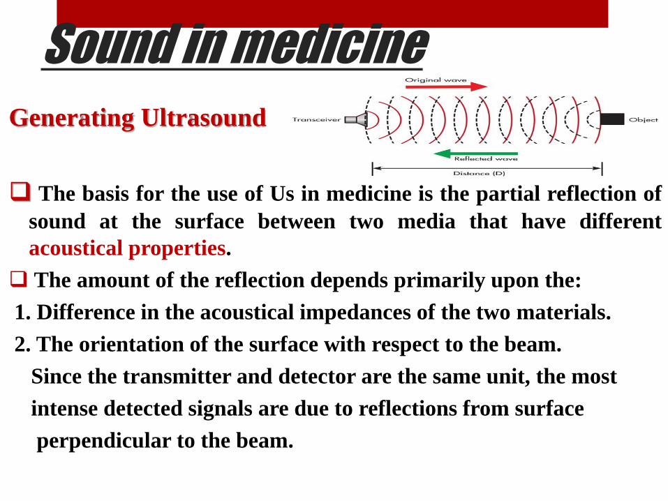
Generating Ultrasound
The basis for the use of Us in medicine is the partial reflection of
sound at the surface between two media that have different
acoustical properties.
The amount of the reflection depends primarily upon the:
1. Difference in the acoustical impedances of the two materials.
2. The orientation of the surface with respect to the beam.
Since the transmitter and detector are the same unit, the most
intense detected signals are due to reflections from surface
perpendicular to the beam.

Sound in medicine
The Ultrasound Ultrasound Picture of the Body
In this subject, we discuss the use of Ultrasound to
produce pictures for medical diagnosis. Basically an
Ultrasound source sends a beam of pluses of 1to 5 MHz
sound into the body. The time required for the sound
pulses to be reflected (Echo pulse) gives information on
the distances to the various structures or organs in the
path of Ultrasound beam.
3D image

Sound in medicine
The Ultrasound How the image is created…
Millions of sound waves are transmitted every second.
As the waves reflected at different times, the computer
in the Ultrasound machine calculates how far the wave
travelled before being reflected (using d=vt).
Using this information the computer builds up an image
of the inside of the patient.

Sound in medicine The Ultrasound Basic Question Example
Q1// How deep is a baby’s head if it takes 0.05 ms for a
sound wave to reach it in the mother’s uterus? The speed
of sound in body tissue is 1500 m/s.
Solu.// v = 1500 m/s , d= ?
t = 0.05 ms = 0.05 x 10⁻³ s
equation : d=vt
d = 1500 x 0.05 x 10-3
d= 0.075 m = 7.5 cm

Sound in medicine The Ultrasound Q2// The speed of sound in the human body is 1500 m/s. If a fetus is 6 cm below the mother’s skin, how long will it take for the echo to be received.
Solu.//
v = 1500 m/s
d = 6cm d= 0.12 m
t = ?
Equation : t = d/v
t = 0.12/1500
t = 0.00008 s
t = 8 x 10⁻⁵ s = 80 µs. Cross-section image

Sound in medicine The Problems of Using US in
Diagnosis
In many diagnostic uses of Us
1. The echoes are very small signals due to weak reflection and
the absorption of the sound by tissue . The solution for this
problem by amplifying the echo electronically.
2. The lack of resolution, or the ability of the equipment to
detect separate echoes from two objects close together. In
general, structures smaller than the wavelength (λ) cannot be
resolved since (λ= v/f ), where (v) is the velocity of sound and
(f) is the frequency, high frequency sound has shorter
wavelength and allows better resolution than low-frequency
sound.

Sound in medicine
Type of Ultrasound Mode
1-A-scan(Amplitude scan)
To obtain diagnostic information about the depth of
structures in the body, we send pulses of Us into the body
and measure the time required to receive the reflected
sound (echoes) from the various surfaces in it. This
procedure is called the A-scan method of Us diagnosis.
Pulses for A scan work are typically a few microseconds
long. They are usually emitted at 400 – 1000 pulses\ sec.

Sound in medicine The Ultrasound Mode
The applications of A-scan
A-scan in Echoencephalography;
A-scan has been used in the
detection of brain tumors.
Pulses of Us are sent into thin
region of the skull slightly above the ear and echoes from the different structures within the head are displayed on an oscilloscope. The usual producer is to compare the echoes from the left side of the head to those from the right side and to look for a shift in the midline echo. A tumor on one side of the brain tends to shift the midline toward the other side. Generally a shift of more than 3mm for an adult or 2mm for a child is considered abnormal (see fig.b).
Normal brain
Abnormal brain

Sound in medicine
The Ultrasound
A – scan in Ophthalmology:
Application of A- scans in ophthalmology can be divided
into two areas that concerned with obtaining
information for use in the :
1. Diagnoses of eye diseases,
2.Measurements of distances in the eye (Biometry).
At the low power levels used, there is no danger patient's
eye.

The Ultrasound
A – scan in Ophthalmology
With Us it is possible to measure distances in the eye
such as lens thickness, depth from cornea to lens, the
distance to the retina, the thickness of the vitreous
humor, and the curvature of cornea.
Sound in medicine

ound in medicineS
The Ultrasound
A – scan in Ophthalmology
Us frequency of up to 20MHZ are used. These high frequencies
can be used in the eye to produce better resolution since there
is no bone to absorb most of energy and absorption is not
significant because the eye is small.
Ultrasound studies of a detached
retina. CRT shows an echo S from the
anterior sclera, an echo r from the
retina, and an echo S from the sclera
at the back of the eye.
In a normal eye the echo from the
retina would blend with the echo from
the posterior sclera.

Sound in medicine
The Ultrasound Mode
2- B–scan (Brightness scan)
B - scan :- for many clinical purposes, A-scan have been
largely replaced by B-scan. The principles are the same
as for the A-scan except that the transducer is moved.
1. B-scan can provide more information than X-rays.
2. They present less risk.

Sound in medicine The Ultrasound Modes Comparison
B-scan A-scan
The transducer is moved. The transducer is fixed.
Two dimensional views. One dimensional views.
Echo produces a dot on the
oscilloscope.
Echo produces signal on the
oscilloscope.
provide informative about
the internal structure of the
body, eye, liver, breast, hart
and fetus.
Less informative about the
internal structure of the
body.

Sound in medicine The Ultrasound Mode
Ultrasound in Measuring
Motion
Two methods are used to obtain information about motion in
the body with Ultrasound:
3- M–scan (Motion scan)
Which is used to study motion of the hart and to obtain
diagnostic information about heart and heart valves.
The M-scan combines certain features of A-scan and B-scan
The transducer is stationary as in A-scan and the echoes
appear as dots as in the B-scan.

Sound in medicine The Ultrasound Mode
Applications of M-scan:
a- M-scan of the mitral valve:
M- scan are used to obtain
diagnostic information about the heart. The places where the heart can be probed are quite limited because of poor Ultrasound transmission through lung tissue and bone.
The usual method is to put the transducer on the patient's left side, aim it between the ribs over the heart, and tip it at different angles to explore various region of the heart.
The examiner must be familiar with the patterns of specific cardiac echoes to interpret the information.

Sound in medicine
The Ultrasound Mode
Applications of M-scan:
The information of interest is the
rate of closing of the mitral valve.
The rate of closing for a normal valve is indicated by the slope in (next slid Fig. a) in this case the rate of closing is 72 mm/sec.
(next slid Fig. b) is an M-scan showing an abnormality called mitral stenosis (a narrowing of the valve opening).
The reduced slope for mitral stenosis is quite different from the normal slope-the slower the rate of closure, the larger the amount of stenosis.

Sound in medicine Applications of M-scan:

Sound in medicine
The Ultrasound Mode
Applications of M-scan:
b- Accumulation of fluids in the heart sac (pericardial
effusion).

Sound in medicine
The Ultrasound
4- Doppler Technique:
Doppler effect is the perceived frequency of sound emitted by
moving source.
The Doppler technique is used for:
a-Study the blood motion in the circulatory system:
Two transducers are used in the audio mode, one as transmitter
and the other as a receiver.
Continuous Ultrasound wave is sending to the artery with a
frequency (f0).

Sound in medicine Doppler Technique:
When the blood move with a speed V at an angle θ from
the direction of the sound waves of speed ν, the
frequency change fd is:
Where f0 is the frequency of the initial Ultrasonic wave ,V
is the velocity of the blood ,ν is the velocity of sound and θ
is the angle between V and ν, (see Fig.)

Sound in medicine
Doppler Technique:
When the fetal heart is moving, a variation in the
frequency give the fetal heart rate.
The output can be audible or displayed on an
oscilloscope.
b- The Doppler technique is also used to locate the point of the
entry of the umbilical cord into the placenta to detect if there is
bleeding due to misplaced placenta (placenta praevia)or there
is an intrauterine transfusion for Rh incompatibility.
Rh incompatibility


![17.2 Sound Waves: In Halliday and Resnick: Longitudinal waves are sound waves! Chapter 17: [Sound] Waves-(II) Sound waves propagate in gases. Can they.](https://static.fdocuments.us/doc/165x107/56649eb25503460f94bb9375/172-sound-waves-in-halliday-and-resnick-longitudinal-waves-are-sound-waves.jpg)


