Sonography of the proximal hamstring muscle …...PICTORIAL ESSAY Ultrasound Features of the...
16
PICTORIAL ESSAY Ultrasound Features of the Proximal Hamstring Muscle-Tendon-Bone Unit Marco Becciolini, MD , Giovanni Bonacchi, MD, Stefano Bianchi, MD The hamstring muscle complex is made by a group of posterior biarticular thigh muscles, originating at the ischial tuberosity, which extend the hip and flex the knee joint. Proximal hamstring injuries are frequent among athletes, commonly involving their long myotendinous junction during an eccentric contraction. In this pictorial essay, we describe the ultrasound technique to visualize the normal anatomy of the proximal hamstring muscle-tendon-bone complex and present ultrasound findings in patients with traumatic injuries and tendinopathies. Key Words—athlete injury; biceps femoris; hamstring; musculoskeletal; myotendinous injury; tendinopathy; thigh muscles; ultrasound T he hamstring muscle complex comprises a group of posterior biarticular thigh muscles, originating at the ischial tuberosity: the long head of the biceps femoris, semimembranosus, and semitendinosus. These muscles extend the hip and flex the knee joint. 1 Proximal hamstring muscle complex injuries are the most frequent among athletes, commonly involving the proximal myotendinous junction during an eccentric contraction. 2,3 So far, magnetic resonance imaging (MRI) has been considered as the modality of choice to evaluate tendinopathy and injuries. 3–6 In this pictorial essay, our aims are to describe the ultrasound (US) technique for visualizing the proximal hamstring muscle complex and to illustrate US findings in patients with traumatic injuries and tendinopathies. This human study was performed in accordance with the Dec- laration of Helsinki. The study was approved by the Cabinet Ima- gerie Médicale. All parents, guardians, or next of kin provided written informed consent for the minors to participate in the study. All adult participants provided written informed consent to participate in the study. Ultrasound Anatomy Technical Considerations A linear transducer with frequencies between 5 and 14 MHz is required. For a larger body habitus, the use of a lower-frequency transducer might be helpful. 7–9 The patient lies prone with the hip and the knee in a neutral position; flexion of the hip using a pillow may be used to avoid artifacts produced by the gluteal fold. 8 Scanning Technique The examination begins with axial US scans at the level of the ischial tuberosity, which can be palpated, being a good anatomic Received June 13, 2018, from the Misericordia di Pistoia, Pistoia, Italy (M.B., G.B.); Cabinet Imagerie Médicale, Geneva, Switzerland (S.B.). Manuscript accepted for publication July 13, 2018. Address correspondence to Marco Becciolini, MD, Misericordia di Pistoia, Via Bonellina 1, 51100 Pistoia, Italy. E-mail: [email protected] Abbreviations MRI, magnetic resonance imaging; US, ultrasound doi:10.1002/jum.14804 © 2018 by the American Institute of Ultrasound in Medicine | J Ultrasound Med 2018; 00:1–16 | 0278-4297 | www.aium.org
Transcript of Sonography of the proximal hamstring muscle …...PICTORIAL ESSAY Ultrasound Features of the...

PICTORIAL ESSAY
Ultrasound Features of the ProximalHamstring Muscle-Tendon-Bone UnitMarco Becciolini, MD , Giovanni Bonacchi, MD, Stefano Bianchi, MD
The hamstring muscle complex is made by a group of posterior biarticular thighmuscles, originating at the ischial tuberosity, which extend the hip and flex theknee joint. Proximal hamstring injuries are frequent among athletes, commonlyinvolving their long myotendinous junction during an eccentric contraction. Inthis pictorial essay, we describe the ultrasound technique to visualize the normalanatomy of the proximal hamstring muscle-tendon-bone complex and presentultrasound findings in patients with traumatic injuries and tendinopathies.
Key Words—athlete injury; biceps femoris; hamstring; musculoskeletal;myotendinous injury; tendinopathy; thigh muscles; ultrasound
T he hamstring muscle complex comprises a group of posteriorbiarticular thigh muscles, originating at the ischial tuberosity:the long head of the biceps femoris, semimembranosus, and
semitendinosus. These muscles extend the hip and flex the knee joint.1
Proximal hamstring muscle complex injuries are the most frequentamong athletes, commonly involving the proximal myotendinousjunction during an eccentric contraction.2,3 So far, magnetic resonanceimaging (MRI) has been considered as the modality of choice toevaluate tendinopathy and injuries.3–6 In this pictorial essay, our aimsare to describe the ultrasound (US) technique for visualizing theproximal hamstring muscle complex and to illustrate US findings inpatients with traumatic injuries and tendinopathies.
This human study was performed in accordance with the Dec-laration of Helsinki. The study was approved by the Cabinet Ima-gerie Médicale. All parents, guardians, or next of kin providedwritten informed consent for the minors to participate in thestudy. All adult participants provided written informed consent toparticipate in the study.
Ultrasound Anatomy
Technical ConsiderationsA linear transducer with frequencies between 5 and 14 MHz isrequired. For a larger body habitus, the use of a lower-frequencytransducer might be helpful.7–9 The patient lies prone with the hipand the knee in a neutral position; flexion of the hip using a pillowmay be used to avoid artifacts produced by the gluteal fold.8
Scanning TechniqueThe examination begins with axial US scans at the level of theischial tuberosity, which can be palpated, being a good anatomic
Received June 13, 2018, from the Misericordiadi Pistoia, Pistoia, Italy (M.B., G.B.); CabinetImagerie Médicale, Geneva, Switzerland(S.B.). Manuscript accepted for publicationJuly 13, 2018.
Address correspondence to Marco Becciolini,MD, Misericordia di Pistoia, Via Bonellina1, 51100 Pistoia, Italy.
E-mail: [email protected]
AbbreviationsMRI, magnetic resonance imaging; US,ultrasound
doi:10.1002/jum.14804
© 2018 by the American Institute of Ultrasound in Medicine | J Ultrasound Med 2018; 00:1–16 | 0278-4297 | www.aium.org

landmark. The long head of the biceps femoris andthe semitendinosus muscle originate from a conjointtendon at the medial and posterior aspects of theischial tuberosity, whereas the semimembranosustendon has a more lateral and anterior origin(Figure 1A).1,9 To improve image quality, we sug-gest using a slight lateral approach (Figure 1, A andB), and the patient should be informed that thetransducer may have to be pressed hardly; thismaneuver is also recommended in symptomaticpatients to reproduce pain: so-called sonopalpa-tion.10,11 The examiner should remember to slowlytilt the transducer to avoid mistaking anisotropy,
which is present when a tendon is not scanned per-pendicularly, for tendinopathy.12,13 This action isneeded because the conjoint and semimembranosustendons have different courses so that in a US scan,one can appear hypoechoic to the other; in doubtfuland subtle cases, a comparison with the oppositeside is advised. Lateral to the hamstring muscle com-plex tendon, the sciatic nerve, a fascicular and flat-tened structure, can be easily shown: it descendsbetween and deep to the long head of the bicepsfemoris and the semimembranosus tendon, superfi-cial to the adductor magnus muscle.14 A smallernerve departing from the sciatic nerve, the posterior
Figure 1. Hamstring muscle complex origin anatomy. A–C, Proximal-to-distal axial US scans (left), as described in the US anatomy part ofthe article, with MRI comparison (right); amt indicates adductor magnus tendon; ct, conjoint tendon; gm, gluteus maximus; it, ischial tuber-osity; smt, semimembranosus tendon; sn, sciatic nerve; and st, semitendinosus muscle.
Becciolini et al—Ultrasound Features of the Proximal Hamstring Muscle Complex
2 J Ultrasound Med 2018; 00:1–16

Figure 3. Proximal anatomy of the hamstring muscle complex. A–C, Proximal-to-distal axial US scans (left), as described in the US anatomypart of the article, with MRI comparison (right). Dashed lines in A indicate the Cohen triangle; am, adductor magnus muscle; arrowhead,semimembranosus membrane; arrows, semitendinosus raphe; ct, conjoint tendon; lbf, long head of biceps muscle; sm, semimembranosusmuscle; smt, semimembranosus tendon; sn, sciatic nerve; and st, semitendinosus muscle.
Figure 2. Axial color Doppler US scan at the level of the ischial tuberosity (it) in a healthy 22-year-old man. In this slender volunteer, US is ableto show a small branch of the inferior gluteal artery (a), interposed between the sciatic nerve (sn) and a small honeycomb structure (arrows),consistent with the posterior femoral cutaneous nerve. Note the close relationship of the nerve and the proximal tendons of the hamstring mus-cle complex, which can explain its irritation in proximal hamstring tendinopathy. The semimembranosus tendon (smt) in this view appearsslightly hypoechoic because of anisotropy; ct indicates conjoint tendon; and gm, gluteus maximus.

femoral cutaneous nerve, may be sometimesdepicted as well in healthy and slender patients,accompanied by the inferior gluteal artery
(Figure 2); it distally passes superficial to the longhead of the biceps femoris and gradually surfaces upto the cutaneous layer.15,16
Figure 4. Images from a 10-year-old boy who had sudden posterior pain while playing soccer. A and B, Axial and sagittal US scansobtained 4 days later. The cortical surface of the ischial tuberosity (it) of the affected side (left) appears irregular (arrows), and the hamstringmuscle complex proximal origin (asterisks) looks swollen compared with the healthy side (right). Ultrasound palpation was painful. Clinicaland US findings are compatible with a minimal apophyseal injury without tendon displacement. The patient was treated conservatively witha good outcome; ct indicates conjoint tendon; gm, gluteus maximus; and smt, semimembranosus tendon.
Figure 5. Images from a 20-year-old man evaluated for chronic pain at the level of the left ischial tuberosity (it). Comparative longitudinalUS scans depict a large calcification casting an acoustic shadow (arrow). The finding is consistent with a previous avulsion, as referred bythe patient who recalled a hamstring muscle complex origin injury at the age of 12 years; ct indicates conjoint tendon; gm, gluteus maximus;and smt, semimembranosus tendon.
Becciolini et al—Ultrasound Features of the Proximal Hamstring Muscle Complex
4 J Ultrasound Med 2018; 00:1–16

The examination continues with axial caudal USscans. At the distal medial tip of the ischial tuberosity,another tendon originates: the adductor magnus,which also has a wider pubofemoral insertion; becauseof this anatomy, it is also called the “mini-hamstring”(Figure 1B).17 Below the ischial tuberosity, the semi-tendinosus muscle is the first to appear, medially to theconjoint tendon; at this level, the semimembranosustendon starts to travel medially (Figure 1C). Caudally,the second muscle to appear is the long head of thebiceps femoris; here, a good anatomic landmark, theCohen triangle,8 is composed of the aponeurosis of theconjoint tendon, the sciatic nerve, and the semimem-branosus tendon (Figure 3A). Considering the con-joint tendon, according to a dissection study on56 hamstring muscle complexes by van der Madeet al,1 the total length of the semitendinosus tendon is12.3 ± 3.6 cm with a myotendinous junction of12.2 cm, whereas the long head of the biceps femoristendon is 19.6 ± 4.1cm with a myotendinous junctionof 14.6 cm. The semimembranosus tendon continues
to descend between the semitendinosus muscle andthe adductor magnus muscle, giving a “membrane”medially from which the first muscular fascicle of thesemimembranosus muscle originates. At this level, asigmoid raphe inside the semitendinosus muscle, whichtravels the muscle from proximal-medial to distal-lat-eral, is another key anatomic landmark for a US evalua-tion of the hamstring muscle complex (Figure 3B).8 Atthe middle third of the thigh, the semimembranosustendon reaches the muscle (Figure 3C); its total lengthaccording to van der Made et al1 is 24.3 ± 3.9 cm witha myotendinous junction of 14.9 cm. Usually justbelow this level, the long head of the biceps femoris,the semitendinosus muscle, and the semimembranosusmuscle belly have a similar size.8 Anatomic variations,such as the absence of the semimembranosus muscle,or a separate origin of the long head of the bicepsfemoris and semitendinosus tendons, have beenreported.2 We perform longitudinal-axis scans of thehamstring muscle complex in cases of disorders to con-firm it and to show its craniocaudal involvement.
Figure 6. Posttraumatic tendinopathy related to a partial-thickness tear with remodeling of the hamstring muscle complex origin in a 35-year-old soccer player, evaluated 2 months after an indirect trauma for persistent pain. A and C, Axial and sagittal US scans (left) of the left ischialtuberosity (it) show a swollen and inhomogeneous (arrows) conjoint tendon (ct) and semimembranosus tendon (smt) compared with thehealthy contralateral side (right). B and D, Corresponding MRI (B, axial; D, coronal); gm indicates gluteus maximus; and sn, sciatic nerve.
Becciolini et al—Ultrasound Features of the Proximal Hamstring Muscle Complex
J Ultrasound Med 2018; 00:1–16 5

Limitations of USUltrasound has some limitations in evaluations ofthe posterior thigh in older and obese patientsbecause of muscular fatty degeneration andincreased thickness of the subcutaneous fat,7,8 espe-cially at the level of the ischial tuberosity region; inthese patients, it may be helpful to search for theother described anatomic landmarks (eg, Cohen tri-angle and semitendinosus raphe) and then to movethe transducer cranially to the ischial tuberosity.The distal course of the hamstring muscle complexgoes beyond the purpose of this pictorial essay, aswell as the anatomy of the short head of the bicepsfemoris.
Ultrasound Findings
Apophyseal InjuriesThe weakest part of the hamstring bone-tendon-muscle unit is linked to the patient’s age2: in children,it is the apophyseal region of the ischial tuberosity;therefore, injuries may lead to an avulsion of a bonyfragment, which can be assessed by radiography.Ultrasound is able to confirm the injury, depicting theavulsed fragment as well as the retracted tendon.18
Ultrasound is particularly helpful in mild cases, inwhich the apophysis is nondisplaced, by showingirregularities in the ischial tuberosity and cartilagecompared with the contralateral side (Figure 4).19
Figure 7. Partial tear of the semimembranosus tendon (smt) in a 28-year-old soccer player with tendinopathy. A, Long-axis view of the hamstringmuscle complex proximal tendon (right) compared with the healthy contralateral side (left) shows a tear in the right semimembranosus tendon (smt),highlighted by a hypoechoic-to-anechoic area related to edema or blood products (asterisks); the conjoint tendon (ct) is also swollen and heteroge-neous, reflecting tendinopathy. B, Axial view of the right hamstring muscle complex proximal tendon confirms the findings; gm indicates gluteusmaximus; and it, ischial tuberosity.
Becciolini et al—Ultrasound Features of the Proximal Hamstring Muscle Complex
6 J Ultrasound Med 2018; 00:1–16

Magnetic resonance imaging should be performed indoubtful cases and, as in the case of a complete ten-don rupture, when there is a retraction and surgery isan option.2,5,8 Chronic avulsion injuries may result inposttraumatic heterotopic ossification (Figure 5).18
TendinopathiesTendinopathies may be found in young adults andare usually related to trauma with microtears(Figure 6), which may lead to partial or completetears (Figure 7); however, tendon injuries are more
Figure 8. Subcomplete tear of the hamstring muscle complex proximal tendons in a 75-year-old patient with tendinosis. The patient wasreferred for sudden pain 3 days previously after falling while gardening on uneven ground. A, Axial US scan at the level of the ischial tuber-osity (it). The conjoint tendon (ct) and semimembranosus tendon (smt) show fiber disruption associated with a hypoechoic-to-anechoicarea of edema or blood products (asterisk) highlighting the tear. B, Corresponding long-axis view of the hamstring muscle complex origin;am indicates adductor magnus; and gm, gluteus maximus.
Figure 9. Complete avulsion of the hamstring muscle complex origin tendons, which are retracted distally, in a 68-year-old patient who fellwhile walking downstairs and was referred for a snap below the buttock. A US examination was done 5 days later. A and B, Longitudinaland axial US scans at the level of the ischial tuberosity (it) show the retracted proximal tendons. The retraction is measured (between cali-pers); am indicates adductor magnus muscle; asterisks, hematoma; ct, conjoint tendon; gm, gluteus maximus; lbf, long head of the bicepsmuscle; and smt, semimembranosus tendon. C, Note clinically how the hematoma has spread distally.
Becciolini et al—Ultrasound Features of the Proximal Hamstring Muscle Complex
J Ultrasound Med 2018; 00:1–16 7

frequent in older adults because of tendon degenera-tion (Figure 8).2,3 When a complete avulsion isshown, the distance between the ischial tuberosityand the retracted tendon should be reported(Figure 9); surgery may be considered if there is dis-placement of greater than 2 cm.3 In tendinosis, thetendon loses its normal fibrillar pattern, becomingthickened and hypoechoic on a US evaluation. Ultra-sound findings are similar in patients with a chronicpartial tear, as it may coexist (Figure 6).13,20 Ham-string syndrome is a clinical entity described by Pura-nen and Orava21 as pain in the lower gluteal area withcaudal irradiation to the posterior thigh, which isrelated to hamstring muscle complex proximal
tendinopathy, commonly seen in athletes. Hamstringsyndrome may be associated with sciatic involvement.Pain with sitting may be also related to posterior fem-oral cutaneous nerve entrapment because of its closerproximity to the hamstring muscle complex proximaltendons, as this sensory nerve provides the innerva-tion for the inferolateral gluteal region and posterioraspect of the thigh, the superomedial perineal region,and the skin at the popliteal fossa.14,16,22
Painful snapping of the hip, also called coxa salt-ans, might be subsequent to proximal hamstring mus-cle complex injury and tendinopathy, caused bysubluxation of the conjoint tendon over the ischialtuberosity. Dynamic US has been shown to be capa-ble of depicting this kind of snapping during hipflexion.23
The examiner should keep in mind that tendino-pathy may affect only a tract, possibly the intramuscu-lar distal part of the tendons, leading to nonspecificposterior thigh pain, and therefore should check thetendons in their whole course with axial US scans:only a systematic and comparative evaluation allowsthe diagnosis necessary for the athlete to plan rehabil-itation and avoid injuries (Figures 10 and 11).
Calcifications related to tendinopathy and chronicinjuries may also involve the hamstring muscle com-plex tendons (Figure 12).6 Calcific tendinopathy, dueto deposition of calcium hydroxyapatite crystals withinthe tendons, which is common in the rotator cuff, hasalso been described at the level of the proximal ham-string muscle complex tendons (Figure 13) and maybe the cause of atraumatic acute ischial pain, likely dur-ing the resorptive phase.9,24
Myotendinous Junction and Proximal MuscleInjuriesIn young athletes, the weakest point of the hamstringmuscle complex is represented by the myotendinousjunction; in these patients, a powerful eccentric con-traction with the hip and the knee on extension maycause a partial (Figures 14 and 15) or a complete(Figure 16) tear. These injuries are frequent in waterskiers and football players.2–4,8 Clinically, the patientsrecall sudden onset of pain after specific indirecttrauma. A US examination should be performed atleast 48 hours after the trauma; otherwise, the mas-sive soft tissue edema/blood products may producearchitectural distortion, which makes a detailed
Figure 10. Images from a 25-year-old soccer player evaluated formild chronic pain at the proximal posterior thigh distal to the ischialtuberosity. The patient did not recall a hamstring injury. A and B,Transverse and sagittal US scans (left) at the proximal posteriorthigh show slight thickening of the distal part of the conjoint ten-don (between calipers) at the level of the myotendinous junction,without fissurations, compared with the healthy contralateral side(right). The conjoint tendon at the ischial tuberosity appeared nor-mal (not shown); am indicates adductor magnus muscle; lbf, longhead of the biceps muscle; sn, sciatic nerve; and st, semitendino-sus muscle.
Becciolini et al—Ultrasound Features of the Proximal Hamstring Muscle Complex
8 J Ultrasound Med 2018; 00:1–16
evaluation difficult.25 Ultrasound findings include apoorly defined hyperechoic or heterogeneous areawith or without muscular architectural alterations, thepresence of an anechoic hematoma, and a completetear with muscular retraction, which can be confirmedby dynamic imaging during muscle contraction.8,25
Myotendinous junction tear classification is not pre-sented in this pictorial essay. Delayed-onset musclesoreness refers to a benign condition in which symp-toms (muscular tenderness, soreness, or stiffness)develop approximately 24 to 48 hours after strenuousexercise, with a peak at 2 or 3 days and resolutionusually within 7 to 10 days. Ultrasound may depict
diffuse or focal hyperechogenicity and an increasedsize of the involved muscle(s), likely correlated withedema, which can be shown by MRI (Figure 17).2,8,26
Color Doppler imaging is widely used to assesstendinopathy20; however, in our practice, we find itnot effective for evaluation of proximal hamstringmuscle complex tendons because of their deepcourse: the needed transducer pressure will artificiallyhide neovascularity present in tendinosis. Instead, wefind that it has an important role in follow-up of myo-tendinous junction tears, in which the disappearanceof hyperemia indicates a subacute-to-chronic phase ofinjury (Figure 18).8,25
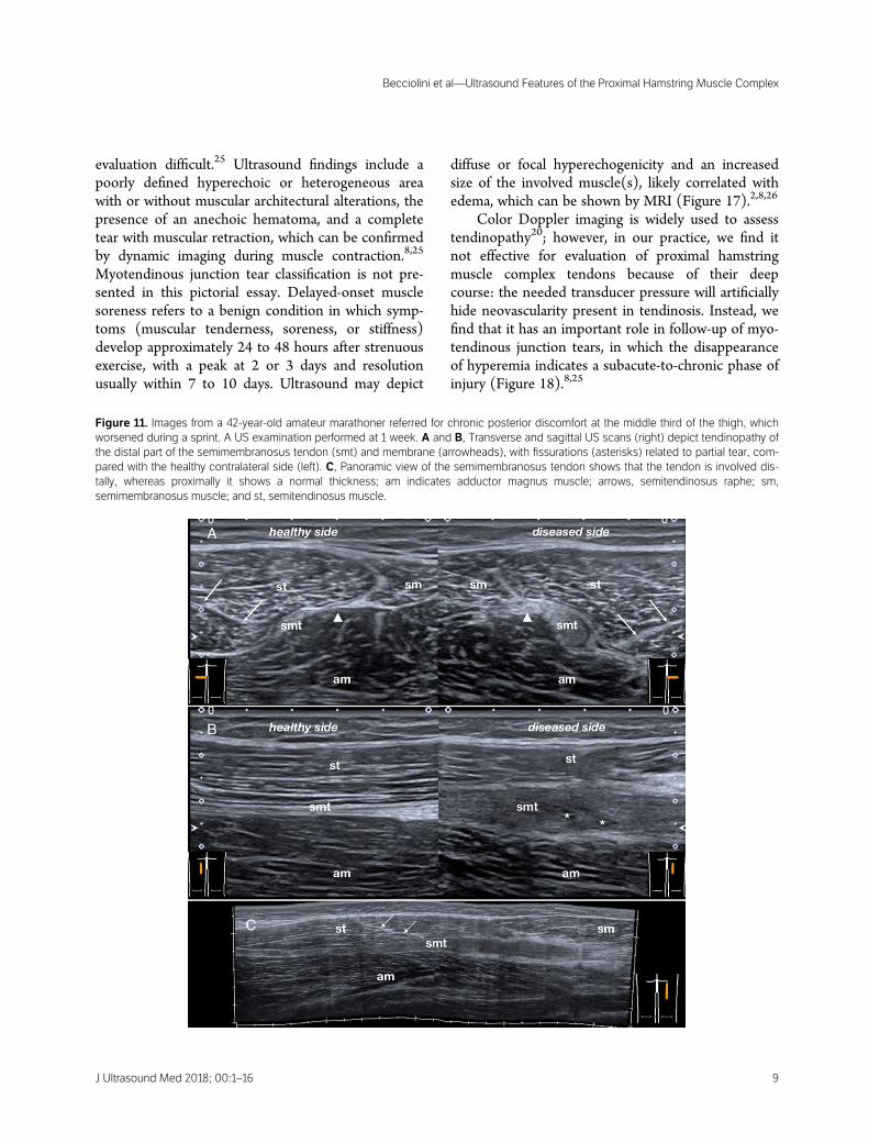
Figure 11. Images from a 42-year-old amateur marathoner referred for chronic posterior discomfort at the middle third of the thigh, whichworsened during a sprint. A US examination performed at 1 week. A and B, Transverse and sagittal US scans (right) depict tendinopathy ofthe distal part of the semimembranosus tendon (smt) and membrane (arrowheads), with fissurations (asterisks) related to partial tear, com-pared with the healthy contralateral side (left). C, Panoramic view of the semimembranosus tendon shows that the tendon is involved dis-tally, whereas proximally it shows a normal thickness; am indicates adductor magnus muscle; arrows, semitendinosus raphe; sm,semimembranosus muscle; and st, semitendinosus muscle.
Becciolini et al—Ultrasound Features of the Proximal Hamstring Muscle Complex
J Ultrasound Med 2018; 00:1–16 9

Figure 12. Images from a 24-year-old athlete referred for indirect trauma at the posterior thigh during a sprint. A, Transverse US scan at15 days depicts the tear, involving the myotendinous junction of the long head of the biceps femoris (lbf ) as well as the intramuscular partof the conjoint tendon (ct). A small hematoma is shown (asterisk). B, Corresponding sagittal US scan confirms the injury. C and D, Trans-verse and sagittal US scans obtained during follow-up at 2 months show the disappearance of the hematoma; the conjoint tendon is thick-ened and hypoechoic; along its course, a calcification (arrowheads) with a faint acoustic shadow can be seen as an outcome of thetrauma; sn indicates, sciatic nerve; and st, semitendinosus muscle.
Figure 13. Calcific tendinopathy of the hamstring muscle complex origin tendons and the adductor magnus tendon (amt) in a 58-year-oldwoman presenting for acute posterior hip pain in the absence of a trauma. A, axial US scan at the level of the ischial tuberosity (it) showscalcific deposits (arrows) in part with a faint acoustic shadow and in part “slurry.” Symptoms were likely related to the colliquation of the cal-cification with resultant local inflammatory changes. Dashed lines in A indicate the sagittal US planes in B and C; ct, conjoint tendon; gm,gluteus maximus; and smt, semimembranosus tendon.

Figure 14. Images from a 26-year-old soccer player who sustained indirect trauma at the proximal third of the posterior thigh while kicking,causing him to leave the game. A, Transverse US scan (left) shows the tear, located at the long head of the biceps femoris (lbf ) myotendi-nous junction, compared with the healthy contralateral side (right). The Cohen triangle is easily depicted. B, Corresponding sagittalextended view confirms the injury, also involving the middle third of the conjoint tendon (ct); am indicates adductor magnus muscle; aster-isks, hematoma; gm, gluteus maximus; smt, semimembranosus tendon; and st, semitendinosus muscle.
Figure 15. Images from a 23-year-old athlete referred for indirect trauma of the posterior medial thigh while sprinting. A and B, Axial and lon-gitudinal US views, obtained 3 days later at the middle third of the thigh, show the tear, involving the semimembranosus (sm) myotendinousjunction in the proximity of its membrane (arrowheads); am indicates adductor magnus muscle; arrows, semitendinosus raphe; asterisks,hematoma; smt, semitendinosus tendon; and st, semitendinosus muscle.
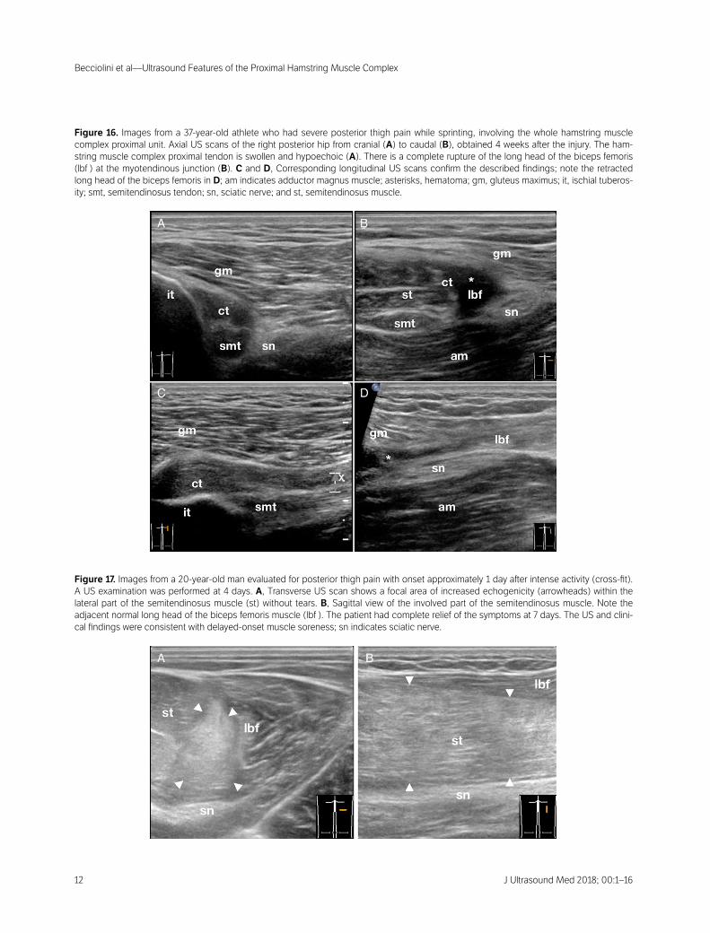
Figure 16. Images from a 37-year-old athlete who had severe posterior thigh pain while sprinting, involving the whole hamstring musclecomplex proximal unit. Axial US scans of the right posterior hip from cranial (A) to caudal (B), obtained 4 weeks after the injury. The ham-string muscle complex proximal tendon is swollen and hypoechoic (A). There is a complete rupture of the long head of the biceps femoris(lbf ) at the myotendinous junction (B). C and D, Corresponding longitudinal US scans confirm the described findings; note the retractedlong head of the biceps femoris in D; am indicates adductor magnus muscle; asterisks, hematoma; gm, gluteus maximus; it, ischial tuberos-ity; smt, semitendinosus tendon; sn, sciatic nerve; and st, semitendinosus muscle.
Figure 17. Images from a 20-year-old man evaluated for posterior thigh pain with onset approximately 1 day after intense activity (cross-fit).A US examination was performed at 4 days. A, Transverse US scan shows a focal area of increased echogenicity (arrowheads) within thelateral part of the semitendinosus muscle (st) without tears. B, Sagittal view of the involved part of the semitendinosus muscle. Note theadjacent normal long head of the biceps femoris muscle (lbf ). The patient had complete relief of the symptoms at 7 days. The US and clini-cal findings were consistent with delayed-onset muscle soreness; sn indicates sciatic nerve.
Becciolini et al—Ultrasound Features of the Proximal Hamstring Muscle Complex
12 J Ultrasound Med 2018; 00:1–16
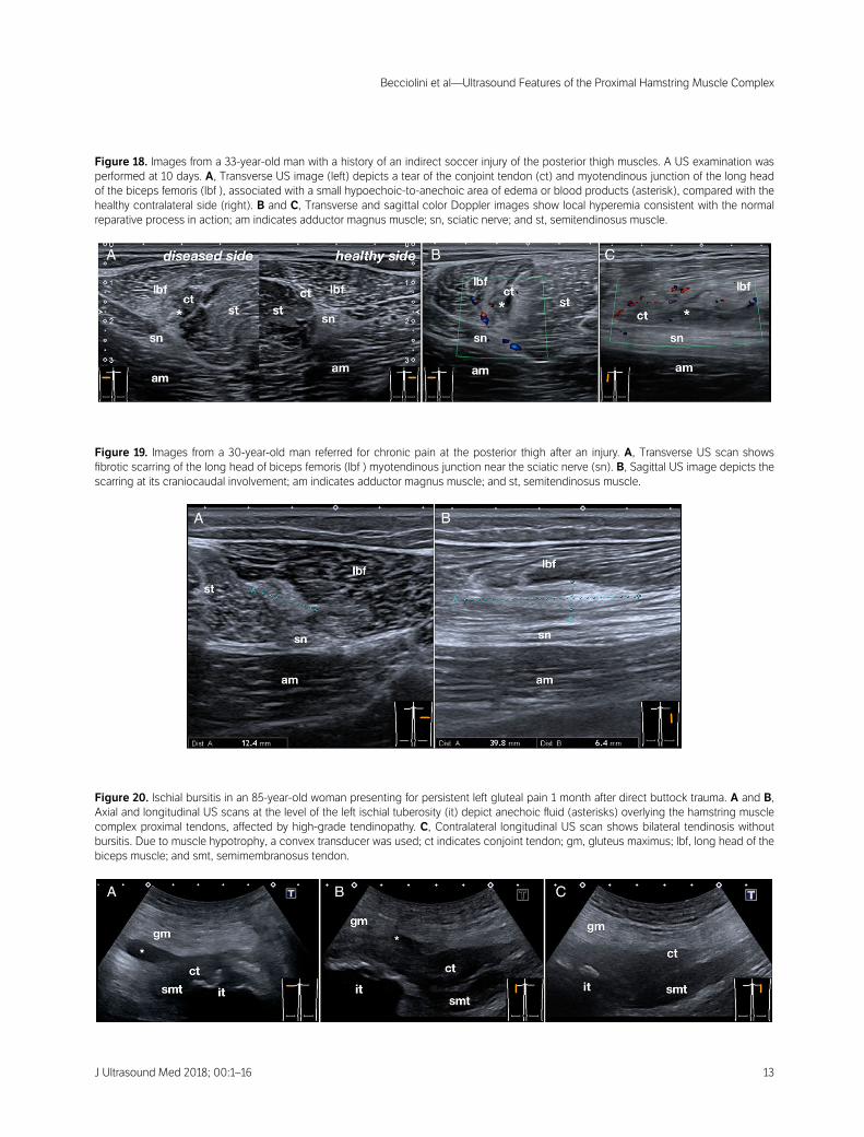
Figure 18. Images from a 33-year-old man with a history of an indirect soccer injury of the posterior thigh muscles. A US examination wasperformed at 10 days. A, Transverse US image (left) depicts a tear of the conjoint tendon (ct) and myotendinous junction of the long headof the biceps femoris (lbf ), associated with a small hypoechoic-to-anechoic area of edema or blood products (asterisk), compared with thehealthy contralateral side (right). B and C, Transverse and sagittal color Doppler images show local hyperemia consistent with the normalreparative process in action; am indicates adductor magnus muscle; sn, sciatic nerve; and st, semitendinosus muscle.
Figure 19. Images from a 30-year-old man referred for chronic pain at the posterior thigh after an injury. A, Transverse US scan showsfibrotic scarring of the long head of biceps femoris (lbf ) myotendinous junction near the sciatic nerve (sn). B, Sagittal US image depicts thescarring at its craniocaudal involvement; am indicates adductor magnus muscle; and st, semitendinosus muscle.
Figure 20. Ischial bursitis in an 85-year-old woman presenting for persistent left gluteal pain 1 month after direct buttock trauma. A and B,Axial and longitudinal US scans at the level of the left ischial tuberosity (it) depict anechoic fluid (asterisks) overlying the hamstring musclecomplex proximal tendons, affected by high-grade tendinopathy. C, Contralateral longitudinal US scan shows bilateral tendinosis withoutbursitis. Due to muscle hypotrophy, a convex transducer was used; ct indicates conjoint tendon; gm, gluteus maximus; lbf, long head of thebiceps muscle; and smt, semimembranosus tendon.
Becciolini et al—Ultrasound Features of the Proximal Hamstring Muscle Complex
J Ultrasound Med 2018; 00:1–16 13

A possible complication of a myotendinous junc-tion tear is muscle scarring. On a US scan, the fibrosisis depicted as a poorly defined hyperechoic spiculatedarea. The sciatic nerve’s relationship with the fibroticarea should be evaluated to rule out adhesions(Figure 19).8
Hamstring Injury Mimics: Differential DiagnosisThe ischial bursa is an inconstant adventitial bursasuperficial to the hamstring muscle complex proximaltendons at the ischial tuberosity. Ischial bursitis isusually related to chronic irritation or trauma andoften leads to buttock pain; it appears as a fluid struc-ture that may have irregular walls and internal septa
(Figure 20).9,27 Deep venous thrombosis simulating ahamstring muscle complex injury and causing diffuseposterior thigh swelling has been described; US isable to show deep venous thrombosis and exclude atear.28
Among uncommon causes of pain near the ham-string muscle complex origin, tumors must not beneglected (Figure 21). Ultrasound may show soft tis-sue masses and may suggest their benign or malignnature, but its main role is to differentiate betweencystic and solid masses and to guide biopsy.11 Whenevaluating deep masses, we recommend completingthe examination with a convex transducer, as it ismore informative in showing a possible relationship
Figure 21. Images from a 55-year-old patient with Ollier syndrome presenting for suspected “hamstring syndrome.” A and B, Ultrasoundscans show a hypoechoic mass (pound signs) with internal calcification and a hard pattern on strain elastography (C). D, A convex trans-ducer was used to analyze its deep aspect; it can be seen arising from the femur (f ). An internal color signal was shown on Doppler imag-ing; pulsed wave Doppler imaging revealed arterial flux with a high resistive index. E, Corresponding axial contrast-enhanced T1-weightedturbo spin echo MRI shows the mass displacing the hamstring muscle complex origin tendons and sciatic nerve (sn) medially. Biopsyrevealed that the mass was a chondrosarcoma; am indicates adductor magnus muscle; ct; conjoint tendon; gm, gluteus maximus; smt;semimembranosus tendon; and st, semitendinosus muscle.
Becciolini et al—Ultrasound Features of the Proximal Hamstring Muscle Complex
14 J Ultrasound Med 2018; 00:1–16

with the bone and vascularity on Doppler imaging(Figure 21D). The use of strain elastography mayhelp improve the sensitivity of US for suspectingmalignant lesions because of their increased stiffness(Figure 21C).29 However, when a deep mass is sus-pected on a US scan, contrast-enhanced MRI has tobe performed (Figure 21E).30
Conclusions
Ultrasound is able to show the normal architecture ofthe proximal hamstring muscle complex as well as theappearance of bone-tendon-muscle unit injuries. Dueto the long extension of the conjoint and semimem-branosus tendons, the examination should be startedat the ischial tuberosity and extended caudally to themiddle third of the posterior thigh, adding a compara-tive scan to confirm pathologic findings. Magneticresonance imaging should be considered in the pres-ence of a discrepancy between clinical and US find-ings and in a case of proximal tendon avulsion topossibly plan surgery; it is also advised in elite ath-letes as a complementary examination.
References
1. van der Made AD, Wieldraaijer T, Kerkhoffs GM, et al. The ham-string muscle complex. Knee Surg Sports Traumatol Arthrosc 2015;23:2115–2122.
2. Beltran L, Ghazikhanian V, Padron M, Beltran J. The proximalhamstring muscle-tendon-bone unit: a review of the normal anat-omy, biomechanics, and pathophysiology. Eur J Radiol 2012; 81:3772–3779.
3. Linklater JM, Hamilton B, Carmichael J, Orchard J, Wood DG.Hamstring injuries: anatomy, imaging, and intervention. SeminMusculoskelet Radiol 2010; 14:131–161.
4. Dimmick S, Linklater JM. Imaging of acute hamstring musclestrain injuries. Semin Musculoskelet Radiol 2017; 21:415–432.
5. Koulouris G, Connell D. Hamstring muscle complex: an imagingreview. Radiographics 2005; 25:571–586.
6. Zissen MH, Wallace G, Stevens KJ, Fredericson M, Beaulieu CF.High hamstring tendinopathy: MRI and ultrasound imaging andtherapeutic efficacy of percutaneous corticosteroid injection. AJRAm J Roentgenol 2010; 195:993–998.
7. Lutterbach-Penna RA, Kalume-Brigido M, Morag Y, Boon T,Jacobson JA, Fessell DP. Ultrasound of the thigh: focal,
compartmental, or comprehensive examination? AJRAm J Roentgenol 2014; 203:1085–1092.
8. Bianchi S, Brasseur JL, Morvan G, Pesquer L, Luong DH. Muscles.In: Beggs I (ed). Musculoskeletal Ultrasound. Philadelphia, PA: Lip-pincott Williams & Wilkins; 2013:254–257.
9. Lee RKL, Griffith JF, Ng AWH, Hung ELK. Sonographic exami-nation of the buttock. J Clin Ultrasound 2013; 41:546–555.
10. Nazarian LN. The top 10 reasons musculoskeletal sonography isan important complementary or alternative technique to MRI. AJRAm J Roentgenol 2008; 190:1621–1626.
11. Jacoson JA, Fessel DP. Soft tissue masses. In: Beggs I (ed). Muscu-loskeletal Ultrasound. Philadelphia, PA: Lippincott Williams & Wil-kins; 2013:178–220.
12. Fornage BD. The hypoechoic normal tendon: a pitfall. J UltrasoundMed 1987; 6:19–22.
13. Lee KS. Musculoskeletal sonography of the tendon. J UltrasoundMed 2012; 31:1879–1884.
14. Hung CY, Hsiao MY, Özçakar L, et al. Sonographic tracking ofthe lower limb peripheral nerves: a pictorial essay and video dem-onstration. Am J Phys Med Rehabil 2016; 95:698–708.
15. Chang KV, Wu WT, Lew HL, Özçakar L. Ultrasound imagingand guided injection for the lateral and posterior hip. Am J PhysMed Rehabil 2018; 97:285–291.
16. Meng S, Lieba-Samal D, Reissig LF, et al. High-resolution ultra-sound of the posterior femoral cutaneous nerve: visualization andinitial experience with patients. Skeletal Radiol 2015; 44:1421–1426.
17. Broski SM, Murthy NS, Krych AJ, Obey MR, Collins MS. Theadductor magnus “mini-hamstring”: MRI appearance and potentialpitfalls. Skeletal Radiol 2016; 45:213–219.
18. Stevens MA, El-Khoury GY, Kathol MH, Brandser EA, Chow S.Imaging features of avulsion injuries. Radiographics 1999; 19:655–672.
19. Martinoli C, Garello I, Marchetti A, et al. Hip ultrasound. Eur JRadiol 2012; 81:3824–3831.
20. Robinson P. Sonography of common tendon injuries. AJRAm J Roentgenol 2009; 193:607–618.
21. Puranen J, Orava S. The hamstring syndrome. Am J Sports Med1988; 16:517–521.
22. Dellon AL. Pain with sitting related to injury of the posterior femo-ral cutaneous nerve. Microsurgery 2015; 35:463–468.
23. Spencer-Gardner LS, Pourcho AM, Smith J, Krych AJ. Atypicalcoxa saltans due to partial proximal hamstring avulsion: a case pre-sentation highlighting the role for dynamic sonography. PM R2015; 7:1102–1105.
24. Holt PD, Keats TE. Calcific tendinitis: a review of the usual andunusual. Skeletal Radiol 1993; 22:1–9.
25. Brasseur JL, Renoux J, Crema MD, et al. Lésions musculaires:l’approche échographique. J Radiol Diagn Interv 2017; 98:252–266.
Becciolini et al—Ultrasound Features of the Proximal Hamstring Muscle Complex
J Ultrasound Med 2018; 00:1–16 15

26. Longo V, Jacobson JA, Fessell DP, Mautner K. Ultrasound findingsof delayed-onset muscle soreness. J Ultrasound Med 2016; 35:2517–2521.
27. Wisniewski SJ, Hurdle M, Erickson JM, Finnoff JT, Smith J.Ultrasound-guided ischial bursa injection: technique and position-ing considerations. PM R 2014; 6:56–60.
28. Lutterbach-Penna RA, Kalume-Brigido M, Robertson BL,Jacobson JA, Girish G, Fessell DP. Deep vein thrombosis
simulating hamstring injury on sonography. J Ultrasound Med2012; 31:660–662.
29. Magarelli N, Carducci C, Bucalo C, et al. Sonoelastography forqualitative and quantitative evaluation of superficial soft tissuelesions: a feasibility study. Eur Radiol 2014; 24:566–573.
30. Noebauer-Huhmann I, Weber MA, Lalam R, et al. Soft tissuetumors in adults: ESSR-approved guidelines for diagnostic imaging.Semin Musculoskelet Radiol 2015; 19:475–482.
Becciolini et al—Ultrasound Features of the Proximal Hamstring Muscle Complex
16 J Ultrasound Med 2018; 00:1–16