Sonographic investigation of flow patterns in the perfused human placenta and their modulation by...
10
Sonographic Investigation of Flow Patterns in the Perfused Human Placenta and Their Modulation by Vasoactive Agents with Enhanced Visualization by the Ultrasound Contrast Agent Albunex Jacques S. Abramowicz, MD, 1,2 Daniel B. Phillips, PhD, 2 Lynn N. Jessee, MS, 1 Harold Levene, PhD, 3 Kevin J. Parker, PhD, 4 Richard K. Miller, PhD 1,5,6 1 Department of Obstetrics and Gynecology, University of Rochester, 601 Elmwood Avenue, Rochester, New York 14642 2 Department of Radiology, University of Rochester, 601 Elmwood Avenue, Rochester, New York 14642 3 Molecular BioSystems, Inc., 10030 Barnes Canyon Road, San Diego, California 92121-2789 4 Department of Electrical Engineering, University of Rochester, 601 Elmwood Avenue, Rochester, New York 14642 5 Department of Environmental Medicine, University of Rochester, 601 Elmwood Avenue, Rochester, New York 14642 6 Environmental Health Sciences Center, University of Rochester, 601 Elmwood Avenue, Rochester, New York 14642 Received 29 January 1999; accepted 29 June 1999 ABSTRACT: Purpose. Our objective was to demon- strate sonographically the flow distribution in the cir- culation of human placentae as well as the sensitivity of the human fetal capillary bed to vasoconstriction and dilatation. Methods. Five human full-term placental lobules were maintained in vitro with fetal and maternal flow. Commercial ultrasound scanners were used for imag- ing. Albunex (1 ml bolus) was administered to the fe- tal “artery” to monitor patterns of flow. U46619 (1 ml, 10 -6 M; a thromboxane agonist and potent vasocon- strictor) and/or nitroglycerin (a potent vasodilator) were added to the fetal artery. Results. Following the addition of U46619, mean “fetal pressures” rapidly rose from 23.2 ± 0.8 to 118 ± 2.9 mm Hg (mean ± standard error of mean; p < 0.001); venous flow rates decreased. As demonstrated by color Doppler imaging, flow markedly changed from a pattern of general distribution throughout the lobule to flow only near the chorionic plate. Color persis- tence was 94.4 ± 6.5 seconds with Albunex after nitro- glycerin and 39.8 ± 3.4 seconds with Albunex after injection of U46619 (p < 0.001). Nitroglycerin had no effect when injected by itself but returned “con- stricted” flow to a “normal” pattern when injected af- ter U46619. Conclusions. The contrast medium Albunex im- proved visualization of the fetal circulation throughout the lobule. Flow in the human placental capillary bed can be regionally manipulated throughout the placen- tal lobule by vasomodulators and monitored by Al- bunex-enhanced sonographic examination. © 1999 John Wiley & Sons, Inc. J Clin Ultrasound 27:513–522, 1999. Keywords: ultrasound contrast agents; human pla- centa; perfusion; blood flow; ultrasonography T he placenta is a complex entity fulfilling the functions of many major fetal organs and al- lowing for communication between 2 separate or- ganisms (mother and embryo/fetus). In the hu- man, the placenta is of the discoidal villous, hemomonochorial type. 1 In addition to the placen- ta’s critical role in utero, the fetal circulation in Correspondence to: J. S. Abramowicz © 1999 John Wiley & Sons, Inc. CCC 0091-2751/99/090513-10 VOL. 27, NO. 9, NOVEMBER/DECEMBER 1999 513
Transcript of Sonographic investigation of flow patterns in the perfused human placenta and their modulation by...
Sonographic Investigation of Flow Patternsin the Perfused Human Placenta and TheirModulation by Vasoactive Agents withEnhanced Visualization by the UltrasoundContrast Agent Albunex
Jacques S. Abramowicz, MD,1,2 Daniel B. Phillips, PhD,2 Lynn N. Jessee, MS,1 Harold Levene, PhD,3
Kevin J. Parker, PhD,4 Richard K. Miller, PhD1,5,6
1 Department of Obstetrics and Gynecology, University of Rochester, 601 Elmwood Avenue,Rochester, New York 146422 Department of Radiology, University of Rochester, 601 Elmwood Avenue, Rochester, New York 146423 Molecular BioSystems, Inc., 10030 Barnes Canyon Road, San Diego, California 92121-27894 Department of Electrical Engineering, University of Rochester, 601 Elmwood Avenue,Rochester, New York 146425 Department of Environmental Medicine, University of Rochester, 601 Elmwood Avenue,Rochester, New York 146426 Environmental Health Sciences Center, University of Rochester, 601 Elmwood Avenue,Rochester, New York 14642
Received 29 January 1999; accepted 29 June 1999
ABSTRACT: Purpose. Our objective was to demon-strate sonographically the flow distribution in the cir-culation of human placentae as well as the sensitivityof the human fetal capillary bed to vasoconstrictionand dilatation.
Methods. Five human full-term placental lobuleswere maintained in vitro with fetal and maternal flow.Commercial ultrasound scanners were used for imag-ing. Albunex (1 ml bolus) was administered to the fe-tal “artery” to monitor patterns of flow. U46619 (1 ml,10−6 M; a thromboxane agonist and potent vasocon-strictor) and/or nitroglycerin (a potent vasodilator)were added to the fetal artery.
Results. Following the addition of U46619, mean“fetal pressures” rapidly rose from 23.2 ± 0.8 to 118 ±2.9 mm Hg (mean ± standard error of mean; p < 0.001);venous flow rates decreased. As demonstrated bycolor Doppler imaging, flow markedly changed from apattern of general distribution throughout the lobuleto flow only near the chorionic plate. Color persis-tence was 94.4 ± 6.5 seconds with Albunex after nitro-
glycerin and 39.8 ± 3.4 seconds with Albunex afterinjection of U46619 (p < 0.001). Nitroglycerin had noeffect when injected by itself but returned “con-stricted” flow to a “normal” pattern when injected af-ter U46619.
Conclusions. The contrast medium Albunex im-proved visualization of the fetal circulation throughoutthe lobule. Flow in the human placental capillary bedcan be regionally manipulated throughout the placen-tal lobule by vasomodulators and monitored by Al-bunex-enhanced sonographic examination. © 1999John Wiley & Sons, Inc. J Clin Ultrasound 27:513–522,1999.
Keywords: ultrasound contrast agents; human pla-centa; perfusion; blood flow; ultrasonography
The placenta is a complex entity fulfilling thefunctions of many major fetal organs and al-
lowing for communication between 2 separate or-ganisms (mother and embryo/fetus). In the hu-man, the placenta is of the discoidal villous,hemomonochorial type.1 In addition to the placen-ta’s critical role in utero, the fetal circulation in
Correspondence to: J. S. Abramowicz
© 1999 John Wiley & Sons, Inc. CCC 0091-2751/99/090513-10
VOL. 27, NO. 9, NOVEMBER/DECEMBER 1999 513

the human placenta under in vitro perfusion canbe considered a prototype for other human capil-lary beds. Flow distribution in the human pla-centa has been described in laboratory investiga-tions and by invasive imaging methods such asscintigraphy.2 Local regulation of blood flowthrough the human fetoplacental vascular bed isunder the influence of some autacoids, particu-larly angiotensin II and nitric oxide.3,4 Exogenoussubstances such as U46619, a thromboxane ago-nist, have also been shown to alter placental cir-culation.3–6 The objective of this study was todemonstrate sonographically the flow distribu-tion in the perfused human placenta in vitro. An-other goal was to demonstrate that the sensitivityof the fetal capillary bed to vasoconstriction andvasodilatation can be characterized via sono-graphic visualization using an ultrasound con-trast agent (UCA). To our knowledge, this is thefirst such attempt to characterize blood flow dy-namics in the perfused placenta in this fashion.
MATERIALS AND METHODS
Five human placentae from healthy, uncompli-cated, term pregnancies were obtained followingdelivery. A cotyledon was selected and connected(using 5-French umbilical catheters) to a mater-nal and a fetal pump per previously describedmethods.7,8 The cotyledon was placed fetal side(chorionic plate) down in a water bath heated to37°C. A catheter was introduced and sutured inplace in a fetal vein and another in a fetal arteryon the fetal side, and 2 catheters were introducedblindly into the maternal side (decidual plate).Figure 1 demonstrates the laboratory setup. Forthe present study, we used an open system (ie,without recirculation). Fetal and maternal flowswere at 3 and 15 ml/minute, respectively. Placen-tal functions that can be studied through thisperfusion model include hemodynamics, trans-placental transport, cellular uptake, endocrinefunction, and metabolism. Criteria for effectivedual perfusion have been published previously8,9;
FIGURE 1. Schematic representation of the laboratory setup. FA, fetal “artery”; FV, fetal “vein”; MA, maternal “artery”; MV, maternal “vein.”
ABRAMOWICZ ET AL
514 JOURNAL OF CLINICAL ULTRASOUND
those most commonly employed are oxygen con-sumption, glucose consumption, lactate produc-tion, human chorionic gonadotropin production,net fetal oxygen transfer, fetal pressure, and flowrates. Hemodynamic control consisted of instan-taneous pressure readings with recording every 5seconds and maintenance of fetal arterial flow at3 ml/minute. The pressures that were obtaineddid not correspond to physiologic systolic and di-astolic pressures in humans but were an indica-tion of flow pressure in the system.
Commercially available APOGEE 800 or ATL-HDI 3000 ultrasound scanners (Advanced Tech-nology Laboratories, Bothell, WA) equipped witha multi-frequency transducer were used for imag-ing. Optimal visualization was at a frequency of 5MHz. Doppler settings were as follows: pulse rep-etition frequency, 1–2 kHz; wall filter, 50 Hz;sample volume, 1 mm. The transducer was intro-duced in a plastic bag containing ultrasound gelat room temperature, and the bag was applieddirectly to the placental surface (maternal side),being supported by a movable mechanical arm soas not to cause pressure on the placental surface.In color Doppler mode, the transducer was moveduntil an area of flow could be demonstrated andwas then fixed in position. Manipulation of flowrates (on/off) in the maternal and fetal pumpsdemonstrated the presence in the same frame ofboth maternal and fetal flows. Color and Dopplersignals were obtained but appeared weak to the
naked eye. This is probably due to the fact thatthe perfusate did not contain red blood cells, thescattering agent normally detected by Doppler so-nography.
The ultrasound contrast medium Albunex (Mo-lecular Biosystems, Inc., San Diego, CA) was in-jected into the arterial side of the fetal circuit (FAin Figure 1) over 1 minute. Albunex consists of 5%aqueous human albumin, sonicated under heatand pressure, yielding a solution of microspheresmeasuring 3–5 microns at a concentration of 4 ×10⁸/ml when rediluted. One-milliliter boluseswere injected into the fetal circuit. Fetal perfus-ate was collected from the fetal vein (FV in Figure1), and flow pressure was monitored in the fetalartery. Two vasoactive substances were also em-ployed. First, U46619, a thromboxane agonist anda potent vasoconstrictor, was injected into the fe-tal artery. The dosage employed (1 ml, 10−6 M)was pharmacologic and corresponded to amountspreviously used by other authors in perfusion ex-periments.5,6 Albunext was added after 1 minute.After 5 more minutes, 1 ml of nitroglycerin, a po-tent vasodilator, was injected, and again Albunexwas added after 1 minute. After a “resting” periodof 1 hour and with all experimental settings dem-onstrating “native” values, nitroglycerin was in-jected again (without priming by U46619), fol-lowed by Albunex.
To evaluate the efficacy of the addition of theUCA in assessing placental perfusion patterns,
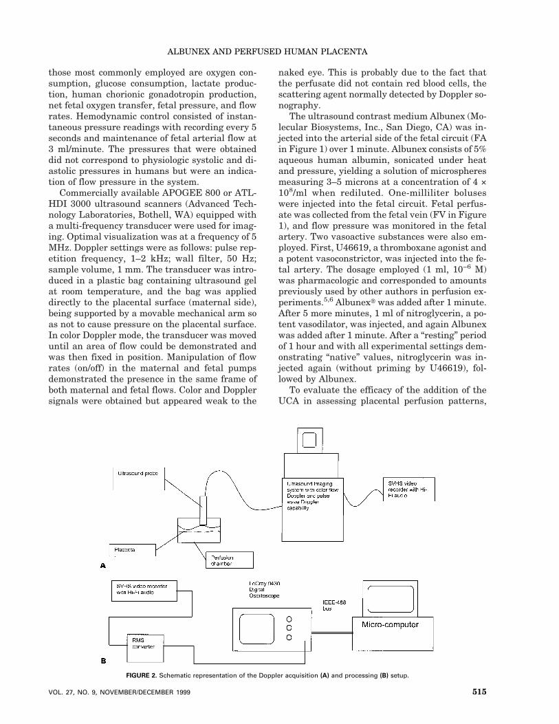
FIGURE 2. Schematic representation of the Doppler acquisition (A) and processing (B) setup.
ALBUNEX AND PERFUSED HUMAN PLACENTA
VOL. 27, NO. 9, NOVEMBER/DECEMBER 1999 515

pulsed Doppler measurements were made at cho-sen sites. A quantitative measure of flow rate wasprovided by observing the root-mean-square(rms) value of the spectral Doppler audio output.The addition of the UCA provided the scatterersnecessary for Doppler velocity detection. For rela-tively dilute concentrations of such scatterers, thescattered intensity is roughly proportional totheir concentration; this permits evaluation offlow in a region of interest when a given concen-tration of the UCA is being delivered at a constantrate of infusion. The change in Doppler intensitycan be related to the change in volume flowrate.10,11 The precise relationship depends on anumber of factors, particularly the stability of thescattering characteristics of the UCA over time inresponse to parameters such as the infusion pres-sure and the qualities of the infusate itself suchas gas solubilities and surfactant properties.Nonetheless, for a constant infusion of UCA, achange in volume flow will be reflected in achange in the observed Doppler signal intensity.
A region of active flow was determined by po-sitioning both the transducer and Doppler gate in
a region of observable Doppler signal intensity asobserved on the Doppler spectral display that cor-responded to starting and stopping of the fetalperfusion pump. The Doppler spectral displayand audio signal were recorded on an S-VHSvideo recorder with the automatic gain controldisabled and with constant gain setting. Epochsof flow measurements were recorded in this man-ner as injections of the UCA were applied withand without prior injection of the vasoactiveagents of interest. The audio signals were thenanalyzed off-line by measuring the variation ofthe rms value of the audio signal level over time.The Doppler gate included a region of vasculatureof varying orientation such that the left and rightchannels of the Doppler signal provided signals ofequivalent amplitude. The rms value was mea-sured and converted into a decibel value (10 timesthe common logarithm of the voltage squared) uti-lizing a circuit based on an AD637 monolithicrms-to-dB converter (Analog Devices, Norwood,MA). The output voltage of the circuit was re-corded with a 9430 Digital Oscilloscope (LeCroy,Chestnut Ridge, NY), and the data were trans-
FIGURE 3. Imaging with no contrast medium to establish anatomy and location of the decidual and chorionic plates. (A) Both maternal and fetalpumps off. (B) Maternal pump off; fetal pump on. (C) Maternal pump on; fetal pump off. (D) Both maternal and fetal pumps on.
ABRAMOWICZ ET AL
516 JOURNAL OF CLINICAL ULTRASOUND
ferred to a personal computer for storage and fur-ther analysis. A diagram of the Doppler acquisi-tion and analysis process is shown in Figure 2.The duration of color persistence was determinedby observing the appearance of color and measur-ing the amount of time that color was present.
Paired t-tests were used to evaluate pressurechanges and color persistence with or without theaddition of vasoactive substances. A p value below0.05 was considered statistically significant.
RESULTS
Before beginning the ultrasound component ofthis study, the placental perfusions met all of thepreviously established physiologic criteria.8 Forthe Albunex portion of the experiment, an opencircuit was used on the fetal side. Thus, not allphysiologic parameters could be documented, butthis was of no relevance for the present experi-ments. Those documented during the perfusionwere fetal venous flow rate, system pressure, PO2,PCO2, and pH.
Imaging with No Contrast MediumIntermittently stopping the maternal and/or fetalpumps permitted demonstration of the 2 circula-tions. Both the spectral and color Doppler signals,as previously mentioned, were demonstrable butweak. Figure 3 shows the presence or absence offlow with either 1 or both pumps on or off.
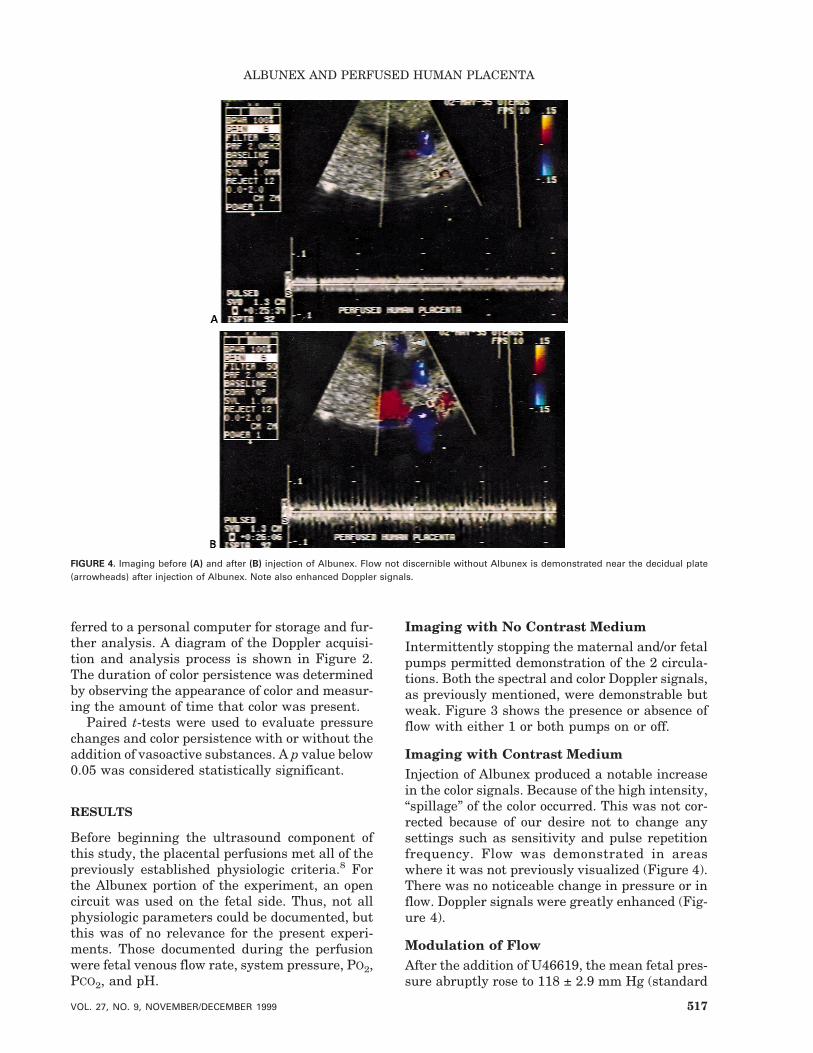
Imaging with Contrast MediumInjection of Albunex produced a notable increasein the color signals. Because of the high intensity,“spillage” of the color occurred. This was not cor-rected because of our desire not to change anysettings such as sensitivity and pulse repetitionfrequency. Flow was demonstrated in areaswhere it was not previously visualized (Figure 4).There was no noticeable change in pressure or inflow. Doppler signals were greatly enhanced (Fig-ure 4).
Modulation of FlowAfter the addition of U46619, the mean fetal pres-sure abruptly rose to 118 ± 2.9 mm Hg (standard
FIGURE 4. Imaging before (A) and after (B) injection of Albunex. Flow not discernible without Albunex is demonstrated near the decidual plate(arrowheads) after injection of Albunex. Note also enhanced Doppler signals.
ALBUNEX AND PERFUSED HUMAN PLACENTA
VOL. 27, NO. 9, NOVEMBER/DECEMBER 1999 517

error of mean) from a baseline of 20 mm Hg; themean pressure was 23.2 ± 0.8 mm Hg after Al-bunex alone (p < 0.0001). The addition of Albunexafter U46619 caused a small increase in pressure,presumably because of some washout effect of theU46619 still in the tubing. Within 1 minute ofU46619 injection, fetal flow, as demonstrated bythe amount of fetal perfusate collected at the fetalvenous side, began to decrease and was absentafter 2 minutes. The addition of nitroglycerin rap-idly returned the pressure to baseline. Figure 5 isthe graphic representation of 1 experiment. WhenAlbunex was added to the system after injectionof U46619, imaging (Figure 6A) demonstratedflow only in the area of the chorionic plate (ie,larger vessels), as opposed to the generalized flow(ie, chorionic and decidual plates as well as pla-cental substance) seen with no vasomodulator(Figure 3B). After the addition of nitroglycerin,flow was again demonstrated in all placental re-gions (Figure 6B). No changes in pressure or flowwere noticeable when nitroglycerin was injectedalone. By qualitative evaluation, the Doppler sig-nals appeared weaker with Albunex after U46619(Figure 6A) and stronger after Albunex alone(Figure 4B) or Albunex after nitroglycerin (Fig-ure 6B).
Flow Analysis
Our semiquantitative estimation of the pres-ence of color demonstrated the shortest per-sistence (39.8 ± 3.4 seconds) when Albunexwas added after injection of U46619 and thelongest (94 ± 6.5 seconds) when Albunex wasadded after injection of nitroglycerin (p < 0.0001).The persistences for Albunex alone and Albunexafter nitroglycerin were not significantly differ-ent. A plot of a typical UCA injection studyis shown in Figure 7A. The abscissa is time, andthe ordinate indicates Doppler signal intensity indecibels. A plot of a UCA flow study after injec-tion of the vasoactive modulator U46619 isshown in Figure 7B. A plot of a UCA flow studyfollowing the injection of the vasodilator nitro-glycerin is shown in Figure 7C. The change inthe time course and decrease in magnitude ofthe measured Doppler signals are evident afterinjection of the vasoconstrictor U46619. Afterinjection of the vasodilator nitroglycerin,the time course and magnitude of the Doppler sig-nals are similar to those before U46619 was in-jected, indicating restoration of the initial flowrates.
FIGURE 5. Fetal arterial pressure and fetal venous flow rate with injection of vasomodulators and Albunex for a sample experiment. Controlrepresents injection of Albunex only (flow rates in the control experiments did not change). White boxes, fetal venous flow rate; line with squares,control; line with triangles, vasomodulators.
ABRAMOWICZ ET AL
518 JOURNAL OF CLINICAL ULTRASOUND

DISCUSSION
One of the major functions of the placenta is sub-stance transfer from the maternal circulation tothe fetus. Placental morphology can be examinedtoday by several imaging techniques such as ra-diography,12 MRI,13 and sonography.2 Placentalfunction is more difficult to evaluate directly. Onecan examine fetal well-being and growth as anassumption of a properly functioning placenta,but the reverse is not necessarily so: poor fetalgrowth may have its origin in maternal or intrin-sic fetal disorders, as well as “placental insuffi-ciency.”
Doppler imaging is a technique allowing evalu-ation of uterine and umbilical circulation but isless effective in evaluation of the placental circu-lation per se. Alterations in downstream hemody-namics are reflected by changes in Doppler veloc-ity waveforms, as demonstrated in laboratorystudies.14,15 Abnormal Doppler tracings havebeen correlated with placental vascular pathology
in morphologic studies.16–18 This is particularlyobvious in cases with absent end-diastolic flow,when severe placental morphologic changes oc-cur, such as decreased weight, decreased basalarea, perivillous microfibrinous deposits, and vil-lous fibrosis.19,20 Changes in the Doppler charac-terization of umbilical artery blood flow velocityhave been described in multiple publications, par-ticularly in cases of intrauterine growth restric-tion.21–24 Alterations in vessel tone or actualchanges in the placental vascular tree have beencorrelated with Doppler tracings.25,26 Placentalvessels lack local innervation but are clearly re-sponsive to autacoids,4,5,27 particularly U46619,apparently via thromboxane A2 receptor activa-tion.6,28,29 New ultrasound techniques such ascolor and amplitude-based Doppler (color Dopplerimaging and power Doppler imaging) as well asthe use of UCAs may allow visualization of intra-placental circulation and verify previously pub-lished studies on placental vascularization.2,30,31
FIGURE 6. Modulation of flow. (A) Injection of Albunex after addition of U46619. Note absence of flow in the decidual plate region (arrowhead).(B) After addition of nitroglycerin. Flow is generalized.
ALBUNEX AND PERFUSED HUMAN PLACENTA
VOL. 27, NO. 9, NOVEMBER/DECEMBER 1999 519
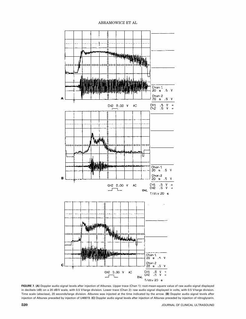
FIGURE 7. (A) Doppler audio signal levels after injection of Albunex. Upper trace (Chan 1): root-mean-square value of raw audio signal displayedin decibels (dB) on a 20 dB/V scale, with 0.5 V/large division. Lower trace (Chan 2): raw audio signal displayed in volts, with 0.5 V/large division.Time scale (abscissa), 20 seconds/large division. Albunex was injected at the time indicated by the arrow. (B) Doppler audio signal levels afterinjection of Albunex preceded by injection of U46619. (C) Doppler audio signal levels after injection of Albunex preceded by injection of nitroglycerin.
ABRAMOWICZ ET AL
520 JOURNAL OF CLINICAL ULTRASOUND
We have demonstrated in a laboratory settingthat maternal and fetal circulation can be exam-ined by color Doppler imaging. The addition of aUCA allows noninvasive Doppler measurementsof flow modulation due to vasoactive agents in theperfused placenta in a quantitative manner. Useof pulsed Doppler imaging permits evaluation offlow changes at specified sites within the placen-tal tissue. By utilizing available ultrasound scan-ning systems that provide simultaneous B-modeimaging, color Doppler imaging, and pulsedDoppler velocity measurement, it is possible toassess the regional distribution of vasoactiveagents’ effects in the perfusion model. In particu-lar, color Doppler imaging can be used to guidethe placement of the pulsed Doppler samplinggate for quantitative measurements, and place-ment is facilitated by the use of a UCA. The back-scatter enhancement provided by the UCA canalso be used to determine the distribution of theflow modulation by analysis of the real-time B-mode images. Areas of enhanced contrast appearas areas of increased brightness over time as theinjection of the UCA proceeds. Measuring thebrightness (or echogenicity) variation over timecan provide another assessment of flow distribu-tion and time course. This can be used to studyflow variations at specific locations or over aspecified region of the placental tissue. As de-scribed by Schwarz et al,32 video-densitometricanalysis of contrast-enhanced B-mode images re-quires consideration of the imaging system’svideo-processing parameters as well as the at-tenuation properties of both the tissue of interestand the UCA agent, including the effect of its con-centration. The addition of a UCA may allowdocumentation of flow in areas that are beyondthe sensitivity of the usual instruments becauseof low velocity and low signal-to-noise ratios innon–contrast-enhanced situations. Whether aUCA was used should be indicated when describ-ing “absent” flow by color Doppler imaging. In ad-dition, quantification of placental insufficiencysecondary to vascular changes33 may be possiblein the future by measuring the amount of contrastmedium taken up or cleared.
Even though our investigations were done invitro, they demonstrate that the placental bed cir-culation can be monitored to identify not only cir-culatory patterns but, more important, continu-ing changes in circulatory patterns as a first stepto assessing pathophysiology that may be associ-ated with fetal compromise. Quantification ofthese changes will be critical to evaluating suchpathophysiology. We have recently begun using amore quantitative method of estimation of color
intensity and duration using a frame grabber andcolor analysis software.
In conclusion, it is possible to characterizechanges in the human placental blood flow usingcolor Doppler imaging, spectral analysis, andUCAs. The next step is to characterize the patho-physiology leading to fetal compromise.
ACKNOWLEDGMENTS
We thank the patients, physicians, and nursingstaff on Labor and Delivery at Highland Hospitaland Strong Memorial Hospital for their assis-tance in providing placentae; Molecular Biosys-tems, Inc., for providing Albunex; and Dr. XucaiChen from the Cardiology Unit of the Departmentof Medicine at the University of Rochester for dis-cussions on the analysis of the Doppler data andthe rms-to-dB circuit.
REFERENCES
1. Benirschke K, Kaufmann P. Pathology of the hu-man placenta. 3rd ed. New York: Springer-Verlag;1995.
2. Panigel M, Abramowicz JS, Miller RK. Techniques:biophysical methods for assessment of placentalfunction. In: Sastry BVR, editor. Placental phar-macology. Boca Raton, FL: CRC Press; 1996. p.1–22.
3. Clifton VL, Read MA, Leitch IM, et al. Corticotro-pin-releasing hormone–induced vasodilation in thehuman fetal-placental circulation: involvement ofthe nitric oxide–cyclic guanosine 38,58-monophos-phate-mediated pathway. J Clin Endocrinol Metab1995;80:2888.
4. King RG, Gude NM, Dilulio JL, et al. Regulation ofhuman placental fetal vessel tone: role of nitric ox-ide. Reprod Fertil Dev 1995;7:1407.
5. Maguire MH, Howard RB, Hosokawa T, et al. Ef-fects of some autacoids and humoral agents on hu-man fetoplacental vascular resistance: candidatesfor local regulation of fetoplacental blood flow. Tro-phoblast Research 1988;3:203.
6. Myatt L, Eis AL, Kossejans W, et al. Autocoid syn-thesis and action in abnormal placental flows re-viewed: causative vs. compensatory roles. Tropho-blast Research 1998;11:315.
7. Miller RK, Wier PJ, Maulik D, et al. Human pla-centa in vitro: characterization during 12 hours ofdual perfusion. Contrib Gynecol Obstet 1985;13:77.
8. Miller RK, Wier PJ, Perez-D’Gregorio R, et al. Hu-man dual placental perfusions—criteria for toxicityevaluations. Methods in Toxicology 1993;3B:246.
9. di Sant’Agnese PA, DeMesy Jensen K, Miller RK,et al. Long term human placental lobule perfu-sion—an ultrastructural study. Trophoblast Re-search 1987;2:549.
10. Schwarz KQ, Bezante GP, Chen X, et al. Quanti-
ALBUNEX AND PERFUSED HUMAN PLACENTA
VOL. 27, NO. 9, NOVEMBER/DECEMBER 1999 521

tative echo contrast measurement by Doppler so-nography. Ultrasound Med Biol 1993;4:289.
11. Chen X, Schwarz KQ, Phillips D, et al. A math-ematical model for the assessment of hemodynamicparameters using quantitative contrast echocardi-ography. IEEE Trans Biomed Eng 1998;6:754.
12. Freese UE. The fetal maternal circulation of theplacenta. I. Histomorphologic plastoid injectionand x-ray cinematographic studies on human pla-centas. Am J Obstet Gynecol 1966;94:354.
13. Panigel M, Dixon T, Constantidinis I, et al. Fastscan magnetic resonance imaging and Doppler ul-trasonography of uteroplacental hemodynamics inthe rhesus monkey. J Med Primatol 1993;22:393.
14. Maulik D, Saini VD, Wanda NC, et al. Dopplerevaluation of fetal hemodynamics. Ultrasound MedBiol 1982;8:705.
15. Surat DR, Adamson SL. Downstream determi-nants of pulsatility of the mean velocity waveformin the umbilical arteries as predicted by a com-puter model. Ultrasound Med Biol 1996;22:707.
16. Giles WB, Trudinger BJ, Baird PJ. Fetal umbilicalartery flow velocity waveforms and placental resis-tance: pathological correlation. Br J Obstet Gynae-col 1985;92:31.
17. Kingdom JC, Burrell SJ, Kaufmann P. Pathologyand clinical implications of abnormal umbilical ar-tery Doppler waveforms. Ultrasound Obstet Gyne-col 1997;9:271.
18. Bruch JF, Sibony O, Benali K, et al. Computerizedmorphometry of umbilical vessels from pregnan-cies with intrauterine growth retardation and ab-normal umbilical artery Doppler. Hum Pathol1997;28:1139.
19. Jimenez E, Vogel M, Arabin B, et al. Correlation ofultrasonographic measurement of the utero-pla-cental and fetal blood flow with the morphologicaldiagnosis of placental function. Trophoblast Re-search 1988;3:325.
20. Mitra SC, Venkataseshan VS, von Hagen S, et al.Morphometric study of the placental vessels andits correlation with umbilical artery Doppler flow.Obstet Gynecol 1997;89:238.
21. Bracero LA, Beneck D, Kirshenbaum N, et al.Doppler velocimetry and placental disease. Am JObstet Gynecol 1989;161:388.
22. Joern H, Klein A, Kuehlwein H, et al. Critical com-
parison of indices and threshold values for assess-ing placenta performance using Doppler ultra-sound. Ultrasound Med Biol 1997;23:1179.
23. Laurin J, Lingman G, Marsal K, et al. Fetal bloodflow in pregnancies complicated by intrauterinegrowth retardation. Obstet Gynecol 1987;69:895.
24. Salafia CM, Pezzullo JC, Minior VK, et al. Placen-tal pathology of absent and reversed end diastolicflow in growth-restricted fetuses. Obstet Gynecol1997;90:830.
25. Fok RY, Pavlova Z, Benirschke K, et al. The corre-lation of arterial lesions with umbilical arteryDoppler velocimetry in the placentas of small-for-dates pregnancies. Obstet Gynecol 1990;75:578.
26. Jackson MR, Walsh AJ, Morrow RJ, et al. Reducedplacental villous tree elaboration in small-for-gestational-age pregnancies: relationship with um-bilical artery Doppler waveforms. Am J Obstet Gy-necol 1995;172:518.
27. Myatt L, Brewer AS, Langdon G, et al. Attenuationof the vasoconstrictor effects of thromboxane andendothelin by nitric oxide in the human fetal-placental circulation. Am J Obstet Gynecol 1992;166:244.
28. Boura ALA, Gute NM, King RG, et al. Character-ization of thromboxane A2 receptors in the humanfetal placental vessel and umbilical veins. Clin ExpPharmacol Physiol 1986;13:83.
29. Adamson SL, Whiteley KJ, Langille BL. Endothe-lin-1 constricts fetoplacental microcirculation anddecreases fetal O2 consumption in sheep. Am JPhysiol 1996;270:H16.
30. Abramowicz JS. Ultrasound contrast media andtheir use in obstetrics and gynecology. UltrasoundMed Biol 1997;23:1287.
31. Kaufmann P, Miller RK, editors. Placental vascu-larization and blood flow—basic research and clini-cal application. New York: Plenum Medical; 1988.
32. Schwarz KQ, Chen X, Steinmetz S. Methods forquantifying ultrasound backscatter and two-dimensional video intensity: implications for con-trast-enhanced sonography. J Am Soc Echocar-diogr 1998;2:155.
33. Sheppard BL, Bonnar J. The maternal blood sup-ply to the placenta in pregnancy complicated byintrauterine fetal growth retardation. TrophoblastResearch 1988;3:69.
ABRAMOWICZ ET AL
522 JOURNAL OF CLINICAL ULTRASOUND