Sonic Hedgehog Signaling and Development of the Dentition fileJournal of Biology Developmental...
15
Journal of Biology Developmental Review Sonic Hedgehog Signaling and Development of the Dentition Maisa Seppala 1,2 , Gareth J. Fraser 3 , Anahid A. Birjandi 1 , Guilherme M. Xavier 1,2 and Martyn T. Cobourne 1,2, * 1 Centre for Craniofacial and Regenerative Biology, King’s College London Dental Institute, Floor 27, Guy’s Hospital, London SE1 9RT, UK; [email protected] (M.S.); [email protected] (A.A.B.); [email protected] (G.M.X.) 2 Department of Orthodontics, King’s College London Dental Institute, Floor 22, Guy’s and St Thomas’ NHS Foundation Trust, London SE1 9RT, UK 3 Department of Animal and Plant Sciences, Alfred Denny Building, University of Sheffield, Sheffield S10 2TN, UK; g.fraser@sheffield.ac.uk * Correspondence: [email protected]; Tel.: +44-207-188-8028; Fax: +44-207-188-1674 Academic Editors: Henk Roelink and Simon J. Conway Received: 4 May 2017; Accepted: 28 May 2017; Published: 31 May 2017 Abstract: Sonic hedgehog (Shh) is an essential signaling peptide required for normal embryonic development. It represents a highly-conserved marker of odontogenesis amongst the toothed vertebrates. Signal transduction is involved in early specification of the tooth-forming epithelium in the oral cavity, and, ultimately, in defining tooth number within the established dentition. Shh also promotes the morphogenetic movement of epithelial cells in the early tooth bud, and influences cell cycle regulation, morphogenesis, and differentiation in the tooth germ. More recently, Shh has been identified as a stem cell regulator in the continuously erupting incisors of mice. Here, we review contemporary data relating to the role of Shh in odontogenesis, focusing on tooth development in mammals and cartilaginous fishes. We also describe the multiple actions of this signaling protein at the cellular level. Keywords: odontogenesis; initiation; patterning; dental mesenchymal stem cells; tooth number 1. Introduction Sonic hedgehog (Shh) signaling plays a key role in multiple developmental processes in vertebrates [1–3]. Shh is a highly-conserved marker for tooth development amongst all the toothed vertebrates [4–9]. The mammalian tooth is a site of dynamic Shh signaling activity, which begins in the dental lamina during initiation, and continues in well-defined regions of the tooth germ epithelium during subsequent coronal and root morphogenesis [8–13]. Several early investigations highlighted the importance of Shh signal transduction during mammalian tooth development, which includes the specification of boundaries between odontogenic and non-odontogenic epithelium during initiation, as well as cell cycle regulation, differentiation, and morphogenesis in the established tooth germ [9,14–17]. More recently, studies have identified a role for Shh in promoting morphogenetic movement in the early tooth bud through cell polarization, influencing both cuspal morphogenesis and root development during the later stages of development, and playing a key role in defining tooth number within the mature dentition [18–20]. Here, we review contemporary data relating to the role of Shh during odontogenesis in jawed vertebrates, describe the multiple actions of this signaling protein at the cellular level, and highlight the more recent identification of Shh as a potential primitive stem cell regulator in the murine incisor [21]. J. Dev. Biol. 2017, 5, 6; doi:10.3390/jdb5020006 www.mdpi.com/journal/jdb
Transcript of Sonic Hedgehog Signaling and Development of the Dentition fileJournal of Biology Developmental...
Journal of
BiologyDevelopmental
Review
Sonic Hedgehog Signaling and Development ofthe Dentition
Maisa Seppala 1,2, Gareth J. Fraser 3, Anahid A. Birjandi 1, Guilherme M. Xavier 1,2 andMartyn T. Cobourne 1,2,*
1 Centre for Craniofacial and Regenerative Biology, King’s College London Dental Institute, Floor 27,Guy’s Hospital, London SE1 9RT, UK; [email protected] (M.S.);[email protected] (A.A.B.); [email protected] (G.M.X.)
2 Department of Orthodontics, King’s College London Dental Institute, Floor 22, Guy’s and St Thomas’ NHSFoundation Trust, London SE1 9RT, UK
3 Department of Animal and Plant Sciences, Alfred Denny Building, University of Sheffield,Sheffield S10 2TN, UK; [email protected]
* Correspondence: [email protected]; Tel.: +44-207-188-8028; Fax: +44-207-188-1674
Academic Editors: Henk Roelink and Simon J. ConwayReceived: 4 May 2017; Accepted: 28 May 2017; Published: 31 May 2017
Abstract: Sonic hedgehog (Shh) is an essential signaling peptide required for normal embryonicdevelopment. It represents a highly-conserved marker of odontogenesis amongst the toothedvertebrates. Signal transduction is involved in early specification of the tooth-forming epitheliumin the oral cavity, and, ultimately, in defining tooth number within the established dentition. Shhalso promotes the morphogenetic movement of epithelial cells in the early tooth bud, and influencescell cycle regulation, morphogenesis, and differentiation in the tooth germ. More recently, Shh hasbeen identified as a stem cell regulator in the continuously erupting incisors of mice. Here, we reviewcontemporary data relating to the role of Shh in odontogenesis, focusing on tooth development inmammals and cartilaginous fishes. We also describe the multiple actions of this signaling protein atthe cellular level.
Keywords: odontogenesis; initiation; patterning; dental mesenchymal stem cells; tooth number
1. Introduction
Sonic hedgehog (Shh) signaling plays a key role in multiple developmental processes invertebrates [1–3]. Shh is a highly-conserved marker for tooth development amongst all the toothedvertebrates [4–9]. The mammalian tooth is a site of dynamic Shh signaling activity, which beginsin the dental lamina during initiation, and continues in well-defined regions of the tooth germepithelium during subsequent coronal and root morphogenesis [8–13]. Several early investigationshighlighted the importance of Shh signal transduction during mammalian tooth development, whichincludes the specification of boundaries between odontogenic and non-odontogenic epithelium duringinitiation, as well as cell cycle regulation, differentiation, and morphogenesis in the established toothgerm [9,14–17]. More recently, studies have identified a role for Shh in promoting morphogeneticmovement in the early tooth bud through cell polarization, influencing both cuspal morphogenesisand root development during the later stages of development, and playing a key role in defining toothnumber within the mature dentition [18–20]. Here, we review contemporary data relating to the role ofShh during odontogenesis in jawed vertebrates, describe the multiple actions of this signaling proteinat the cellular level, and highlight the more recent identification of Shh as a potential primitive stemcell regulator in the murine incisor [21].
J. Dev. Biol. 2017, 5, 6; doi:10.3390/jdb5020006 www.mdpi.com/journal/jdb
J. Dev. Biol. 2017, 5, 6 2 of 15
2. Shh Pathway Components and Signaling Activity
Shh transcription is consistently restricted to the epithelial component of the developing toothgerm, from initiation through to root development (Figure 1). However, the secreted signaling proteinhas been shown to diffuse within the surrounding epithelium and mesenchyme of the tooth, influencingreciprocal epithelial–mesenchymal signaling between these two embryonic tissue components in bothmice and humans [9,13,16]. Shh signals through a complex pathway, with the availability and activity ofsecreted ligand modified at multiple levels in both producing and receiving cells in a tissue-dependentmanner. The post-translational modification of Shh involves the addition of cholesterol [22,23] andpalmitate [24] groups to the newly synthesized pre-protein. Cholesterol limits the movement of theprocessed ligand, and creates a higher concentration gradient close to the signal source [25]. Palmitateis required for the formation of a multimeric complex that facilitates Shh diffusion over multiple celllayers [26]. In addition, Shh creates a self-regulatory feedback loop by inducing the transcriptionof signaling components, such as the receptor Patched1 (Ptch1), Glioma-associated oncogene familymembers Gli1-2, and the negative regulator Hedgehog-interacting protein (Hhip1). The target genes canbe found in both Shh-expressing cells and surrounding tissues, demarcating regions where autocrineand paracrine signaling is active [9,27,28]. This multifaceted regulation of Shh is an essential part ofdefining the correct tissue responses, as it acts as a morphogen whose concentration gradient elicitsdifferent tissue responses and cell fates based on their distance from the signal source [28–30].
J. Dev. Biol. 2017, 5, 6 2 of 15
Shh transcription is consistently restricted to the epithelial component of the developing tooth germ, from initiation through to root development (Figure 1). However, the secreted signaling protein has been shown to diffuse within the surrounding epithelium and mesenchyme of the tooth, influencing reciprocal epithelial–mesenchymal signaling between these two embryonic tissue components in both mice and humans [9,13,16]. Shh signals through a complex pathway, with the availability and activity of secreted ligand modified at multiple levels in both producing and receiving cells in a tissue-dependent manner. The post-translational modification of Shh involves the addition of cholesterol [22,23] and palmitate [24] groups to the newly synthesized pre-protein. Cholesterol limits the movement of the processed ligand, and creates a higher concentration gradient close to the signal source [25]. Palmitate is required for the formation of a multimeric complex that facilitates Shh diffusion over multiple cell layers [26]. In addition, Shh creates a self-regulatory feedback loop by inducing the transcription of signaling components, such as the receptor Patched1 (Ptch1), Glioma-associated oncogene family members Gli1-2, and the negative regulator Hedgehog-interacting protein (Hhip1). The target genes can be found in both Shh-expressing cells and surrounding tissues, demarcating regions where autocrine and paracrine signaling is active [9,27,28]. This multifaceted regulation of Shh is an essential part of defining the correct tissue responses, as it acts as a morphogen whose concentration gradient elicits different tissue responses and cell fates based on their distance from the signal source [28–30].
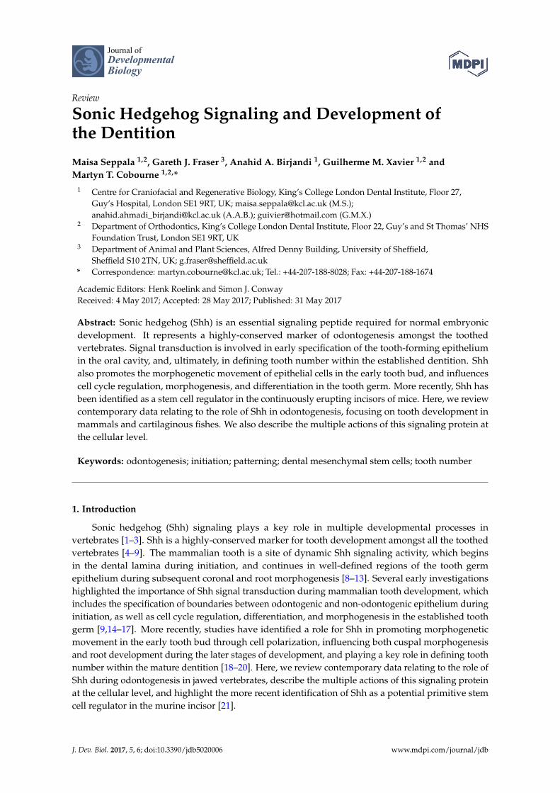
Figure 1. Schematic representation of Shh transcription in the developing tooth (red). (A) Initiation; (B) Early bud stage (at the tip of the bud in enamel knot precursor cells); (C) Early cap stage (in the primary enamel knot); (D) Late cap stage (in the secondary enamel knots); (E) Bell stage (in the internal enamel epithelium, pre-ameloblasts and stratum intermedium); (F) During root development (in Hertwig’s epithelial root sheath).
In recent years, the primary cilium has been identified as an important region of the cell that coordinates a number of molecular signaling pathways, including Hedgehog and Wingless-type MMTV integration site family (Wnt) [31]. During signaling, pathway activation occurs following the binding of Shh to the primary receptor Ptch1 [32–34] (Figure 2). In the resting state, Ptch1 accumulates at the cilium, and inhibits transduction through the indirect suppression of Smoothened (Smo), a G-protein coupled receptor-like protein essential for transduction [35,36]. Activated Smo then accumulates at the ciliary membrane [37], and effects downstream intracellular signaling through the processing of Gli1-3 transcription factors [33,38,39]. Following pathway activation, Ptch1 rapidly accumulates on the cell surface to buffer signal activity in a ligand-dependent manner [40,41]. The relative availability of bound and unbound receptor is further regulated through the Shh-mediated transcriptional repression of its own co-receptors. These include the GPI-linked membrane glycoprotein Growth arrest-specific 1 (Gas1) [42], as well as the Ig/fibronectin single-pass membrane-spanning cell adhesion proteins Cdon (cell adhesion associated, oncogene regulated) and Boc (cell adhesion associated, oncogene regulated) [43,44]. Although a vast amount of previous and ongoing
Figure 1. Schematic representation of Shh transcription in the developing tooth (red). (A) Initiation;(B) Early bud stage (at the tip of the bud in enamel knot precursor cells); (C) Early cap stage (in theprimary enamel knot); (D) Late cap stage (in the secondary enamel knots); (E) Bell stage (in theinternal enamel epithelium, pre-ameloblasts and stratum intermedium); (F) During root development(in Hertwig’s epithelial root sheath).
In recent years, the primary cilium has been identified as an important region of the cell thatcoordinates a number of molecular signaling pathways, including Hedgehog and Wingless-type MMTVintegration site family (Wnt) [31]. During signaling, pathway activation occurs following the bindingof Shh to the primary receptor Ptch1 [32–34] (Figure 2). In the resting state, Ptch1 accumulates at thecilium, and inhibits transduction through the indirect suppression of Smoothened (Smo), a G-proteincoupled receptor-like protein essential for transduction [35,36]. Activated Smo then accumulates atthe ciliary membrane [37], and effects downstream intracellular signaling through the processing ofGli1-3 transcription factors [33,38,39]. Following pathway activation, Ptch1 rapidly accumulates on thecell surface to buffer signal activity in a ligand-dependent manner [40,41]. The relative availability ofbound and unbound receptor is further regulated through the Shh-mediated transcriptional repressionof its own co-receptors. These include the GPI-linked membrane glycoprotein Growth arrest-specific 1(Gas1) [42], as well as the Ig/fibronectin single-pass membrane-spanning cell adhesion proteinsCdon (cell adhesion associated, oncogene regulated) and Boc (cell adhesion associated, oncogene

J. Dev. Biol. 2017, 5, 6 3 of 15
regulated) [43,44]. Although a vast amount of previous and ongoing research has identified multipleintriguing features of this pathway, a complete understanding of the biochemistry remains elusive.
J. Dev. Biol. 2017, 5, 6 3 of 15
research has identified multiple intriguing features of this pathway, a complete understanding of the biochemistry remains elusive.
Figure 2. A simple diagrammatic representation of the (known) key events in Shh signaling at the primary cilium. In the absence of signal (OFF), Ptch1 accumulates at the ciliary membrane and represses Smo. GLI proteins are sequestered by Kif7 and Suppressor-of-fused (SuFu), and phosphorylated through the activity of Gpr161, PKA, CK1 and GSK3B. The phosphorylated GLI proteins are proteolytically processed into truncated repressor forms, which act to inhibit transcriptional activation of the pathway. The signaling pathway is activated (ON) through the binding of Shh to Ptch1, which is facilitated by Cdo, Boc and Gas1 co-receptors. The receptor complex is internalized and degraded, which leads to the accumulation of Smo in the ciliary membrane, where it is phosphorylated and interacts with Ellis-van Creveld (EvC) proteins in the basal component of the cilium. The Kif7, SuFu, and GLI complex moves to the tip of the cilium where GLI is concentrated and then transported back to the cytosol where it enters the nucleus as a full length activator of transcriptional targets.
3. Shh Induces Invagination of the Early Dental Lamina
During mouse embryonic day (E)11 Shh is expressed in the early dental placodes [8] and has an inverse relationship with Wnt7b, which is expressed in the non-odontogenic epithelium and inhibits Shh. These expression domains define the sites where teeth will and will-not form [17]. Subsequently, the Shh-expressing dental placode undergoes strong vertically-orientated cell division and becomes histologically visible. The placode further stratifies into basal (deeper) and multiple suprabasal (superficial) layers to produce an epithelial thickening, which invaginates into the underlying mesenchyme at around E11.5 and progresses to an early bud by E12.5 [45,46]. After a short period of quiescence, Shh expression becomes localized at the tip of the established tooth bud [9,15].
The repeated expression of Shh in epithelium of the developing tooth prompted early studies investigating the role of this signal pathway during tooth development [9,14–16,47]. One of the first investigations demonstrated that the application of ectopic Shh was capable of stimulating crude invagination of both odontogenic and non-odontogenic oral epithelium [9]. These data proved consistent with findings from mandibular explant culture experiments, where the opposite effect was achieved following the inhibition of Shh using the 5E1 blocking antibody [14]. Specifically, a loss of Shh function from E10.5 resulted in the early arrest of tooth development at the epithelial thickening stage, associated with the down-regulation of Ptch1 and a lack of proliferation in deeper regions of the epithelial thickening. Shh mutant mice display severe defects associated with the first branchial arch [48,49], which makes them redundant as a model to study tooth development. To overcome this,
Figure 2. A simple diagrammatic representation of the (known) key events in Shh signaling atthe primary cilium. In the absence of signal (OFF), Ptch1 accumulates at the ciliary membraneand represses Smo. GLI proteins are sequestered by Kif7 and Suppressor-of-fused (SuFu), andphosphorylated through the activity of Gpr161, PKA, CK1 and GSK3B. The phosphorylated GLIproteins are proteolytically processed into truncated repressor forms, which act to inhibit transcriptionalactivation of the pathway. The signaling pathway is activated (ON) through the binding of Shh toPtch1, which is facilitated by Cdo, Boc and Gas1 co-receptors. The receptor complex is internalized anddegraded, which leads to the accumulation of Smo in the ciliary membrane, where it is phosphorylatedand interacts with Ellis-van Creveld (EvC) proteins in the basal component of the cilium. The Kif7,SuFu, and GLI complex moves to the tip of the cilium where GLI is concentrated and then transportedback to the cytosol where it enters the nucleus as a full length activator of transcriptional targets.
3. Shh Induces Invagination of the Early Dental Lamina
During mouse embryonic day (E)11 Shh is expressed in the early dental placodes [8] and has aninverse relationship with Wnt7b, which is expressed in the non-odontogenic epithelium and inhibitsShh. These expression domains define the sites where teeth will and will-not form [17]. Subsequently,the Shh-expressing dental placode undergoes strong vertically-orientated cell division and becomeshistologically visible. The placode further stratifies into basal (deeper) and multiple suprabasal(superficial) layers to produce an epithelial thickening, which invaginates into the underlyingmesenchyme at around E11.5 and progresses to an early bud by E12.5 [45,46]. After a short period ofquiescence, Shh expression becomes localized at the tip of the established tooth bud [9,15].
The repeated expression of Shh in epithelium of the developing tooth prompted early studiesinvestigating the role of this signal pathway during tooth development [9,14–16,47]. One of thefirst investigations demonstrated that the application of ectopic Shh was capable of stimulatingcrude invagination of both odontogenic and non-odontogenic oral epithelium [9]. These data provedconsistent with findings from mandibular explant culture experiments, where the opposite effect wasachieved following the inhibition of Shh using the 5E1 blocking antibody [14]. Specifically, a loss ofShh function from E10.5 resulted in the early arrest of tooth development at the epithelial thickeningstage, associated with the down-regulation of Ptch1 and a lack of proliferation in deeper regions ofthe epithelial thickening. Shh mutant mice display severe defects associated with the first branchialarch [48,49], which makes them redundant as a model to study tooth development. To overcome this,mice have been generated using Cre-LoxP-mediated recombination to remove Shh function in the early
J. Dev. Biol. 2017, 5, 6 4 of 15
oral epithelium from around E11.75 using a Keratin-14 (K14) promoter. Interestingly, these mice didform epithelial buds at E12.5, but they were associated with wider and shallower invaginations whencompared to control teeth [15]. Collectively, these studies provided strong evidence of an inductiveand mitogenic role for Shh during the early stages of tooth development.
More recently, the specific requirements for Shh during the progression from placode to bud havebeen further investigated using explant culture and confocal imaging [20]. A temporal 24-h inhibitionand over-expression of Shh and Fibroblast growth factor (Fgf) signaling in mandibular cultures at E10.5and 11.5 have revealed distinct and independent functions for these pathways in the early tooth germ.The inhibition of Fgf signaling at E11.5 using the pan-Fgf receptor inhibitor SU5402 results in a failureof epithelial stratification and decreased levels of proliferation, with reduced depth in the placodalregion. Similarly, Fgf inhibition at E12.5 also produces smaller tooth buds, but even though there is lessdownward growth, the width of the tooth bud remains unaffected, indicating that Fgf signals driveproliferation, epithelial stratification, and the generation of suprabasal cells, but are likely dispensablefor invagination. This study also showed that even though Shh has previously been shown to induceproliferation during the early stages of tooth development, it is more likely to be Fgf signaling that isrequired and sufficient to drive vertically-orientated cell division at the dental thickening stage. Theinhibition of Shh using the Smo-antagonist cyclopamine at E10.5 and 11.5 had no effect on stratificationor proliferation, but the tooth buds were shallower and wider. Cellular analysis revealed that thesetooth germs lacked columnar-shaped cells and elongated nuclei in basal and suprabasal regions, withthe difference most significant in shoulder regions of the invaginating dental lamina. When Shh activitywas amplified using the Shh-agonist purmorphamine, the results were narrowed tooth bud necks thatinvaginated to a much deeper level in comparison to controls. Changes in cellular and nuclear shapeare indicative of motile cell behavior, and Shh appears to coordinate the morphogenetic movement ofcells required for normal invagination of the tooth germ. Significantly, this is contradictory to previousstudies—as no changes in proliferation levels were noted—and it suggests an important role for Shhspecifically influencing the cellular rearrangements required for driving the invagination of the earlytooth germ [34].
4. Shh Governs Early Molar Morphogenesis
After the initial thickening stage, Shh is upregulated at E13.5 in the epithelial bud tip. The intensityof Shh expression subsequently increases a day later at the cap stage, when the primary enamel knotappears [9,12]. At the same time, Ptc1, Gli1-2, and Smo are all expressed in partially overlappingdomains within both the dental epithelium and the surrounding mesenchyme, but their expression isexcluded from the enamel knot [9].
The primary and secondary enamel knots are transient signaling centers [12] that express anumber of signaling molecules, including Fgf3, 4, and 9; Bone morphogenetic protein (Bmp) 2, 4, and 7;Wnt10a and 10b; and Shh [12,50–53] and act to define tooth shape [12,54–57]. At E14.5, K14-Cre; Shhconditional knock-out molars show continued dental development, but morphogenesis is significantlydisrupted. At this stage, the molars are around 25 per cent smaller than wild type and are dysmorphic,as they fail to obtain a normal cap stage form. Interestingly, these molars also show signs of disruptedbucco–lingual polarisation, as only the remnants of a lingual protrusion are observed. However, Fgf4,Bmp2 and Wnt10b are all expressed in the enamel knot of these teeth, whilst ectopic Wnt10b and Lef1expression extends lingually. This suggests that the loss of Shh results in a partial change of cell fate,from proliferative to a less proliferative enamel knot-like population on the lingual side. In addition,E14.5 tooth germs are fused with the oral epithelium and lack any clear dental cord connecting the oralepithelium with the invaginating tooth germ. Thereafter, Shh appears to drive polarisation of the toothin a bucco–lingual axis and induce proliferation in a differential manner, taking part in determiningtooth shape [15]. This is supported by the stronger expression of Ptch1 on the lingual side of the capstage outer enamel epithelium (OEE) [9,15]. Furthermore, a molecular investigation of conditionalknockouts shows the intact expression of Bmp4, Msx1, and Pax9, providing evidence that during the

J. Dev. Biol. 2017, 5, 6 5 of 15
start of morphogenesis, Shh is dispensable for their induction. In contrast, Pitx1 is downregulated andappears to be under the regulation of Shh [15].
The loss of Ellis-van Creveld (EvC) function also results in fusion of the first and second molars,reduced size of the first molar in comparison to the second and single root formations. In addition, EvCappears to be involved in a symmetrical bucco–lingual response to Shh during molar development.Intriguingly, EvC mutant mice show more severe defects on the buccal side rather than the lingual,distinguishing them from K14-Cre; Shh mice. This may be due to the different origins of the buccaland lingual tissues from the ectoderm and endoderm, respectively; as patients with EvC syndrome donot present with any known anomalies associated with endoderm-derived tissues [58,59]. At E14.5,the asymmetric molar phenotype is accompanied by a buccal shift in expression of Shh, Wnt10b, andLef1-expressing enamel knot cells and a lingual shift of Fgf4. Although Shh expression remains robustin the epithelium, Ptch1 and Gli1 are absent from the surrounding mesenchyme, underlining thatShh signaling is disrupted in EvC mutants [58]. When EvC forms a complex with EvC2 it localises tothe cilium, where it interacts with Smo and controls dissociation of the negative Hedgehog regulatorSuppressor-of-fused (SuFu) from Gli3, and thus Gli3 movement into the cilia (see Figure 2) [60].
5. A Lack of Shh Results in a Failure of Ameloblast Polarization
The process of cytodifferentiation begins in the tooth germ at the bell stage, around E18 in amouse. At this stage, the future tooth consists of an epithelial enamel organ (EEO) and the dentalmesenchyme. The EEO is a complex structure consisting of the inner enamel epithelium (IEE),secondary enamel knots, and the OEE, which is separated from the IEE by an internally-locatedstratum intermedium (SI) and stellate reticulum (SR). Polarized pre-odontoblasts situated adjacentto the IEE within the mesenchymal compartment differentiate and secrete predentin, which inducesthe IEE to differentiate into pre-ameloblasts [61,62]. Shh is strongly expressed in the IEE with Ptch1in the underlying mesenchyme just prior to the differentiation of the pre-odontoblasts [16,63]. Theinductive signal for odontoblast differentiation is known to originate from the IEE, and thereafter thespatio-temporal expression pattern of Shh and Ptch1 implicates Shh as having a role in transmitting therequired signal between these two tissue components, as well as being a marker for pre-ameloblasts.
Regardless of the strong expression of Shh in the IEE, K14-Cre; Shh mice do have differentiatedpre-odontoblasts and pre-ameloblasts in the developing tooth at E19.5, with these cells expressingknown markers that demonstrate functionality. Furthermore, a secreted enamel and dentin matrixis evident between the two cell populations. However, the cellular architecture is disrupted, as theodontoblasts fail to form a continuous monolayer, and only a proportion of these cells polarise andelongate. Similarly, the differentiating ameloblasts remain small and cuboidal, in contrast to theelongated and polarised cell shapes seen in control tooth germs [15]. The conditional removal of Shhresponsiveness exclusively from the oral epithelium by inactivation of Smo under the K14 promoter(K14-Cre; Smo) results in the normal polarisation of the mesenchymally-derived pre-odontoblasts,but disrupted polarisation of the pre-ameloblasts, and hypocellularity of the SR and SI. An abnormalcytoskeletal organisation of the pre-ameloblasts is seen, associated with a lack of Ptch2 and Gli1accumulation in the intracellular space on the basal side. This implicates a possible role for thestratum intermedium in providing a guiding signal for the subcellular trafficking of proteins thattakes place during polarisation. In addition, K14-Cre; Smo mice show an absence of β-tubulinaccumulation, and a polarized cytoplasmic localisation of E-Cadherin and Zonula Occludens-1 (ZO-1).This provides evidence of a lack of specialised junctional complexes that, together with microtubules,are important for establishing the cell polarity and the apical and basolateral domains. Further analysishas demonstrated reduced proliferation as well as early loss of the phosphorylated histone H3 (PH-H3)protein (a known marker of mitosis) and the Shh-induced cyclinD1. This indicates that decreasedproliferation levels could be a consequence of premature exit from the cell cycle, and cytodifferentiationof ameloblast progenitors before predentin secretion. This suggests that Shh regulates the cell cycle andexerts its mitogenic role by promoting cyclin D1 transcription to control the G1/S transition [16]. These
J. Dev. Biol. 2017, 5, 6 6 of 15
studies have highlighted how Shh guides cytoskeletal organisation and promotes the arrangement ofcell–cell junctional complexes, not only at sites of mesenchymal–epithelial interaction, but also withinepithelial cell populations.
6. Disrupted Shh Signaling Causes Molar Fusion and Altered Cusp Morphology
The signaling molecules expressed in the enamel knot regulate morphogenesis by diffusingthrough the surrounding epithelial and mesenchymal tissues, where they are responsible for definingappropriate molecular and cellular responses. These molecular signals can positively and negativelyinteract at multiple levels to polarise and pattern tissues.
An elaborate model, based on Alan Turing’s reaction–diffusion principles guiding patternformation, has been redrawn to potentially explain cuspal patterning by Shh and Wnt signaling [18].At E14.5, Shh, Wnt10a, and Wnt10b are similarly expressed in the enamel knot and secreted laterallyto the surrounding tissues [50]. Whilst Wnt induces its own transcription factor Lef1 in immediateproximity to its source and positively regulates Shh via Fgf3 and Fgf4, Shh ligand has the ability todiffuse faster than Wnt, and travels outside of the Wnt-created Lef1 zone, where it has an opposingeffect. Shh induces the expression of the Wnt inhibitor Wise, indirectly limiting the activity of Wnt andsimultaneously creating the odontogenic Lef1 and non-odontogenic Wise zones [16,18,64–66]. In thisShh–Wise–Wnt feedback model, Wnt acts as an activator; Shh a mediator; and Wise an inhibitor [18].Consistently, a loss of Wise function [54] and multiple mouse models with reduced Shh signalingactivity [15,16,18] have severely disrupted cuspal patterning in the molar dentition, manifesting asenlarged and fused molar teeth with multiple smaller cusps.
The importance of temporal Shh signaling in molar development has more recently beeninvestigated by blocking Shh activity in utero at different time-points between E10–18 with 5E1intra-peritoneal injection. When injections are carried out at E10 or 12, the non-odontogenic field isabsent only between first and second molars, which are fused together. If injections are carried out atE16 the same effect moves further distally in the dental arch, with the second and third molars fused.This correlates with the sequential development of these teeth, progressing along the mesio–distalaxis. This phenotype is associated with altered Wnt signaling domains between the fusing molars,as demonstrated by an expanded Sp5 expression across the first and second molars, and in contrast tocontrols that demonstrated the separate molar domains of Sp5. Interestingly, reduced levels of Shhand increased levels of Wnt were associated with the accelerated development of the second molars incomparison to controls [18].
7. Shh Influences Tooth Root Development
Shh and constituent members of the signalling pathway are also expressed during earlydevelopment of the molar tooth root in the mouse. Specifically, Shh transcripts localise to theepithelial cells of Hertwig’s epithelial root sheath, whilst Smo, Ptch1, and Gli are expressed in the apicalmesenchyme of the associated dental papilla and follicle [11]. The analysis of mesenchymal dysplasia(mes) mice, which harbour an abnormal C-terminus associated with Ptch1, has shown repressedproliferation, shorter roots and a disturbed molar eruption [67]. More recently, it has been shown thatregulating appropriate levels of Shh signaling is crucial for the normal growth and development ofthe root, with both reduced and increased signal levels producing reduced levels of proliferation, andultimately, root length. Moreover, levels of Hedgehog activity are regulated by nuclear factor I/C(Nfic), which binds Hhip1 in the apical papilla [68]. Signifcantly, Nfic mutant mice also develop shortmolar roots [69].
8. Shh and Its Role in Defining the Tooth Formula
In contrast to humans, mice have only a single incisor and three molars in each dental quadrant,separated by a non-odontogenic diastema. However, at the earliest stages of odontogenesis, multipleepithelial thickenings accompanied by the expression of Shh are identifiable in the diastema region.

J. Dev. Biol. 2017, 5, 6 7 of 15
These vestigial swellings only progress to a thickening or rudimentary bud stage, and thereafter regressvia apoptosis with the simultaneous disappearance of Shh expression [70–74]. Interestingly, severalmouse models with disrupted Shh, Wnt, Fgf, and Bmp signaling levels develop supernumerary teeth.These teeth typically arise from the most posterior rudimentary swelling, referred to as R2, and whichduring normal mouse development fuse with the main cap stage’s first molar [71,73]. Although thesepremolar-like supernumeraries have been described as evolutionary remnants (the non-odontogenicdiastema has been a prevailing feature of mice for over 50 million years) [19,55,73], they provide anexcellent model to study how different signaling cascades interact to control tooth formula, by eitherinducing or suppressing the progression of tooth development, and defining the boundaries betweenodontogenic and non-odontogenic fields.
Whereas a reduction or loss of Shh signaling activity has been associated with molarfusion [15,16,18], increased activity in the diastema can result in altered molar field patterning andsupernumerary tooth formation (Figure 3) [19,55]. Indeed, multiple knockout mouse models haveidentified the upregulation of Shh in the diastema, which as a primary or secondary response resultsin supernumerary tooth formation [38,47,54], and places Shh as a downstream target [54,55]. Thesestudies demonstrate how a complex regulatory network—consisting of its own as well as othermolecular signaling cascade members—is responsible for defining the pivotal threshold levels of Shhfor the normal progression of tooth development.
J. Dev. Biol. 2017, 5, 6 7 of 15
several mouse models with disrupted Shh, Wnt, Fgf, and Bmp signaling levels develop supernumerary teeth. These teeth typically arise from the most posterior rudimentary swelling, referred to as R2, and which during normal mouse development fuse with the main cap stage’s first molar [71,73]. Although these premolar-like supernumeraries have been described as evolutionary remnants (the non-odontogenic diastema has been a prevailing feature of mice for over 50 million years) [19,55,73], they provide an excellent model to study how different signaling cascades interact to control tooth formula, by either inducing or suppressing the progression of tooth development, and defining the boundaries between odontogenic and non-odontogenic fields.
Whereas a reduction or loss of Shh signaling activity has been associated with molar fusion [15,16,18], increased activity in the diastema can result in altered molar field patterning and supernumerary tooth formation (Figure 3) [19,55]. Indeed, multiple knockout mouse models have identified the upregulation of Shh in the diastema, which as a primary or secondary response results in supernumerary tooth formation [38,47,54], and places Shh as a downstream target [54,55]. These studies demonstrate how a complex regulatory network—consisting of its own as well as other molecular signaling cascade members—is responsible for defining the pivotal threshold levels of Shh for the normal progression of tooth development.
Figure 3. Disrupted Shh signaling and early tooth development. (A–C) Para-sagittal sections of the developing maxillary and mandibular molar dentition in (A) E16.5 Wild type; (B) E16.5 CreER™; Shh mutant; (C) E15.5 Gas1 mutant embryos. In the wild type embryo, the first molars (m1) are at the late cap/early bell stage of development and the second molars (m2) are at the early cap stage. In CreER™; Shh mutant embryos, Shh signaling has been abrogated from E10.5 following the administration of Tamoxifen, with fusion between the first and second mandibular molars (m1/m2). In the Gas 1 mutant, m1 are at the late cap stage and the m2 tooth germs are just beginning to form an early cap. However, a supernumerary premolar tooth (sn) is present in mesial to m1 in both quadrants.
Shh also negatively regulates transcription of the co-receptors Gas1, Cdon, and Boc, which are typically expressed at some distance from the signal source, and act in a reciprocal manner to Ptch1 [42,75]. In peripheral regions of the Shh signaling domain where protein levels are low, the increased transcription of these co-receptors is thought to promote signaling in receiving cells, and help achieve appropriate threshold levels for signal transduction in the peripheral regions of the signaling domain [42,76–79]. Interestingly, Gas1 seems to influence Shh in a context-dependent manner: it positively regulates signaling in the facial midline and neural tube [42,76,78], whilst seemingly having an opposite inhibitory role in the early jaw [19,47]. During early tooth development, Gas1 is abundant in the surrounding mesenchyme of early tooth germs and in the non-odontogenic diastema, expressed mainly in a reciprocal manner to Ptch1, and sharing only small areas of overlapping expression in the peripheral regions of their respective domains. When Gas1 is over-expressed in the non-odontogenic diastema it downregulates Ptch1, which suggests an important early regulatory mechanism that restricts Shh activity to the tooth-forming regions of the mandible [47]. This is consistent with the molar phenotype of Gas1 mutant mice, which demonstrates how an absence of Gas1 allows the diffusion of Shh protein further away from its source. This results at E12.5 in the presence of ectopic protein in the diastema, and an accompanied increased expression of Ptch1. Significantly, Gas1 mutant mice present with single premolar-like supernumeraries located mesial to
Figure 3. Disrupted Shh signaling and early tooth development. (A–C) Para-sagittal sections of thedeveloping maxillary and mandibular molar dentition in (A) E16.5 Wild type; (B) E16.5 CreER™; Shhmutant; (C) E15.5 Gas1 mutant embryos. In the wild type embryo, the first molars (m1) are at the latecap/early bell stage of development and the second molars (m2) are at the early cap stage. In CreER™;Shh mutant embryos, Shh signaling has been abrogated from E10.5 following the administration ofTamoxifen, with fusion between the first and second mandibular molars (m1/m2). In the Gas 1 mutant,m1 are at the late cap stage and the m2 tooth germs are just beginning to form an early cap. However,a supernumerary premolar tooth (sn) is present in mesial to m1 in both quadrants.
Shh also negatively regulates transcription of the co-receptors Gas1, Cdon, and Boc, whichare typically expressed at some distance from the signal source, and act in a reciprocal mannerto Ptch1 [42,75]. In peripheral regions of the Shh signaling domain where protein levels are low, theincreased transcription of these co-receptors is thought to promote signaling in receiving cells, and helpachieve appropriate threshold levels for signal transduction in the peripheral regions of the signalingdomain [42,76–79]. Interestingly, Gas1 seems to influence Shh in a context-dependent manner: itpositively regulates signaling in the facial midline and neural tube [42,76,78], whilst seemingly havingan opposite inhibitory role in the early jaw [19,47]. During early tooth development, Gas1 is abundantin the surrounding mesenchyme of early tooth germs and in the non-odontogenic diastema, expressedmainly in a reciprocal manner to Ptch1, and sharing only small areas of overlapping expression in theperipheral regions of their respective domains. When Gas1 is over-expressed in the non-odontogenicdiastema it downregulates Ptch1, which suggests an important early regulatory mechanism thatrestricts Shh activity to the tooth-forming regions of the mandible [47]. This is consistent with the

J. Dev. Biol. 2017, 5, 6 8 of 15
molar phenotype of Gas1 mutant mice, which demonstrates how an absence of Gas1 allows thediffusion of Shh protein further away from its source. This results at E12.5 in the presence of ectopicprotein in the diastema, and an accompanied increased expression of Ptch1. Significantly, Gas1 mutantmice present with single premolar-like supernumeraries located mesial to the first molars, whichfurther supports the concept of Gas1 acting as an additional inhibitor required for limiting Shh activityto the tooth-forming regions [19]. The high-level regulation of Shh signaling in the tooth is furtherdemonstrated by the strong expression of Hhip1 in the peripheral odontogenic mesenchyme [27]. Hhip1encodes a membrane glycoprotein that can also bind and sequester Shh [80], although to date it onlyseems to retain an essential function in root development [68]. Primary cilia protein components alsogenerally exert a negative regulatory effect on Shh activity, and seem to play a role in repressing toothformation. Mice mutants for the cilia intraflagellar transport (IFT) protein (IFT88 or Polaris) showan upregulation of Shh signaling activity, and form ectopic premolar-like teeth in the diastema [19].However, the phenotypic consequences of a disrupted ciliary function have to be interpreted withinthe wider context of their potential effect on other signal pathways, including Wnt transduction.
The Sprouty (Spr) gene family encode universal receptor tyrosine kinase signaling (RTK) inhibitorsthat are induced by Fgfs, but which inhibit Fgf signaling to provide an important negative feedbackregulatory mechanism [81]. Fgf s are found at multiple sites in the developing tooth and at multiplestages, with expression domains extending into the dental epithelium. There, they become localised tothe enamel knot (Fgf4 and Fgf9) and surrounding mesenchyme (Fgf3 and Fgf10), including the diastema(Fgf10). Interestingly, Spr2 and Spr4 mutant mice form premolar-like diastema supernumeraries similarto those seen in Gas1 and Polaris mutants [55]. Predictably, Spr2 and Spr4 mutants demonstrateectopic Fgf activity, which spatio-temporally correlates with the formation of these supernumeraries.There is also the continued expression of Shh in R2 at E14.5, providing further evidence that Fgfs actupstream of Shh in the tooth and positively regulate its activity. Whereas Spr4 is found exclusivelyin the surrounding mesenchyme, Spr2 localizes more prominently to the bud epithelium. However,some activity is also present in the mesenchyme surrounding the developing first molar. Thereafter,Spr2 is considered to be required for the indirect inhibition of Fgf-induced Shh expression, and innormal development acts to suppress the development of the first molar [55]. In addition, Fgf andWnt pathways form an important reciprocal signaling network between the epithelium and themesenchyme at this stage, because Fgf4 transcription relies on Wnt-induced Lef1 [65].
Interestingly, even though diastema teeth develop in Spr2 and Spr4 mutants, they do notsignificantly affect first molar cusp morphology, indicating that Sprouty gene activity is more notablyinvolved in defining tooth number rather than cusp patterning [55]. This is distinct from Wise-mediatedloss of Wnt inhibition, which results in both the development of diastema supernumerary teethwith variable penetrance and altered cusp morphology [54]. The relationship between Shh andWnt signaling during supernumerary tooth formation has been further investigated by generatingWise+/−; Shh/GFPCre/+ compound mice, which exhibit reduced Shh dosage. These mice also display asupernumerary tooth phenotype that can be rescued by increasing Shh levels through a partial loss ofPtch1 function, or reducing Wnt signaling levels by reducing Lrp6. In turn, when Shh is downregulated,Wnt signaling is upregulated, indicative of a Shh negative–feedback loop required for controllingthe levels of Wnt signaling. Altogether, there is strong evidence available to suggest that the relativeexpression levels of Shh and Wnt play a major role in coordinating multiple patterning processes,and that an appropriate balance between these pathways is crucial for the normal progression oftooth development.
9. Shh as a Regulator of Dental Stem Cells
Mouse incisors grow continuously throughout life, and have provided a unique model to studythe potential surface markers, anatomical location and role of stem cells in tooth development. Thecervical loop is a component of the enamel organ that disappears in molars, but prevails in incisorsand enables enamel formation to continue in adult mice. Actively proliferating transit amplifying cells
J. Dev. Biol. 2017, 5, 6 9 of 15
(TACs) are located close to the cervical loop, and are thought to represent the location of an epithelialstem cell reservoir [82,83]. Murine incisor epithelial stem cells can be identified in the incisor based onthe expression of specific surface markers, which include Gli1, Bmi1 and Sox2 [83,84]. Investigations inthe murine incisor using Gli1-LacZ reporter mice have found such expression (in addition to dentalepithelial cells and labial post-mitotic odontoblasts) specifically in the mesenchyme surrounding theneurovascular bundle (NVB) in both arterioles and their surrounding nerves [84]. Interestingly, thisexpression was not induced by Shh originating from the dental epithelium, but Shh secreted by sensoryneurons of the trigeminal ganglion, whose axons also innervate the inferior alveolar nerve and adultlower incisors. In the incisor mesenchyme, Gli1+ cells strongly overlap with H2BGFP-labelled quiescentstem cells, supporting Gli1+ as being a stem cell marker. The Gli1+ and H2BGFP double-labelledcells do not express many of the common stem cell-related surface markers such as CD105, CD73,NG2, CD146, CD44, or Sca1. However, culture-based studies using Gli1-CE; Tdtomato mice haveshown that these cells rapidly lose Gli1 expression when communication with the NVB is lost,and can subsequently differentiate into different MSC subpopulations that express CD146, CD105,Sca1, and CD73. In addition, upon hard tissue injury, Gli1+ cells can give rise to odontoblasts [21].Similarly to Shh being critical for an osteoblast lineage commitment [85], it may also be essentialfor an odontogenic commitment of incisor stem cells through primitive Gli1+ cells giving rise topericytes, which consecutively upon injury are stimulated to differentiate into odontoblasts. Althoughthe inhibition of Shh signaling in mice fed for one month with a Hedgehog-antagonist did reducedentin formation, it did not have any significant impact on mesenchymal cell proliferation, apoptosis,or the number of label-retaining slow cycling stem cells [84]. Therefore, Shh derived from sensoryneurons has a key role in both murine adult incisor homeostasis and hard tissue tissue repair, but is notrequired for stem cell proliferation and maintenance. Significantly, Gli1+ cells are also found elsewherein the body surrounding the arteries, and raise the possibility that they could also mark stem cells inother organs [86].
10. Shh as a Universal Marker of Vertebrate Tooth Development
Although the majority of tooth development research has focused on the murine dentition,it is clear that Shh is also a conserved marker of tooth development, from initiation through tomorphogenesis, amongst all toothed vertebrates including fish, reptiles, and humans [4–9]. Even inthe phylogenetically basal cartilaginous fishes, the Chondrichthyes (which include sharks, rays andholocephalans), shh is a key epithelial marker of tooth development and morphogenesis [6,7]. In theembryonic shark (Scyliorhinus canicula), shh is expressed in a restricted band along the oral jaw priorto tooth initiation [6] (Figure 4). This expression is an indicator of emerging dental competence inthe pre-lamina epithelium or odontogenic band [6,87]. This early shh expression in the odontogenicband is obvious across vertebrate taxa, and demarcates the spatial restriction of tooth territories.The comparative appearance of shh expression in this region also confirms a variability that highlightsthe early diversity of tooth patterning among vertebrate clades [88].
Following initial establishment of the odontogenic band and the first appearance of shh expression,the first teeth in the shark jaw manifest as superficial thickened placodes in a similar pattern ofemergence to the teeth of other vertebrates, such as reptiles and mammals. During this process of sharktooth placode maturation, shh expression persists within the epithelial placode (the early tooth bud)as the underlying mesenchyme begins to condense and trigger the cap stage of tooth morphogenesis(Figure 4). Here, the epithelial component surrounds the mesenchymal core or dental papilla in acharacteristic form that appears to be highly conserved among all vertebrates. At this stage, shhexpression becomes clearly localized to the apical cells of the tooth bud (Stage 32; Figure 4), whichis indicative of the regions attributed to the enamel knot signaling centres that govern tooth shapein mammals [6]. In the shark, defined regions of shh expression (and the expression of other knownenamel knot markers) in the presumptive primary enamel knot cells show a uniformity, and thereforea conservation of primary enamel knot signaling among vertebrates. This indicates that not only

J. Dev. Biol. 2017, 5, 6 10 of 15
are the early phases of vertebrate tooth initiation conserved, but also that the later stages of toothmorphogenesis, and therefore the genetic programme (including Shh signaling) for general vertebratetooth development, is an evolutionarily stable process.J. Dev. Biol. 2017, 5, 6 10 of 15
Figure 4. Conserved shh expression in shark tooth initiation and successional morphogenesis. Expression of shh in the odontogenic band marks the onset of dental competence in the shark (Scyliorhinus stellaris) lower jaw (A) and upper jaw (B). Development of the first tooth and subsequent morphogenesis in Scyliorhinus canicula is superficial with shh expression restricted to the dental epithelium at the apical cusp, lower jaw (C) and upper jaw (D). Dental lamina epithelium continues to invaginate lingual to the first tooth, from which new successional teeth will develop in the lower jaw (E) and upper jaw (F). New successional teeth in the shark continue to express shh during morphogenesis (E,F). Note shh is not expressed in the epithelial dental lamina away from developing teeth, and its expression is restricted to the enamel knot-like cells at the apex of each new tooth throughout morphogenesis. False colour, (magenta) gene expression; nuclear counterstain DAPI (white).
Following initial establishment of the odontogenic band and the first appearance of shh expression, the first teeth in the shark jaw manifest as superficial thickened placodes in a similar pattern of emergence to the teeth of other vertebrates, such as reptiles and mammals. During this process of shark tooth placode maturation, shh expression persists within the epithelial placode (the early tooth bud) as the underlying mesenchyme begins to condense and trigger the cap stage of tooth morphogenesis (Figure 4). Here, the epithelial component surrounds the mesenchymal core or dental papilla in a characteristic form that appears to be highly conserved among all vertebrates. At this stage, shh expression becomes clearly localized to the apical cells of the tooth bud (Stage 32; Figure 4), which is indicative of the regions attributed to the enamel knot signaling centres that govern tooth shape in mammals [6]. In the shark, defined regions of shh expression (and the expression of other known enamel knot markers) in the presumptive primary enamel knot cells show a uniformity, and therefore a conservation of primary enamel knot signaling among vertebrates. This indicates that not only are the early phases of vertebrate tooth initiation conserved, but also that the later stages of tooth morphogenesis, and therefore the genetic programme (including Shh signaling) for general vertebrate tooth development, is an evolutionarily stable process.
Following the establishment of the first tooth and the subsequent territories of the elasmobranch first generation dentition, the dental epithelial cells lingual to the first tooth continue to proliferate and invaginate lingually into the mesenchyme. This invagination oral to the jaw cartilages produces the early dental lamina, which is the continuous jaw-wide epithelial unit that plays a crucial role in
Figure 4. Conserved shh expression in shark tooth initiation and successional morphogenesis.Expression of shh in the odontogenic band marks the onset of dental competence in the shark(Scyliorhinus stellaris) lower jaw (A) and upper jaw (B). Development of the first tooth and subsequentmorphogenesis in Scyliorhinus canicula is superficial with shh expression restricted to the dentalepithelium at the apical cusp, lower jaw (C) and upper jaw (D). Dental lamina epithelium continues toinvaginate lingual to the first tooth, from which new successional teeth will develop in the lower jaw (E)and upper jaw (F). New successional teeth in the shark continue to express shh during morphogenesis(E,F). Note shh is not expressed in the epithelial dental lamina away from developing teeth, andits expression is restricted to the enamel knot-like cells at the apex of each new tooth throughoutmorphogenesis. False colour, (magenta) gene expression; nuclear counterstain DAPI (white).
Following the establishment of the first tooth and the subsequent territories of the elasmobranchfirst generation dentition, the dental epithelial cells lingual to the first tooth continue to proliferate andinvaginate lingually into the mesenchyme. This invagination oral to the jaw cartilages produces theearly dental lamina, which is the continuous jaw-wide epithelial unit that plays a crucial role in theproduction of successional teeth, and houses progenitor cells for successional tooth development [89].Sharks, and their cartilaginous relatives (the elasmobranchs), offer a novel tooth and jaw complex tostudy the locations where teeth develop and attach superficially to jaw cartilages via connective tissuesrather than being anchored in a bony jaw as seen in bony vertebrates (Osteichthyans). This conditionin elasmobranchs permits a unique flexibility during tooth development, and importantly duringthe production of multiple tooth generations, where each tooth position along the jaw has a relatedlingual succession of teeth (family) at various stages of development housed within a continuous dentallamina. Sharks are polyphyodont vertebrates with continuous tooth regeneration occurring throughoutlife [89]; therefore, several generations of teeth are present ahead of functions within the epithelialdental lamina (Figure 4). As each new tooth generation is initiated within the deeply invaginated distal

J. Dev. Biol. 2017, 5, 6 11 of 15
tip of the dental lamina (successional lamina), shh is expressed within the dental epithelium, whereit becomes more restricted to the cells of the primary enamel-like cell clusters as the tooth matures(Figure 4). Intriguingly, although shh is expressed in the odontogenic band, and therefore the earlydental epithelium essential for first generation tooth formation, in non-mammalian polyphyodontspecies it is not expressed within the dental lamina (aside from the cells associated with the cusps ofdeveloping teeth). Importantly, it is absent from regions of the dental lamina that are actively initiatingnew tooth generation, the successional lamina. This characteristic of polyphyodonty is observed inteleost fishes [90], chondrichthyan fishes [6], and in reptilian models [5]. This suggests that althoughshh—and more generally, Hedgehog signaling—is a requirement for the normal development of firstgeneration teeth, it is not essential to the subsequent development of successional tooth families innon-mammalian polyphyodont animals. This indicates a crucial role for Shh signaling during firsttooth initiation. However, after the dental epithelium becomes competent for tooth formation, thewider dental epithelium (dental lamina, and successional lamina) is no longer dependent on shh fortooth production.
11. Conclusions
The current data suggests a key role for Shh signaling in tooth development that is complex, actsat multiple stages, coordinates various cellular processes and is relevant for multiple species. Theseprocesses are independently regulated and are involved in inducing Shh-mediated proliferation, cellpolarisation, differentiation, morphogenesis and patterning. Shh interacts with several other signalingpathways at the molecular level, particularly Fgf and Wnt, to ensure the normal progression of toothdevelopment up to and beyond the bud stage. In addition, signal transduction not only activatesdownstream target genes, but also inhibits pathway activity. This provides an important regulatorymechanism to ensure that signal levels achieve the appropriate cellular response.
Acknowledgments: M.S. and G.M.X. in the laboratory of M.T.C. are supported by the European OrthodonticSociety and Academy of Medical Sciences (M.S.) and Academy of Medical Sciences (G.M.X.). The laboratory ofG.J.F. is supported by the Natural Environment Research Council (NERC) Standard Grant NE/K014595/1 andLeverhulme Trust Research Grant RPG-211. We thank Liam Rasch for performing the in situ hybridization shownin Figure 4.
Conflicts of Interest: The authors declare no conflict of interest.
References
1. Briscoe, J.; Therond, P.P. The mechanisms of hedgehog signalling and its roles in development and disease.Nat. Rev. Mol. Cell Biol. 2013, 14, 418–431. [CrossRef] [PubMed]
2. Ingham, P.W.; McMahon, A.P. Hedgehog signaling in animal development: Paradigms and principles.Genes Dev. 2001, 15, 3059–3087. [CrossRef] [PubMed]
3. Xavier, G.M.; Seppala, M.; Barrell, W.; Birjandi, A.A.; Geoghegan, F.; Cobourne, M.T. Hedgehog receptorfunction during craniofacial development. Dev. Biol. 2016, 415, 198–215. [CrossRef] [PubMed]
4. Buchtova, M.; Handrigan, G.R.; Tucker, A.S.; Lozanoff, S.; Town, L.; Fu, K.; Diewert, V.M.; Wicking, C.;Richman, J.M. Initiation and patterning of the snake dentition are dependent on sonic hedgehog signaling.Dev. Biol. 2008, 319, 132–145. [CrossRef] [PubMed]
5. Handrigan, G.R.; Richman, J.M. Autocrine and paracrine shh signaling are necessary for toothmorphogenesis, but not tooth replacement in snakes and lizards (squamata). Dev. Biol. 2010, 337, 171–186.[CrossRef] [PubMed]
6. Rasch, L.J.; Martin, K.J.; Cooper, R.L.; Metscher, B.D.; Underwood, C.J.; Fraser, G.J. An ancient dental geneset governs development and continuous regeneration of teeth in sharks. Dev. Biol. 2016, 415, 347–370.[CrossRef] [PubMed]
7. Smith, M.M.; Fraser, G.J.; Chaplin, N.; Hobbs, C.; Graham, A. Reiterative pattern of sonic hedgehogexpression in the catshark dentition reveals a phylogenetic template for jawed vertebrates. Proc. Biol. Sci.2009, 276, 1225–1233. [CrossRef] [PubMed]

J. Dev. Biol. 2017, 5, 6 12 of 15
8. Bitgood, M.J.; McMahon, A.P. Hedgehog and bmp genes are coexpressed at many diverse sites of cell-cellinteraction in the mouse embryo. Dev. Biol. 1995, 172, 126–138. [CrossRef] [PubMed]
9. Hardcastle, Z.; Mo, R.; Hui, C.C.; Sharpe, P.T. The shh signalling pathway in tooth development: Defects ingli2 and gli3 mutants. Development 1998, 125, 2803–2811. [PubMed]
10. Iseki, S.; Araga, A.; Ohuchi, H.; Nohno, T.; Yoshioka, H.; Hayashi, F.; Noji, S. Sonic hedgehog is expressed inepithelial cells during development of whisker, hair, and tooth. Biochem. Biophys. Res. Commun. 1996, 218,688–693. [CrossRef] [PubMed]
11. Khan, M.; Seppala, M.; Zoupa, M.; Cobourne, M.T. Hedgehog pathway gene expression during earlydevelopment of the molar tooth root in the mouse. Gene Expr. Patterns 2007, 7, 239–243. [CrossRef] [PubMed]
12. Vaahtokari, A.; Åberg, T.; Jernvall, J.; Keranen, S.; Thesleff, I. The enamel knot as a signaling center in thedeveloping mouse tooth. Mech. Dev. 1996, 54, 39–43. [CrossRef]
13. Hu, X.; Zhang, S.; Chen, G.; Lin, C.; Huang, Z.; Chen, Y.; Zhang, Y. Expression of shh signaling molecules inthe developing human primary dentition. BMC Dev. Biol. 2013, 13, 11. [CrossRef] [PubMed]
14. Cobourne, M.T.; Hardcastle, Z.; Sharpe, P.T. Sonic hedgehog regulates epithelial proliferation and cellsurvival in the developing tooth germ. J. Dent. Res. 2001, 80, 1974–1979. [CrossRef] [PubMed]
15. Dassule, H.R.; Lewis, P.; Bei, M.; Maas, R.; McMahon, A.P. Sonic hedgehog regulates growth andmorphogenesis of the tooth. Development 2000, 127, 4775–4785. [PubMed]
16. Gritli-Linde, A.; Bei, M.; Maas, R.; Zhang, X.M.; Linde, A.; McMahon, A.P. Shh signaling within the dentalepithelium is necessary for cell proliferation, growth and polarization. Development 2002, 129, 5323–5337.[CrossRef] [PubMed]
17. Sarkar, L.; Cobourne, M.; Naylor, S.; Smalley, M.; Dale, T.; Sharpe, P.T. Wnt/shh interactions regulateectodermal boundary formation during mammalian tooth development. Proc. Natl. Acad. Sci. USA 2000, 97,4520–4524. [CrossRef] [PubMed]
18. Cho, S.W.; Kwak, S.; Woolley, T.E.; Lee, M.J.; Kim, E.J.; Baker, R.E.; Kim, H.J.; Shin, J.S.; Tickle, C.; Maini, P.K.;et al. Interactions between shh, sostdc1 and wnt signaling and a new feedback loop for spatial patterning ofthe teeth. Development 2011, 138, 1807–1816. [CrossRef] [PubMed]
19. Ohazama, A.; Haycraft, C.J.; Seppala, M.; Blackburn, J.; Ghafoor, S.; Cobourne, M.; Martinelli, D.C.; Fan, C.M.;Peterkova, R.; Lesot, H.; et al. Primary cilia regulate shh activity in the control of molar tooth number.Development 2009, 136, 897–903. [CrossRef] [PubMed]
20. Li, J.; Chatzeli, L.; Panousopoulou, E.; Tucker, A.S.; Green, J.B. Epithelial stratification and placodeinvagination are separable functions in early morphogenesis of the molar tooth. Development 2016, 143,670–681. [CrossRef] [PubMed]
21. Zhao, H.; Feng, J.; Seidel, K.; Shi, S.; Klein, O.; Sharpe, P.; Chai, Y. Secretion of shh by a neurovascularbundle niche supports mesenchymal stem cell homeostasis in the adult mouse incisor. Cell Stem Cell 2014,14, 160–173. [CrossRef] [PubMed]
22. Porter, J.A.; Ekker, S.C.; Park, W.J.; von Kessler, D.P.; Young, K.E.; Chen, C.H.; Ma, Y.; Woods, A.S.; Cotter, R.J.;Koonin, E.V.; et al. Hedgehog patterning activity: Role of a lipophilic modification mediated by thecarboxy-terminal autoprocessing domain. Cell 1996, 86, 21–34. [CrossRef]
23. Porter, J.A.; von Kessler, D.P.; Ekker, S.C.; Young, K.E.; Lee, J.J.; Moses, K.; Beachy, P.A. The product ofhedgehog autoproteolytic cleavage active in local and long-range signalling. Nature 1995, 374, 363–366.[CrossRef] [PubMed]
24. Pepinsky, R.B.; Zeng, C.; Wen, D.; Rayhorn, P.; Baker, D.P.; Williams, K.P.; Bixler, S.A.; Ambrose, C.M.;Garber, E.A.; Miatkowski, K.; et al. Identification of a palmitic acid-modified form of human sonic hedgehog.J. Biol. Chem. 1998, 273, 14037–14045. [CrossRef] [PubMed]
25. Saha, K.; Schaffer, D.V. Signal dynamics in sonic hedgehog tissue patterning. Development 2006, 133, 889–900.[CrossRef] [PubMed]
26. Zeng, X.; Goetz, J.A.; Suber, L.M.; Scott, W.J., Jr.; Schreiner, C.M.; Robbins, D.J. A freely diffusible form ofsonic hedgehog mediates long-range signalling. Nature 2001, 411, 716–720. [CrossRef] [PubMed]
27. Cobourne, M.T.; Sharpe, P.T. Expression and regulation of hedgehog-interacting protein during early toothdevelopment. Connect. Tissue Res. 2002, 43, 143–147. [CrossRef] [PubMed]
28. Cohen, M.; Kicheva, A.; Ribeiro, A.; Blassberg, R.; Page, K.M.; Barnes, C.P.; Briscoe, J. Ptch1 and gli regulateshh signalling dynamics via multiple mechanisms. Nat. Commun. 2015, 6, 6709. [CrossRef] [PubMed]
J. Dev. Biol. 2017, 5, 6 13 of 15
29. Fan, C.M.; Porter, J.A.; Chiang, C.; Chang, D.T.; Beachy, P.A.; Tessier-Lavigne, M. Long-range sclerotomeinduction by sonic hedgehog: Direct role of the amino-terminal cleavage product and modulation by thecyclic amp signaling pathway. Cell 1995, 81, 457–465. [CrossRef]
30. Harfe, B.D.; Scherz, P.J.; Nissim, S.; Tian, H.; McMahon, A.P.; Tabin, C.J. Evidence for an expansion-basedtemporal shh gradient in specifying vertebrate digit identities. Cell 2004, 118, 517–528. [CrossRef] [PubMed]
31. Bangs, F.; Anderson, K.V. Primary cilia and mammalian hedgehog signaling. Cold Spring Harb. Perspect. Biol.2017, 9. [CrossRef] [PubMed]
32. Goodrich, L.V.; Milenkovic, L.; Higgins, K.M.; Scott, M.P. Altered neural cell fates and medulloblastoma inmouse patched mutants. Science 1997, 277, 1109–1113. [CrossRef] [PubMed]
33. Haycraft, C.J.; Banizs, B.; Aydin-Son, Y.; Zhang, Q.; Michaud, E.J.; Yoder, B.K. Gli2 and gli3 localize to ciliaand require the intraflagellar transport protein polaris for processing and function. PLoS Genet. 2005, 1, e53.[CrossRef] [PubMed]
34. Tukachinsky, H.; Kuzmickas, R.P.; Jao, C.Y.; Liu, J.; Salic, A. Dispatched and scube mediate the efficientsecretion of the cholesterol-modified hedgehog ligand. Cell Rep. 2012, 2, 308–320. [CrossRef] [PubMed]
35. Taipale, J.; Cooper, M.K.; Maiti, T.; Beachy, P.A. Patched acts catalytically to suppress the activity ofsmoothened. Nature 2002, 418, 892–897. [CrossRef] [PubMed]
36. Zhang, X.M.; Ramalho-Santos, M.; McMahon, A.P. Smoothened mutants reveal redundant roles for shh andihh signaling including regulation of l/r asymmetry by the mouse node. Cell 2001, 105, 781–792. [CrossRef]
37. Corbit, K.C.; Aanstad, P.; Singla, V.; Norman, A.R.; Stainier, D.Y.; Reiter, J.F. Vertebrate smoothened functionsat the primary cilium. Nature 2005, 437, 1018–1021. [CrossRef] [PubMed]
38. Aberger, F.; Ruiz, I.A.A. Context-dependent signal integration by the gli code: The oncogenic load, pathways,modifiers and implications for cancer therapy. Semin. Cell Dev. Biol. 2014, 33, 93–104. [CrossRef] [PubMed]
39. Hui, C.C.; Angers, S. Gli proteins in development and disease. Annu. Rev. Cell. Dev. Biol. 2011, 27, 513–537.[CrossRef] [PubMed]
40. Casali, A.; Struhl, G. Reading the hedgehog morphogen gradient by measuring the ratio of bound tounbound patched protein. Nature 2004, 431, 76–80. [CrossRef] [PubMed]
41. Chen, Y.; Struhl, G. Dual roles for patched in sequestering and transducing hedgehog. Cell 1996, 87, 553–563.[CrossRef]
42. Martinelli, D.C.; Fan, C.M. Gas1 extends the range of hedgehog action by facilitating its signaling. Genes Dev.2007, 21, 1231–1243. [CrossRef] [PubMed]
43. Kang, J.S.; Gao, M.; Feinleib, J.L.; Cotter, P.D.; Guadagno, S.N.; Krauss, R.S. Cdo: An oncogene-, serum-,and anchorage-regulated member of the ig/fibronectin type iii repeat family. J. Cell Biol. 1997, 138, 203–213.[CrossRef] [PubMed]
44. Kang, J.S.; Mulieri, P.J.; Hu, Y.; Taliana, L.; Krauss, R.S. Boc, an ig superfamily member, associates with cdoto positively regulate myogenic differentiation. EMBO J. 2002, 21, 114–124. [CrossRef] [PubMed]
45. Biggs, L.C.; Mikkola, M.L. Early inductive events in ectodermal appendage morphogenesis. Semin. CellDev. Biol. 2014, 25–26, 11–21. [CrossRef] [PubMed]
46. Pispa, J.; Thesleff, I. Mechanisms of ectodermal organogenesis. Dev. Biol. 2003, 262, 195–205. [CrossRef]47. Cobourne, M.T.; Miletich, I.; Sharpe, P.T. Restriction of sonic hedgehog signalling during early tooth
development. Development 2004, 131, 2875–2885. [CrossRef] [PubMed]48. Chiang, C.; Litingtung, Y.; Lee, E.; Young, K.E.; Corden, J.L.; Westphal, H.; Beachy, P.A. Cyclopia and defective
axial patterning in mice lacking sonic hedgehog gene function. Nature 1996, 383, 407–413. [CrossRef][PubMed]
49. Yamagishi, C.; Yamagishi, H.; Maeda, J.; Tsuchihashi, T.; Ivey, K.; Hu, T.; Srivastava, D. Sonic hedgehog isessential for first pharyngeal arch development. Pediatr. Res. 2006, 59, 349–354. [CrossRef] [PubMed]
50. Dassule, H.R.; McMahon, A.P. Analysis of epithelial-mesenchymal interactions in the initial morphogenesisof the mammalian tooth. Dev. Biol. 1998, 202, 215–227. [CrossRef] [PubMed]
51. Kettunen, P.; Karavanova, I.; Thesleff, I. Responsiveness of developing dental tissues to fibroblast growthfactors: Expression of splicing alternatives of fgfr1, -2, -3, and of fgfr4; and stimulation of cell proliferationby fgf-2, -4, -8, and -9. Dev. Genet. 1998, 22, 374–385. [CrossRef]
52. Kettunen, P.; Laurikkala, J.; Itaranta, P.; Vainio, S.; Itoh, N.; Thesleff, I. Associations of fgf-3 and fgf-10 withsignaling networks regulating tooth morphogenesis. Dev. Dyn. 2000, 219, 322–332. [CrossRef]
J. Dev. Biol. 2017, 5, 6 14 of 15
53. Sarkar, L. The Wnt Signalling Pathway in Tooth Development. Ph.D. Thesis, University of London, London,UK, 1999.
54. Ahn, Y.; Sanderson, B.W.; Klein, O.D.; Krumlauf, R. Inhibition of wnt signaling by wise (sostdc1) and negativefeedback from shh controls tooth number and patterning. Development 2010, 137, 3221–3231. [CrossRef][PubMed]
55. Klein, O.D.; Minowada, G.; Peterkova, R.; Kangas, A.; Yu, B.D.; Lesot, H.; Peterka, M.; Jernvall, J.; Martin, G.R.Sprouty genes control diastema tooth development via bidirectional antagonism of epithelial-mesenchymalfgf signaling. Dev. Cell 2006, 11, 181–190. [CrossRef] [PubMed]
56. Pummila, M.; Fliniaux, I.; Jaatinen, R.; James, M.J.; Laurikkala, J.; Schneider, P.; Thesleff, I.; Mikkola, M.L.Ectodysplasin has a dual role in ectodermal organogenesis: Inhibition of bmp activity and induction of shhexpression. Development 2007, 134, 117–125. [CrossRef] [PubMed]
57. Murashima-Suginami, A.; Takahashi, K.; Sakata, T.; Tsukamoto, H.; Sugai, M.; Yanagita, M.; Shimizu, A.;Sakurai, T.; Slavkin, H.C.; Bessho, K. Enhanced bmp signaling results in supernumerary tooth formation inusag-1 deficient mouse. Biochem. Biophys. Res. Commun. 2008, 369, 1012–1016. [CrossRef] [PubMed]
58. Nakatomi, M.; Hovorakova, M.; Gritli-Linde, A.; Blair, H.J.; MacArthur, K.; Peterka, M.; Lesot, H.;Peterkova, R.; Ruiz-Perez, V.L.; Goodship, J.A.; et al. Evc regulates a symmetrical response to shh signalingin molar development. J. Dent. Res. 2013, 92, 222–228. [CrossRef] [PubMed]
59. Ruiz-Perez, V.L.; Blair, H.J.; Rodriguez-Andres, M.E.; Blanco, M.J.; Wilson, A.; Liu, Y.N.; Miles, C.; Peters, H.;Goodship, J.A. Evc is a positive mediator of ihh-regulated bone growth that localises at the base ofchondrocyte cilia. Development 2007, 134, 2903–2912. [CrossRef] [PubMed]
60. Yang, C.; Chen, W.; Chen, Y.; Jiang, J. Smoothened transduces hedgehog signal by forming a complex withevc/evc2. Cell Res. 2012, 22, 1593–1604. [CrossRef] [PubMed]
61. Sharpe, P.T. Neural crest and tooth morphogenesis. Adv. Dent. Res. 2001, 15, 4–7. [CrossRef] [PubMed]62. Thesleff, I.; Hurmerinta, K. Tissue interactions in tooth development. Differentiation 1981, 18, 75–88.
[CrossRef] [PubMed]63. Koyama, E.; Yamaai, T.; Iseki, S.; Ohuchi, H.; Nohno, T.; Yoshioka, H.; Hayashi, Y.; Leatherman, J.L.;
Golden, E.B.; Noji, S.; et al. Polarizing activity, sonic hedgehog, and tooth development in embryonic andpostnatal mouse. Dev. Dyn. 1996, 206, 59–72. [CrossRef]
64. Gritli-Linde, A.; Lewis, P.; McMahon, A.P.; Linde, A. The whereabouts of a morphogen: Direct evidencefor short- and graded long-range activity of hedgehog signaling peptides. Dev. Biol. 2001, 236, 364–386.[CrossRef] [PubMed]
65. Kratochwil, K.; Galceran, J.; Tontsch, S.; Roth, W.; Grosschedl, R. Fgf4, a direct target of lef1 and wnt signaling,can rescue the arrest of tooth organogenesis in lef1(-/-) mice. Genes Dev. 2002, 16, 3173–3185. [CrossRef][PubMed]
66. Liu, F.; Chu, E.Y.; Watt, B.; Zhang, Y.; Gallant, N.M.; Andl, T.; Yang, S.H.; Lu, M.M.; Piccolo, S.;Schmidt-Ullrich, R.; et al. Wnt/beta-catenin signaling directs multiple stages of tooth morphogenesis.Dev. Biol. 2008, 313, 210–224. [CrossRef] [PubMed]
67. Nakatomi, M.; Morita, I.; Eto, K.; Ota, M.S. Sonic hedgehog signaling is important in tooth root development.J. Dent. Res. 2006, 85, 427–431. [CrossRef] [PubMed]
68. Liu, Y.; Feng, J.; Li, J.; Zhao, H.; Ho, T.V.; Chai, Y. An nfic-hedgehog signaling cascade regulates tooth rootdevelopment. Development 2015, 142, 3374–3382. [CrossRef] [PubMed]
69. Steele-Perkins, G.; Butz, K.G.; Lyons, G.E.; Zeichner-David, M.; Kim, H.J.; Cho, M.I.; Gronostajski, R.M.Essential role for nfi-c/ctf transcription-replication factor in tooth root development. Mol. Cell. Biol. 2003, 23,1075–1084. [CrossRef] [PubMed]
70. Lesot, H.; Peterkova, R.; Viriot, L.; Vonesch, J.L.; Tureckova, J.; Peterka, M.; Ruch, J.V. Early stages of toothmorphogenesis in mouse analyzed by 3d reconstructions. Eur. J. Oral Sci. 1998, 106 (Suppl. S1), 64–70.[CrossRef] [PubMed]
71. Peterkova, R.; Hovorakova, M.; Peterka, M.; Lesot, H. Three-dimensional analysis of the early developmentof the dentition. Aust. Dent. J. 2014, 59 (Suppl. S1), 55–80. [CrossRef] [PubMed]
72. Peterkova, R.; Peterka, M.; Viriot, L.; Lesot, H. Dentition development and budding morphogenesis.J. Craniofac. Genet. Dev. Biol. 2000, 20, 158–172. [PubMed]
73. Peterková, R.; Peterka, M.; Viriot, L.; Lesot, H. Development of the vestigial tooth primordia as part of mouseodontogenesis. Connect. Tissue Res. 2002, 43, 120–128. [CrossRef] [PubMed]

J. Dev. Biol. 2017, 5, 6 15 of 15
74. Viriot, L.; Lesot, H.; Vonesch, J.L.; Ruch, J.V.; Peterka, M.; Peterkova, R. The presence of rudimentaryodontogenic structures in the mouse embryonic mandible requires reinterpretation of developmental controlof first lower molar histomorphogenesis. Int. J. Dev. Biol. 2000, 44, 233–240. [PubMed]
75. Tenzen, T.; Allen, B.L.; Cole, F.; Kang, J.S.; Krauss, R.S.; McMahon, A.P. The cell surface membrane proteinscdo and boc are components and targets of the hedgehog signaling pathway and feedback network in mice.Dev. Cell. 2006, 10, 647–656. [CrossRef] [PubMed]
76. Allen, B.L.; Tenzen, T.; McMahon, A.P. The hedgehog-binding proteins gas1 and cdo cooperate to positivelyregulate shh signaling during mouse development. Genes Dev. 2007, 21, 1244–1257. [CrossRef] [PubMed]
77. Okada, A.; Charron, F.; Morin, S.; Shin, D.S.; Wong, K.; Fabre, P.J.; Tessier-Lavigne, M.; McConnell, S.K. Bocis a receptor for sonic hedgehog in the guidance of commissural axons. Nature 2006, 444, 369–373. [CrossRef][PubMed]
78. Seppala, M.; Depew, M.J.; Martinelli, D.C.; Fan, C.M.; Sharpe, P.T.; Cobourne, M.T. Gas1 is a modifier forholoprosencephaly and genetically interacts with sonic hedgehog. J. Clin. Investig. 2007, 117, 1575–1584.[CrossRef] [PubMed]
79. Zhang, W.; Kang, J.S.; Cole, F.; Yi, M.J.; Krauss, R.S. Cdo functions at multiple points in the sonic hedgehogpathway, and cdo-deficient mice accurately model human holoprosencephaly. Dev. Cell 2006, 10, 657–665.[CrossRef] [PubMed]
80. Chuang, P.T.; McMahon, A.P. Vertebrate hedgehog signalling modulated by induction of a hedgehog-binding protein. Nature 1999, 397, 617–621. [CrossRef] [PubMed]
81. Reich, A.; Sapir, A.; Shilo, B. Sprouty is a general inhibitor of receptor tyrosine kinase signaling. Development1999, 126, 4139–4147. [PubMed]
82. Harada, H.; Kettunen, P.; Jung, H.S.; Mustonen, T.; Wang, Y.A.; Thesleff, I. Localization of putative stemcells in dental epithelium and their association with notch and fgf signaling. J. Cell Biol. 1999, 147, 105–120.[CrossRef] [PubMed]
83. Juuri, E.; Saito, K.; Ahtiainen, L.; Seidel, K.; Tummers, M.; Hochedlinger, K.; Klein, O.D.; Thesleff, I.; Michon, F.Sox2+ stem cells contribute to all epithelial lineages of the tooth via sfrp5+ progenitors. Dev. Cell 2012, 23,317–328. [CrossRef] [PubMed]
84. Seidel, K.; Ahn, C.P.; Lyons, D.; Nee, A.; Ting, K.; Brownell, I.; Cao, T.; Carano, R.A.; Curran, T.; Schober, M.;et al. Hedgehog signaling regulates the generation of ameloblast progenitors in the continuously growingmouse incisor. Development 2010, 137, 3753–3761. [CrossRef] [PubMed]
85. Rodda, S.J.; McMahon, A.P. Distinct roles for hedgehog and canonical wnt signaling in specification,differentiation and maintenance of osteoblast progenitors. Development 2006, 133, 3231–3244. [CrossRef][PubMed]
86. Passman, J.N.; Dong, X.R.; Wu, S.P.; Maguire, C.T.; Hogan, K.A.; Bautch, V.L.; Majesky, M.W. A sonichedgehog signaling domain in the arterial adventitia supports resident sca1+ smooth muscle progenitorcells. Proc. Natl. Acad. Sci. USA 2008, 105, 9349–9354. [CrossRef] [PubMed]
87. Fraser, G.J.; Graham, A.; Smith, M.M. Conserved deployment of genes during odontogenesis acrossosteichthyans. Proc. Biol. Sci. 2004, 271, 2311–2317. [CrossRef] [PubMed]
88. Fraser, G.J.; Bloomquist, R.F.; Streelman, J.T. A periodic pattern generator for dental diversity. BMC Biol.2008, 6, 32. [CrossRef] [PubMed]
89. Martin, K.J.; Rasch, L.J.; Cooper, R.L.; Metscher, B.D.; Johanson, Z.; Fraser, G.J. Sox2+ progenitors in sharkslink taste development with the evolution of regenerative teeth from denticles. Proc. Natl. Acad. Sci. USA2016, 113, 14769–14774. [CrossRef] [PubMed]
90. Fraser, G.J.; Bloomquist, R.F.; Streelman, J.T. Common developmental pathways link tooth shape toregeneration. Dev. Biol. 2013, 377, 399–414. [CrossRef] [PubMed]
© 2017 by the authors. Licensee MDPI, Basel, Switzerland. This article is an open accessarticle distributed under the terms and conditions of the Creative Commons Attribution(CC BY) license (http://creativecommons.org/licenses/by/4.0/).