Some Research and Clinical Evidence in support of ... - The Trigeminocervical Complex... · The...
26
Some Research and Clinical Evidence in support of Cervicogenic Headaches
Transcript of Some Research and Clinical Evidence in support of ... - The Trigeminocervical Complex... · The...

Some Research and Clinical Evidence
in support of Cervicogenic Headaches
Embriologically ldquoThe head is formed from the first two cervical segments (except the mandible which is formed by the third) The first and second cervical vertebra are also derived from these two segments Hence on anatomical grounds lesions of the occipito-atlanto-axial joints may set up pain felt to spread to any part of the headrdquo - James Cyriax (1975)
The Trigeminocervical Complex (TCC) ldquoThe trigeminocervical complex and migraine current concepts and synthesisrdquo [Bartsch T Goadsby PJ Institute of Neurology Queen Square London WC1N 3BG UK] Abstract Neurones in the trigeminocervical complex are the major relay neurones for nociceptive afferent input from the meninges and cervical structures therefore they are the neural substrates of head pain This review highlights the importance of two basic mechanisms in headache physiology convergence of nociceptive afferents and sensitization of trigeminocervical neurones These physiologic findings have clinical correlates such as hypersensitivity and spread and referral of pain frequently seen in patients with primary headache such as migraine Special reference is made to the influence of structures from the upper cervical spine in generating and contributing to migraine headaches The pathophysiology and functional relevance of these basic mechanisms to headaches is discussed in the context of recent experimental findings with regard to pain processing
Curr Pain Headache Rep 2003 Oct7(5)371-6
The Trigeminocervical Complex (TCC) ldquoMigraine and the neck new insights from basic datardquo [Bartsch T Department of Neurology University of Kiel Schittenhelmstr 10 24105 Kiel Germany tbartschneurologieuni-kielde Abstract The clinical presentation of pain in patients with migraine showing spread and referral of pain throughout the trigeminal and cervical innervation territories accompanied by hyperalgesia and allodynia indicates a dynamic trigemino-cervical interaction The physiologic mechanisms may be convergence of trigemino-cervical afferents and central sensitization of trigemino-cervical neurons leading to dynamic neuroplastic changes during migraine This review highlights the clinical phenotype and mechanisms of how nociceptive input from neck structures of the upper cervical spine are integrated into the trigemino-cervical system The nociceptive input into the spinal cord also is subject to a modulation by segmental mechanisms in the spinal cord and by inhibitory projections from brain stem structures such as the periaqueductal gray The functional relevance of these basic mechanisms is discussed with reference to recent studies using neurostimulation of afferent nerves aiming at pain modulation in patients with migraine Curr Pain Headache Rep 2005 Jun9(3)191-6
The Trigeminocervical Complex (TCC) ldquoMASSAGING OVER THE GREATER OCCIPITAL NERVE REDUCES THE INTENSITY OF MIGRAINE ATTACKSrdquo ndash ldquoEvidence for inhibitory trigemino-cervical convergence mechanismsrdquo [Elcio Juliato Piovesan12 Fabrizio Di Stani3 Pedro Andreacute Kowacs1 Rogeacuterio Andrade Mulinari2 Victor Hugo Radunz2 Marco Utiumi2 Eder B Muranka2 Mario Luiz Giublin1 Lineu Ceacutesar Werneck12] ABSTRACT - Activation of the trigemino-cervical system constitutes one of the first steps in the genesis of migraine The objective of this study was to confirm the presence of trigemino-cervical convergence mechanisms and to establish whether such mechanisms may also be of inhibitory origin We describe a case of a 39-years-old woman suffering from episodic migraine who showed a significant improvement in her frontal headache during migraine attacks if the greater occipital nerve territory was massaged after the appearance of static mechanical allodynia (cortical sensitization) We review trigemino-cervical convergence and diffuse nociceptive inhibitory control (DNIC) mechanisms and suggest that the convergence mechanisms are not only excitatory but also inhibitory
Arq Neuropsiquiatr 200765(3-A)599-604
Trigeminocervical complex C1 C2 C3
ldquoThe intermingled impulses then travel up to the cortex of the brain The cortex is unable to distinguish the precise area from which the impulses arose so information from the C1-C3 neck structures are indiscernible from trigeminal impulses In other words there is the classic neurologic condition of lsquoreferred painrdquo (Rothbart 1996) The trigeminocervical nucleus incorporates the marginal zone the substantia gelatinosa and the nucleus proprius of the grey matter of the cervical spinal cord and the homologous divisions of the trigeminal nucleus In both the cord and the trigeminal nucleus these areas are the main centres involved in the transmission of nociceptive information ie pain Therefore the trigeminocervical nucleus can be viewed as the nociceptive nucleus for the entire head and neck (Bogduk Grieves)
Sympathetic Nervous System innervation of the Head
Head and neck nerve plexuses Most of the sympathetic nerve supply to the head and neck is derived from the superior cervical ganglion of the sympathetic chain (figure165) Postganglionic axons of sympathetic nerves form plexuses that extend superiorly to the head and inferiorly to the neck The plexuses give off branches to supply sweat glands in the skin smooth muscle in skeletal and skin blood vessels and the smooth muscle of the arrector pili Axons from the plexuses also join branches of the trigeminal nerves (cranial nerve V) to supply the skin of the face the salivary glands the iris and the ciliary muscles of the eye
From Anatomy and Physiology Sixth Edition SeeleyminusStephensminusTate The McGrawminusHill Companies 2004
Cervicogenic Headache wwwpainmanagementroundsorg 2004 Volume 1 Issue 8 By DAVID M BIONDI DO
bull ldquoCervicogenic headacherdquo covers a wide variety of symptoms or headache classifications
bull It includes pain and other symptoms that come from a variety of sources in the neck muscle joint ligament vascular and neurological
bull The area of focus is on all of the tissues and structures that make up what we would call the upper cervical spine (Occiput-C1-2-3) the trigeminocervical complex and the trigeminal nerve (amp and all that it innervates)
Cervicogenic Headache ldquoThe pathophysiology and source of pain in this condition have been debatedhellip but it is believed to be referred from one or more muscular neurogenic osseous articular and vascular structures in the neckhellip The trigeminocervical nucleus is an area of the upper cervical spinal cord where sensory nerve fibers in the descending tract of the trigeminal nerve (trigeminal nucleus caudalis) are believed to interact with sensory fibers from the upper cervical roots This functional convergence of upper cervical and trigeminal sensory pathways allows the bidirectional referral of painful sensations between the neck and trigeminal sensory receptive fields of the face and headrdquo
Cervicogenic Headache
bull This classification allows us to include tension headaches migraines or any class of headaches that involves the neck-shoulder girdle andor head amp face as amenable to manual therapies
TABLE 2 Clinical characteristics of cervicogenic headache
bull Unilateral head or face pain without sideshift the pain may occasionally be bilateral bull Pain localized to the occipital frontal temporal or orbital regions bull Moderate-to-severe pain intensity bull Intermittent attacks of pain lasting hours to days constant pain or constant pain with superimposed attacks of pain bull Pain is generally deep and non-throbbing in character throbbing may occur when migraine attacks are superimposed
continuedhellip bull Head pain is triggered by neck movement sustained or awkward neck postures digital pressure to the suboccipital C2 C3 or C4 regions or over the greater occipital nerve valsalva cough or sneeze might also trigger pain bull Restricted active and passive neck range of motion neck stiffness bull Associated signs and symptoms can be similar to typical migraine accompaniments including nausea vomiting photophobia phonophobia and dizziness others include ipsilateral blurred vision lacrimation and conjunctival injection or ipsilateral neck shoulder or arm pain
wwwpainmanagementroundsorg 2004 Volume 1 Issue 8 By DAVID M BIONDI DO
Neurological Structures bull Trigeminocervical Complex bull Suboccipital nerve (dorsal ramus of C1) innervates
the occipital-atlanto joint (O-C1) bull C2 spinal nerve gives rise to the greater occipital
nerve and its dorsal root ganglion innervate the atlantoaxial (C1-2) and C2-3 zygapophyseal joints ndash C2 neuralgia is typically described as a deep or dull pain that
usually radiates from the occipital to parietal temporal frontal and periorbital regions
ndash A sharp or shock-like pain is often superimposed over the constant pain
ndash Ipsilateral eye lacrimation and conjunctival injection are common associated signs
ndash Arterial or venous compression of the C2 spinal nerve or its dorsal root ganglion have been suggested as a cause for C2 neuralgia in some cases
Greater Occipital Nerve (C2)
Lesser Occipital
Nerve (C2-3)
Third Occipital
Nerve (C3)
Neurological Structures
bull Third occipital nerve (dorsal ramus C3) has a close anatomic proximity to and innervates the C2-3 zygapophyseal joint ndash This joint and the third occipital nerve appear most vulnerable
to trauma from acceleration-deceleration (ldquowhiplashrdquo) injuries of the neck
ndash Pain from the C2-3 zygapophyseal joint is referred to the occipital region but is also referred to the frontotemporal and periorbital regions
Fig 5 Distribution of innervation -C2P Posterior ramus of C2 -C2A anterior ramus of C2 -C3A and C3P anterior (A) and posterior (P) rami of C3
Jaw Pinch-roll for C2
Eyebrow Pinch-roll - C2-3 facet also skin thickened ipsilaterally
From ldquoCervicogenic Headacherdquo by Robert Maigne MD
Neurological Structures ldquoSensory afferent nerve fibers from upper cervical regions have been observed to enter the spinal column by way of the spinal accessory nerve before entering the dorsal spinal cordhellip It is believed that the close association between sensorimotor fibers of the spinal accessory nerve and spinal sensory nerves allows for a functional exchange of somatosensory proprioceptive and nociceptive information from the trapezius sternocleidomastoid and other cervical muscles to converge in the trigeminocervical nucleus ultimately resulting in the referral of pain to trigeminal sensory fields of the head and facerdquo which means rarr
wwwpainmanagementroundsorg 2004 Volume 1 Issue 8 By DAVID M BIONDI DO
Referred Pain ndash TrPrsquos The inter-relationship between the upper cervical sensory nerves their passage into and through the trigeminocervical complex and the Accessory Nerve (Cranial Nerve XI) which supplies the Upper Trapezius amp Sternocleidomastoid muscles etc goes a long way to explain the neurological connections that Under- lies the Myofascial Trigger Point(TrP) referral patterns into the head and face
Cranial Nerve XI Accessory
Figure XI from Table 132
On average the total curve of the lordosis from O-C7 is 40⁰ The C1-C2 vertebrae are shaped in such a way that they provide most of this curvature
Fig 1 The distribution of pain following stimulation of the zygapophysial joints indicated
from ldquoThe anatomy and pathophysiology of neck painrdquo by Nikolai Bogduk Phys Med Rehabil Clin N Am 14 (2003) 455ndash472
Assessment amp CHx Red Flags bull Nuchal rigidity (Px on moving neckhead) bull Pain while swallowing bull Severe trauma bull SS of heart or organ dysfunctionimpairment bull Changes of Levels of Consciousness (LOC) bull Dysphasia (speech impairments since onset) bull CNS signs (sensation motor reflexes) or ANS SS
such as pilomotor changes in gland secretions or other tissue texture changes
bull Hx RA Downrsquos Syndrome alcohol or drug abuse
- Some Research and Clinical Evidencein support of Cervicogenic Headaches
- Slide Number 2
- The Trigeminocervical Complex (TCC)
- The Trigeminocervical Complex (TCC)
- The Trigeminocervical Complex (TCC)
- Slide Number 6
- Slide Number 7
- Sympathetic Nervous System innervation of the Head
- Cervicogenic Headachewwwpainmanagementroundsorg 2004 Volume 1 Issue 8 By DAVID M BIONDI DO
- Cervicogenic Headache
- Cervicogenic Headache
- Slide Number 12
- Slide Number 13
- Neurological Structures
- Slide Number 15
- Slide Number 16
- Neurological Structures
- Slide Number 18
- Neurological Structures
- Referred Pain ndash TrPrsquos
- Cranial Nerve XI Accessory
- Slide Number 22
- Slide Number 23
- Slide Number 24
- Slide Number 25
- Assessment amp CHx
-
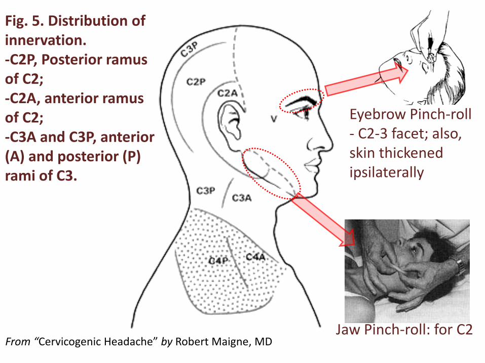