
Some pathological, biochemical and hematological ... tilapia (Oreochromis niloticus) following...
10
Journal of American Science 2010;6(10) http://www.americanscience.org [email protected] 542 Some pathological, biochemical and hematological investigations on Nile tilapia (Oreochromis niloticus) following chronic exposure to edifenphos pesticide. A.Y. Gaafar 1* , E.M. El-Manakhly 2 , M.K. Soliman 3 , H. Soufy 1 , Mona S. Zaki 1 , Safinaz G. Mohamed 4 and Shahenaz M. Hassan 5 . 1 Veterinary Research Division, National Research Centre, Cairo, Egypt. 2 Faculty of Veterinary Medicine - Alexandria University, Edfina, Egypt. 3 Faculty of Veterinary Medicine - Behira University, Bostan City, Egypt. 4 National Institute of Oceanography and Fisheries, Alexandria, Egypt. 5 Animal Health Research Institute, Alexandria branch, Egypt. * [email protected] Abstract: Nile tilapia is the main cultured species in Egypt; the Egyptian fish farms are irrigated with agricultural drainage which contains pesticides residues or their metabolites which may affect fish. This study concerned with the pathologic and clinicopathologic findings due to chronic exposure to the organophosphate fungicide edifenphos on Nile tilapia Oreochromis niloticus. Eight weeks exposure to 1/10 96 hours LC 50 (0.1 ppm) led to adverse effect on some serum parameters including AST, ALT, ALP, cholinesterase activity, total protein, blood urea nitrogen and creatinine. Also some of hematological parameters such as RBCs count, Hb content and blood indices were affected negatively. Histopathological investigations revealed various degrees of pathological lesions in different organs like gills, hepatopancreas, spleen, kidney, brain, and others. From this study it was obvious that edifenphos caused harmful effects on Nile tilapia fish. [Journal of American Science 2010;6(10):542-551]. (ISSN: 1545-1003). Keywords: Edifenphos, Hinosan, Oreochromis niloticus, tilapia, organophosphate, histopathology. 1. Introduction Chemical pesticides are well recognized as an economic approach to control pests, at the same time such chemicals are highly toxic to other species in the environment. Now there is growing concern worldwide over the indiscriminate use of such chemicals, which result in environmental pollution and toxicity risk to nontarget organisms. Most of pesticides find their way into rivers, lakes and pond, and have been found to be highly toxic not only to fishes but also to the organisms which contribute to the food chain of fishes (Anees, 1975). The organophosphate fungicide edifenphos (Hinosan) is cutinase inhibitors and displays a specific antipenetrant action, but in practice its therapeutic activity may also involve direct fungitoxicity (Sisler, 1986). As organophosphate pesticide, it causes irreversible inhibition of the cholinesterase enzymes (Haddad and Winchester, 1983). A lot of fish toxicity studies; including histopathological and clinicopathological studies; were conducted upon organophosphate pesticides. For instance (Jauch 1980) confirmed gill pathology in Herotilapia multispinosa and Tilapia leucostica ranging from hyperplasia and separation of the respiratory epithelium in the secondary lamellae with congestion and numerous telangiectasis, while studying the toxic effects of Lebaycid R on fish gills. Also (Joshi and Desai 1981) assessed that exposure of O. mossambica to monocrotophos increased activity of acid and alkaline phosphatase and correlatedthis result with necrosis of hepatic and renal tissues. Meanwhile (Prasada and Ramana 1984) stated the increase of activity levels of (AST), (ALT) and (ATPase) in muscle, gill, hepatopancreas and brain tissues of methyl parathion exposed fish. While (El-Zahaby 1986) stated that acid and alkaline phosphatase activities showed a slight elevation in mucosal intestinal epithelial cells of cyolane-injected Clarias Lazera. (El-Sheikh et al. 1990) assessed the biochemical changes of (AST) and (ALT) and acid and alkaline phosphatase in Oreochromis niloticus fingerlings exposed to a nonlethal concentration of Ofunac and Sumithion for a period of 28 days. Also (El-Aulaimi et al. 1994) recorded abnormalities in some hematological parameters; (RBCs) count, (Hb) content, (Ht) value and (MCV) in Sarotherodon
-
Upload
truongduong -
Category
Documents
-
view
219 -
download
1
Transcript of Some pathological, biochemical and hematological ... tilapia (Oreochromis niloticus) following...
Journal of American Science 2010;6(10)
http://www.americanscience.org [email protected] 542
Some pathological, biochemical and hematological investigations on Nile tilapia (Oreochromis niloticus) following chronic exposure to
edifenphos pesticide.
A.Y. Gaafar1*, E.M. El-Manakhly2, M.K. Soliman3, H. Soufy1, Mona S. Zaki1, Safinaz G. Mohamed4 and Shahenaz M. Hassan5.
1Veterinary Research Division, National Research Centre, Cairo, Egypt. 2Faculty of Veterinary Medicine - Alexandria University, Edfina, Egypt.
3Faculty of Veterinary Medicine - Behira University, Bostan City, Egypt. 4National Institute of Oceanography and Fisheries, Alexandria, Egypt.
5Animal Health Research Institute, Alexandria branch, Egypt. *[email protected]
Abstract: Nile tilapia is the main cultured species in Egypt; the Egyptian fish farms are irrigated with agricultural drainage which contains pesticides residues or their metabolites which may affect fish. This study concerned with the pathologic and clinicopathologic findings due to chronic exposure to the organophosphate fungicide edifenphos on Nile tilapia Oreochromis niloticus. Eight weeks exposure to 1/10 96 hours LC50 (0.1 ppm) led to adverse effect on some serum parameters including AST, ALT, ALP, cholinesterase activity, total protein, blood urea nitrogen and creatinine. Also some of hematological parameters such as RBCs count, Hb content and blood indices were affected negatively. Histopathological investigations revealed various degrees of pathological lesions in different organs like gills, hepatopancreas, spleen, kidney, brain, and others. From this study it was obvious that edifenphos caused harmful effects on Nile tilapia fish. [Journal of American Science 2010;6(10):542-551]. (ISSN: 1545-1003). Keywords: Edifenphos, Hinosan, Oreochromis niloticus, tilapia, organophosphate, histopathology. 1. Introduction
Chemical pesticides are well recognized as an economic approach to control pests, at the same time such chemicals are highly toxic to other species in the environment. Now there is growing concern worldwide over the indiscriminate use of such chemicals, which result in environmental pollution and toxicity risk to nontarget organisms.
Most of pesticides find their way into rivers, lakes and pond, and have been found to be highly toxic not only to fishes but also to the organisms which contribute to the food chain of fishes (Anees, 1975).
The organophosphate fungicide edifenphos (Hinosan) is cutinase inhibitors and displays a specific antipenetrant action, but in practice its therapeutic activity may also involve direct fungitoxicity (Sisler, 1986). As organophosphate pesticide, it causes irreversible inhibition of the cholinesterase enzymes (Haddad and Winchester, 1983). A lot of fish toxicity studies; including histopathological and clinicopathological studies; were conducted upon organophosphate pesticides.
For instance (Jauch 1980) confirmed gill pathology in Herotilapia multispinosa and Tilapia leucostica ranging from hyperplasia and separation of the respiratory epithelium in the secondary lamellae with congestion and numerous telangiectasis, while studying the toxic effects of Lebaycid R on fish gills. Also (Joshi and Desai 1981) assessed that exposure of O. mossambica to monocrotophos increased activity of acid and alkaline phosphatase and correlatedthis result with necrosis of hepatic and renal tissues. Meanwhile (Prasada and Ramana 1984) stated the increase of activity levels of (AST), (ALT) and (ATPase) in muscle, gill, hepatopancreas and brain tissues of methyl parathion exposed fish. While (El-Zahaby 1986) stated that acid and alkaline phosphatase activities showed a slight elevation in mucosal intestinal epithelial cells of cyolane-injected Clarias Lazera. (El-Sheikh et al. 1990) assessed the biochemical changes of (AST) and (ALT) and acid and alkaline phosphatase in Oreochromis niloticus fingerlings exposed to a nonlethal concentration of Ofunac and Sumithion for a period of 28 days. Also (El-Aulaimi et al. 1994) recorded abnormalities in some hematological parameters; (RBCs) count, (Hb) content, (Ht) value and (MCV) in Sarotherodon
Journal of American Science 2010;6(10)
http://www.americanscience.org [email protected] 543
galilaeus exposed to dimethoate. The data obtained here revealed marked reduction in (RBCs) count, (Hb) contents and (Ht) values in fish exposed to dimethoate. While (MCV) displayed significant increase in its values. Also (Sherif and Eisa 1994) exposed Tilapia zillii to sublethal concentrations of chlorpyrifos and recorded histopathological changes in the gills such as, dilation of the blood vessels, tilangectasis in the secondary lamellae with slight proliferative changes of the epithelial covering, and hepatopancreas showed vacuolar degeneration and necrosis of hepatocytes together with activation of pancreatic acini.
While somewhat few studies are conducted using edifenphos, (El-Gendy et al. 1996) stated the depressive effect of edifenphos on the activities of acetylcholinesterase (AChE), adenosine triphosphatase (ATPase) and glutathion-S-transferase (GST) with the elevation of catalase activity in tissues of O. niloticus. Also (Aly 1996) found the same results in addition to decrease of serum protein and reduced response of splenocytes to mitogens. In the same year (Ramadhan 1996) investigated the probable genotoxic effects of edifenphos on common carp fish and found a significant changes in the relative proportions of protein fractions in the electrophoretic patterns of hepatopancreas and brain esterase isozyme of the fish.
On the same way (Rezq-Allah et al. 1997) revealed the same results with edifenphos on the electropherograms of sarcoplasmic proteins of common carp (Cyprinus carpio). Again (El-Gendy et al. 1998) investigated the effects of 1/1000 field recommended concentration of edifenphos on the immune response and protein contents of O. niloticus and found that The cell mediated immune response assessed by proliferative response of splenocytes to mitogens; phytohemagglutinin (PHA) and concanavalin A (Con A) for T cell and lipopolysaccharide (LPS) for B cell decreased significantly in terms of the level of stimulation index in the treated fish and reached maximal depression after 4 weeks.
Humoral immunity assessed as splenic antibody plaque forming cells (PFC) measured after 5 days in vitro immunization to sheep erythrocytes (SRBC's) were suppressed in a concentration dependent pattern of the used pesticide. 2. Material and Methods
Edifenphos is presented as 50% solution (Hinosan EC 50® Bayer). The acute 96 hours LC50 value of edifenphos in Nile tilapia was 1 ppm (Gaafar 2005).
The levels of serum acetyl cholinesterase activity, alkaline phosphatase, transaminases, total protein, total serum bilirubin, blood urea nitrogen and serum creatinine were estimated by using kits obtained from BioMérieux – France.
A total No. of 150 fish (80±20 g. B.W); were divided into 2 groups. The 1st group was 100 fish exposed to 1/10 LC50 of edifenphos (0.1 mg/ℓ) were divided into four (50 liters). The 2nd (control group) was 50 fish placed in two (50 liters) aquaria. The experiment lasted for 8 weeks, collection of blood samples and scarification of fish were done on weekly interval.
Blood samples were collected from the caudal vein of each fish, 1 ml of each blood sample was mixed wis anticoagulant for estimation of the blood parameters: Total erythrocytic count (Kanaev, 1985), Hemoglobin content, Hematocrit value (PCV) (Tietz, 1976), Mean Corpuscular Volume (MCV), Mean Corpuscular Hemoglobin (MCH), Mean Corpuscular Hemoglobin Concentration (MCHC) (Hrubec et al., 2000). Also another one ml of blood was left to coagulate, centrifuged and serum was collected to evaluate the following biochemical parameters: Serum cholinesterase activity, alkaline phosphatase, transaminases: aspartate aminotransferase (AST) and alanine aminotransferase (ALT), total serum protein, total serum bilirubin, blood urea Nitrogen and creatinine, according to the methods mentioned by the Kits manufacturer.
One ml citrated blood was collected to perform the following haematological parameters: Following necropsy, the specimens were collected from sacrificed fish. Tissue specimen from the skin with the underlying musculature, gills, liver, spleen, intestine, brain, heart and kidneys were taken weekly from 5 fish of treated and 3 of control group.
Tissue specimens were rapidly fixed in 10% neutral buffered formalin. The fixed specimens were processed through the conventional paraffin embedding technique. Paraffin blocks were prepared, from which 5 microns thick sections were obtained. These sections were stained by Hematoxyline and Eosin (H&E) according to the method described by Culling (1983).
Statistical analysis
Statistical analysis of the obtained data was by using the SPSS 11 computer program (SPSS Inc. Chicago, Illinois, USA). Using analysis of variance (One-way ANOVA). 3. Results and Discussion: Clinical signs:
Respiratory distress after 3 days, nervous
Journal of American Science 2010;6(10)
http://www.americanscience.org [email protected] 544
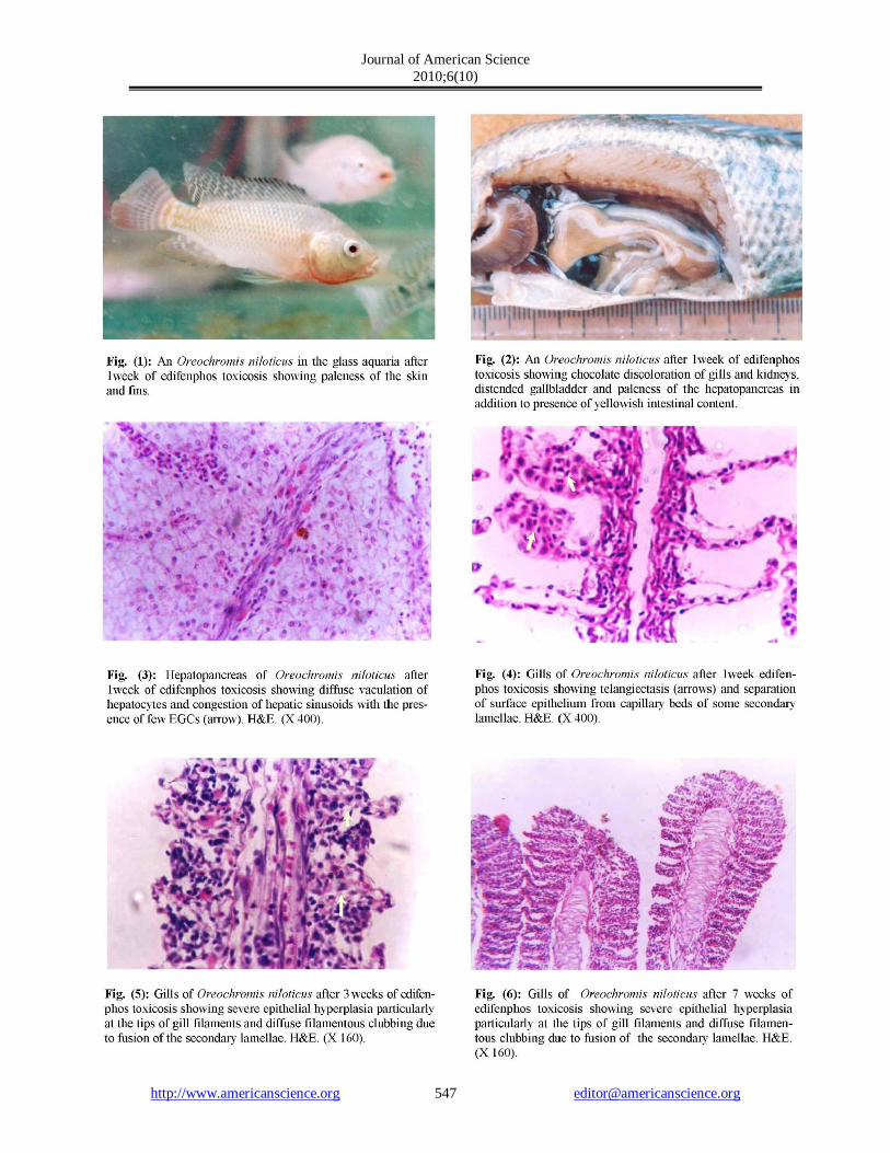
manifestations, including hyperexcitability by erratic movements appeared after 6 days. Paleness of the gills appeared after 1 week. After 12 days there was paleness of whole body surface (Fig.1) with slimness. Severe convulsive reflexes upon stimulation were abundant after 3 weeks. After 5 weeks the fish shows lethargy and slowing of all movements.
Biochemical parameters:
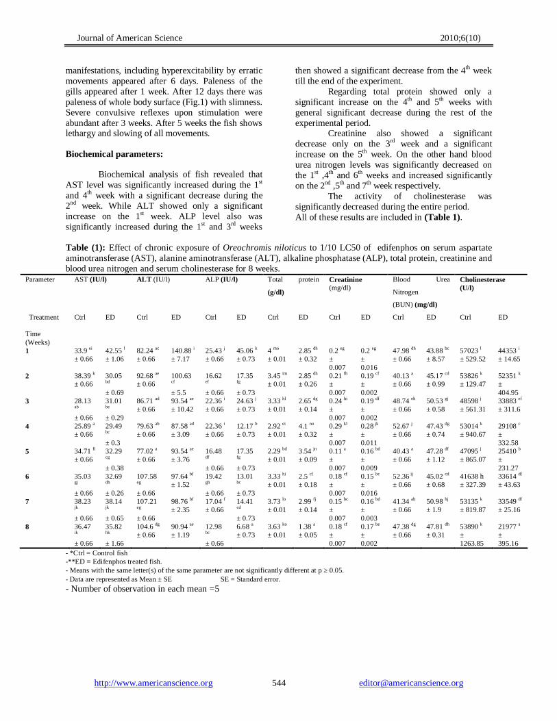
Biochemical analysis of fish revealed that AST level was significantly increased during the 1st and 4th week with a significant decrease during the 2nd week. While ALT showed only a significant increase on the 1st week. ALP level also was significantly increased during the 1st and 3rd weeks
then showed a significant decrease from the 4th week till the end of the experiment.
Regarding total protein showed only a significant increase on the 4th and 5th weeks with general significant decrease during the rest of the experimental period.
Creatinine also showed a significant decrease only on the 3rd week and a significant increase on the 5th week. On the other hand blood urea nitrogen levels was significantly decreased on the 1st ,4th and 6th weeks and increased significantly on the 2nd ,5th and 7th week respectively.
The activity of cholinesterase was significantly decreased during the entire period. All of these results are included in (Table 1).
Table (1): Effect of chronic exposure of Oreochromis niloticus to 1/10 LC50 of edifenphos on serum aspartate aminotransferase (AST), alanine aminotransferase (ALT), alkaline phosphatase (ALP), total protein, creatinine and blood urea nitrogen and serum cholinesterase for 8 weeks.
Parameter AST (IU/l) ALT (IU/l) ALP (IU/l) Total protein
(g/dl)
Creatinine (mg/dl)
Blood Urea
Nitrogen
(BUN) (mg/dl)
Cholinesterase (U/l)
Treatment Time (Weeks)
Ctrl ED Ctrl ED Ctrl ED Ctrl ED Ctrl ED Ctrl ED Ctrl ED
1 33.9 ei ± 0.66
42.55 l ± 1.06
82.24 ac ± 0.66
140.88 i ± 7.17
25.43 j ± 0.66
45.06 k ± 0.73
4 mo ± 0.01
2.85 dh ± 0.32
0.2 eg ± 0.007
0.2 eg ± 0.016
47.98 dh ± 0.66
43.88 bc ± 8.57
57023 l ± 529.52
44353 i ± 14.65
2 38.39 k ± 0.66
30.05 bd ± 0.69
92.68 ae ± 0.66
100.63 cf ± 5.5
16.62 ef ± 0.66
17.35 fg ± 0.73
3.45 im ± 0.01
2.85 dh ± 0.26
0.21 fh ± 0.007
0.19 cf ± 0.002
40.13 a ± 0.66
45.17 cd ± 0.99
53826 k ± 129.47
52351 k ± 404.95
3 28.13 ab ± 0.66
31.01
be ± 0.29
86.71 ad ± 0.66
93.54 ae ± 10.42
22.36 i ± 0.66
24.63 j ± 0.73
3.33 hl ± 0.01
2.65 dg ± 0.14
0.24 hi ± 0.007
0.19 df ± 0.002
48.74 eh ± 0.66
50.53 gj ± 0.58
48598 j ± 561.31
33883 ef ± 311.6
4 25.89 a ± 0.66
29.49 bc ± 0.3
79.63 ab ± 0.66
87.58 ad ± 3.09
22.36 i ± 0.66
12.17 b ± 0.73
2.92 ei ± 0.01
4.1 no ± 0.32
0.29 kl ± 0.007
0.28 jk ± 0.011
52.67 j ± 0.66
47.43 dg ± 0.74
53014 k ± 940.67
29108 c ± 332.58
5 34.71 fi ± 0.66
32.29 cg ± 0.38
77.02 a ± 0.66
93.54 ae ± 3.76
16.48 df ± 0.66
17.35
fg ± 0.73
2.29 bd ± 0.01
3.54 jn ± 0.09
0.11 a ± 0.007
0.16 bd ± 0.009
40.43 a ± 0.66
47.28 df ± 1.12
47095 j ± 865.07
25410 b ± 231.27
6 35.03 gj ± 0.66
32.69 dh ± 0.26
107.58 eg ± 0.66
97.64 bf ± 1.52
19.42 gh ± 0.66
13.01 bc ± 0.73
3.33 hi ± 0.01
2.5 cf ± 0.18
0.18 cf ± 0.007
0.15 bc ± 0.016
52.36 ij ± 0.66
45.02 cd ± 0.68
41638 h ± 327.39
33614 df ± 43.63
7 38.23 jk ± 0.66
38.14 jk ± 0.65
107.21 eg ± 0.66
98.76 bf ± 2.35
17.04 f ± 0.66
14.41 cd ± 0.73
3.73 lo ± 0.01
2.99 fj ± 0.14
0.15 bc ± 0.007
0.16 bd ± 0.003
41.34 ab ± 0.66
50.98 hj ± 1.9
53135 k ± 819.87
33549 df ± 25.16
8 36.47 ik ± 0.66
35.82 hk ± 1.66
104.6 dg ± 0.66
90.94 ae ± 1.19
12.98 bc ± 0.66
6.68 a ± 0.73
3.63 ko ± 0.01
1.38 a ± 0.05
0.18 cf ± 0.007
0.17 be ± 0.002
47.38 dg ± 0.66
47.81 dh ± 0.31
53890 k ± 1263.85
21977 a ± 395.16
- *Ctrl = Control fish -**ED = Edifenphos treated fish. - Means with the same letter(s) of the same parameter are not significantly different at p ≥ 0.05. - Data are represented as Mean ± SE SE = Standard error.
- Number of observation in each mean =5
Journal of American Science 2010;6(10)
http://www.americanscience.org [email protected] 545
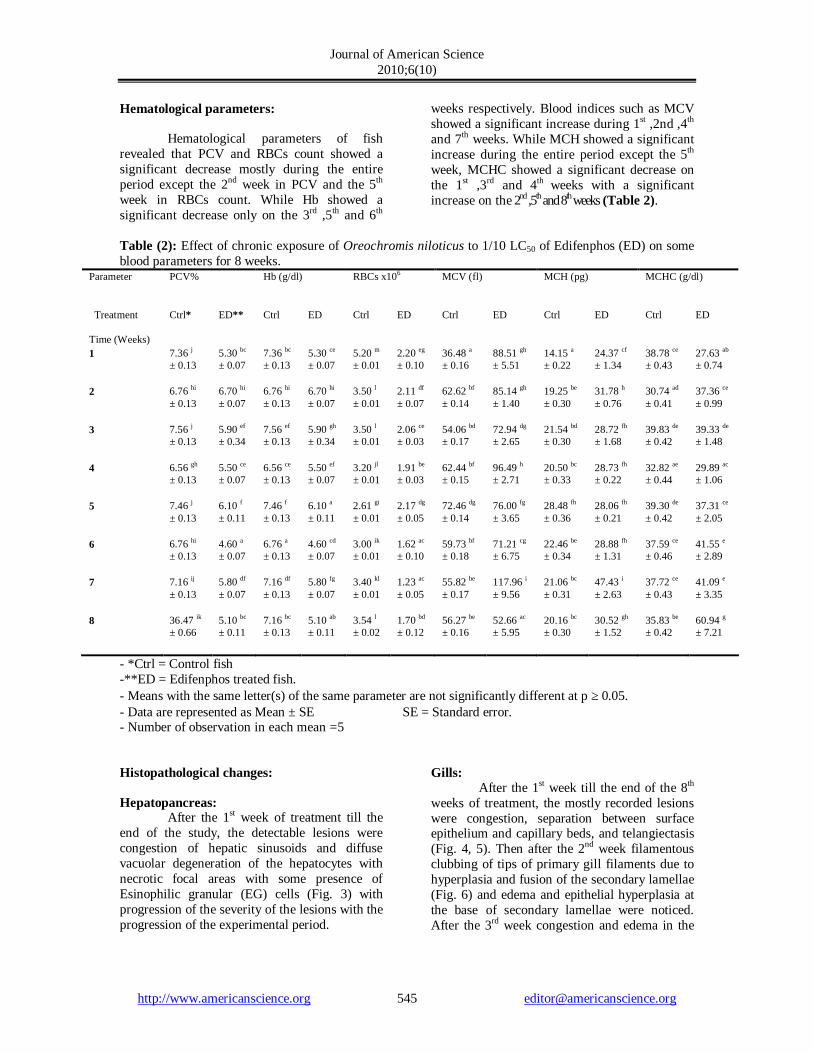
Hematological parameters:
Hematological parameters of fish revealed that PCV and RBCs count showed a significant decrease mostly during the entire period except the 2nd week in PCV and the 5th week in RBCs count. While Hb showed a significant decrease only on the 3rd ,5th and 6th
weeks respectively. Blood indices such as MCV showed a significant increase during 1st ,2nd ,4th and 7th weeks. While MCH showed a significant increase during the entire period except the 5th week, MCHC showed a significant decrease on the 1st ,3rd and 4th weeks with a significant increase on the 2nd ,5th and 8th weeks (Table 2).
Table (2): Effect of chronic exposure of Oreochromis niloticus to 1/10 LC50 of Edifenphos (ED) on some blood parameters for 8 weeks.
Parameter PCV% Hb (g/dl) RBCs x106 MCV (fl) MCH (pg) MCHC (g/dl)
Treatment Time (Weeks)
Ctrl* ED** Ctrl ED Ctrl ED Ctrl ED Ctrl ED Ctrl ED
1 7.36 j ± 0.13
5.30 bc ± 0.07
7.36 bc ± 0.13
5.30 ce ± 0.07
5.20 m ± 0.01
2.20 eg ± 0.10
36.48 a ± 0.16
88.51 gh ± 5.51
14.15 a ± 0.22
24.37 cf ± 1.34
38.78 ce ± 0.43
27.63 ab ± 0.74
2 6.76 hi ± 0.13
6.70 hi ± 0.07
6.76 hi ± 0.13
6.70 hi ± 0.07
3.50 l ± 0.01
2.11 df ± 0.07
62.62 bf ± 0.14
85.14 gh ± 1.40
19.25 be ± 0.30
31.78 h ± 0.76
30.74 ad ± 0.41
37.36 ce ± 0.99
3 7.56 j ± 0.13
5.90 ef ± 0.34
7.56 ef ± 0.13
5.90 gh ± 0.34
3.50 l ± 0.01
2.06 ce ± 0.03
54.06 bd ± 0.17
72.94 dg ± 2.65
21.54 bd ± 0.30
28.72 fh ± 1.68
39.83 de ± 0.42
39.33 de ± 1.48
4 6.56 gh ± 0.13
5.50 ce ± 0.07
6.56 ce ± 0.13
5.50 ef ± 0.07
3.20 jl ± 0.01
1.91 be ± 0.03
62.44 bf ± 0.15
96.49 h ± 2.71
20.50 bc ± 0.33
28.73 fh ± 0.22
32.82 ae ± 0.44
29.89 ac ± 1.06
5 7.46 j ± 0.13
6.10 f ± 0.11
7.46 f ± 0.13
6.10 a ± 0.11
2.61 gi ± 0.01
2.17 dg ± 0.05
72.46 dg ± 0.14
76.00 fg ± 3.65
28.48 fh ± 0.36
28.06 fh ± 0.21
39.30 de ± 0.42
37.31 ce ± 2.05
6 6.76 hi ± 0.13
4.60 a ± 0.07
6.76 a ± 0.13
4.60 cd ± 0.07
3.00 ik ± 0.01
1.62 ac ± 0.10
59.73 bf ± 0.18
71.21 cg ± 6.75
22.46 be ± 0.34
28.88 fh ± 1.31
37.59 ce ± 0.46
41.55 e ± 2.89
7 7.16 ij ± 0.13
5.80 df ± 0.07
7.16 df ± 0.13
5.80 fg ± 0.07
3.40 kl ± 0.01
1.23 ac ± 0.05
55.82 be ± 0.17
117.96 i ± 9.56
21.06 bc ± 0.31
47.43 i ± 2.63
37.72 ce ± 0.43
41.09 e ± 3.35
8 36.47 ik ± 0.66
5.10 bc ± 0.11
7.16 bc ± 0.13
5.10 ab ± 0.11
3.54 l ± 0.02
1.70 bd ± 0.12
56.27 be ± 0.16
52.66 ac ± 5.95
20.16 bc ± 0.30
30.52 gh ± 1.52
35.83 be ± 0.42
60.94 g ± 7.21
- *Ctrl = Control fish -**ED = Edifenphos treated fish. - Means with the same letter(s) of the same parameter are not significantly different at p ≥ 0.05. - Data are represented as Mean ± SE SE = Standard error. - Number of observation in each mean =5
Histopathological changes:
Hepatopancreas: After the 1st week of treatment till the
end of the study, the detectable lesions were congestion of hepatic sinusoids and diffuse vacuolar degeneration of the hepatocytes with necrotic focal areas with some presence of Esinophilic granular (EG) cells (Fig. 3) with progression of the severity of the lesions with the progression of the experimental period.
Gills: After the 1st week till the end of the 8th
weeks of treatment, the mostly recorded lesions were congestion, separation between surface epithelium and capillary beds, and telangiectasis (Fig. 4, 5). Then after the 2nd week filamentous clubbing of tips of primary gill filaments due to hyperplasia and fusion of the secondary lamellae (Fig. 6) and edema and epithelial hyperplasia at the base of secondary lamellae were noticed. After the 3rd week congestion and edema in the
Journal of American Science 2010;6(10)
http://www.americanscience.org [email protected] 546
gill arch were observed. The before-mentioned lesions continued till the end of the experiment. Kidneys:
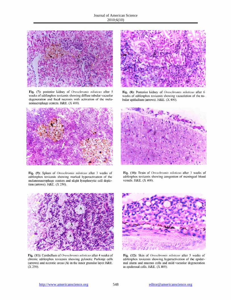
Alterations exhibited in the posterior kidney of Oreochromis niloticus during the 8 weeks of the experiment and directly after the 1st week were congestion, diffuse cloudy swelling and hyaline droplet degeneration of renal tubules with depletion of interstitial hemopoietic tissue and activation of melanomacrophage centers (Fig. 7,8). Moreover, the anterior kidney revealed severe activation of melanomacrophage centers with necrosis and depletion of the haemopoietic tissues. Spleen:
The histopathologic examination from the beginning of the 1st till the end of the 8th week revealed activation of melanomacrophage centers with diffuse reduction of the spleenic haemopoietic tissues. From the 2nd week hyperplasia of spleenic ellipsoid and multifocal necrotic areas surrounded by the activated melanomacrophage centers were common (Fig. 9). Brain:
The brain revealed that there was severe congestion of cerebral blood vessels after the 2nd week till the end of the experimental period, with concurrent congestion of meningeal blood vessels (Fig. 10) with neuronal degeneration after the 3rd week there were pyknotic Purkinje cells and necrotic areas in the inner granular layer of the cerebellum (Fig. 11). Skin:
After the 2nd week it showed mild vacuolar degeneration of epidermal cells, proliferation of club cell and hyperactivation of the melanophores (Fig. 12) from then till the end of experimental period. Intestine:
After the 1st week of treatment, the histopathologic examination revealed that there was esinophilic granular (EG) cell infiltration in the submucosa then after the 2nd week epithelial degeneration and submucosal edema were observed till the end of the 8 weeks.
In this study, the fishes were exposed to edifenphos concentration equal to 1/10 96 hours LC50 (0.1 ppm), the clinical signs and post mortem changes of edifenphos were in the form of nervous manifestations. Respiratory distress reflected by congestion then paleness of the gills, chocolate discolouration of most internal organs and severe distention of gall bladder, cachexia
with prominent paleness of whole body surface with slimness. These results supports that edifenphos has the same nervous toxic effect of its chemical group the organophosphate as described by Jauch (1980) and Joshi and Desai (1981) with unique anemic action leading to cachexia with prominent paleness of whole body surface as observed by El-Aulaimi et al. (1994) while investigating dimethoate.
The results of serum biochemistry revealed a significant increase in serum ALT and AST and ALP at the beginning of the experiment, these findings supported the hypothesis that the increased serum transaminases (ALT and AST) may reflect hepatic toxicity which leads to extensive liberation of the enzymes into the blood circulation (Daabees et al., 1992). Then after that, the enzymes suffered from a significant decrease in their levels. ALP level also showed significant increase then significant decrease towards the end of the experiment. The decrease in activity of AST, ALT and ALP in fish exposed to pesticides was also reported by different authors (El-Boushy, 1994 and Begum, 2004). Agius and Coushman (1986) linked the increased activity of ALP in fish to the increased catabolic tissue breakdown in melanomacrophage centers.
Saeed (1983) attributed the decrease in liver transaminases activity to the decrease in protein content in serum and tissues due to the resultant hepatic necrosis of fish exposed to pesticides. Also the severe hepatic necrosis leads to lack of cells from which the enzymes are produced. These findings indicated that decrease in liver function was occurred which may lead to major dangerous sequelae in body metabolism.
Total serum proteins showed general significant decrease after exposure to edifenphos. This may be due to liver damage where most of plasma protein synthesis usually occurs in the liver, this result agreed with that of Singh et al. (1998).
Creatinine also showed some sort of significant increase after the 5th week. Also blood urea nitrogen levels generally showed significant increase. These results supported that edifenphos exerts harmfull effects on kidney tissue.
Acetyle cholinesterase (ACHase) Showed total decrease in activity, this agreed with many authors (Gosselin, 1984). This may
Journal of American Science 2010;6(10)
http://www.americanscience.org [email protected] 549
be attributed to that edifenphos is a potent anticholinesterase which results in accumulation of acetylecholine at the synapsis of neurons leading to nervous manifestations. Decreased ACHase activity may be used as boindicator of pollution by such pesticides in the environment.
The hematological changes during the chronic toxicosis of edifenphos showed a significant decrease mostly during the entire period in Hb, RBCs count and PCV, this reveals the prominent anemic effect of edifenphos which is confirmed by the results of the blood indices. Which in turns revealed the hemolytic effect of edifenphos, and may explain the chocolate discoloration of parynchymatus organs, as hemoglobin may be converted into methemoglobin with resultant hemolysis and reduced blood oxygen carrying capacity which accumulates with the irritant effect of edifenphos causing respiratory distress to the fish? The severity of anemia also is magnified by the hypoproteinemic effect showed by edifenphos. The haemolytic and destructive effects of the pesticides on blood cells was supported by El-Boushy (1994) and Robert (2001).
The marked decrease in RBCs count was in agreement with those reported by other workers, Rani et al. (1987) and Venkateshwarthlu et al. (1990) proved these changes in blood parameters in catfish intoxicated with the organophosphate pesticide dichlorvos.
The histopathological changes during the chronic toxicosis of edifenphos were various. The hepatic tissue showed congestion with various degrees of degenerative changes starting firstly with granular degeneration then vacuolar degeneration with progression towards hepatic cell necrosis after 1 week of exposure. These changes may be attributed to direct toxic effect of edifenphos on hepatocytes since the hepatopancreas is the site of detoxification of all types of toxins and chemicals (Robert, 2001).
Congestion and various degrees of pathological harm in gills were evident. The firstly observed lesion was lamellar edema which is frequent following exposure to chemical pollutants. Complete edematous separation of the respiratory epithelium of primary and secondary lamellae with necrosis of lamellar epithelial cells and severe, often lethal, r espiratory and osmoregulatory distr ess may supervene (Yang and Albright, 1992).
Also severe epithelial proliferation of secondary gill lamellae, which resulted as a response of the malpighian cells to chemical irritation, as they migrate distally, often in the early stages, resulting in an accumulation of cells at the leading edge of the secondary lamella, progression of this migration leads to lamellar fusion and terminal lamellar clubbing (Robert, 2001). This may be attributed to that edifenphos has a direct effect on gill filaments as cytotoxic and irritating substance which resulted in proliferation and fusion of secondary lamellae. Moreover, gills are important not only for gaseous exchange but also for osmoregulation and excretion of toxic waste products (Robert, 2001), thus any harm in the gills leads to impairment of such vital functions revealing respiratory distress, impaired osmoregulation and retention of toxic wastes . Hyperplasia may in some situations represent an adaptation by the organism to protect underlying tissues from any irritant. However, increased thickness of the epithelial layers including mucous cell hyperplasia and fusion of adjacent secondary lamellae as the result of hyperplasia will not only decrease the surface area available for oxygen extraction but also will increase the oxygen diffusion distance between water and blood (Kumaraguru et al., 1982). Also exposure to pollutants, including pesticides can cause rupture of the retaining pillar, or pilaster cells, which normally join the dorsal surface of secondary lamellae to the ventral one. The result will be dilation of the lamellar capillary and pooling of the blood, thrombosis and eventually fibrosis. Fusion with adjacent lamella, leads to the telangiectasis which is a characteristic pathological change of the gill associated with physical or chemical trauma (Robert, 2001).
The skin of the fish showed varied degrees of vacuolar degeneration of epidermal cells, proliferation of club cell and hyperactivation of the melanophores this may be attributed to that being in contact with edifenphos causes direct cytotoxic and irritating effect on dermal cells
The renal tissue of posterior kidney exhibited congestion, diffuse granular and vacuolar degenerative changes and focal hyaline droplet degeneration after 1 week of exposure and the marked depletion in haemopoietic elements which was evident in spleen and anterior and posterior kidney were probably caused by direct cytotoxic effect of edifenphos.
Journal of American Science 2010;6(10)
http://www.americanscience.org [email protected] 550
As neurotoxin, edifenphos caused degenerative effect on the brain tissue as revealed in this study appeared as severe congestion of cerebral blood vessels with neuronal degeneration led to necrotic areas in the brain tissue.
The activation of melanomacrophage centers either in spleen, hepatopancreas, or anterior and posterior kidney was a prominent and constant lesion. It is quite known as an unusual sequel to infection or irritation in fish belonging to fish immune response (Robert, 2001).
From the results of the present work, it can be concluded that: Edifenphos should be listed under the highly toxic pollutants to Oreochromis niloticus fish even at sublethal dose (1 ppm) where it may cause toxicity or death, not only to rice fungi but also to fish. Prolonged exposure of Oreochromis niloticus to low doses of edifenphos pesticide (0.1 ppm) caused damage in kidney, liver, spleen and gills tissues. Corresponding Authors A.Y. Gaafar Veterinary Research Division, National Research Centre, Cairo, Egypt. [email protected] 4. References 1. Agius, C., and Coushman, W., (1986):
Induction of enhanced alkaline phosphatase activity in the melano-macrophage centers of Oreochromis aureus (Steindachner) through starvation and vaccination. Journal of Fish Biology, 28:87-92.
2. Aly, N. M. (1996): Interaction of pesticide with biological processes in aquatic organisms. Ph.D. Thesis in Chemistry of Pesticides, Faculty of Agriculture Alexandria University.
3. Anees, M.A. (1975): Acute toxicity of four organophosphorus insecticides for a fresh-water teleost Channa punctatus (Bloch). Pakistan Journal of Zoology, 7(2): 135-141.
4. Begum, G. (2004): Carbofuran insecticide induced biochemical alterations in liver and muscle tissues of the fish Clarias batrachus(linn) and recovery response, Aquatic Toxicology. 66:83–92.
5. Culling, C.F. (1983): Handbook of Histopathological and Histochemical Staining Techniques. 3rd Ed.; Butterworth - London.
6. Daabees, A. Y.; El- Damiaty, N.A.; Soliman, S.A. and El-Toweissy, M.Y. (1992): Comparative action of three synthetic pesticides on serum, liver and brain of the
freshwater fish Clarias lazera. Journal of Egyptian German Society for Zoology. 9(A). Comparative Physiology, 105-119.
7. El-Aulaimi, I.A. et al., (1994): Hematological abnormalities induced by dimethoate exposure to freshwater fish (Sarotherodon galilaeus). Journal of the Egyptian German Society of Zoology: Comparative Physiology. 14(A): 407-419.
8. El-Boushy, M.A. (1994): The effect of molluscicides polluted on the blood picture and serum biochemical parameters in Clarias lazera. M.V.Sc. Thesis, Faculty of Veterinary Medecine, Suez-Canal University.
9. El-Gendy, K.S.; Aly, N.M. and El-Sebae, A.H. (1998): Effects of edifenphos and glyphosate on the immune response and protein biosynthesis of bolti fish (Tilapia nilotica). Journal of Environmental Science and Health, Part. B: Pesticide Food Contamination and Agricultural Wastes. 33B (2)135-149.
10. El-Gendy, K.S.; Aly, N.M.; Saber, N. and El-Sebae, A.A. (1996): Toxicological effects of some pesticides on Tilapia nilotica Alexandria Science Exchanges. 17(3):243-251.
11. El-Sheikh, et al., (1990): Biochemical responses in Oreochromis niloticus L. fingerlings exposed to non-lethal concentration of certain organophosphorus insecticides. Annals of Agricultural Science. Special Issue. 565-573.
12. El-Zahaby, E., (1986): Experimental studies on the effect of organophosphorus insecticides on the enzymatic activities of acid and alkaline phosphatases in fish ileal mucosal cells. The Egyptian Journal of Histology. 9 (l):107-114.
13. Eurell, T.E.; Lewis, S.D.H. and Grumbles, L.C. (1978): Comparison of selected diagnostic tests for detection of motile Aeromonas septicemia in fish. American Journal of Volume Research 39(8):1384-1386.
14. Gaafar, A.Y. (2005): Pathologic and clinicopathologic effect of carbofuran and edifenphos on Nile tilapia and monosex tilapia. M.V.Sc. Thesis, Faculty of Veterinary Medecine, Alexandria University.
15. Gosselin, R.E.; Smith R.P. and Hodge, H.C. (1984): Clinical Toxicology of Commercial Products. 5th ed. Baltimore, London: Williams and Wilkins. II-305.
16. Haddad, L.M. and Winchester, J.F. (1983): Clinical Management of Poisoning and Drug Overdosage. Philadelphia, PA: W.B. Saunders Co. 711
17. Hawk, P.B.; Oscar, B.L. and Summerson, W. (1965): Hawk's physiological chemistry. 14th Ed. London Journal; and A.Churchill .L.T.D. 401
18. Hrubec, T.C.; Cardinale, J.L.; Smith, S.A. (2000): Hematology and Plasma Chemistry Reference Intervals for Cultured Tilapia (Oreochromis Hybrid) Veterinary Clinical Pathology, 29.(1):7-12.
Journal of American Science 2010;6(10)
http://www.americanscience.org [email protected] 551
19. Jauch, D. (1980): Invsetigations on toxic effects of the organophosphate Lebaycid R on respiration and circulation of cichlid fishes Herotilapia multispinosa (Guenther 1898) and Tilapia leucostica (Trewavas 1933). Part 1 and Part 2. Tuebingen Frg Fakultaet Fuer Biologie. 46, 49.
20. Joshi, U.M. and Desai, A.K. (1981): Effect of Sublethal Concentration of Monocrotophos on Acid and Alkaline Phosphatase Activities in the Tissue of Fresh Water Fish Tilapia mossambica .Journal of Animal Morphology and Physiology. 28(1-2):221-228.
21. Kanaev, A.E. (1985): Veterinary Hygiene in Fish Farming. Moscow pp., 140-194.
22. Kumaraguru, A.K.; Beamish, F.W.H. & Ferguson, H.W. (1982): Direct and ciyculatory paths of Permethrin (NRDC-143) causing histopathological changes in the gills of rainbow trout, Salmo gairdneri Richardson. Journal of Fish Biology. 20: 87-91.
23. Prasada, R.S.; Ramana, R.V. (1984): Tissue specific alteration of aminotransferases and total ATPase in the fish (Tilapia mossambica) under methyl parathion impact.Toxicological Letters. 20 (1): 53
24. Ramadhan, Ashraf Abdel-Rahman (1996): Genetic studies on common carp fish. M.Sc. Thesis in Genetics, Faculty of Agriculture Zagazig University.
25. Rani, V.J.S.; Venkateshwarlu, P.; Zanaiah, C. andPrasad, M. (1987): Effect of Dichlorovos DDVP on certain blood parameters of the teleost Clarias batrachus linn. Indian Journal of Comparative Animal Physiology, 5(1): 18-21.
26. Rezq-Allah, E. H., et al. (1997): Effect of some pesticides on the sarcoplasmic protein fractionation of common carp (Cyprinus carpio). Egyptian Journal of Agricultural Research. 75.(1):225-246.
27. Robert, R.J. (2001): Fish Pathology. 3rd Ed., Bailliere Tindall, London, Philadelphia, Sydney, Tokyo, Tornonto.
28. Saeed, R.M.A. (1983): Biochemical and physiological studies on the effect of some pesticides on the Nile fish Clarias lazera. Ph.D. Thesis, Faculty of Girls Ain Shams University.
29. Sherif, M.M. and Eisa, M.E. (1994): Histopathological alterations in the cichlid fish Tilapia zillii exposed chronically to the organophosphorus insecticide chlorpyrifos. Journal of the Egyptian Society of Toxicology. (13):13-16.
30. Singh, R.K. and Sharma, B. (1998): Carbofuran-induced biochemical changes in Clarias batrachus. Pesticide Science 53(4):285-290.
31. Sisler, H.D. (1986): Control of fungal diseases by compounds acting as antipenetrants. Crop Protection. 5(5):306-313.
32. SPSS 11, (2001): SPSS for windows standard version- release 11.0.0,SPSS Inc., LEAD Technologies Inc.
33. Tietz, N.W. (1976): Textbook of Clinical Chemistry. Saunders, pp. 1536.
34. Venkateshwarthlu, P.; Rani, V.J.S.; Janaiah, C. and Prasad, M.S.K. (1990): Effect of endosulfan and kelthane on hematology and serum biochemical parameters of the teleost Clarias batrachus linn. Indian Journal of Comparative Animal Physiology, 8(1): 8-13.
35. Yang, C.Z. and Albright, L.J. (1992): Effects of the harmful diatom Cheatoceros concavicornis on respiration of rainbow trout Onchorynchus mykiss. Disease of Aquatic Organisms. 19:51-55.
7/8/2010