Some observations on special structures in the cell walls ... · the leaf~epiderm of Rhoeo discolor...
7
964 ments obtained with a HILGER E 1. spectrograph. The measurements are made not on the absorption line but on the emission in the arc. The symbols A, RR, R, rand no symbol indicate 5 classes of absorption. The A lines have the strongest absorption, the lines the faintest absorption. A few lines indicated I are thorium I lines. In conclusion the writers wish to ex pre ss their appreciation to Professor P. ZEEMAN for his interest during this investigation. October 1938. Laboratory "Physica" of the University of Amsterdam. Botany. - Same abservatians on special structures in the cel! walls of plants. By B. J. D. MEEUSE. (Communicated by Prof. G. VAN lTERSON Jr.). (Communicated at the meeting of October 29, 1938.) Bringing tangential sections of the epiderm of the hypocotyl of H elianthus annuus L. into astrong solution of chlor zinc iodine (specific weight 1.87. content of free iodine 0.4 9 per 100 cm il ), we observed af ter some time in the outer cell walls a very regular design of fine blue lines, usually running accurately parallel in the transverse direction of the cells, against an evenly grey background (fig. 1). 75 !' Helianthlls anmms, chlor zinc iodine preparation; outer walls of the epiderm of a young hypocotyl in top view. Magn. 1000.
Transcript of Some observations on special structures in the cell walls ... · the leaf~epiderm of Rhoeo discolor...
964
ments obtained with a HILGER E 1. spectrograph. The measurements are made not on the absorption line but on the emission in the arc. The symbols A, RR, R, rand no symbol indicate 5 classes of absorption. The A lines have the strongest absorption, the no~symbol lines the faintest absorption. A few lines indicated I are thorium I lines.
In conclusion the writers wish to ex pre ss their appreciation to Professor P. ZEEMAN for his interest during this investigation.
October 1938.
Laboratory "Physica" of the University of Amsterdam.
Botany. - Same abservatians on special structures in the cel! walls of plants. By B. J. D. MEEUSE. (Communicated by Prof. G. VAN
lTERSON Jr.).
(Communicated at the meeting of October 29, 1938.)
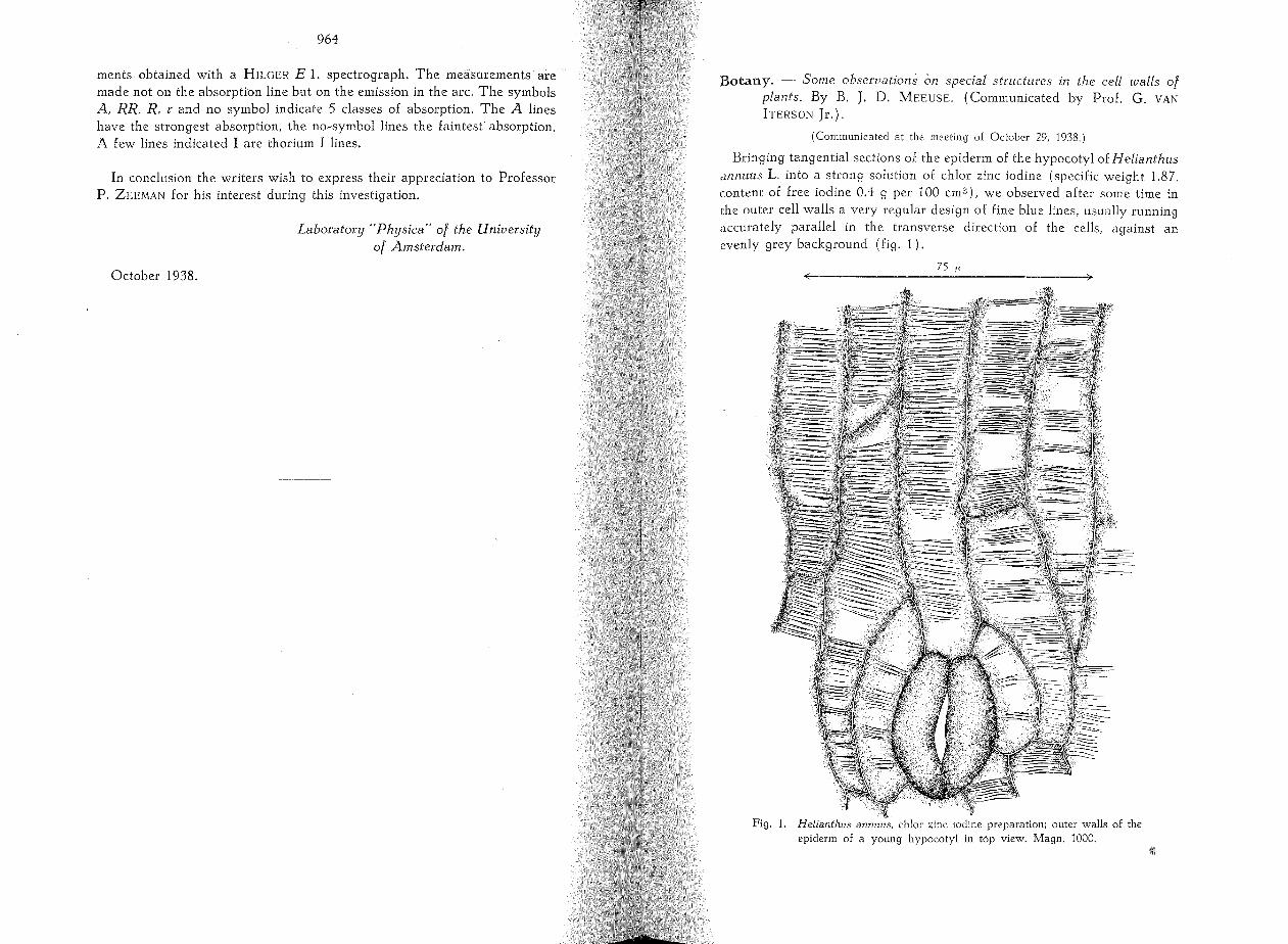
Bringing tangential sections of the epiderm of the hypocotyl of H elianthus annuus L. into astrong solution of chlor zinc iodine (specific weight 1.87. content of free iodine 0.4 9 per 100 cm il ), we observed af ter some time in the outer cell walls a very regular design of fine blue lines, usually running accurately parallel in the transverse direction of the cells, against an evenly grey background (fig. 1).
75 !'
Helianthlls anmms, chlor zinc iodine preparation; outer walls of the epiderm of a young hypocotyl in top view. Magn. 1000.
966
NÄGELI (1), STRASBURGER (2 and 3) and particularly CORRENS (4) made an elaborate investigation on transverse striation in the outer ceU waU of the epiderm of Hyacinthus orientalis L.; the ,latter observed this also in some other epiderms. In 1933 HEYN (5) observed a sttiation in the epiderm of the Avena~coleoptile and published microphotographic repro~ ductions of it.
1. General occurrence of the structural peculiarity.
I found that by a correct application of chlor zinc iodine the above~ mentioned structure or one that is related to it may be made visible in the cell wa lIs of numerous plants. For this purpose should be used a concent~ rated solution of the reagent. The sections ought to be thin and by preference first laid in water, but in that case they must be freed from most of the water by means of filter~paper before the reagent is added. The observations should be made shortly af ter the reagent has penetrated into the cell walls, since otherwise the background, against which the blue lines stand out, will be stained as weIL We warn against the possibility th at the observation is impeded by the folds which frequently occur in the cuticle and which - as will become apparent later on - are in no way connected with the structures under discussion.
Observations on ceU walls in top view, made for a hundred species of plants, induced me to draw the following conclusions:
a. Sttiation does not occur only in outer walls of the epiderm but is also found in inner walls of the epiderm and in the walls of collenchyma (e.g. in collenchyma of the stalk of Commelina virginica L.), in walls of parenchyma cells (e.g. in parenchyma of the stalk of Candollea adnata (R. Br.), F. v. Muel!.) and in walls of bast fibres (e.g. in fibres of the f1ower~stalk of Gerbera J amesonii Baker).
b. There is a great difference between the perceptibility of the striation in different objects. Sometimes the striation may be observed even without staining with chlor zinc iodine (CORRENS made his observations for the greater part on unstained preparations). This applies in particular to sttiation in epiderms, e.g. the epiderm of the flower~stalk of Milla uniflora R. Grah. (see fig. 5). In other cases the striation is hardly visible; to these belong the object mentioned in the beginning and further the outer wall of the leaf~epiderm of Rhoeo discolor Hance.
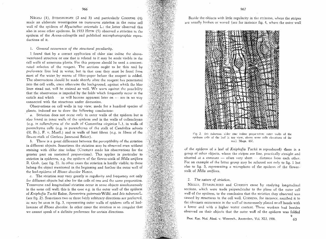
c. The sttiation may vary greatly in regularity and frequency not only for different objects but also for the cells of one and the same preparation. Transverse and longitudinal striation occur in some objects simultaneously in the same ceU wall; this is the case e.g. in the outer wall of the epiderm of Kniphofia Tuckii Baker, Sanseviera guinensis Willd. and Iris tuberosa L. (see fig. 2). Sometimes two or three fairly arbitrary directions are preferred,
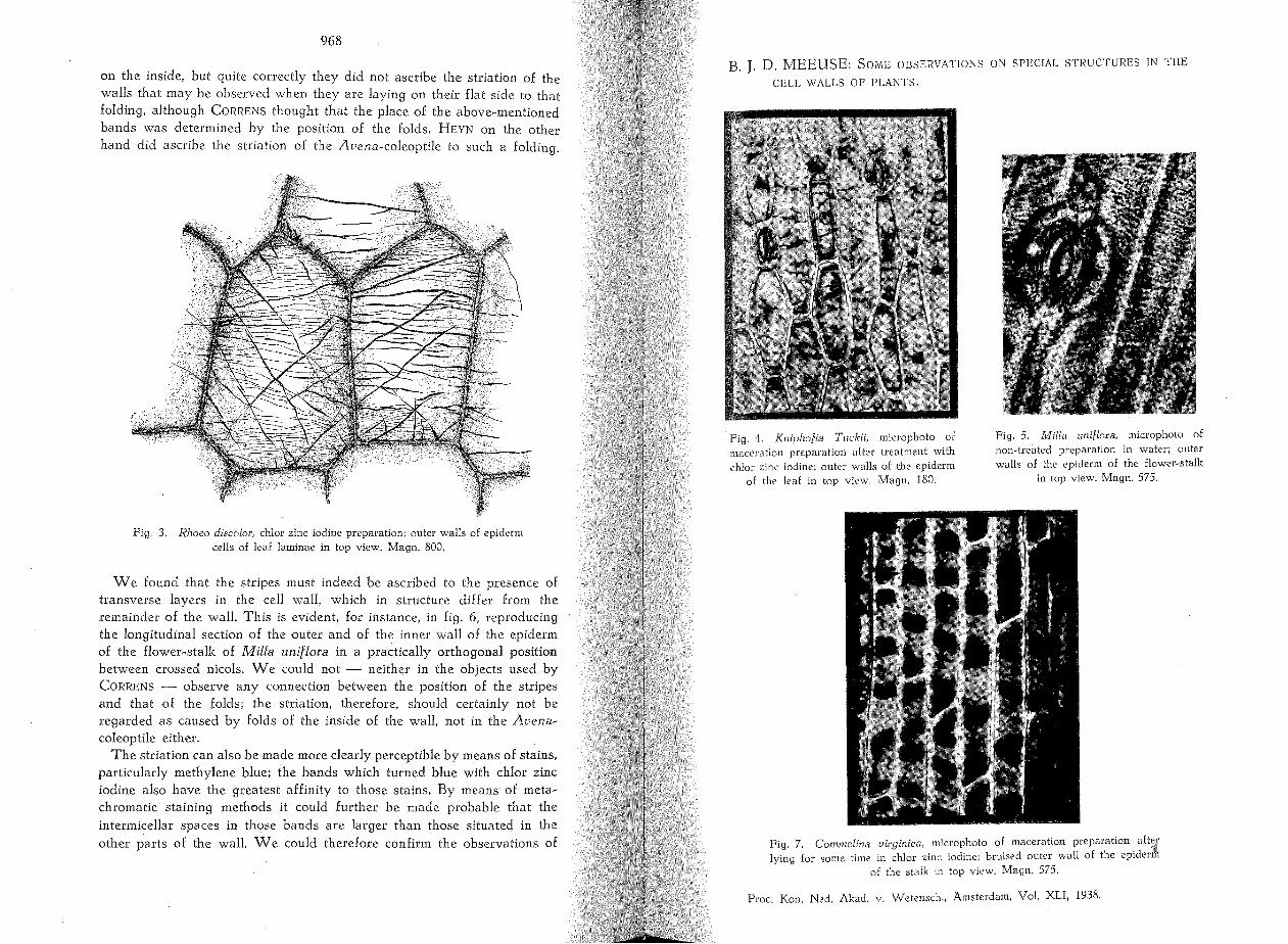
as may be seen in fig. 3, representing outer walls of epiderm cells of leaf~ laminae of Rhoeo discolor. In other cases the sttiation is so irregular that
we cannot speak of a definite preference for certain directions.
967
Beside the objects with little regulatity in the striation, where the strip es are usuaUy broken or waved (see for instance fig. 4, where the outer wall
Fig. 2. Iris tuberosa, chlor zinc iodine preparation; outer walls of the epiderm cells of the leaf in top view, ab ave same cells elevations of the
walL Magn. 450.
of the epiderm of a leaf of Kniphofia Tuckii is reproduced) there is a group of other objects, where the stripes are fine, practically straight and situated at a constant - of ten very short - distance from each other. For an example of the latter group may be referred not only to fig. 1 but also to fig. 5, representing a microphoto of the epiderm of the flower·· stalk of Milia uniflora.
2. The natur,e of striation.
NÄGELI, STRASBURGER and CORI~ENS came by studying longitudinal sections, which were made perpendicular to the plane of the outer ceU waU of the epiderm, to the conclusion that the striation they observed was caused by structures in the cell walL CORRENS, for instance, asctibed it to the alternate occurrence in the waU of transversely placed small bands with
a lower and with a higher water content. These wor kers had besides observed on their objects that the outer wall of the epiderm was folded
III Proc. Kon. Ned. Akad. v. Wetenseh., Amsterdam, Vol. XLI, 1938. 63
968
on the inside, but quite correctly they did not ascribe the striation of the walls that may be observed when they are laying on their flat side to that folding, although CORRENS thought th at the place of the above~mentioned bands was determined by the position of the folds. HEYN on the other hand did ascribe the striation of the Avena~coleoptile to such a folding.
Fig. 3. Rhoeo discoloc, chlor zinc iodine preparation; outer walls of epiderm cells: of leaf laminae in top view. Magn. 800.
We found that the stripes must indeed be ascribed to the pres en ce of transverse layers in the cell wall, which in structure differ from the remainder of the walL This is evident, for instance, in fig. 6, reproducing the longitudinal section of the outer and of the inner wall of the epiderm of the flower~stalk of Milla uniflora in a practically orthogonal position between crossed nicols. We could not - neither in the objects used by CORRENS - observe any connection between the position of the stripes and that of the folds; the striation, therefore, should certainly not be regarded as caused by folds of the inside of the wall, not in the Avena~ coleoptile either.
The striation can a]so be made more clearly perceptible by means of stains, particularly methylene blue; the bands which turned blue with chlor zinc iodine also have the greatest affinity to those stains. By means of meta~ chromatic staining methods it could further be made probable that the intermicellar spaces in those bands are larger than those situated in the other parts of the walL We could therefore confirm the observations of
B. J. D. MEEUSE: SOME OBSERVATIONS ON SPECIAL STRUCTURES IN THE
CELL WALLS OF PLANTS.
Fig. 4. Kniphofia Tl1ckii, microphoto of maceration preparation af ter treatment with chlor zinc iodine; outer walls of the epiderm
of the !eaf in top view. Magn. 180.
Fig. 5. Milla llnifloea, microphoto of non,treated preparation in water; outer walls of the epiderm of the flower,stalk
in top view. Magn. 575.
Fig. 7. Commclina vieginica, microphoto of maceration preparation. aft\i{ lying for same time in chlor zinc iodine; bruised outer wall of the eplderm
of the stalk in top view. Magn. 575.
Proc. Kon. Ned. Akad. v. Wetenseh., Amsterdam, Vol. XLI, 1938.

969
CORRENS, pointing to a different water content in the bands and outside the bands. Finally, according to a method of TH. KERR - .this method has been communicated by BAILEY and VESTAL (6) - we produced iodine crystals in the wall and then sometimes observed an orientation of very
Fig. 6. Milia unit/ot'a, non-treated preparation in practically orthogonal position between crossed nicols; made perpendicular to the wall surface in
the longitudinal direction of the fIower-stalk. Magn. 800.
small crystals into rows in the direction of the strip es. However, larger iodine crystals placed themselves with their long axis perpendicular to those stripes.
Taking all this into consideration, we regard the stripedesigns as the result of the presence in the wall of bands perpendicular to the plane of the wal1, while we consider the structure of the cellulose in those bands to be "looser" than in the places outside the bands. It will be clear that such like bands show a similarity in nature with the slipplanes which have been observed in the walls of some fibres (e.g. in flax and ramie) and on which in particular "'ON HÖHNEL made elaoorate researches.
~3*

970
This view is in agreement with the observation that the chlor zinc iodine from the strip es may penetrate further into the wall. This is particularly clear if only few strip es occur in the wal!. A similar case has been repro~ duced in fig. 7; the strip es have changed there into wide bluebands.
3. The inlluence ol mechanical actions on the genesis of the striation.
There is no doubt about it that the striation in numerous cases has to be considered as an artefact. We draw this conclusion from the following observations:
a. In many cases, in preparations where we observed the walls laying on their flat side, we could state that a blue stripe or that a wide bundie of small blue strip es was running continuously through the upper walls of two or of more contiguous cells (this may, for instance, be perceived in many places in fig. 2).
b. For various objects the direction of striation appeared to be determined by the direction in which the section had been made and then
Fig. 8. Rhoco discolor. chlor zinc iodine preparation; outer wal! of the epiderm of the midrib on the lower si de of the leaf in top view. section cut approximately in the direction perpendicular to the fine striations. Magn. 575.
971
also the striations in the upper walls of contiguous cells usually commun~ icated. We refer to fig. 8, reproducing a section wh ere the knife had cut through the tissue in the direction perpendicular to the fine striation.
c. Tangential sections of the epiderm often showed a stronger striation on two sides, namely where the knife had touched the object first and where it had touched last. The cells on the sides and in the centre of such a section were then far less strongly striated. If such a section on the object~glass was cut into halves with a sharp scalpel, the sections in deed showed more stripes close to the new planes of intersection than originally was the case, but yet far less than on the first~mentioned ends. This may be explained by the great damage done by introduction and removal of the razor and the smaller damage by the scalpel.
d. Of ten the walls of thick sections showed less striation than thin ones. e. Wh en a thick section was bruised by means of a pincette, it some~
times showed more stripes than a similar section which had not been bruised.
I. Af ter picking 2 holes (with a cactus~needle) in the outer wall of the same cell of the epiderm, sometimes stripes were formed running from one hole to the other.
Over against all these convincing observations th ere are, however, also cases where it was by no means immediately clear that the striation is an artefact. In Avena coleoptiles, for example, the striation was observed just as well when those coleoptiles as a whole were placed in chlor zinc iodine under the microscope and the same holds gQod for the membranous bracts of some Liliaceae (e.g. for Scilla spec., Hyacinthus spec.). If the epiderms of Rhoeo discolor and Helianthus annuus were isolated by careful maceration, they displayed the striation just as weIl as when sections were treated with chlor zinc iodine. Moreover, in some objects the direction of striation could not be influenced by the direction of cutting.
The distinction between these two groups mainly coincided with the one I made at the end of § 1.
Nevertheless, I am convinced that even for this last group of cases the striation may be considered an artificial product and that there is a relation between the fineness and the regularity of the striations, which are characteristic of these cases, and the difficulties met with in trying to influence the direction of these striations mechanically. This wiJl be explained in the next paragraph.
4. Relation bet ween the direction of striation and the optical properties of the wal!.
In all cases where the direction of striation could only with difficulty or not at all be influenced by mechanical actions I confirmed an observation already made by CORRENS for the few objects studied by him, namely that in the walls observed in top~view the direction of the strip es is perpendicular to the long axis of the "active refraction index ellipse" in those walls.

972
Por the outer walls of the epiderm cells of the hypocotyls of Helianthus annuus (fig. 1) and for the epiderm cells of the flower~stalk of Milia uniflora (fig. 5) th at long axis is consequently situated in the longitudinal direct ion of the cells (i.e. in the longitudinal direction of the plant organ). In the elongated epiderm cells of the bulbils of Ornithogalum longebrac~ teatum Jacq. frequently a longitudinal striation was observed; the long axis of the refraction index ellipse lay th en in the transversal direction of the cell.
Moreover, I noticed that the striati;n generally was finer and more regular and that its direction was more difficult to influence according as the double refraction of the waIl, in which the striation occurred, was stronger.
The cell walls in which the striation is irregular and may be easily influenced as a rule proved to possess a weak double refraction and the long axis of the refractiön index ellipse had no definite pref eren ce for a special direction.
IE we add to this that the striation is particularly clear in cell walls containing a large amount of cellulose and also that the fine structure of striation is not yet visible in young ceIl walls, which in top~view do not yet show double refraction, but that it sets in when the cell walls, af ter starting of the longitudinal growth, have become distinctly double refractive, then we must of necessity draw the following conclusion: 1;,he striations must be regarded as pI aces of fracture in those walls which contain a large quantity of more or less parallel oriented, crystalline cellulose micelles and which consequently to a certain extent may be considered as thin crystaI plates; according as the order of the cellulose micelles is more perfect, the waII layer will show the more the brittleness of "Chinese porcelain".
This view induced me to consider the fine, regular striations also as artefacts and to assume that these striations set in very easily and even by taking many precautions can hardly be avoided. It became indeed apparent th at sometimesthe inner walls of very carefully - by maceration -isolated epiderm cell waIls, which originally were practically free from striation, after pencilling with a brush displayed a striation figure. We consider it possible, therefore, that striation sometimes is produced in cell walls by movements of the walls which are the result of air currents. Similarly tensions in the waIls, occurring during growth, might play a part in the formation of such striations.
The phenomenon that the planes of fracture occur specially perpendicular to the direction, which is preferred by the long cellulose molecules and the long ceIIulose micelles respectively, is undoubtedly due to the fact that long molecules have astrong tendency to place themselves parallel to each other and to keep each other in that position, for which we refer to J. H. DE BOER (7).
Por the present we are not able to explain the great regularity of the fine striations. However, we wish to point out that our observations and
973
views do not lead to the "Querhauttheorie" of LÜDTKE (9), which has been contested by GRIFFIOEN (8) on good grounds. The striations shouId not be regarded as a "Fremdhaut", but as "Gitteraufsplitterungen" and "Fibrillenbrüche" in the sense of SAUTER (10).
The occasionaIly simultaneous preference for more than one direction by the striation in the same cell wal!, which we discussed in § 1 and of which we reproduced an example in fig. 3, may perhaps be explained by a stratiform construction of ceIl walls, in which case we must assume that the long axis of the micelles in the different layers prefers different directions.
FinaIly we revert to the similarity between our conception concerning the nature of the striations and that of the character of the slipplanes in fibre~walls. We mentioned th is similarity in § 2, but by what we discussed in this paragraph it has become even more conspicuous. Of these slipplanes are characteristic "exchanges" in the course of the walls as weil as the occurrence of stripes, which form an angle of about 45° or 125° with the axis of the fibre and frequently cross each other. These peculiarities were not observed in the striations discussed here. We wish, however, to point out th at those structures in the fibre~walls are caused by mechanica! influences on thick~walled, concave, cylindrical bodies, whereas in our cases we have to deal with fractures in thin, plate~like walls, which are commonly reinforced by walls perpendicular to them and in their turn are sometimes connected by inner walls. Besides, the order of the molecules and micelles in the curved fibre~walls is probably different from that in the flat walls discussed here.
5. The possible significanee of the structure for a phenomenon observed by W. HOFMEISTER and for the grrowth of the cel! wal!.
In 1860 HOFMEISTER (11) described the phenomenon that numerous fast growing shoots af ter shaking wither and bend (this bending is not due to weakness for it is also distinctly perceptible in the treated shoots af ter they have been turned upside down). The same phenomenon occurs if on such shoots we tap rapidly with a rod. If we do this on the ligneous part of the shoot, the young top bends in the direction from where the tapping came. PRILLIEUX (12) thought to have demonstrated that the bending may be ascribed to a purely mechanical process; he observed namely that a sm all leaden rod, fixed in astrong wooden stick, bent when he tapped on the stick and that this took pI ace in the same direction as the one in which the shoots bent under similar conditions. However, PRILLIEUX did not explain the loss of turgor. Moreover, if we consider which processes in the experiment with the leaden rod set in, we find th at the similarity with what is shown by the living object is only very superficiaI. So far the "HOFMEISTER phenomenon" has not yet been
explained. Self~evidently we assumed now that by the shaking and tapping on a
\!I

974
shoot striation would arise in the cell walls of the epiderm. These numerous disturbances of the crystal structure would make the waIls more extensible in longitudinal direction. By the mechanical operations also a real extension in that direction wiIl be brought about. The epiderm belongs to the so~called "passive tissues", in the sense of J. SACHS, which tissues during the growth of the stalk are kept in a condition of tension by the active tissues. Wh en this ten sion is removed rather suddenly, the active tissue wiIl not have a chance to extend sufficiently quickly in longitudinal direction and the stalk wiIl relax. If the disturbances in the wall structure occur one~sidedly in the staIk, the latter will bend.
Indeed I found that, af ter violent shaking of leaves of Rhoeo discolor, of staIks of Commelina virginica and of some other objects, astrong increase of the number of strip es may be observed in the outer waIl of the epiderm ceIls. Sometimes the strip es in these waIlswere afterwards so 11llmerOUS that in chlor zinc iodine almost immediately a complete blue coloration of the walls set in.
Meanwhile it is by no means certain th at, if the HOFMEISTER phenomenon is produced by one~sided tapping on a shoot, we have an equally strong mechanical influence as was the case in the just mentioned shaking experiments. On the outer and inner walls of epiderms, extremely carefully isolated by maceration from staIks which showed bending after tapping, I observed hardly any striations, whereas particularly on such walls af ter isolation striation became visible af ter I had pencilled them with a brush.
Consequently we cannot maintain the above~mentioned explanation of the HOFMEISTER phenomenon, but we con si der th is part of our investigation not yet definitely concluded.
FinaIly we wish to suggest as a hypothetical possibility that the structures observed by us play a part in the growth of the ceH walls and the tissues. It is quite weIl conceivable th at celI walls in the places of the disturbances in their regular structure are stretched more easily by the turgor pressure than elsewhere and that subsequently by preference in these places new ceH wall substance is deposited.
Deltt, October 1938.
Laboratory tor Technical Botany ot the University College ot Technology.
LITERATURE.
1. NÄGELI. c.. Ueb. den inner en Bau der veget. Zellmembranen. Sitz.ber. München, 7 Mai 1864.
,2. STRAS BURGER, E., Ueb. den Bau und das Wachstum der Zellhäute. Jena (1882). 3. , Ueb. das Wachstum vegetabilischer Zellhäute. HistoJ. Beitr .. 2, Jena (1889). 4. CORRENS, c., Zur Kenntnis der inner en Struktur der vegetabi.Jischen Zellmembranen.
Jahrb. wiss. Bot., 23, 254-338 (1892). 5. HEYN. A. N. J., Further investig. on the mechanism of cell e1ongation etc. L Proto
plasma, 19,78-96 (1933).
975
6. BAILEY, 1. W. and VESTAL, MARY R., The Orientation of Cellulose in the Secondary Wall of Tracheary Cells. J. ARNOLO Arbor .. 18, 185-196 (1937) .
7. DE BOER, J. H., Trans. Faraday Soc., 32, 1 (1936). 8. GRIFFIOEN, K, Ueb. Quellungsbilder verschiedener Faserarten und deren Bedeutung
für die Faserstruktur. Planta, 24, 584-601 (1935). 9. LÜOTKE, M., Unters. üb. Aufbau und Bildung der pflanz1. Zellmembran und ihrer
stofflichen Komponenten. Bioch. Z .. 223, 1-61 (1931), and elsewhere. 10. SAUTER, E., Beitr. zur Röntgenographie und Morphologie der Cellulose 1. u. Ir.
Z. physik. Chem. B 35, 83-129 (1937). 11. HOFMEISTER, W., Ueb. die Beugungen saftreicher Pflanzentheilen nach Erschüt
terung. Jahrb. wiss. Bot., 2, 237-266 (1860). 12. PRILLIEUX, E., Etude sur les courbures que produisent les secouses sur les jeunes
pousses des végétaux. Ann. sei. nat. Bot .. (5) 9, 248-266 (1868).