
Solulink Bioconjugation Primer Bioconjugation Primer—January 2013 ... Conjugation and...
14
1 Solulink Bioconjugation Primer—January 2013 + 1 858.625.0670 Solulink Bioconjugation Primer Conjugation and immobilization of biomolecules has historically been very problematic for a variety of reasons; primarily because there are few covalent bond-forming reactions that proceed in water that can be engineered to link biomolecules together. A wide variety of methods to conjugate and immobilize biomolecules are extensively described in Bioconjugate Techniques, but most of these methods are difficult to perform, stoichiometrically inefficient, and result in low conjugate yields. It is important to understand the desired characteristics of the ideal bioconjugation chemistry. The following list presents many criteria that need to be fulfilled to produce the ideal bioconjugation technology: Introduction: Bioconjugation is the linking of two biomolecules to form a hybrid, the bioconjugate, which retains the properties of each individual component, yet yielding a single entity with two complementary functions. Biomolecules exist and function in aqueous environments; therefore, the preparation of bioconjugates is primarily about “chemistry in water.” Any suitable bioconjugate chemistry must be compatible with such an environment, while at the same time preserving the biological activity or function of the biomolecules. Conjugates are generally formed through the addition of separate but reactively complementary functional groups to each of the two biomolecules. The two modified biomolecules are then mixed together to form the desired bioconjugate. Figure 1. Bioconjugate Techniques, Greg T. Hermanson, Academic Press, 2008.
Transcript of Solulink Bioconjugation Primer Bioconjugation Primer—January 2013 ... Conjugation and...
1
Solulink Bioconjugation Primer—January 2013
+ 1 858.625.0670
Solulink Bioconjugation Primer
Conjugation and immobilization of biomolecules has
historically been very problematic for a variety of reasons;
primarily because there are few covalent bond-forming
reactions that proceed in water that can be engineered to
link biomolecules together. A wide variety of methods
to conjugate and immobilize biomolecules are extensively
described in Bioconjugate Techniques, but most of these
methods are difficult to perform, stoichiometrically inefficient,
and result in low conjugate yields.
It is important to understand the desired characteristics of the
ideal bioconjugation chemistry.
The following list presents many criteria that need to be
fulfilled to produce the ideal bioconjugation technology:
Introduction: Bioconjugation is the linking of two biomolecules to form a hybrid, the bioconjugate, which
retains the properties of each individual component, yet yielding a single entity with two complementary
functions. Biomolecules exist and function in aqueous environments; therefore, the preparation of
bioconjugates is primarily about “chemistry in water.” Any suitable bioconjugate chemistry must be
compatible with such an environment, while at the same time preserving the biological activity or
function of the biomolecules. Conjugates are generally formed through the addition of separate
but reactively complementary functional groups to each of the
two biomolecules. The two modified biomolecules are then
mixed together to form the desired bioconjugate.
Figure 1. Bioconjugate Techniques, Greg T. Hermanson,
Academic Press, 2008.
2
For more information visit www.solulink.com
a) Linkers must be incorporated on biomolecules in a mild,
controllable manner
b) The inherent biological function of the biomolecules must
be unaffected after modification and conjugation
c) The conjugation reaction occurs directly upon mixing the
two modified biomolecules, preferably not requiring
addition of an oxidant, reductant, or metal
d) Modified biomolecules are stable over extended periods
e) Formation of the covalent linkage is stable under a broad
pH range and at elevated temperatures
f) Quantification of both incorporated linkers and
final conjugate is readily performed, i.e, they are
spectrophotometrically traceable
g) Conjugation occurs in buffered aqueous solutions, at a
physiological pH
h) Stoichiometrically efficient (e.g., 1:1)
i) Fast reaction kinetics
j) No undesirable covalent side reactions during modification
k) No electrostatic/hydrophobic interactions
l) Linkers can be incorporated on a variety of biomolecules,
including oligos and peptides, through solid phase synthesis
The classical method used to prepare covalent conjugates makes
use of the maleimido/thiol coupling pair.
Disadvantages of the maleimido/thiol pair include:
a) The aqueous instability (hydrolysis) of the linker functional
group (i.e., maleimide) on biomolecules during conjugation
b) The need to protect the thiol group
c) The slow kinetics of the reaction
d) The need to activate the protected thiol group with a strong
reducing agent (e.g., DTT or TCEP)
e) The need to activate the protected thiol group by addition
of a strong reducing agent (e.g., DTT)
f) The potential for undesirable homo-dimers via disulfide
bridge formation
g) Cleavage and/or partial fragmentation of proteins by
reduction of disulfide bonds
h) Reduced or inactive protein function
Next
The solution... v v
3
Solulink Bioconjugation Primer—January 2013
+ 1 858.625.0670
The Solulink Solution
Solulink has developed a bioconjugate chemistry that more
mildly, more efficiently, and more reproducibly conjugates
and immobilizes proteins, peptides, oligonucleotides,
carbohydrates, and polymers.
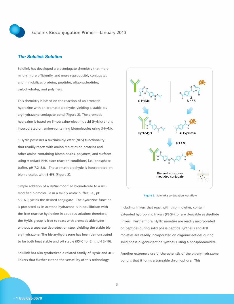
This chemistry is based on the reaction of an aromatic
hydrazine with an aromatic aldehyde, yielding a stable bis-
arylhydrazone conjugate bond (Figure 2). The aromatic
hydrazine is based on 6-hydrazino-nicotinic acid (HyNic) and is
incorporated on amine-containing biomolecules using S-HyNic .
S-HyNic possesses a succinimidyl ester (NHS) functionality
that readily reacts with amino moieties on proteins and
other amine-containing biomolecules, polymers, and surfaces
using standard NHS ester reaction conditions, i.e., phosphate
buffer, pH 7.2–8.0. The aromatic aldehyde is incorporated on
biomolecules with S-4FB (Figure 2).
Simple addition of a HyNic-modified biomolecule to a 4FB-
modified biomolecule in a mildly acidic buffer, i.e., pH
5.0–6.0, yields the desired conjugate. The hydrazine function
is protected as its acetone hydrazone is in equilibrium with
the free reactive hydrazine in aqueous solution; therefore,
the HyNic group is free to react with aromatic aldehydes
without a separate deprotection step, yielding the stable bis-
arylhydrazone. The bis-arylhydrazone has been demonstrated
to be both heat stable and pH stable (95OC for 2 hr, pH 2–10).
Solulink has also synthesized a related family of HyNic and 4FB
linkers that further extend the versatility of this technology;
Figure 2. Solulink’s conjugation workflow.
including linkers that react with thiol moieties, contain
extended hydrophilic linkers (PEG4), or are cleavable as disulfide
linkers. Furthermore, HyNic moieties are readily incorporated
on peptides during solid phase peptide synthesis and 4FB
moieties are readily incorporated on oligonucleotides during
solid phase oligonucleotide synthesis using a phosphoramidite.
Another extremely useful characteristic of the bis-arylhydrazone
bond is that it forms a traceable chromophore. This
4
For more information visit www.solulink.com
chromophore absorbs at 354 nm with a molar extinction
coefficient of 29,000 (Figure 1 inset). This unique property allows
the researcher to (1) follow the linking or labeling reactions
in real time on a spectrophotometer, (2) directly quantify the
number of linkages formed in the conjugate, and (3) visualize or
trace conjugate fractions during FPLC or HPLC purification.
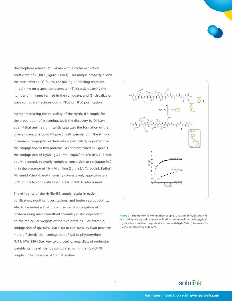
Further increasing the versatility of the HyNic/4FB couple for
the preparation of bioconjugates is the discovery by Dirksen
et al.1,2 that aniline significantly catalyzes the formation of the
bis-arylhdyrazone bond (Figure 3, with permission). The striking
increase in conjugate reaction rate is particularly important for
the conjugation of two proteins. As demonstrated in Figure 3,
the conjugation of HyNic-IgG (1 mol. equiv.) to 4FB-BSA (1.5 mol.
equiv.) proceeds to nearly complete conversion to conjugate in 2
hr in the presence of 10 mM aniline (Solulink’s TurboLink Buffer).
Maleimido/thiol–based chemistry converts only approximately
50% of IgG to conjugate when a 1:4 IgG:BSA ratio is used.
The efficiency of the HyNic/4FB couple results in easier
purification, significant cost savings, and better reproducibility.
Also to be noted is that the efficiency of conjugation of
proteins using maleimido/thiol chemistry is also dependent
on the molecular weights of the two proteins. For example,
conjugation of IgG (MW 150 kDa) to HRP (MW 45 kDa) proceeds
more efficiently than conjugation of IgG to phycoerythrin
(R-PE; MW 250 kDa). Any two proteins, regardless of molecular
weights, can be efficiently conjugated using the HyNic/4FB
couple in the presence of 10 mM aniline.
Figure 3. The HyNic/4FB conjugation couple. Ligation of HyNic and 4FB with aniline-catalyzed hydrazone ligation between 6-hydrazinopyridyl (HyNic)-functionalized peptide 4 and benzaldehyde 5 (left) followed by UV-VIS spectroscopy (340 nm).
5
Solulink Bioconjugation Primer—January 2013
+ 1 858.625.0670
HyNic/4FB Conjugation Couple Advantages
The many significant advantages to Solulink’s HyNic/4FB–based
conjugation technology include:
a) An efficient conjugation chemistry
b) No metals, reducing, or oxidizing agents are required
c) Fast conjugation kinetics catalyzed by aniline
d) Conjugate bond is stable to extremes of heat and pH
e) Conjugate bond forms a chromophore that can be used
to follow reactions in real time and quantify the number
of linkages in a conjugate
f) Versatile: proteins, oligos, and peptides can be rapidly
conjugated to each other and to surfaces
g) Direct conjugate bond formation under mild buffer
conditions
h) Excellent retention of inherent biological function of
biomolecules following controlled modification and
conjugation
i) 4FB linker can be incorporated on oligonucleotides during
solid phase synthesis
j) HyNic linker can be incorporated on peptides during solid
phase synthesis
Conjugation Examples
To demonstrate the versatility and breadth of utility of
Solulink’s HyNic/4FB conjugation couple, we show examples of
the preparation of protein-protein, peptide-oligonucleotide,
and protein-oligonucleotide conjugates (Figures 4, 5, 6). More
detailed descriptions can be found in the Solulink white papers.
Protein-Protein Conjugation
Solulink has taken advantage of the efficiency of conjugation
of two proteins using the HyNic/4FB couple in the presence of
aniline to develop an All-in-One Conjugation kit that allows
preparation of IgG-HRP, IgG-AlkPhos, and IgG-R-PE conjugates
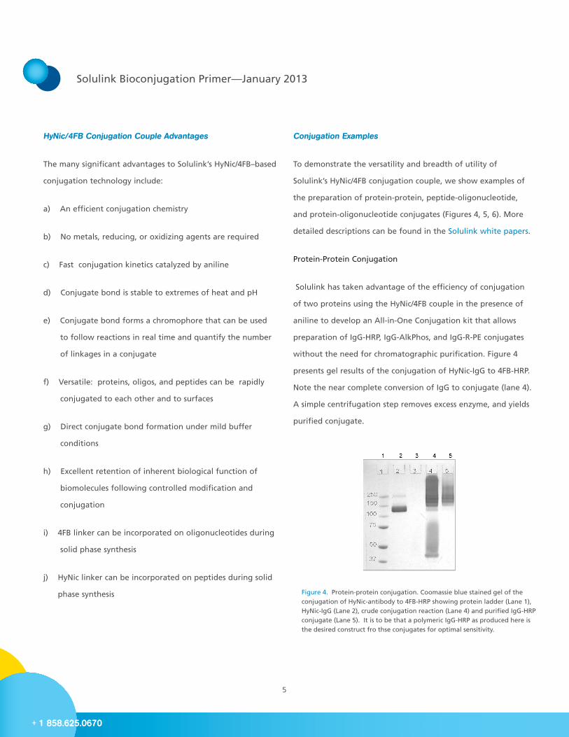
without the need for chromatographic purification. Figure 4
presents gel results of the conjugation of HyNic-IgG to 4FB-HRP.
Note the near complete conversion of IgG to conjugate (lane 4).
A simple centrifugation step removes excess enzyme, and yields
purified conjugate.
Figure 4. Protein-protein conjugation. Coomassie blue stained gel of the conjugation of HyNic-antibody to 4FB-HRP showing protein ladder (Lane 1), HyNic-IgG (Lane 2), crude conjugation reaction (Lane 4) and purified IgG-HRP conjugate (Lane 5). It is to be that a polymeric IgG-HRP as produced here is the desired construct fro thse conjugates for optimal sensitivity.
6
For more information visit www.solulink.com
Protein-Oligonucleotide Conjugation
The ability to efficiently and reproducibly prepare antibody-
oligonucleotide conjugates has limited their exploitation in
multiplexed diagnostic asssays.
Solulink’s HyNic/4FB bioconjugation couple, as applied to
the conjugation of oligonucleotides with antibodies, is
stoichiometrically efficient and high yielding, converting >95%
antibody to antibody-oligonucleotide conjugate (Figure 7).
Furthermore, conjugations of oligomers of 20–60 nucleotides are
conjugated with equal efficiency. The method is extremely mild,
as no metals, reductants, or oxidants are used in the conjugation
step. Further enhancing the efficiency of conjugation is the
use of aniline as a reaction catalyst. In a standard conjugation
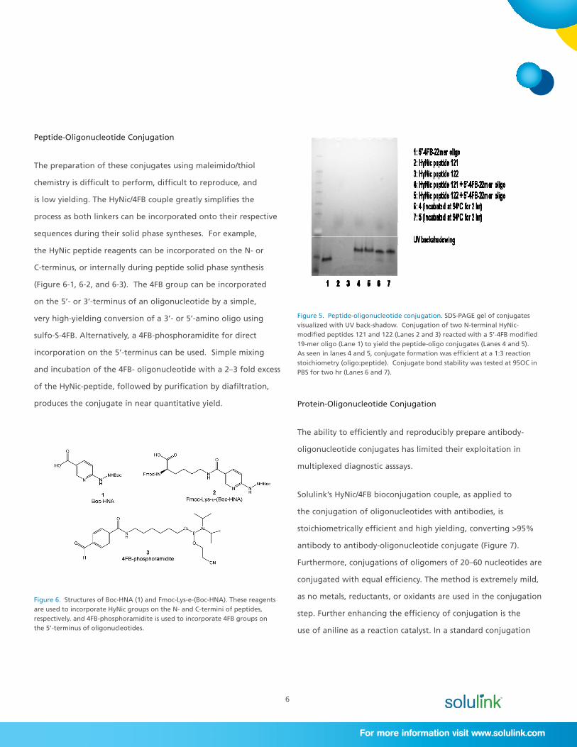
Peptide-Oligonucleotide Conjugation
The preparation of these conjugates using maleimido/thiol
chemistry is difficult to perform, difficult to reproduce, and
is low yielding. The HyNic/4FB couple greatly simplifies the
process as both linkers can be incorporated onto their respective
sequences during their solid phase syntheses. For example,
the HyNic peptide reagents can be incorporated on the N- or
C-terminus, or internally during peptide solid phase synthesis
(Figure 6-1, 6-2, and 6-3). The 4FB group can be incorporated
on the 5’- or 3’-terminus of an oligonucleotide by a simple,
very high-yielding conversion of a 3’- or 5’-amino oligo using
sulfo-S-4FB. Alternatively, a 4FB-phosphoramidite for direct
incorporation on the 5’-terminus can be used. Simple mixing
and incubation of the 4FB- oligonucleotide with a 2–3 fold excess
of the HyNic-peptide, followed by purification by diafiltration,
produces the conjugate in near quantitative yield.
Figure 6. Structures of Boc-HNA (1) and Fmoc-Lys-e-(Boc-HNA). These reagents are used to incorporate HyNic groups on the N- and C-termini of peptides, respectively. and 4FB-phosphoramidite is used to incorporate 4FB groups on the 5’-terminus of oligonucleotides.
Figure 5. Peptide-oligonucleotide conjugation. SDS-PAGE gel of conjugates visualized with UV back-shadow. Conjugation of two N-terminal HyNic-modified peptides 121 and 122 (Lanes 2 and 3) reacted with a 5’-4FB modified 19-mer oligo (Lane 1) to yield the peptide-oligo conjugates (Lanes 4 and 5). As seen in lanes 4 and 5, conjugate formation was efficient at a 1:3 reaction stoichiometry (oligo:peptide). Conjugate bond stability was tested at 95OC in PBS for two hr (Lanes 6 and 7).
7
Solulink Bioconjugation Primer—January 2013
+ 1 858.625.0670
protocol, 5 equivalents of 4FB-oligonucleoitde are used,
resulting in the conjugation of 2–3 oligonucleotides per
antibody.
In a second breakthrough for antibody-oligonucleotide
conjugation, we developed a method to purify the conjugate
by adsorption to a proprietary magnetic affinity matrix that
allows removal of excess 4FB-oligonucleoitde, followed by
elution of the purified conjugate using mild elution buffers.
The overall yield of the antibody-oligonucleotide conjugate
is 30–50% based on antibody recovery. The conjugate is
>95% free from unconjugated HyNic-antibody and 4FB-
oligonucleotide. Multiple conjugates can be prepared
simultaneously, satisfying the requirement for the use of
this protocol to prepare multiple antibody-oligonucleotide
conjugates for highly multiplexed detection of antigens.
Traceable Biotin and Digoxigenin Labeling of Biomolecules with
ChromaLink™ Linkers
Accurate and controlled incorporation of haptens such as biotin
and digoxigenin on biomolecules continues to be problematic
due to the inherent lack of an internal chromophore on these
tags. To overcome this problem, Solulink incorporated its
UV-traceable (354 nm) bis-arylhydrazone chromophore into
the linker arm in both biotin and digoxigenin modification
reagents, the ChromaLink™ Biotin and ChromaLink™
Digoxigenin (Figure 8). Spectrophotometric quantification
of the incorporation of these chromophoric haptens is
straightforward and highly reproducible. Simply measuring the
A280 and A354 absorbance of the modified proteins yields both
the protein concentration and number of incorporated labels
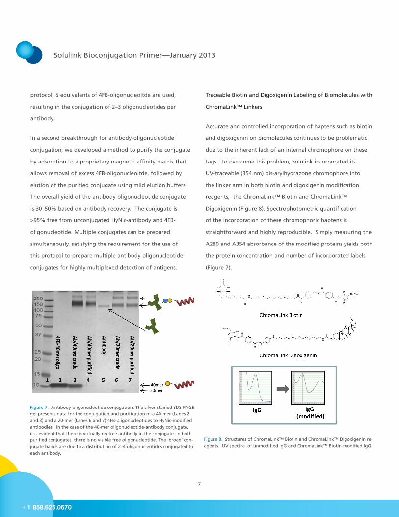
(Figure 7).
Figure 7. Antibody-oligonucleotide conjugation. The silver stained SDS-PAGE gel presents data for the conjugation and purification of a 40-mer (Lanes 2 and 3) and a 20-mer (Lanes 6 and 7) 4FB-oligonucleotides to HyNic-modified antibodies. In the case of the 40-mer oligonucleotide-antibody conjugate, it is evident that there is virtually no free antibody in the conjugate. In both purified conjugates, there is no visible free oligonucleotide. The ‘broad’ con-jugate bands are due to a distribution of 2–4 oligonucleotides conjugated to each antibody.
Figure 8. Structures of ChromaLink™ Biotin and ChromaLink™ Digoxigenin re-agents. UV spectra of unmodified IgG and ChromaLink™ Biotin-modified IgG.
8
For more information visit www.solulink.com
Biomolecule Immobilization
Immobilization of proteins, especially antibodies, is critically
important in many biological applications; including
immunoprecipitation, cell enrichment, antigen purification, and
immunodetection. For this reason, researchers often require that
proteins be immobilized on either a magnetic or non-magnetic
surface.
There are two main methods used to immobilize proteins: 1)
immobilization of biotinylated proteins on streptavidin-modified
surfaces or 2) direct covalent immobilization on activated
surfaces. Solulink has engineered its conjugation chemistry to
offer both alternatives within the NanoLink™ and MagnaLink™
family of products.
For antibody labeling, Solulink designed and produced the
ChromaLink™ Biotin and ChromaLink™ Digoxigenin One-Shot
Kits , which include everything required to label, purify, and
quantify incorporation of these labels from a single 100 µg
quantity of antibody. Solulink has also developed the more
flexible ChromaLink™ Biotin Protein Labeling Kit, used in the
controlled traceable biotinylation of any protein ranging from
20 to 200 kDa in size, in concentrations ranging from 0.25 to 10
mg/mL.
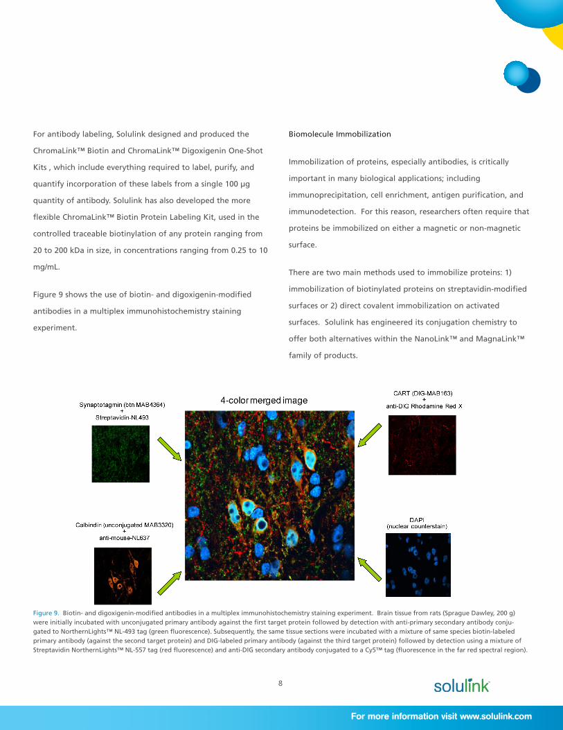
Figure 9 shows the use of biotin- and digoxigenin-modified
antibodies in a multiplex immunohistochemistry staining
experiment.
Figure 9. Biotin- and digoxigenin-modified antibodies in a multiplex immunohistochemistry staining experiment. Brain tissue from rats (Sprague Dawley, 200 g) were initially incubated with unconjugated primary antibody against the first target protein followed by detection with anti-primary secondary antibody conju-gated to NorthernLights™ NL-493 tag (green fluorescence). Subsequently, the same tissue sections were incubated with a mixture of same species biotin-labeled primary antibody (against the second target protein) and DIG-labeled primary antibody (against the third target protein) followed by detection using a mixture of Streptavidin NorthernLights™ NL-557 tag (red fluorescence) and anti-DIG secondary antibody conjugated to a Cy5™ tag (fluorescence in the far red spectral region).
9
Solulink Bioconjugation Primer—January 2013
+ 1 858.625.0670
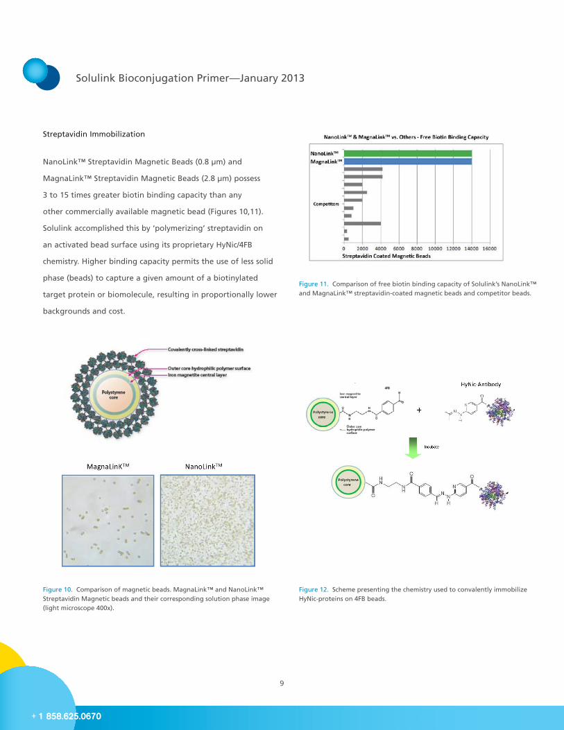
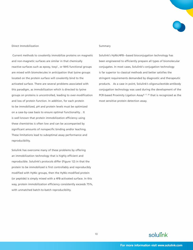
Streptavidin Immobilization
NanoLink™ Streptavidin Magnetic Beads (0.8 µm) and
MagnaLink™ Streptavidin Magnetic Beads (2.8 µm) possess
3 to 15 times greater biotin binding capacity than any
other commercially available magnetic bead (Figures 10,11).
Solulink accomplished this by ‘polymerizing’ streptavidin on
an activated bead surface using its proprietary HyNic/4FB
chemistry. Higher binding capacity permits the use of less solid
phase (beads) to capture a given amount of a biotinylated
target protein or biomolecule, resulting in proportionally lower
backgrounds and cost.
Figure 10. Comparison of magnetic beads. MagnaLink™ and NanoLink™ Streptavidin Magnetic beads and their corresponding solution phase image (light microscope 400x).
Figure 11. Comparison of free biotin binding capacity of Solulink’s NanoLink™ and MagnaLink™ streptavidin-coated magnetic beads and competitor beads.
Figure 12. Scheme presenting the chemistry used to convalently immobilize HyNic-proteins on 4FB beads.
10
For more information visit www.solulink.com
Direct Immobilization
Current methods to covalently immobilize proteins on magnetic
and non-magnetic surfaces are similar in that chemically
reactive surfaces such as epoxy, tosyl , or NHS functional groups
are mixed with biomolecules in anticipation that lysine groups
located on the protein surface will covalently bind to the
activated surface. There are several problems associated with
this paradigm, as immobilization which is directed to lysine
groups on proteins is uncontrolled, leading to over-modification
and loss of protein function. In addition, for each protein
to be immobilized, pH and protein levels must be optimized
on a case-by-case basis to ensure optimal functionality. . It
is well known that protein immobilization efficiency using
these chemistries is often low and can be accompanied by
significant amounts of nonspecific binding and/or leaching.
These limitations lead to suboptimal assay performance and
reproducibility.
Solulink has overcome many of these problems by offering
an immobilization technology that is highly efficient and
reproducible. Solulink’s protocols differ (Figure 12) in that the
protein to be immobilized is first controllably and reproducibly
modified with HyNic groups, then the HyNic-modified protein
(or peptide) is simply mixed with a 4FB-activated surface. In this
way, protein immobilization efficiency consistently exceeds 75%,
with unmatched batch-to-batch reproducibility.
Summary
Solulink’s HyNic/4FB–-based bioconjugation technology has
been engineered to efficiently prepare all types of biomolecular
conjugates. In most cases, Solulink’s conjugation technology
is far superior to classical methods and better satisfies the
stringent requirements demanded by diagnostic and therapeutic
products. As a case in point, Solulink’s oligonucleotide-antibody
conjugation technology was used during the development of the
PCR-based Proximity Ligation Assay8, 17, 20 that is recognized as the
most sensitive protein detection assay.
11
Solulink Bioconjugation Primer—January 2013
+ 1 858.625.0670
Citations
1. Schlingemann J, Leijon M, Yacoub A et al. (2010) Novel
means of viral antigen identification: Improved detection of
avian influenza viruses by proximity ligation. J Virol Methods
v163, I 1:116-122.
2. Dirksen A and Dawson PE (2008) Rapid Oxime and
Hydrazone Ligations with Aromatic Aldehydes for Biomolecular
Labeling. Bioconjugate Chem 19:2543-2548.
3. Ma Z, Li Q, An H et al. (2009) Targeting HER signaling
with Neuregulin’s Heparin-binding Domain. J Biol Chem
13:284(46):32108-15 Epub Aug 28.
4. Hansen RR, Johnson LM, Bowman CN (2009) Visual,
base-specific detection of nucleic acid hybridization using
polymerization-based amplification. Anal Biochem v386, I. 2:285-287.
5. Kwong GA, Radu CG, Hwang K et al. (2009) Modular Nucleic
Acid Assembled p/MHC Microarrays for Multiplexed Sorting of
Antigen-Specific T Cells. J Am Chem Soc 131 (28):9695–9703.
6. Sharma S, Dominguez A, Manrique S et al. (2008) Systemic
Targeting of CpG-ODN to the Tumor Microenvironment with
Anti-neu-CpG Hybrid Molecule and T Regulatory Cell Depletion
Induces Memory Responses in BALB-neuT Tolerant Mice. Cancer
Res 68(18): 7530-40.
7. Sikes D, Hansen R, Johnson L et al. (2008) Using polymeric
materials to generate anamplified response to molecular
recognition events. Nat Mater 7:52-6.
8. Fredriksson S, Horecka J, Brustugun OT et al. (2008)
Multiplexed Proximity Ligation Assays to Profile Putative Plasma
Biomarkers Relevant to Pancreatic and Ovarian Cancer. Clin
Chem 54:582-589.
9. Liu G, Dou S, Rusckowski M et al. (2008) An experimental
and theoretical evaluation of the influence of pretargeting
antibody on the tumor accumulation of effector. Mol Cancer
Ther 7:1025-1032.
10. Sharma S, Dominguez AL, Manrique SZ et al. (2008)
Systemic targeting of CpG-ODN to the tumor microenvironment
with anti-neu-CpG hybrid molecule and T regulatory cell
depletion induces memory responses in BALB-neuT tolerant
mice. Cancer Res 15;68(18):7530-40.
11. Chaturvedi A, Dorward D, Pierce SK (2008) The B Cell
Receptor Governs the Subcellular Location of Toll-like Receptor
9 Leading to Hyperresponses to DNA-Containing Antigens.
Immunity v28, I. 6:799-809.
12. Faintuch BL, Teodoro R, Duatti A et al. (2008)
Radiolabeled bombesin analogs for prostate cancer diagnosis:
preclinical studies. Nucl Med Bio v35, I. 4:401-411.
13. Liu G, Dou S, Yin D et al. (2007) A Novel Pretargeting
Method for Measuring Antibody Internalization in Tumor Cells.
Cancer Biother Radio 22:33-39.
14. Levashova Z, Backer J, Backer M et al. (2007) Direct
labeling of single-chain VEGF (sc-VEGF) with Tc99m. J Nucl Med
48 (Supplement 2) 181P.

12
For more information visit www.solulink.com
15. Wong CH, Mruk DD, Lee W et al. (2007) Targeted
and reversible disruption of the blood-testis barrier by an FSH
mutant-occludin peptide conjugate. FASEB J 21:438.
16. Darmanis S, Kähler A, Spångberg L et al. (2007) Self-
assembly of proximity probes for flexible and modular proximity
ligation assays. Biotechniques 43:443-450.
17. Fredriksson S, Dixon W, Ji H et al. (2007) Multiplexed
protein detection by proximity ligation for cancer biomarker
validation. Nat Methods 4:327.
18. He J, Liu G, Dou S et al. (2007) An improved method
for covalently conjugating morpholino oligomers to antitumor
antibodies. Bioconjugate Chem 18:983-8.
19. Vardar-Schara G, Krab I, Yi G et al. (2007) A
homogeneous fluorometric assay platform based on novel
synthetic proteins. Biochem Biophy Res Co 361:103-108.
20. Jarvius M, Paulsson J, Weibrecht I et al. (2007) In situ
detection of phosphorylated PDGF receptor β using a generalized
proximity ligation method. Mol Cell Proteomics 6:1500-9.
21. Rennen HJ, Laverman P, van Eerd JE et al. (2007) PET
imaging of infection with a HYNIC-conjugated LTB4 antagonist
labeled with F-18 via hydrazone formation. Nucl Med Biol v34, I.
6:691-695.
22. Bailey RC, Kwong GA, Radu CG et al. (2007) DNA-
Encoded Antibody Libraries: A Unified Platform for Multiplexed
Cell Sorting and Detection of Genes and Proteins. J Amer Chem Soc
129:1959-1967.
23. Dirksen A, Dirksen S, Hackeng TM et al. (2006) Nucleophilic
Catalysis of Hydrazone Formation and Transimination: Implications
for Dynamic Covalent Chemistry. J Am Chem Soc 128:15602-3.
24. Dirksen A, Hackeng TM, Dawson PE (2006) Nucleophilic
Catalysis of Oxime Ligation. Angew Chem Int Ed 45:7581-7584.
25. Schallmeiner E, Oksanen E, Ericsson O et al. (2006) Sensitive
protein detection via triple-binder proximity ligation assays. Nat
Methods 4:135-137.
26. Buhl A, Metzger JH, Heegaard NHH et al. (2006) Novel
Biosensor-Based Analytic Device for the Detection of Anti-Double-
Stranded DNA Antibodies. Clin Chem 10:1373.
27. Dadachova E, Moadel T, Schweitzer AD et al. (2006)
Radiolabeled Melanin-Binding Peptides Are Safe and Effective in
Treatment of Human Pigmented Melanoma in a Mouse Model of
Disease. Cancer Biother Radio 21:117-129.
28. Wong CH, Mruk DD, Lee WM et al. (2006) Targeted and
reversible disruption of the blood-testis barrier by an FSH mutant-
occludin peptide conjugate. FASEB J 10:1096.
29. Kubler-Kielb J, Liu TY, Mocca C et al. (2006) Additional
Conjugation Methods and Immunogenicity of Bacillus anthracis Poly-
-D-Glutamic Acid-Protein Conjugates. Infect Immun 74:4744-4749.
30. Nakamoto K, Wang S, Jenison RD et al. (2006) Linkage
disequilibrium blocks, haplotype structure, and htSNPs of human
CYP7A1 gene. BMC Genetics 7:29.
13
Solulink Bioconjugation Primer—January 2013
+ 1 858.625.0670
31. Steinberg-Tatman, G, Huynh M, Barker D et al. (2006)
Synthetic Modification of Silica Beads That Allows for Sequential
Attachment of Two Different Oligonucleotides. Bioconjugate
Chem 17:841-848.
32. Liu G, Dou S, He J et al. (2006) Radiolabeling of MAG3-
morpholino oligomers with 188Re at high labeling efficiency
and specific radioactivity for tumor pretargeting. Appl Radiat
Isotopes 64:971.
33. Liu G, Dou S, Mardirossian G et al. (2006) Successful
Radiotherapy of Tumor in Pretargeted Mice by 188Re-
Radiolabeled Phosphorodiamidate Morpholino Oligomer, a
Synthetic DNA Analogue. Clin Cancer Res 12:4958.
34. Decristoforo C, Faintuch-Linkowski B, Rey A et al. (2006)
[99mTc]HYNIC-RGD for imaging integrin βvβ3 expression. Nucl
Med Bio v33 I. 8:945-952.
35. Lee Y, Jeong JM, Kim HW et al. (2006) An improved
method of 18F peptide labeling: hydrazone formation with
HYNIC-conjugated c(RGDyK). Nucl Med Biol, v33 I. 5:677-683.
36. Achilles K and Kiedrowski V (2005) Kinetic model studies
on the chemical ligation of oligonucleotides via hydrazone
formation. Bioorg Med Chem Lett 15:3806.
37. Kline MC, Duewer DL, Redman JW et al. (2004) Results
from the NIST 2004 DNA Quantitation Study. J Forensic Sci 50:1.
38. Faintuch BL, Santos RLSR, Souza ALFM et al. (2005)
99mTc-HYNIC-Bombesin (7-14)NH2: Radiochemical Evaluation
with Co-ligands EDDA (EDDA = Ethylenediamine-N,N’-diacetic
Acid), Tricine, and Nicotinic Acid, Synthesis and Reactivity.
Inorganic, Metal-Organic, and Nano-Metal Chemistry 35:43.
39. Zhong X, Leng L, Beitin A et al. (2005) Simultaneous
detection of microsatellite repeatsand SNPs in the macrophage
migration inhibitory factor (MIF) gene by thin-film biosensor
chips and application to rural field studies. Nuc Acids Res 33:e121.
40. Walker GF, Fella C, Pelisek J et al. (2005) Toward
Synthetic Viruses: Endosomal pH-Triggered Deshielding of
Targeted Polyplexes Greatly Enhances Gene Transfer in vitro and
in vivo. Molec Ther 11:418–425.
41. Daftarian P, Sharan R, Haq W et al. (2005) Novel
conjugates of epitope fusion peptides with CpG-ODN display
enhanced immunogenicity and HIV recognition. Vaccine v23, I.
26:3453-3468.
42. Achilles K and Kiedrowski GV (2005) Kinetic model
studies on the chemical ligation of oligonucleotides via
hydrazone formation. Bioorg Med Chem Lett v15, I. 4:1229-1233.
43. Kozlov IA, Melnyk PC, Stromsborg KE et al. (2004)
Efficient Strategies for the Conjugation of Oligonucleoitdes
to Antibodies Enabling Highly Sensitive Protein Detection.
Biopolymers 73:621-30.
44. Chen Y. Aveyard J, Wilson R (2004) Gold and
silver nanoparticles functionalized with known numbers of
oligonucleotides per particle for DNA detection. Chem Commun
2804-2805.
14
For more information visit www.solulink.com
45. Zhong X, Reynolds R, Kidd JR et al. (2003) Single-nucleotide
polymorphism genotyping on optical thin-film biosensor chips. P
Natl Acad Sci USA 100:11559-11564.
46. Hartmann DM, Heller M, Esener SC et al. (2002) Selective
DNA attachment of micro- and nanoscale particles to substrates. J
Mater Res 17:473- 478.
47. Graham KAN, Wang Q, Eisenhut M et al. (2002) A general
method for functionalizing both the C- and N-terminals of Tyr3-
octreotate. Tetrahedron Lett v43, I. 29:5021-5024.


![Radiosynthesis and Bioconjugation of [18 F]FPy5yne - Triumf](https://static.fdocuments.us/doc/165x107/6203aba2da24ad121e4c1a6b/radiosynthesis-and-bioconjugation-of-18-ffpy5yne-triumf.jpg)
