
Solitary Langerhans' Cell Occipital Lobe: A Case · PDF fileTllrkish Nellrosllrgery 12: 102 -...
5
Tllrkish Nellrosllrgery 12: 102 - 106, 2002 0:1/111'1: llllmcmllini His'incy'osi!; the Histiocytosis of Case Report Langerhans' Cell Lobe: A Solitary Occipital Oksipital Lob Soliter Langerhans Hiicre Histiyositozu: Bir Olgunun Sunumu EMiN OZYURT, SOHRET Au OGUZOGLU, AHMET HiLMi KAYA, T ANER T ANRIVERDi, TiBET KA(iRA University of Istanbul, Cerrahpasa Medical Faculty, Department of Neurosurgery (EO, SAO, AHK, TT, TK) Received: 24.12.2001 ¢::> Accepted: 5.3.2002 Abstract: Objective: Langerhans' cell histiocytosis is a disease that can involve many organs or remain localized to one organ. The hypothalamus is the most common site of solitary involvement of the central nervous system, and other locations have only been reported rarely. This report describes an unusual case of solitary Langerhans' cell histiocytosis of the occipital lobe, and discusses the clinical findings, radiological features, and treatments for this condition. Methods: A 4-year-old girl presented with epilepsy of unknown origin. Electroencephalography and diagnostic imaging identified a lesion in her left occipital lobe. The patient was surgically treated with transoccipital lesionectomy and posterior mesial temporal resection. Res lilt s: The histopathological diagnosis was lymphoplasmacytic infiltration of the leptomeninges and the underlying brain. Postoperatively, the patient had transient hemiparesia for 3 weeks, but recovered well otherwise. At 10 months post-surgery, there was no clinical or radiological evidence of recurrence. No adjuvant therapy was needed. COllclllsion: The small number of similar cases in the literature provide little information on which to base decisions about adjuvant therapy after surgical excision. Our patient's outcome, and the results in most cases with long follow-up indicate that total excision of the lesion is satisfactory and sufficient treatment. Ozet: Amof: Langerhans hucre histiyositozu bir veya bird en <;ok orgam tutan bir hastalIkt!r. Merkezi sinir sisteminde en s!k hipotalamusta soliter tutulum gbri.ili.ir ve diger balgelerde daha nadirdir. Bu yaz!da occipital Langerhans hucre histiyositozu olan bir v<tkanm klinikopatolojik, radyolojik a<;!dan tart!?J!mas! ama<;lanml~tJr. YOlltem: Dart ya~mda kIz <;ocugu epilepsi nedeni ile klinigimize ba~vurdu. Elektroensefalografisi ve garunh.ileme tetkikleri somasmda saptanan oksipitallob yerle~imli tUmoral lezyonu transoksipital yolla eksize edilerek, posterior mesial temporal balgeye de eksizyon uygulandli. B 1I1glli 0 r: Histopatolojik <;a!J~ma sonucunda leptomeninkslerde ve altmdaki beyin dokusunda lenfoplazmositik infiltrasyon saptandl. Cerra hi somas! hastada 3 hafta suren ge<;ici hemiparezi gari.ildi.i. Cerrahiden 10 ay soma yap!lan kontrolde hastada klinik ve radyolojik ol<trak rekurrens bulgusuna rastlanmadl. Diger yardlmCl tedavilere gerek duyulmadl. SOllllf: Cerrahiden sonra yard Jn1CI tedavilere gerek duyulup duyulmamasma karar verdirebilecek kadar literaturde yeterli vaka bildirilmemesine ragmen, total olarak <;!kanlan vakalarda cerrahinin yeterli ve tatmin edici oldugunu du~unmekteyiz. Key words: Central nervous system, Langerhans' cell histiocytosis, solitary lesion Anahtar Kelimeler: Merkezi sinir sistem, langerhans hucre histiyositozu, soliter lezyon. 102
Transcript of Solitary Langerhans' Cell Occipital Lobe: A Case · PDF fileTllrkish Nellrosllrgery 12: 102 -...

Tllrkish Nellrosllrgery 12: 102 - 106, 2002 0:1/111'1: llllmcmllini His'incy'osi!;
theHistiocytosis ofCase Report
Langerhans' CellLobe: A
SolitaryOccipital
Oksipital Lob Soliter Langerhans Hiicre Histiyositozu: BirOlgunun Sunumu
EMiN OZYURT, SOHRET Au OGUZOGLU, AHMET HiLMi KAYA,
T ANER T ANRIVERDi, TiBET KA(iRA
University of Istanbul, Cerrahpasa Medical Faculty, Department of Neurosurgery (EO, SAO, AHK, TT, TK)
Received: 24.12.2001 ¢::> Accepted: 5.3.2002
Abstract: Objective: Langerhans' cell histiocytosis is adisease that can involve many organs or remain localizedto one organ. The hypothalamus is the most common siteof solitary involvement of the central nervous system, andother locations have only been reported rarely. This reportdescribes an unusual case of solitary Langerhans' cellhistiocytosis of the occipital lobe, and discusses the clinicalfindings, radiological features, and treatments for thiscondition.
Methods: A 4-year-old girl presented with epilepsy ofunknown origin. Electroencephalography and diagnosticimaging identified a lesion in her left occipital lobe. Thepatient was surgically treated with transoccipitallesionectomy and posterior mesial temporal resection.Res lilt s: The histopathological diagnosis waslymphoplasmacytic infiltration of the leptomeninges andthe underlying brain. Postoperatively, the patient hadtransient hemiparesia for 3 weeks, but recovered wellotherwise. At 10 months post-surgery, there was no clinicalor radiological evidence of recurrence. No adjuvanttherapy was needed.COllclllsion: The small number of similar cases in the
literature provide little information on which to basedecisions about adjuvant therapy after surgical excision.Our patient's outcome, and the results in most cases withlong follow-up indicate that total excision of the lesion issatisfactory and sufficient treatment.
Ozet: Amof: Langerhans hucre histiyositozu bir veyabird en <;ok orgam tutan bir hastalIkt!r. Merkezi sinirsisteminde en s!k hipotalamusta soliter tutulum gbri.ili.irve diger balgelerde daha nadirdir. Bu yaz!da occipitalLangerhans hucre histiyositozu olan bir v<tkanmklinikopatolojik, radyolojik a<;!dan tart!?J!mas!ama<;lanml~tJr.
YOlltem: Dart ya~mda kIz <;ocugu epilepsi nedeni ileklinigimize ba~vurdu. Elektroensefalografisi vegarunh.ileme tetkikleri somasmda saptanan oksipitallobyerle~imli tUmoral lezyonu transoksipital yolla eksizeedilerek, posterior mesial temporal balgeye de eksizyonuygulandli.B 1I1glli 0 r: Histopatolojik <;a!J~ma sonucunda
leptomeninkslerde ve altmdaki beyin dokusundalenfoplazmositik infiltrasyon saptandl. Cerra hi somas!hastada 3 hafta suren ge<;ici hemiparezi gari.ildi.i.Cerrahiden 10 ay soma yap!lan kontrolde hastada klinikve radyolojik ol<trak rekurrens bulgusuna rastlanmadl.Diger yardlmCl tedavilere gerek duyulmadl.SOllllf: Cerrahiden sonra yard Jn1CI tedavilere gerekduyulup duyulmamasma karar verdirebilecek kadarliteraturde yeterli vaka bildirilmemesine ragmen, totalolarak <;!kanlan vakalarda cerrahinin yeterli ve tatminedici oldugunu du~unmekteyiz.
Key words: Central nervous system, Langerhans' cellhistiocytosis, solitary lesion
Anahtar Kelimeler: Merkezi sinir sistem, langerhanshucre histiyositozu, soliter lezyon.
102
Turkish Neurosurgery 12: 102 - 106, 2002
INTRODUCTION
Langerhans' cell histiocytosis (LCH) is a raredisease characterized by clonal proliferation ofhistiocytes in various tissues (20).The central nervoussystem (CNS) is a rarely affected, and neurologicaldysfunction occurs in only 1-4% of patients withLCH. When the disease does involve the CNS, thepituitary-hypothalamic axis and the cerebellum arethe areas most frequently affected. Solitary CNSinvolvement outside the hypothalamus and withoutinfiltration of the meningeal structures and thecranial vault is rare. To date, only 14 cases have beenreported in the literature 0,3,7,8,10,11,13,18). In thispaper, we describe a case of solitary LCH of theoccipital lobe, and discuss the clinical findings,radiological features, and treatments for thiscondition.
CASE
A 4-year-old girl presented to our clinic withepilepsy of unknown origin. Her first seizure hadoccurred 4 months prior to presentation, and was ageneralized tonic attack with status epilepticus thatlasted for 45 minutes. She was started on anti
epileptic medication after this initial event. However,she then experienced three consecutive partialepileptic attacks while on the medication. Each ofthese attacks started with gastrointestinal discomfortand progressed to right-sided tonic-clonic seizuringof the face, arm, and leg that lasted for 5 minutes.
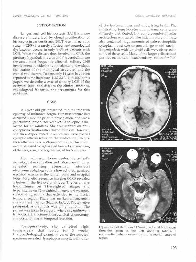
Upon admission to our center, the patient'sneurological examination and laboratory findingsrevealed nothing abnormal. Interictalelectroencephalogra ph y showed disorganizedelectrical activity in the left temporal and occipitallobes. Magnetic resonance imaging (MRI) revealeda lesion in the left occipital lobe. The lesion washypointense on Tl-weighted images andhyperintense on T2-weighted images, and we notedsurrounding edema that extended to the mesialtemporal region. There was marked enhancementafter contrast injection (Figures la, b, c). The tentativepreoperative diagnosis was ganglioglioma. Thepatient was taken to surgery, where she underwentleft occipital craniotomy, transoccipitallesionectomy,and posterior mesial temporal resection.
Postoperatively, she exhibited righthemiparesia that lasted for 3 weeks.Histopathological examination of the surgicalspecimen revealed lymphoplasmacytic infiltration
O=yurl: lufrncrtllliai Hi,fiocylo.<is
of the leptomeninges and underlying brain. Theinfiltrating lymphocytes and plasma cells werediffusely distributed, but some pseudofolliculararchitecture was noted. The inflammatory infiltratealso contained large amounts of pale eosinophiliccytoplasm and one or more large ovoid nuclei.Emperipolesis with lymphoid cells were observed insome of these cells. Many of the larger cells stainedpositive on immunohistochemistry studies for S100
Figures la and lb: Tl- and T2-weighted axial MR imagesshow the lesion in the left occipital lobe, withsurrounding edema extending to the mesial temporalregion,
103

Tlirkish Neliroslirgery 12: 102 - 106, 2002
Figure lc: Striking enhancement of the lesion aftercontrast injection.
and CD68, and showed weak positivity for CD1.Positivity for CD1 was confirmed by studies withpeanut agglutinin, which stained the cells strongly.Immunohistochemical staining for Ki67, a knownmarker of cellular proliferation, highlighted amoderate proportion of the nuclei of the all cellswithin the lesion. All the features mentioned above
were with Langerhans' cell histiocytosis.
A pediatric hematologist consulted the patientduring her postoperative course, and no systemicfindings of the disease were detected. Adjuvanttherapy was not considered. At 10 months postsurgery, she showed no clinical or radiographicevidence of recurrence (Figures 2a, b, c).
DISCUSSION
LCH is a disease characterized by abnormalproliferation of histiocytes in one or many organs.
Figures 2a and 2b: At the tenth postoperative month, Tl- and T2- weighted axial MR images, respectively; 2c: safittal MRimage revealed no recurrence.
104

Turkish Neurosurgery 12: 102 - 106, 2002
The skeletal system, skin, lung, liver, CNS, spleen,reticuloendothelial system, and gastrointestinal tractare the sites most often involved (17). Although thecause of this illness is not clear, immunologicalabnormalities such as decreased suppressor T-cellactivity, increased immunoglobulin synthesis, anddecreased thymic function have been documentedin affected individuals (12,14,15). Severalinvestigators have detected clonal CD1a+ histiocytesin all the LCH lesions they tested, which suggeststhat the condition may be a clonal neoplastic disorderwith highly variable biological behavior and clinicalseverity (21). Recent reviews have proposed furtherstratification of the clinical form of the disease. Oneproposed system is based on extent of disease, withcategories listed as "restricted versus extensive, withor without organ dysfunction" (2). A second is basedon location, with categories of "skin isolated,monostotic, poliostotic, and multisystem disease"(19).
The most common intracranial site of LCH
involvement is the hypothalamus-pituitary axis, andthe clinical presentation in these cases is the triad ofdiabetes insipidus, exophthalmus, and a lytic skulllesion (6,9). The second most frequent intracranialsite is the cerebellopontine pathway (6).
Imaging material from an LCH-CNS studydone on 38 patients in 1997 illustrates the wide arrayof parenchymal and extraparenchymallesions thatcan occur within the CNS (5).Most of the cases (87%)exhibited more than one type of lesion. The MRIappearances of the lesions were not specific for LCH,and the diagnoses could not be made withoutassociated clinical features. Since these lesions can
be located in the gray or white matter, and they mayor may not show contrast enhancement, the list ofdifferential diagnoses for LCH is large. It includessuch diverse pathologies as multiple sclerosis, acutedisseminated encephalomyelitis, leukodystrophy,infection, metastasis, sarcoidosis, infarction, andneoplasia (6). As mentioned above, the finding of asolitary space-occupying LCH lesion in the CNS thatdoes not involve the hypothalamus, meninges, orcranial vault is very rare. To our knowledge, only 14such cases have been reported in the literature(1,3,7,8,10,11,13,18). Most of these lesions were infrontal, temporal, choroidal, parieto-occipital,parietal, and insular locations, and the majority ofpatients had no other systemic findings.
From a surgical point of view, pure solitary nonhypothalamic LCH-CNS lesions appear to be a
Q:yurl: Illlracrallial HislioCl/I,)"i,
distinct form that can be totally excised. As a result,the prognosis is favorable. Recent reports of caseswith follow-up periods as long as 17years have notedsuccess with surgical excision alone, andradiotherapy and chemotherapy have only beenadvocated in situations where subtotal resection was
performed (4,7,13). Among the reports in theliterature, only one describes local recurrence andone describes a fatal outcome (16,18).
In conclusion, there are too few documentedcases of solitary non-hypothalamic LCH-CNS lesionsto make definitive decisions about adjuvant therapy;however, the outcomes in our case and others suggestthat surgical excision with close follow-up is a safeand adequate treatment.
Correspondence: Prof. Dr. Emin OzyurtCerrahpasa Tip FakiiltesiBeyin Cerrahisi ABDP.K. 4, 34301 CerrahpasaIstanbul, TurkeyE-mail: [email protected] Number: 0090-212-6320026
REFERENCES
1. Berkmann M, Yuan Y, Bruck W, Palm KV: SolitaryLangerhans' cell histiocytosis lesion of the parietooccipital lobe: A case report and review of the literature.Clin Neurol Neurosurg 99 (1): 50-5, 1997
2. Egeler RM, D'Angio GJ: Langerhans' cell histiocytosis.J Pediatr 127 (9):1-11,1995
3. Eriksen B, Janinis J, Variakojis D, Winter J, Russel E,Marder R, DalCanto MC: Primary histiocytosis X ofthe parieto-occipital lobe. Hum PathoI19(5): 611-4, 1988
4. Grant GA, Kim DK, Shaw CM, Berger MS: Solitaryeosinophilic granuloma of the temporal lobe: Casereport and review of the literature. Brain Tumor Pathol16(1): 55-59, 1999
5. Grois N, Broadbant V, Favara BF, et al: Report of theHistiocyte Society Workshop on "Centred NervousSystem (CNS) Disease in Langerhans' CellHistiocytosis (LCH)" Med Pediatr On Call 29:73-8,1997
6. Grois NG, Favara BE,Mostbeck GH, Prayer D: CentralNervous System Disease in Langerhans' CellHistiocytosis. In: Egeler RM, D' Angio GJ (eds).Hematology /Oncology Clinics of North America. WBSaunders Company, Philadelphia, 1998, pp. 287-305
7. Hund E, Steiner H, Jonsen 0, Sievert H, Sohl G, EssingM: Treatment of Langerhans' cell histiocytosis. J NeuralSci 171(2):145-52, 1999
8. Itoh H, Waga S, Kojima T, Hoshino T: Solitaryeosinophilic granuloma in the frontal lobe: a casereport. Neurosurgery 30(2): 295-98, 1992
9. Kepes JJ, Kepes M: Predominantly cerebral forms ofhistiocytosis-X. Acta Neuropathol (Bern) 14:77-98, 1969
105

TurkislJ Neurosurgery 12: 102 - 106, 2002
10. Khan A, Fulco JD, Shende A, Rosenthal A, Marc JA:
Focal histiocytosis X of the parietal lobe. Case report. JNeurosurg 52(3): 431-33, 1980
11. Kim EY, ChOl JU, Kim TS, Kim or, Kim KY: Huge
Langerhans' cell histiocytosis granuloma of the choroidplexus in a child with Hand-Schuller Christian disease.Case report. J Neurosurg 83(6): 1080-84, 1995
12. Lahey ME, Heyn R, Ladisch S, Leikin S, Neerhoul R,Newler W, Shere N, Smith D, Hamend D:
Hypergammaglobulinemia in histiocytosis X. J Pediatr107: 572-74, 1985
13. Montine TJ, Hollensead SC, Ellis WG, Martin JS, Moffat
EJ, Burder PC: Solitary eosinophilic granuloma of thetemporal lobe: Case report and long-term follow-upof previous reported cases. Clin Neuropathol13: 225228, 1994
14. Nesbit ME Jr, O'Lary M, Dehner LP, Ramsay NK: Theimmune system and the histiocytosis syndromes. AmJ Pediatr Hematol Oncol3: 141-44, 1981
15. Newton WA, Hammodui AB, Shannon BT: Role of the
thymus in histiocytosis-X. Hematol Oncol Clin NorthAm 1: 63-74, 1987
16. Sivalingam S, Corkill G, Ellis WG, Claiche JR: Focal
Ozyurl: Illtracrallial Hisli,)cl/lo,i"
eosinophilic granuloma of the temporal lobe. Casereport. J Neurosurg 47: 941-45, 1977
17. Vaughan VC, McKay RJ, Behirman RE: NelsonTextbook of Pediatrics, eleventh edition, Philadelphia,WB Saunders Company, pp 1983-86, 1979
18. Vital A, Loisau H, Kantor G, Vital C, Cohadon F:
Primary Langerhans' cell histiocytosis of the centralnervous system with fatal outcome, a case report. JNeurosurg 85: 1156-60, 1996
19. Willis B, Ablin A, Weinberg V, Zoger S, Wara WM,Matthay KK: Disease course and late sequelae ofLangerhans' cell histiocytosis: 25-year experience at theuniversity of California, San Francisco. J Clin Oncol14: 2073-82, 1996
20. Willman CL, Busque L, Griffit BB, Favara BE, McClainKL, Duncan MH, Gilliand DG: Langerhans'-cellhistiocytosis (histiocytosis-X) - A clonal proliferativedisease. N Engl J Med 331: 154-160, 1994
21. Willman CL, Mclain KL: An Update on Clonality,Cytokines, and Viral Etiology in Langerhans' CellHistiocytosis. In: Egeler RM, D' Angio GJ (eds).Hematology /Oncology Clinics of North America, WBSaunders Company, Philadelphia, 1998, pp: 287-305
J Bone Joint Surg Br 2002 Aug;84(6):870-2
Eosinophilic granuloma. A different behaviour tn childrenthan in adults.
Plasschaert F, Craig C, Bell R, Cole WG, Wunder JS,Alman BA.
Localised Langerhans-cell histiocytosis of bone (eosinophilicgranuloma) is a benign tumour-like condition with a variableclinical course. Different forms of treatment have been
reported to give satisfactory results. No recurrences werenoted In the skeletally immature group even after biopsyalone. By contrast, four of 13 skeletally mature patients hada recurrence and required further surgery. This suggests thateosinophilic granuloma has a low rate of recurrence Inskeletally immature patients.
106


