The Human Digestive System. Incisors: Soft palate: Tongue: Uvula:

Journal of Feline Medicine and Surgery (2011) 13, 594e596doi:10.1016/j.jfms.2011.01.018
CASE REPORTSoft palate cyst in a cat
Nathan W Peterson DVM, Dipl ACVECC*, Nicole J Buote DVM, Dipl ACVS
VCA California Animal HospitalVeterinary Specialty Group, 1736 SSepulveda Blvd, Ste A & B, LosAngeles, CA 90025, USA
*Corresponding author. E-mail: npeters
1098-612X/11/080594+03 $36.00/0
A 17-month-old cat presented for frequent swallowing, occasional cough andstertor. Radiographic evaluation revealed a soft tissue mass at the level of the softpalate. Oral examination revealed a soft tissue mass within the soft palate.Surgical resection was performed. Histopathology was consistent with a largedevelopmental cyst. Following surgical management the cat has remainedclinically free of signs for 2 months.
Date accepted: 13 January 2011 � 2011 ISFM and AAFP. Published by Elsevier Ltd. All rights reserved.
The soft palate forms the floor of the nasophar-ynx and the roof of the oropharynx. It is a mo-bile, valve-like structure that separates and
protects the proximal airway during swallowing aswell as closing off the oral cavity during nose breath-ing. The importance of this separation is highlightedby the fact that the mucous membrane of the dorsalsurface of the soft palate is of respiratory type, whiledigestive type is present on the ventral surface.1 Be-cause of this structures important role in protectingthe airway, any disease process that causes inflamma-tion, swelling or damage to the linings may impedenot only swallowing but breathing as well. Althoughpharyngeal masses with similar clinical signs havepreviously been described in cats, to the authors’knowledge there have been no published reports ofa soft palate cyst in a cat.
A 17-month-old indoor only female spayed domes-tic shorthair cat was presented for evaluation of fre-quent swallowing and sonorous breathing withoccasional cough. The owner reported that the sterto-rous sounds were worse when the cat was sleepingand had been present and stable since the time ofadoption 6 weeks prior. No sneezing or nasal dis-charge was reported and the cat exhibited a good ap-petite at home. There was no report of exerciseintolerance or respiratory distress. The cat had beenpreviously treated for presumed pneumonia by the re-ferring veterinarian with amoxicillin, marbofloxacin,prednisolone and lysine, without improvement inclinical signs.
The cat was bright and alert upon presentation. Ini-tial physical examination was within normal limits ex-cept for a grade 2/6 focal systolic murmur and a soft
� 2011 ISFM a
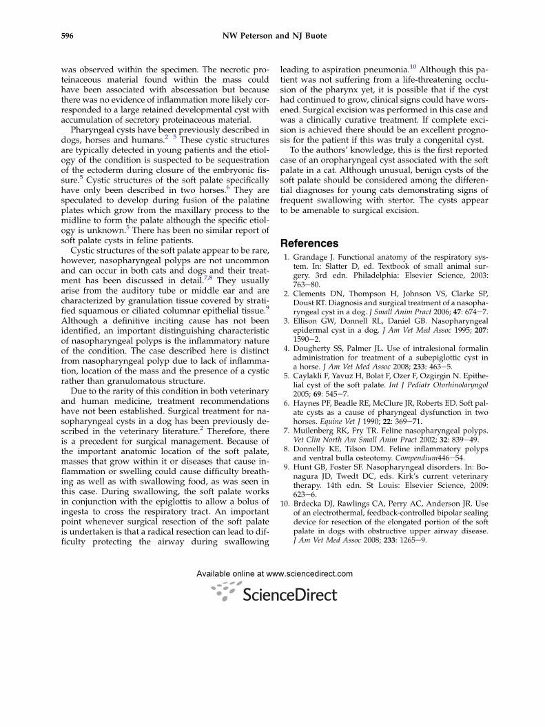
upper respiratory noise on inhalation. A limited non-sedated oral examination was performed and wasunremarkable. Non-sedated orthogonal thoracic ra-diographs were obtained and revealed a mild to mod-erate bronchial pattern. The cardiac silhouette wasconsidered to be normal. A single lateral cervical ra-diograph was obtained and revealed an abnormalsoft tissue opacity in the caudal pharyngeal regionthat appeared to be associated with the caudal aspectof the soft palate (Fig 1). Differential diagnoses for thissoft tissue opacity included soft palate mass, nasopha-ryngeal polyp, foreign body or neoplasia. An echocar-diogram was performed due to the presence of themurmur and revealed no structural heart disease. Acomplete blood count, chemistry panel, thyroid paneland urinalysis were performed and were within nor-mal limits. A feline leukemia and feline immunodefi-ciency antibody test were both negative.
The following day the cat was anesthetized fora complete oral examination with propofol (5 mg/kgIV) and maintained on 1.5e2% isoflurane in oxygen,intravenous fluid support was provided at a rate of40 ml/h (10 ml/kg/h). The examination revealeda firm mass approximately 2 cm� 1 cm within thedorsal and ventral mucosal surfaces of the soft palate.The ventral mucosa of the soft palate had no evidenceof ulceration, damage, erythema, or mass lesions. Noother abnormalities were found in the oropharyngealregion except for a mildly enlarged left pharyngealtonsil. Biopsy or excision of the mass was recommen-ded. The cat recovered uneventfully from this proce-dure and surgery was scheduled for the followingday. The following day the patient was premedicatedwith buprenorphine (0.015 mg/kg IM), and was in-duced with diazepam 0.5 mg/kg IV and propofol4 mg/kg IV. Anesthesia was maintained with 1e2%
nd AAFP. Published by Elsevier Ltd. All rights reserved.

Fig 1. Lateral cervical radiograph reveals a soft tissue opacity associated with the caudal aspect of the soft palate (arrows).
595Soft palate cyst
isoflurane in oxygen and intravenous fluid supportwas provided at a rate of 40 ml/h (10 m/kg). The pa-tient was placed in sternal recumbency, a mouth gagwas placed and tape was placed under both upper ca-nines to aid in visualization of the soft palate. The cau-dal pharynx was packed off with moistened gauzeand a stay suture of 3-0 Monocryl was placed at theventral aspect of the soft palate for retraction. Theventral mucosa of the soft palate was transectedwith metzenbaum scissors which revealed a well cir-cumscribed green, encapsulated mass. The mass wasbluntly and sharply dissected from the soft palate inits entirety (Fig 2). The visible dorsal aspect of thesoft palate appeared edematous and was removedalong with the mass. The mucosal defect was closedin one layer with 4-0 Monocryl in a simple continuouspattern. The remaining soft palate reached the midtonsillar crypts bilaterally. The mass and soft palate
Fig 2. Cross section of the cyst following surgical resec
mucosa were submitted for histopathologic review.Recovery from anesthesia was unremarkable. Dexa-methasone SP (0.1 mg/kg IV) was given to help pre-vent postoperative swelling. There was animmediate improvement in stertor. Post-surgicallythe patient was treated with buprenorphine(0.01 mg/kg IV q 8 h). The patient was dischargedthe following day with tramadol (3 mg/kg PO q 8 has needed). At recheck examination 14 days later,the owner reported complete resolution of clinicalsigns at home. A non-sedated oral exam was per-formed and no obvious abnormalities with the softpalate incision were seen.
Biopsy results revealed mild mucosal hyperplasiawith submucosal edema and focally extensive degener-ate/necrotic cystic material. No inflammation was ob-served in the unaffected submucosa or mucosaltissue. No evidence of malignancy or infectious agents
tion. Note the proteinaceous material within the cyst.
596 NW Peterson and NJ Buote
was observed within the specimen. The necrotic pro-teinaceous material found within the mass couldhave been associated with abscessation but becausethere was no evidence of inflammation more likely cor-responded to a large retained developmental cyst withaccumulation of secretory proteinaceous material.
Pharyngeal cysts have been previously described indogs, horses and humans.2�5 These cystic structuresare typically detected in young patients and the etiol-ogy of the condition is suspected to be sequestrationof the ectoderm during closure of the embryonic fis-sure.5 Cystic structures of the soft palate specificallyhave only been described in two horses.6 They arespeculated to develop during fusion of the palatineplates which grow from the maxillary process to themidline to form the palate although the specific etiol-ogy is unknown.5 There has been no similar report ofsoft palate cysts in feline patients.
Cystic structures of the soft palate appear to be rare,however, nasopharyngeal polyps are not uncommonand can occur in both cats and dogs and their treat-ment has been discussed in detail.7,8 They usuallyarise from the auditory tube or middle ear and arecharacterized by granulation tissue covered by strati-fied squamous or ciliated columnar epithelial tissue.9
Although a definitive inciting cause has not beenidentified, an important distinguishing characteristicof nasopharyngeal polyps is the inflammatory natureof the condition. The case described here is distinctfrom nasopharyngeal polyp due to lack of inflamma-tion, location of the mass and the presence of a cysticrather than granulomatous structure.
Due to the rarity of this condition in both veterinaryand human medicine, treatment recommendationshave not been established. Surgical treatment for na-sopharyngeal cysts in a dog has been previously de-scribed in the veterinary literature.2 Therefore, thereis a precedent for surgical management. Because ofthe important anatomic location of the soft palate,masses that grow within it or diseases that cause in-flammation or swelling could cause difficulty breath-ing as well as with swallowing food, as was seen inthis case. During swallowing, the soft palate worksin conjunction with the epiglottis to allow a bolus ofingesta to cross the respiratory tract. An importantpoint whenever surgical resection of the soft palateis undertaken is that a radical resection can lead to dif-ficulty protecting the airway during swallowing
leading to aspiration pneumonia.10 Although this pa-tient was not suffering from a life-threatening occlu-sion of the pharynx yet, it is possible that if the cysthad continued to grow, clinical signs could have wors-ened. Surgical excision was performed in this case andwas a clinically curative treatment. If complete exci-sion is achieved there should be an excellent progno-sis for the patient if this was truly a congenital cyst.
To the authors’ knowledge, this is the first reportedcase of an oropharyngeal cyst associated with the softpalate in a cat. Although unusual, benign cysts of thesoft palate should be considered among the differen-tial diagnoses for young cats demonstrating signs offrequent swallowing with stertor. The cysts appearto be amenable to surgical excision.
References1. Grandage J. Functional anatomy of the respiratory sys-
tem. In: Slatter D, ed. Textbook of small animal sur-gery. 3rd edn. Philadelphia: Elsevier Science, 2003:763e80.
2. Clements DN, Thompson H, Johnson VS, Clarke SP,Doust RT. Diagnosis and surgical treatment of a nasopha-ryngeal cyst in a dog. J Small Anim Pract 2006; 47: 674e7.
3. Ellison GW, Donnell RL, Daniel GB. Nasopharyngealepidermal cyst in a dog. J Am Vet Med Assoc 1995; 207:1590e2.
4. Dougherty SS, Palmer JL. Use of intralesional formalinadministration for treatment of a subepiglottic cyst ina horse. J Am Vet Med Assoc 2008; 233: 463e5.
5. Caylakli F, Yavuz H, Bolat F, Ozer F, Ozgirgin N. Epithe-lial cyst of the soft palate. Int J Pediatr Otorhinolaryngol2005; 69: 545e7.
6. Haynes PF, Beadle RE, McClure JR, Roberts ED. Soft pal-ate cysts as a cause of pharyngeal dysfunction in twohorses. Equine Vet J 1990; 22: 369e71.
7. Muilenberg RK, Fry TR. Feline nasopharyngeal polyps.Vet Clin North Am Small Anim Pract 2002; 32: 839e49.
8. Donnelly KE, Tilson DM. Feline inflammatory polypsand ventral bulla osteotomy. Compendium446e54.
9. Hunt GB, Foster SF. Nasopharyngeal disorders. In: Bo-nagura JD, Twedt DC, eds. Kirk’s current veterinarytherapy. 14th edn. St Louis: Elsevier Science, 2009:623e6.
10. Brdecka DJ, Rawlings CA, Perry AC, Anderson JR. Useof an electrothermal, feedback-controlled bipolar sealingdevice for resection of the elongated portion of the softpalate in dogs with obstructive upper airway disease.J Am Vet Med Assoc 2008; 233: 1265e9.
Available online at www.sciencedirect.com