
Small-molecule targeted recruitment of a nuclease to ...Small-molecule targeted recruitment of a...
6
Small-molecule targeted recruitment of a nuclease to cleave an oncogenic RNA in a mouse model of metastatic cancer Matthew G. Costales a , Haruo Aikawa a , Yue Li a , Jessica L. Childs-Disney a , Daniel Abegg a , Dominic G. Hoch a , Sai Pradeep Velagapudi a , Yoshio Nakai a , Tanya Khan a , Kye Won Wang b , Ilyas Yildirim b , Alexander Adibekian a , Eric T. Wang c , and Matthew D. Disney a,1 a Department of Chemistry, The Scripps Research Institute, Jupiter, FL 33458; b Department of Chemistry and Biochemistry, Florida Atlantic University, Jupiter, FL 33458; and c Department of Molecular Genetics & Microbiology, University of Florida, Gainesville, FL 32610 Edited by Vern L. Schramm, Albert Einstein College of Medicine, Bronx, NY, and approved December 12, 2019 (received for review August 17, 2019) As the area of small molecules interacting with RNA advances, general routes to provide bioactive compounds are needed as ligands can bind RNA avidly to sites that will not affect function. Small-molecule targeted RNA degradation will thus provide a general route to affect RNA biology. A non–oligonucleotide-containing com- pound was designed from sequence to target the precursor to on- cogenic microRNA-21 (pre–miR-21) for enzymatic destruction with selectivity that can exceed that for protein-targeted medicines. The compound specifically binds the target and contains a heterocycle that recruits and activates a ribonuclease to pre–miR-21 to substoi- chiometrically effect its cleavage and subsequently impede metasta- sis of breast cancer to lung in a mouse model. Transcriptomic and proteomic analyses demonstrate that the compound is potent and selective, specifically modulating oncogenic pathways. Thus, small molecules can be designed from sequence to have all of the func- tional repertoire of oligonucleotides, including inducing enzymatic degradation, and to selectively and potently modulate RNA function in vivo. nucleic acids | RNA | chemical biology | metastatic | cancer R NA structures are key players in important biological pro- cesses and in diseased states. The only general way to target RNA, however, is by using oligonucleotide-based approaches that preferentially target unstructured regions (1). Because RNA biology is often mediated by the structure that it forms (2), approaches to target structured RNAs are advantageous. Small molecules inter- acting with an RNA’s 3D structure could allow specificity in activity. One class of structured RNAs that play roles in human disease biology is noncoding microRNAs (miRs) (3). They are produced from highly structured precursors processed in the nucleus (pri- miRs) and cytoplasm (pre-miRs) by the nucleases Drosha and Dicer, respectively (Fig. 1A). An important example is miR-21 as its expression in solid tumors negatively correlates with survival in triple-negative breast cancer (TNBC) patients (Fig. 1A) (4). Herein, we describe a general strategy to endow small mole- cules to achieve targeted degradation of RNA transcripts through ribonuclease recruitment. These studies demonstrate that sequence- based design can afford small molecules that can target struc- tured RNAs for enzymatic cleavage, a feature previously only known to oligonucleotides. Results and Discussion A sequence-based design approach termed Inforna (Fig. 1B) was used to design small molecules that target the 3D folds in pre– miR-21. Inforna uses the output of folded RNA structures that bind small molecules as determined from a library-versus-library selection (5). This analysis identified a fragment (1, Fig. 2) that bound the miR-21 Dicer site selectively with a K d of 20 μM and inhibited in vitro Dicer processing (SI Appendix, Figs. S1 and S2). Treatment of TNBC cells (MDA-MB-231) with 1 (10 μM) inhibited miR-21 production by 50%, while the levels of pre–miR-21 were increased by 1.3-fold, as expected for a compound that acts by inhibiting Dicer processing (SI Appendix, Fig. S3). Full miR profiling showed that 1 was modestly selective (Fig. 1D). To optimize 1 for avidity, the RNA folds in all miR precursors in the human transcriptome were compared to pre–miR-21 (SI Appendix, Figs. S4–S6). Several miR precursors display the A bulge motif (5′ G AC/3′ C_G; n = 20), yet no other targets con- tained it and the adjacent U bulge (5′ U UG/3′ A_C) (SI Appendix, Figs. S4 and S5). Fortuitously, fragment 1 bound to both sites and assembly of two fragments of 1 to target them in a single com- pound afforded 2 (Figs. 1C and 2 and SI Appendix, Fig. S3), which selectively bound pre–miR-21 with a 20-fold enhancement over 1 (SI Appendix, Fig. S1). Target engagement of 2 to pre–miR-21 in vitro and in MDA- MB-231 cells was confirmed by using Chemical Cross-Linking and Isolation by Pulldown (Chem-CLIP), a strategy that utilizes a proximity-based reaction to cross-link compounds to their cellular targets (6). Cells were treated with the active Chem-CLIP probe or an inactive control probe lacking RNA binding modules (SI Significance The human genome produces RNAs that do not code for pro- tein but play important roles in biology, including causing dis- ease. These RNAs are potential drug targets. Estimates suggest that there are 100-fold more potential RNA than protein drug targets. Despite this potential, small-molecule targeting of hu- man RNA is rare as it is technically challenging. Here we describe a general and fast strategy to design small molecules from se- quence to bind an RNA and subsequently cause its destruction. The approach was proven to destroy a cancer-causing RNA in a mouse model thereby inhibiting metastasis. Armed with these approaches, we can more deeply evaluate the potential of small- molecule therapeutics targeting RNAs. Author contributions: M.G.C., H.A., Y.L., J.L.C.-D., S.P.V., Y.N., and M.D.D. designed re- search; M.G.C., H.A., Y.L., J.L.C.-D., D.A., D.G.H., S.P.V., Y.N., T.K., K.W.W., I.Y., and A.A. performed research; M.G.C., H.A., Y.L., J.L.C.-D., S.P.V., Y.N., and E.T.W. analyzed data; M.D.D. conceived of the ideas and directed the study; and M.G.C., H.A., Y.L., J.L.C.-D., and M.D.D. wrote the paper. Competing interest statement: M.D.D. is a founder of Expansion Therapeutics and M.D.D. and E.T.W. are consultants for Expansion Therapeutics. This article is a PNAS Direct Submission. This open access article is distributed under Creative Commons Attribution-NonCommercial- NoDerivatives License 4.0 (CC BY-NC-ND). Data deposition: The data reported in this paper have been deposited in the Cambridge Crystallographic Data Centre (CCDC), https://www.ccdc.cam.ac.uk/, under reference number CCDS1912054. 1 To whom correspondence may be addressed. Email: [email protected]. This article contains supporting information online at https://www.pnas.org/lookup/suppl/ doi:10.1073/pnas.1914286117/-/DCSupplemental. First published January 21, 2020. 2406–2411 | PNAS | February 4, 2020 | vol. 117 | no. 5 www.pnas.org/cgi/doi/10.1073/pnas.1914286117 Downloaded by guest on April 11, 2020
Transcript of Small-molecule targeted recruitment of a nuclease to ...Small-molecule targeted recruitment of a...
Small-molecule targeted recruitment of a nuclease tocleave an oncogenic RNA in a mouse model ofmetastatic cancerMatthew G. Costalesa, Haruo Aikawaa, Yue Lia, Jessica L. Childs-Disneya, Daniel Abegga, Dominic G. Hocha, Sai PradeepVelagapudia, Yoshio Nakaia, Tanya Khana, Kye Won Wangb, Ilyas Yildirimb, Alexander Adibekiana, Eric T. Wangc,and Matthew D. Disneya,1
aDepartment of Chemistry, The Scripps Research Institute, Jupiter, FL 33458; bDepartment of Chemistry and Biochemistry, Florida Atlantic University,Jupiter, FL 33458; and cDepartment of Molecular Genetics & Microbiology, University of Florida, Gainesville, FL 32610
Edited by Vern L. Schramm, Albert Einstein College of Medicine, Bronx, NY, and approved December 12, 2019 (received for review August 17, 2019)
As the area of small molecules interacting with RNA advances,general routes to provide bioactive compounds are needed asligands can bind RNA avidly to sites that will not affect function.Small-molecule targeted RNA degradationwill thus provide a generalroute to affect RNA biology. A non–oligonucleotide-containing com-pound was designed from sequence to target the precursor to on-cogenic microRNA-21 (pre–miR-21) for enzymatic destruction withselectivity that can exceed that for protein-targeted medicines. Thecompound specifically binds the target and contains a heterocyclethat recruits and activates a ribonuclease to pre–miR-21 to substoi-chiometrically effect its cleavage and subsequently impede metasta-sis of breast cancer to lung in a mouse model. Transcriptomic andproteomic analyses demonstrate that the compound is potent andselective, specifically modulating oncogenic pathways. Thus, smallmolecules can be designed from sequence to have all of the func-tional repertoire of oligonucleotides, including inducing enzymaticdegradation, and to selectively and potently modulate RNA functionin vivo.
nucleic acids | RNA | chemical biology | metastatic | cancer
RNA structures are key players in important biological pro-cesses and in diseased states. The only general way to target
RNA, however, is by using oligonucleotide-based approaches thatpreferentially target unstructured regions (1). Because RNA biologyis often mediated by the structure that it forms (2), approaches totarget structured RNAs are advantageous. Small molecules inter-acting with an RNA’s 3D structure could allow specificity in activity.One class of structured RNAs that play roles in human disease
biology is noncoding microRNAs (miRs) (3). They are producedfrom highly structured precursors processed in the nucleus (pri-miRs) and cytoplasm (pre-miRs) by the nucleases Drosha andDicer, respectively (Fig. 1A). An important example is miR-21as its expression in solid tumors negatively correlates withsurvival in triple-negative breast cancer (TNBC) patients (Fig. 1A)(4). Herein, we describe a general strategy to endow small mole-cules to achieve targeted degradation of RNA transcripts throughribonuclease recruitment. These studies demonstrate that sequence-based design can afford small molecules that can target struc-tured RNAs for enzymatic cleavage, a feature previously onlyknown to oligonucleotides.
Results and DiscussionA sequence-based design approach termed Inforna (Fig. 1B) wasused to design small molecules that target the 3D folds in pre–miR-21. Inforna uses the output of folded RNA structures thatbind small molecules as determined from a library-versus-libraryselection (5). This analysis identified a fragment (1, Fig. 2) thatbound the miR-21 Dicer site selectively with a Kd of 20 μM andinhibited in vitro Dicer processing (SI Appendix, Figs. S1 and S2).Treatment of TNBC cells (MDA-MB-231) with 1 (10 μM) inhibited
miR-21 production by 50%, while the levels of pre–miR-21 wereincreased by 1.3-fold, as expected for a compound that acts byinhibiting Dicer processing (SI Appendix, Fig. S3). Full miRprofiling showed that 1 was modestly selective (Fig. 1D).To optimize 1 for avidity, the RNA folds in all miR precursors
in the human transcriptome were compared to pre–miR-21 (SIAppendix, Figs. S4–S6). Several miR precursors display the Abulge motif (5′ GAC/3′ C_G; n = 20), yet no other targets con-tained it and the adjacent U bulge (5′UUG/3′ A_C) (SI Appendix,Figs. S4 and S5). Fortuitously, fragment 1 bound to both sites andassembly of two fragments of 1 to target them in a single com-pound afforded 2 (Figs. 1C and 2 and SI Appendix, Fig. S3), whichselectively bound pre–miR-21 with a 20-fold enhancement over1 (SI Appendix, Fig. S1).Target engagement of 2 to pre–miR-21 in vitro and in MDA-
MB-231 cells was confirmed by using Chemical Cross-Linkingand Isolation by Pulldown (Chem-CLIP), a strategy that utilizesa proximity-based reaction to cross-link compounds to their cellulartargets (6). Cells were treated with the active Chem-CLIP probeor an inactive control probe lacking RNA binding modules (SI
Significance
The human genome produces RNAs that do not code for pro-tein but play important roles in biology, including causing dis-ease. These RNAs are potential drug targets. Estimates suggestthat there are 100-fold more potential RNA than protein drugtargets. Despite this potential, small-molecule targeting of hu-man RNA is rare as it is technically challenging. Here we describea general and fast strategy to design small molecules from se-quence to bind an RNA and subsequently cause its destruction.The approach was proven to destroy a cancer-causing RNA in amouse model thereby inhibiting metastasis. Armed with theseapproaches, we can more deeply evaluate the potential of small-molecule therapeutics targeting RNAs.
Author contributions: M.G.C., H.A., Y.L., J.L.C.-D., S.P.V., Y.N., and M.D.D. designed re-search; M.G.C., H.A., Y.L., J.L.C.-D., D.A., D.G.H., S.P.V., Y.N., T.K., K.W.W., I.Y., and A.A.performed research; M.G.C., H.A., Y.L., J.L.C.-D., S.P.V., Y.N., and E.T.W. analyzed data;M.D.D. conceived of the ideas and directed the study; and M.G.C., H.A., Y.L., J.L.C.-D., andM.D.D. wrote the paper.
Competing interest statement: M.D.D. is a founder of Expansion Therapeutics and M.D.D.and E.T.W. are consultants for Expansion Therapeutics.
This article is a PNAS Direct Submission.
This open access article is distributed under Creative Commons Attribution-NonCommercial-NoDerivatives License 4.0 (CC BY-NC-ND).
Data deposition: The data reported in this paper have been deposited in the CambridgeCrystallographic Data Centre (CCDC), https://www.ccdc.cam.ac.uk/, under referencenumber CCDS1912054.1To whom correspondence may be addressed. Email: [email protected].
This article contains supporting information online at https://www.pnas.org/lookup/suppl/doi:10.1073/pnas.1914286117/-/DCSupplemental.
First published January 21, 2020.
2406–2411 | PNAS | February 4, 2020 | vol. 117 | no. 5 www.pnas.org/cgi/doi/10.1073/pnas.1914286117
Dow
nloa
ded
by g
uest
on
Apr
il 11
, 202
0
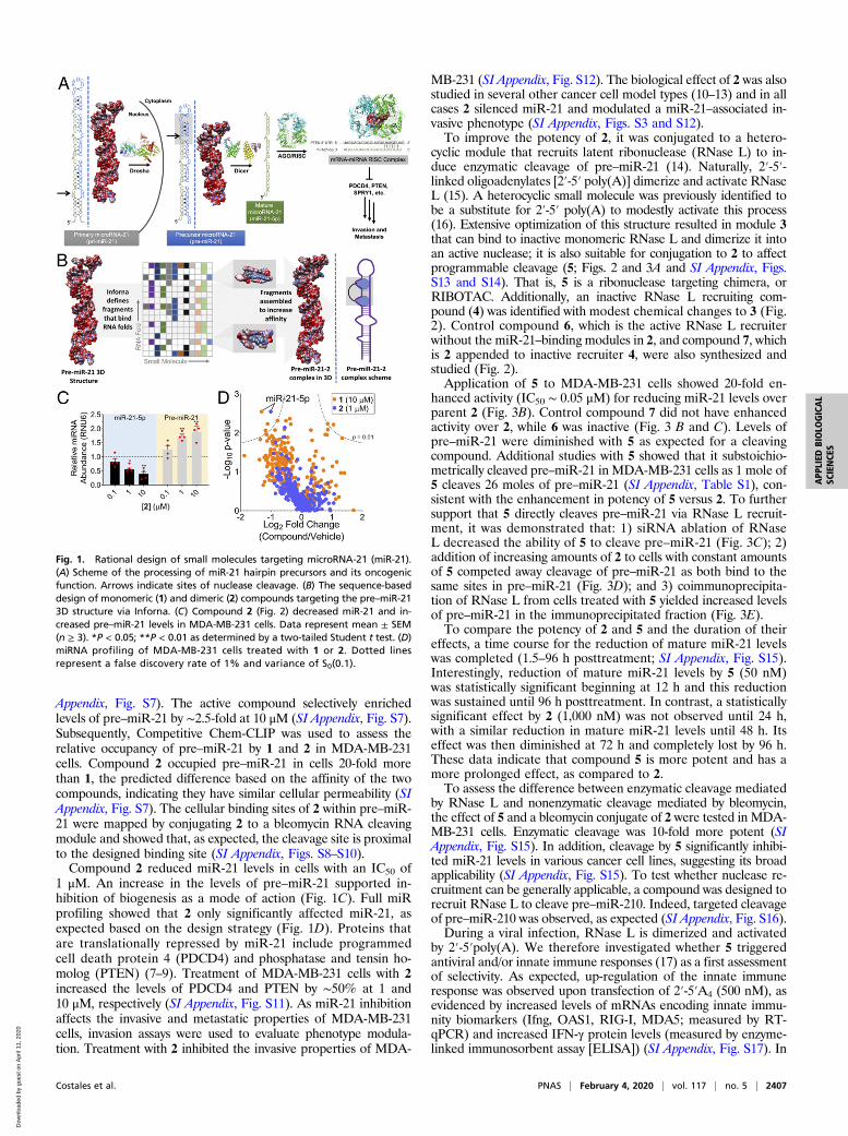
Appendix, Fig. S7). The active compound selectively enrichedlevels of pre–miR-21 by ∼2.5-fold at 10 μM (SI Appendix, Fig. S7).Subsequently, Competitive Chem-CLIP was used to assess therelative occupancy of pre–miR-21 by 1 and 2 in MDA-MB-231cells. Compound 2 occupied pre–miR-21 in cells 20-fold morethan 1, the predicted difference based on the affinity of the twocompounds, indicating they have similar cellular permeability (SIAppendix, Fig. S7). The cellular binding sites of 2 within pre–miR-21 were mapped by conjugating 2 to a bleomycin RNA cleavingmodule and showed that, as expected, the cleavage site is proximalto the designed binding site (SI Appendix, Figs. S8–S10).Compound 2 reduced miR-21 levels in cells with an IC50 of
1 μM. An increase in the levels of pre–miR-21 supported in-hibition of biogenesis as a mode of action (Fig. 1C). Full miRprofiling showed that 2 only significantly affected miR-21, asexpected based on the design strategy (Fig. 1D). Proteins thatare translationally repressed by miR-21 include programmedcell death protein 4 (PDCD4) and phosphatase and tensin ho-molog (PTEN) (7–9). Treatment of MDA-MB-231 cells with 2increased the levels of PDCD4 and PTEN by ∼50% at 1 and10 μM, respectively (SI Appendix, Fig. S11). As miR-21 inhibitionaffects the invasive and metastatic properties of MDA-MB-231cells, invasion assays were used to evaluate phenotype modula-tion. Treatment with 2 inhibited the invasive properties of MDA-
MB-231 (SI Appendix, Fig. S12). The biological effect of 2 was alsostudied in several other cancer cell model types (10–13) and in allcases 2 silenced miR-21 and modulated a miR-21–associated in-vasive phenotype (SI Appendix, Figs. S3 and S12).To improve the potency of 2, it was conjugated to a hetero-
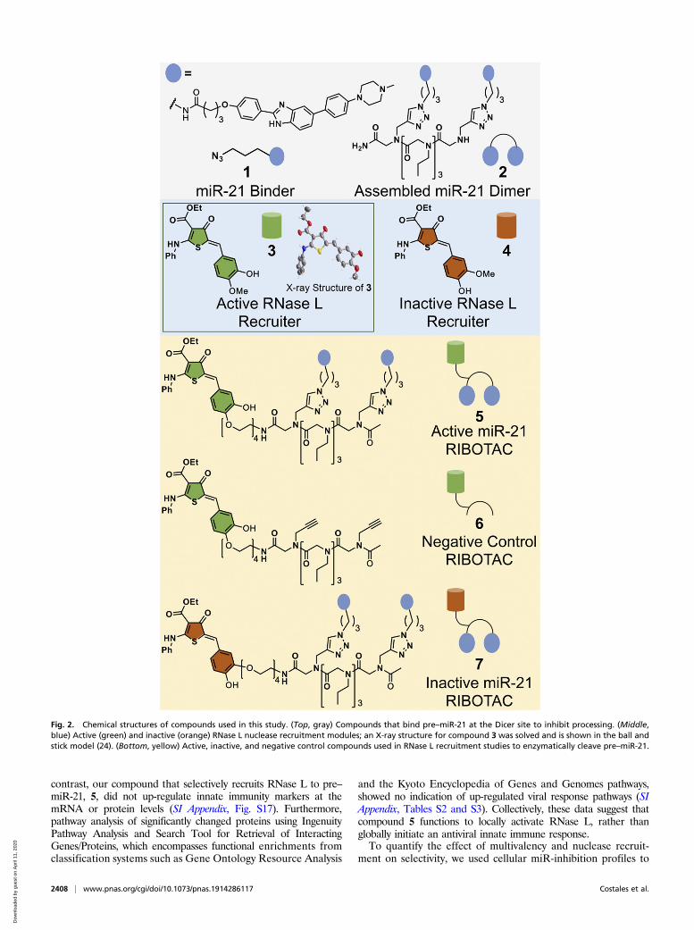
cyclic module that recruits latent ribonuclease (RNase L) to in-duce enzymatic cleavage of pre–miR-21 (14). Naturally, 2′-5′-linked oligoadenylates [2′-5′ poly(A)] dimerize and activate RNaseL (15). A heterocyclic small molecule was previously identified tobe a substitute for 2′-5′ poly(A) to modestly activate this process(16). Extensive optimization of this structure resulted in module 3that can bind to inactive monomeric RNase L and dimerize it intoan active nuclease; it is also suitable for conjugation to 2 to affectprogrammable cleavage (5; Figs. 2 and 3A and SI Appendix, Figs.S13 and S14). That is, 5 is a ribonuclease targeting chimera, orRIBOTAC. Additionally, an inactive RNase L recruiting com-pound (4) was identified with modest chemical changes to 3 (Fig.2). Control compound 6, which is the active RNase L recruiterwithout the miR-21–binding modules in 2, and compound 7, whichis 2 appended to inactive recruiter 4, were also synthesized andstudied (Fig. 2).Application of 5 to MDA-MB-231 cells showed 20-fold en-
hanced activity (IC50 ∼ 0.05 μM) for reducing miR-21 levels overparent 2 (Fig. 3B). Control compound 7 did not have enhancedactivity over 2, while 6 was inactive (Fig. 3 B and C). Levels ofpre–miR-21 were diminished with 5 as expected for a cleavingcompound. Additional studies with 5 showed that it substoichio-metrically cleaved pre–miR-21 in MDA-MB-231 cells as 1 mole of5 cleaves 26 moles of pre–miR-21 (SI Appendix, Table S1), con-sistent with the enhancement in potency of 5 versus 2. To furthersupport that 5 directly cleaves pre–miR-21 via RNase L recruit-ment, it was demonstrated that: 1) siRNA ablation of RNaseL decreased the ability of 5 to cleave pre–miR-21 (Fig. 3C); 2)addition of increasing amounts of 2 to cells with constant amountsof 5 competed away cleavage of pre–miR-21 as both bind to thesame sites in pre–miR-21 (Fig. 3D); and 3) coimmunoprecipita-tion of RNase L from cells treated with 5 yielded increased levelsof pre–miR-21 in the immunoprecipitated fraction (Fig. 3E).To compare the potency of 2 and 5 and the duration of their
effects, a time course for the reduction of mature miR-21 levelswas completed (1.5–96 h posttreatment; SI Appendix, Fig. S15).Interestingly, reduction of mature miR-21 levels by 5 (50 nM)was statistically significant beginning at 12 h and this reductionwas sustained until 96 h posttreatment. In contrast, a statisticallysignificant effect by 2 (1,000 nM) was not observed until 24 h,with a similar reduction in mature miR-21 levels until 48 h. Itseffect was then diminished at 72 h and completely lost by 96 h.These data indicate that compound 5 is more potent and has amore prolonged effect, as compared to 2.To assess the difference between enzymatic cleavage mediated
by RNase L and nonenzymatic cleavage mediated by bleomycin,the effect of 5 and a bleomycin conjugate of 2 were tested in MDA-MB-231 cells. Enzymatic cleavage was 10-fold more potent (SIAppendix, Fig. S15). In addition, cleavage by 5 significantly inhibi-ted miR-21 levels in various cancer cell lines, suggesting its broadapplicability (SI Appendix, Fig. S15). To test whether nuclease re-cruitment can be generally applicable, a compound was designed torecruit RNase L to cleave pre–miR-210. Indeed, targeted cleavageof pre–miR-210 was observed, as expected (SI Appendix, Fig. S16).During a viral infection, RNase L is dimerized and activated
by 2′-5′poly(A). We therefore investigated whether 5 triggeredantiviral and/or innate immune responses (17) as a first assessmentof selectivity. As expected, up-regulation of the innate immuneresponse was observed upon transfection of 2′-5′A4 (500 nM), asevidenced by increased levels of mRNAs encoding innate immu-nity biomarkers (Ifng, OAS1, RIG-I, MDA5; measured by RT-qPCR) and increased IFN-γ protein levels (measured by enzyme-linked immunosorbent assay [ELISA]) (SI Appendix, Fig. S17). In
Fig. 1. Rational design of small molecules targeting microRNA-21 (miR-21).(A) Scheme of the processing of miR-21 hairpin precursors and its oncogenicfunction. Arrows indicate sites of nuclease cleavage. (B) The sequence-baseddesign of monomeric (1) and dimeric (2) compounds targeting the pre–miR-213D structure via Inforna. (C) Compound 2 (Fig. 2) decreased miR-21 and in-creased pre–miR-21 levels in MDA-MB-231 cells. Data represent mean ± SEM(n ≥ 3). *P < 0.05; **P < 0.01 as determined by a two-tailed Student t test. (D)miRNA profiling of MDA-MB-231 cells treated with 1 or 2. Dotted linesrepresent a false discovery rate of 1% and variance of S0(0.1).
Costales et al. PNAS | February 4, 2020 | vol. 117 | no. 5 | 2407
APP
LIED
BIOLO
GICAL
SCIENCE
S
Dow
nloa
ded
by g
uest
on
Apr
il 11
, 202
0
contrast, our compound that selectively recruits RNase L to pre–miR-21, 5, did not up-regulate innate immunity markers at themRNA or protein levels (SI Appendix, Fig. S17). Furthermore,pathway analysis of significantly changed proteins using IngenuityPathway Analysis and Search Tool for Retrieval of InteractingGenes/Proteins, which encompasses functional enrichments fromclassification systems such as Gene Ontology Resource Analysis
and the Kyoto Encyclopedia of Genes and Genomes pathways,showed no indication of up-regulated viral response pathways (SIAppendix, Tables S2 and S3). Collectively, these data suggest thatcompound 5 functions to locally activate RNase L, rather thanglobally initiate an antiviral innate immune response.To quantify the effect of multivalency and nuclease recruit-
ment on selectivity, we used cellular miR-inhibition profiles to
Fig. 2. Chemical structures of compounds used in this study. (Top, gray) Compounds that bind pre–miR-21 at the Dicer site to inhibit processing. (Middle,blue) Active (green) and inactive (orange) RNase L nuclease recruitment modules; an X-ray structure for compound 3 was solved and is shown in the ball andstick model (24). (Bottom, yellow) Active, inactive, and negative control compounds used in RNase L recruitment studies to enzymatically cleave pre–miR-21.
2408 | www.pnas.org/cgi/doi/10.1073/pnas.1914286117 Costales et al.
Dow
nloa
ded
by g
uest
on
Apr
il 11
, 202
0
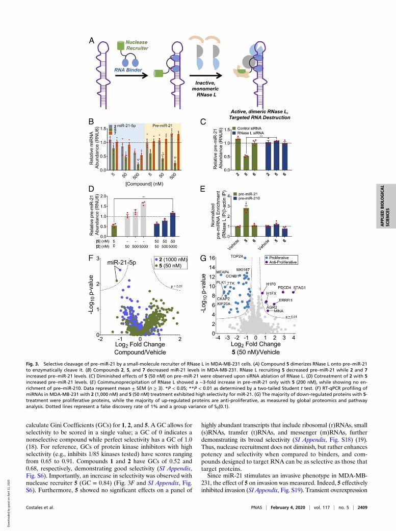
calculate Gini Coefficients (GCs) for 1, 2, and 5. A GC allows forselectivity to be scored in a single value; a GC of 0 indicates anonselective compound while perfect selectivity has a GC of 1.0(18). For reference, GCs of protein kinase inhibitors with highselectivity (e.g., inhibits 1/85 kinases tested) have scores rangingfrom 0.65 to 0.91. Compounds 1 and 2 have GCs of 0.52 and0.68, respectively, demonstrating good selectivity (SI Appendix,Fig. S6). Importantly, an increase in selectivity was observed withnuclease recruiter 5 (GC = 0.84) (Fig. 3F and SI Appendix, Fig.S6). Furthermore, 5 showed no significant effects on a panel of
highly abundant transcripts that include ribosomal (r)RNAs, small(s)RNAs, transfer (t)RNAs, and messenger (m)RNAs, furtherdemonstrating its broad selectivity (SI Appendix, Fig. S18) (19).Thus, nuclease recruitment does not diminish, but rather enhancespotency and selectivity when compared to binders, and com-pounds designed to target RNA can be as selective as those thattarget proteins.Since miR-21 stimulates an invasive phenotype in MDA-MB-
231, the effect of 5 on invasion was measured. Indeed, 5 effectivelyinhibited invasion (SI Appendix, Fig. S19). Transient overexpression
Fig. 3. Selective cleavage of pre–miR-21 by a small-molecule recruiter of RNase L in MDA-MB-231 cells. (A) Compound 5 dimerizes RNase L onto pre–miR-21to enzymatically cleave it. (B) Compounds 2, 5, and 7 decreased miR-21 levels in MDA-MB-231. RNase L recruiting 5 decreased pre–miR-21 while 2 and 7increased pre–miR-21 levels. (C) Diminished effects of 5 (50 nM) on pre–miR-21 were observed upon siRNA ablation of RNase L. (D) Cotreatment of 2 with 5increased pre–miR-21 levels. (E) Coimmunoprecipitation of RNase L showed a ∼3-fold increase in pre–miR-21 only with 5 (200 nM), while showing no en-richment of pre–miR-210. Data represent mean ± SEM (n ≥ 3). *P < 0.05; **P < 0.01 as determined by a two-tailed Student t test. (F) RT-qPCR profiling ofmiRNAs in MDA-MB-231 with 2 (1,000 nM) and 5 (50 nM) treatment exhibited high selectivity for miR-21. (G) The majority of down-regulated proteins with 5-treatment were proliferative proteins, while the majority of up-regulated proteins are anti-proliferative, as measured by global proteomics and pathwayanalysis. Dotted lines represent a false discovery rate of 1% and a group variance of S0(0.1).
Costales et al. PNAS | February 4, 2020 | vol. 117 | no. 5 | 2409
APP
LIED
BIOLO
GICAL
SCIENCE
S
Dow
nloa
ded
by g
uest
on
Apr
il 11
, 202
0
of pre–miR-21 ablated the inhibitory effect of 5, indicating it wasdue to targeting pre–miR-21. Additionally, 5 also decreased inva-siveness broadly in melanoma and lung-cancer cell lines that ex-press miR-21 (SI Appendix, Fig. S19). In contrast, 5 had no effecton invasion in MCF-10a, a model of healthy breast epithelial cellsthat does not appreciably express pre–miR-21. Transient trans-fection of pre–miR-21 into MCF-10a made the cell line invasiveand application of 5 to MCF-10a under these conditions inhibitedinvasion (SI Appendix, Fig. S19).The effect of 5 on the proteome of MBA-MB-231 cells was
studied. Only 47 proteins of 4,181 were significantly affected(Dataset S1). The two most enhanced proteins were PDCD4, adirect target of miR-21, and STAG1, Cohesin subunit SA-1, whichare involved in decreasing cellular proliferation and in protectinggenome integrity, respectively (Fig. 3G and SI Appendix, Fig. S20and Table S2). Pathway analysis of significantly modulated pro-teins found that 5 affected pathways involved in cell division andproliferation and regulation of the cell cycle (SI Appendix, TablesS2 and S3). Generally, proteins involved in genome stabilitywere up-regulated while oncogenes were down-regulated. Im-portantly, the median log fold change of predicted downstreamprotein targets of miR-21 (TargetScanHuman v7.2) (20) follow-ing treatment by 5 was significantly up-regulated relative to allproteins (SI Appendix, Fig. S21). In contrast, no significant shift wasobserved among the downstream protein targets of similarlyexpressed miR-let-7–5p (SI Appendix, Fig. S21). Thus, effects onthe proteome are selective and consistent with that expected uponmiR-21 depletion.To assess the ability of 5 to inhibit metastasis in vivo, we first
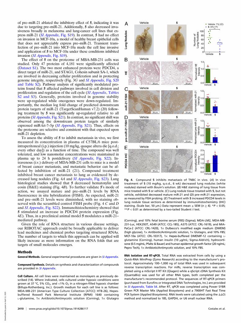
measured its concentration in plasma of C57BL/6 mice post-intraperitoneal (i.p.) injection (10 mg/kg, quaque altera die [q.o.d.;every other day]) as a function of time. The compound was welltolerated, and low nanomolar concentrations were maintained inplasma up to 24 h postdelivery (SI Appendix, Fig. S22). In-travenous (i.v.) delivery of MDA-MB-231 cells to mice is a modelof breast cancer metastasis, and metastatic behavior can be af-fected by inhibition of miR-21 (21). Compound treatmentinhibited breast cancer metastasis to lung as evidenced by de-creased lung nodules (Fig. 4A and SI Appendix, Fig. S23). Lunghistological studies showed that 5 decreased hematoxylin andeosin (H&E) staining (Fig. 4B). To further validate 5’s mode ofaction, we assayed mature and pre–miR-21 levels by RNAfluorescence in situ hybridization (FISH). As expected, miR-21and pre–miR-21 levels were diminished, with no staining ob-served with the scrambled control FISH probe (Fig. 4 C and Dand SI Appendix, Fig. S22). Immunohistochemistry showed that5 stimulated an increase in PDCD4 protein expression (Fig.4E). Thus, in a preclinical animal model 5modulates a miR-21–mediated pathway.Given the role of RNA structure in various disease settings,
our RIBOTAC approach could be broadly applicable to deliverlead medicines and chemical probes targeting structured RNAs.The number of targets to which this approach can be applied willlikely increase as more information on the RNA folds that aretargets of small molecules emerges.
MethodsGeneral Methods. General experimental procedures are given in SI Appendix.
Compound Synthesis. Details on synthesis and characterization of compoundsare provided in SI Appendix.
Cell Culture. All cell lines were maintained as monolayers as previously de-scribed (14). Where indicated, cells cultured under hypoxic conditions weregrown at 37 °C, 5% CO2, and <1% O2 in a nitrogen-filled hypoxic chamber(Billups-Rothenberg, Inc.). Growth medium for each cell line is as follows:MDA-MB-231 (American Type Culture Collection [ATCC]: HTB-26), Hepes-buffered Roswell Park Memorial Institute (RPMI) 1640 containingL-glutamine, 1× Antibiotic/Antimycotic solution (Corning), 1× Glutagro
(Corning), and 10% fetal bovine serum (FBS) (Sigma); MDA-LM2, MDA-MB-231-Luc, HEK293T, A549 (ATCC: CCL-185), A375 (ATCC: CRL-1619), and MIA-PaCa-2 (ATCC: CRL-1420), 1× Dulbecco’s modified eagle medium (DMEM)(high glycose), 1× Antibiotic/Antimycotic solution, 1× Glutagro, and 10% FBS;MCF-10a (ATCC: CRL-10317), 1× Hepes-buffered DMEM/F-12 containing L-glutamine (Corning), human insulin (10 μg/mL; Sigma-Aldrich), hydrocorti-sone (0.5 mg/mL; Pfaltz & Bauer) and human epidermal growth factor (20 ng/mL;Pepro Tech), 1× Antibiotic/Antimycotic solution, and 10% FBS.
RNA Isolation and RT-qPCR. Total RNA was extracted from cells by using aQuick-RNA MiniPrep (Zymo Research) according to the manufacturer’s pro-tocol. Approximately 100–1,000 ng of total RNA was used in subsequentreverse transcription reactions. For miRs, reverse transcription was com-pleted using a miScript II RT Kit (Qiagen) while a qScript cDNA Synthesis Kit(QuantaBio) was used for all other RNA types, both completed per themanufacturer’s recommended protocol. The sequences of RT-qPCR primers(purchased from Eurofins or Integrated DNA Technologies, Inc.) are providedin SI Appendix, Table S4. After RT, qPCR was completed using Power SYBRGreen PCR Master Mix (Applied Biosystems) and a 7900HT Fast Real TimePCR System (Applied Biosystems). RNA levels were calculated using the ΔΔCtmethod and normalized to 18S, GAPDH, or U6 small nuclear RNA.
Fig. 4. Compound 5 inhibits metastasis of TNBC in vivo. (A) In vivotreatment of 5 (10 mg/kg, q.o.d., 6 wk) decreased lung nodules (whitenodules) stained with Bouin’s solution. (B) H&E staining of lung tissue frommice treated with 5 or vehicle. (C) Lung nodule tissue treated with 5, but notvehicle, exhibited decreased mature miR-21 and (D) pre–miR-21 expression,as measured by FISH probing. (E) Treatment with 5 increased PDCD4 levels inlung nodule tissue sections as determined by immunohistochemistry (IHC)staining. (Scale bar, 50 μm.) Data represent mean ± SEM (n ≥ 4). *P < 0.05;**P < 0.01 as determined by a two-tailed Student t test.
2410 | www.pnas.org/cgi/doi/10.1073/pnas.1914286117 Costales et al.
Dow
nloa
ded
by g
uest
on
Apr
il 11
, 202
0
RNA Immunoprecipitation. Immunoprecipitation studies were completed aspreviously described (22), with the following modifications: 1) MDA-MB-231cells (∼70%confluency in 6-well plates) were treated with 200 nM of 2′-5′ A4
or 200 nM 5 (200 nM), prepared in growth medium, for 48 h; 2) cells werelysed in 100 μL of M-PER Buffer supplemented with 80 U RNaseOUTRecombinant Ribonuclease Inhibitor (Invitrogen) and 1× Protease InhibitorMixture III for Mammalian Cells (Research Products International Corp.)according to the manufacturer’s recommendations. Normalized fold changewas calculated using Eq. 1 (22):
Normalized Fold Change = Relative RNA Expression in RNase L fractionRelative RNA Expression in β-actin fraction
.
[1]
Lung Nodule Metastasis Study. Female nonobese diabetic/severe combinedimmunodeficiency (NOD/SCID) mice (n = 8, 5–7 wk) were used for in vivostudies. Mice were purchased from Jackson Laboratory and were housed inthe Scripps Florida vivarium. All experiments using live animals were ap-proved by the Scripps Florida Institutional Animal Care and Use Committee.The MDA-MB-231 cells stably transfected with luciferase (MDA-MB-231-Luc)were harvested by trypsinization, washed twice in phosphate-buffered sa-line (PBS), and counted. A total of 0.8 × 10⁶ cells was i.v. injected into NOD/SCID mice tail veins. Mice were imaged for luciferase activity immediatelyafter injection to exclude any animal that was not successfully xenografted.After cell implantation, the luciferase signal was monitored after injection ofcells every other day to determine initial compound treatment. Mice wereanesthetized and injected intraperitoneally with 100 μL of D-luciferin solu-tion (30 mg/mL in PBS). Imaging was performed with 90-s exposure timeusing a Lago X In Vivo Imager (Spectral Instruments).
After 3 d, the mice were split into two groups with the same mean lu-ciferase signal. The vehicle group was dosed with DMSO/Tween-80/H2O(10/10/80) and the compound treatment group was dosed with 10 mg/kg 5 inDMSO/Tween-80/H2O (10/10/80). Dosing was performed every other day, andthe weight of each mouse was monitored. Luciferase activity was monitoredevery week. After 6 wk of dosing, the mice were euthanized (in accordancewith guidelines provided by the American Veterinarian Medical Association),the lungs were perfused with PBS and harvested. The harvested lungs werefixed in Bouin’s solution (Sigma: HT10132-1L) immediately for less than 24 h.The lung nodule metastases were then counted, and then the fixed lung
tissues were immersed into 50 mL of 10% formalin solution and washed eighttimes over 48 h to remove the Bouin’s solution. Lungs were then given to theHistology Core at Scripps Research Florida to prepare paraffin-embeddedsections for the next staining steps.
Lung Tissue Histology for H&E Staining, miR-21 Staining, and PDCD4 Staining.The tissue samples were processed and embedded in paraffin and sectionedat 3 μm. To assess levels of PDCD4, an anti-PDCD4 (rb) antibody (Abcam;ab51495) was used, diluted to a final concentration of 1:100. The slides werestained with a Leica Bond-Max immunostaining platform using a DAB Refinekit. Negative control slides were stained by the same protocol but withoutapplying the primary antibody. After staining, slides were dehydrated ingraded alcohols, cleared in xylenes, and coverslipped with Cytoseal 60. Allhistology staining (H&E and PDCD4) was performed by the Histology Core atScripps Florida.
Pre- and mature miR-21 were imaged by RNA FISH, as previously described(23), with the following modifications: 1) the prepared paraffin-embeddedsections were first incubated at 60 °C overnight, followed by deparaffini-zation through three consecutive xylene baths (5 min each); 2) custom-synthesized oligonucleotides (0.2 μM) with locked nucleic acid modifica-tions and 3′ end labeling with fluorescein isothiocyanate (FITC) (Qiagen)were used to probe for miR-21, pre–miR-21, or a scrambled control sequencewere incubated with the tissue sections at 37 °C overnight; and 3) post-hybridization, slides were washed three times with 2× saline-sodium citrate(SSC) at room temperature for 15 min each, followed by three washes withPBS for 15 min each.
Where indicated, slides were stained with Mayer’s Hematoxylin Solution(Sigma: MHS1-100ML) per the manufacturer’s protocol.
Images of all slides were obtained using light microscopy on a LeicaDMI3000 B upright fluorescent microscope.
ACKNOWLEDGMENTS. This work was supported by the National Institutesof Health Grants R01 GM97455 and DP1 NS096898 (to M.D.D.) and theAmerican Chemical Society Medicinal Chemistry Predoctoral Fellowship (toM.G.C.). We thank the Nelson Family Fund, Alan J. and Susan A. Fuirst Philan-thropic Fund, and the Frenchman Creek’s Women for Cancer Research. We alsothank Rea Guertler for preliminary experiments, Jon Chen and HaJeung Parkfor molecular modeling, Christiana Teijaro for mass spectrometry, and theScripps Florida X-Ray Crystallography and Histology Core Facilities fortheir technical services.
1. X. Wu, D. P. Bartel, Widespread influence of 3′-end structures on mammalian mRNAprocessing and stability. Cell 169, 905–917.e11 (2017).
2. L. Sun et al., RNA structure maps across mammalian cellular compartments. Nat.Struct. Mol. Biol. 26, 322–330 (2019).
3. D. P. Bartel, MicroRNAs: Genomics, biogenesis, mechanism, and function. Cell 116,281–297 (2004).
4. A. M. Krichevsky, G. Gabriely, miR-21: A small multi-faceted RNA. J. Cell Mol. Med. 13,39–53 (2009).
5. S. P. Velagapudi, S. M. Gallo, M. D. Disney, Sequence-based design of bioactive smallmolecules that target precursor microRNAs. Nat. Chem. Biol. 10, 291–297 (2014).
6. Z. Su et al., Discovery of a biomarker and lead small molecules to target r(GGGGCC)-associated defects in c9FTD/ALS. Neuron 84, 239 (2014).
7. D. Iliopoulos, S. A. Jaeger, H. A. Hirsch, M. L. Bulyk, K. Struhl, STAT3 activation of miR-21 and miR-181b-1 via PTEN and CYLD are part of the epigenetic switch linking in-flammation to cancer. Mol. Cell 39, 493–506 (2010).
8. L. B. Frankel et al., Programmed cell death 4 (PDCD4) is an important functional targetof the microRNA miR-21 in breast cancer cells. J. Biol. Chem. 283, 1026–1033 (2008).
9. N. M. McLoughlin, C. Mueller, T. N. Grossmann, The therapeutic potential of PTENmodulation: Targeting strategies from gene to protein. Cell Chem. Biol. 25, 19–29(2018).
10. M. J. Hendrix et al., Retinoic acid inhibition of human melanoma cell invasion througha reconstituted basement membrane and its relation to decreases in the expression ofproteolytic enzymes and motility factor receptor. Cancer Res. 50, 4121–4130 (1990).
11. M. Seike et al., MiR-21 is an EGFR-regulated anti-apoptotic factor in lung cancer innever-smokers. Proc. Natl. Acad. Sci. U.S.A. 106, 12085–12090 (2009).
12. S. F. Tavazoie et al., Endogenous human microRNAs that suppress breast cancermetastasis. Nature 451, 147–152 (2008).
13. F. Sicard, M. Gayral, H. Lulka, L. Buscail, P. Cordelier, Targeting miR-21 for the therapyof pancreatic cancer. Mol. Ther. 21, 986–994 (2013).
14. M. G. Costales, Y. Matsumoto, S. P. Velagapudi, M. D. Disney, Small molecule targetedrecruitment of a nuclease to RNA. J. Am. Chem. Soc. 140, 6741–6744 (2018).
15. Y. Han et al., Structure of human RNase L reveals the basis for regulated RNA decay inthe IFN response. Science 343, 1244–1248 (2014).
16. C. S. Thakur et al., Small-molecule activators of RNase L with broad-spectrum antiviralactivity. Proc. Natl. Acad. Sci. U.S.A. 104, 9585–9590 (2007).
17. A. Chakrabarti, B. K. Jha, R. H. Silverman, New insights into the role of RNase L ininnate immunity. J. Interferon Cytokine Res. 31, 49–57 (2011).
18. P. P. Graczyk, Gini coefficient: A new way to express selectivity of kinase inhibitorsagainst a family of kinases. J. Med. Chem. 50, 5773–5779 (2007).
19. W. Y. Yang, R. Gao, M. Southern, P. S. Sarkar, M. D. Disney, Design of a bioactive smallmolecule that targets r(AUUCU) repeats in spinocerebellar ataxia 10. Nat. Commun. 7,11647 (2016).
20. V. Agarwal, G. W. Bell, J.-W. Nam, D. P. Bartel, Predicting effective microRNA targetsites in mammalian mRNAs. eLife 4, e05005 (2015).
21. S. Yang, J. J. Zhang, X.-Y. Huang, Mouse models for tumor metastasis. Methods Mol.Biol. 928, 221–228 (2012).
22. M. G. Costales, B. Suresh, K. Vishnu, M. D. Disney, Targeted degradation of a hypoxia-associated non-coding RNA enhances the selectivity of a small molecule interactingwith RNA. Cell Chem. Biol. 26, 1180–1186.e5 (2019).
23. N. Yamamichi et al., Locked nucleic acid in situ hybridization analysis of miR-21 ex-pression during colorectal cancer development. Clin. Cancer Res. 15, 4009–4016 (2009).
24. P. HaJeung et al., ethyl (Z)-5-(3-hydroxy-4-methoxybenzylidene)-4-oxo-2-(phenyl-amino)-4,5-dihydrothiophene-3-carboxylate. The Cambridge Crystallographic DataCentre (CCDC). https://www.ccdc.cam.ac.uk/structures/Search?Ccdcid=1912054&DatabaseToSearch=Published. Deposited 24 April 2019.
Costales et al. PNAS | February 4, 2020 | vol. 117 | no. 5 | 2411
APP
LIED
BIOLO
GICAL
SCIENCE
S
Dow
nloa
ded
by g
uest
on
Apr
il 11
, 202
0


![The conserved Fanconi anemia nuclease Fan1 and the SUMO E3 … · 2017. 2. 23. · FAN1 (Fanconi anemia-associated nuclease 1, or FANCD2/FANCI-associated nuclease 1) [13–18]. Human](https://static.fdocuments.us/doc/165x107/60c9d965c710eb0d72008d0e/the-conserved-fanconi-anemia-nuclease-fan1-and-the-sumo-e3-2017-2-23-fan1-fanconi.jpg)

