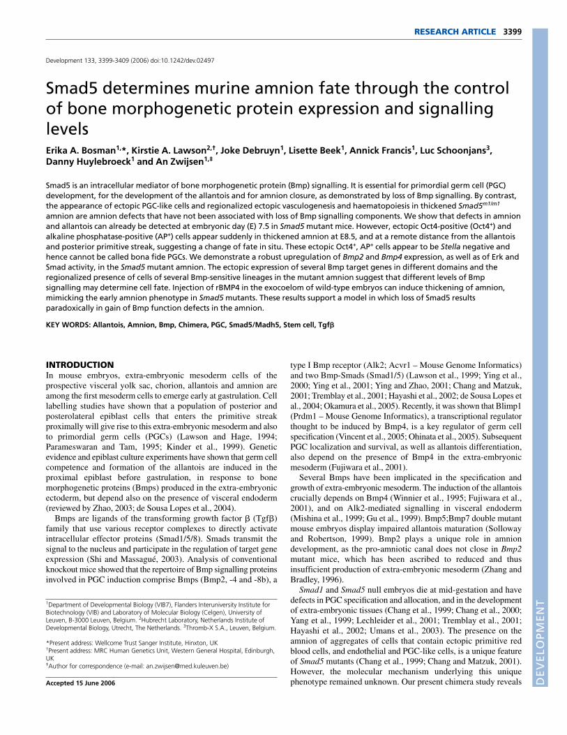
Smad5 determines murine amnion fate through the control of
11
DEVELOPMENT 3399 RESEARCH ARTICLE INTRODUCTION In mouse embryos, extra-embryonic mesoderm cells of the prospective visceral yolk sac, chorion, allantois and amnion are among the first mesoderm cells to emerge early at gastrulation. Cell labelling studies have shown that a population of posterior and posterolateral epiblast cells that enters the primitive streak proximally will give rise to this extra-embryonic mesoderm and also to primordial germ cells (PGCs) (Lawson and Hage, 1994; Parameswaran and Tam, 1995; Kinder et al., 1999). Genetic evidence and epiblast culture experiments have shown that germ cell competence and formation of the allantois are induced in the proximal epiblast before gastrulation, in response to bone morphogenetic proteins (Bmps) produced in the extra-embryonic ectoderm, but depend also on the presence of visceral endoderm (reviewed by Zhao, 2003; de Sousa Lopes et al., 2004). Bmps are ligands of the transforming growth factor (Tgf) family that use various receptor complexes to directly activate intracellular effector proteins (Smad1/5/8). Smads transmit the signal to the nucleus and participate in the regulation of target gene expression (Shi and Massagué, 2003). Analysis of conventional knockout mice showed that the repertoire of Bmp signalling proteins involved in PGC induction comprise Bmps (Bmp2, -4 and -8b), a type I Bmp receptor (Alk2; Acvr1 – Mouse Genome Informatics) and two Bmp-Smads (Smad1/5) (Lawson et al., 1999; Ying et al., 2000; Ying et al., 2001; Ying and Zhao, 2001; Chang and Matzuk, 2001; Tremblay et al., 2001; Hayashi et al., 2002; de Sousa Lopes et al., 2004; Okamura et al., 2005). Recently, it was shown that Blimp1 (Prdm1 – Mouse Genome Informatics), a transcriptional regulator thought to be induced by Bmp4, is a key regulator of germ cell specification (Vincent et al., 2005; Ohinata et al., 2005). Subsequent PGC localization and survival, as well as allantois differentiation, also depend on the presence of Bmp4 in the extra-embryonic mesoderm (Fujiwara et al., 2001). Several Bmps have been implicated in the specification and growth of extra-embryonic mesoderm. The induction of the allantois crucially depends on Bmp4 (Winnier et al., 1995; Fujiwara et al., 2001), and on Alk2-mediated signalling in visceral endoderm (Mishina et al., 1999; Gu et al., 1999). Bmp5;Bmp7 double mutant mouse embryos display impaired allantois maturation (Solloway and Robertson, 1999). Bmp2 plays a unique role in amnion development, as the pro-amniotic canal does not close in Bmp2 mutant mice, which has been ascribed to reduced and thus insufficient production of extra-embryonic mesoderm (Zhang and Bradley, 1996). Smad1 and Smad5 null embryos die at mid-gestation and have defects in PGC specification and allocation, and in the development of extra-embryonic tissues (Chang et al., 1999; Chang et al., 2000; Yang et al., 1999; Lechleider et al., 2001; Tremblay et al., 2001; Hayashi et al., 2002; Umans et al., 2003). The presence on the amnion of aggregates of cells that contain ectopic primitive red blood cells, and endothelial and PGC-like cells, is a unique feature of Smad5 mutants (Chang et al., 1999; Chang and Matzuk, 2001). However, the molecular mechanism underlying this unique phenotype remained unknown. Our present chimera study reveals Smad5 determines murine amnion fate through the control of bone morphogenetic protein expression and signalling levels Erika A. Bosman 1, *, Kirstie A. Lawson 2,† , Joke Debruyn 1 , Lisette Beek 1 , Annick Francis 1 , Luc Schoonjans 3 , Danny Huylebroeck 1 and An Zwijsen 1,‡ Smad5 is an intracellular mediator of bone morphogenetic protein (Bmp) signalling. It is essential for primordial germ cell (PGC) development, for the development of the allantois and for amnion closure, as demonstrated by loss of Bmp signalling. By contrast, the appearance of ectopic PGC-like cells and regionalized ectopic vasculogenesis and haematopoiesis in thickened Smad5 m1/m1 amnion are amnion defects that have not been associated with loss of Bmp signalling components. We show that defects in amnion and allantois can already be detected at embryonic day (E) 7.5 in Smad5 mutant mice. However, ectopic Oct4-positive (Oct4 + ) and alkaline phosphatase-positive (AP + ) cells appear suddenly in thickened amnion at E8.5, and at a remote distance from the allantois and posterior primitive streak, suggesting a change of fate in situ. These ectopic Oct4 + , AP + cells appear to be Stella negative and hence cannot be called bona fide PGCs. We demonstrate a robust upregulation of Bmp2 and Bmp4 expression, as well as of Erk and Smad activity, in the Smad5 mutant amnion. The ectopic expression of several Bmp target genes in different domains and the regionalized presence of cells of several Bmp-sensitive lineages in the mutant amnion suggest that different levels of Bmp signalling may determine cell fate. Injection of rBMP4 in the exocoelom of wild-type embryos can induce thickening of amnion, mimicking the early amnion phenotype in Smad5 mutants. These results support a model in which loss of Smad5 results paradoxically in gain of Bmp function defects in the amnion. KEY WORDS: Allantois, Amnion, Bmp, Chimera, PGC, Smad5/Madh5, Stem cell, Tgf Development 133, 3399-3409 (2006) doi:10.1242/dev.02497 1 Department of Developmental Biology (VIB7), Flanders Interuniversity Institute for Biotechnology (VIB) and Laboratory of Molecular Biology (Celgen), University of Leuven, B-3000 Leuven, Belgium. 2 Hubrecht Laboratory, Netherlands Institute of Developmental Biology, Utrecht, The Netherlands. 3 Thromb-X S.A., Leuven, Belgium. *Present address: Wellcome Trust Sanger Institute, Hinxton, UK † Present address: MRC Human Genetics Unit, Western General Hospital, Edinburgh, UK ‡ Author for correspondence (e-mail: [email protected]) Accepted 15 June 2006
Transcript of Smad5 determines murine amnion fate through the control of
DEVELO
PMENT
3399RESEARCH ARTICLE
INTRODUCTIONIn mouse embryos, extra-embryonic mesoderm cells of theprospective visceral yolk sac, chorion, allantois and amnion areamong the first mesoderm cells to emerge early at gastrulation. Celllabelling studies have shown that a population of posterior andposterolateral epiblast cells that enters the primitive streakproximally will give rise to this extra-embryonic mesoderm and alsoto primordial germ cells (PGCs) (Lawson and Hage, 1994;Parameswaran and Tam, 1995; Kinder et al., 1999). Geneticevidence and epiblast culture experiments have shown that germ cellcompetence and formation of the allantois are induced in theproximal epiblast before gastrulation, in response to bonemorphogenetic proteins (Bmps) produced in the extra-embryonicectoderm, but depend also on the presence of visceral endoderm(reviewed by Zhao, 2003; de Sousa Lopes et al., 2004).
Bmps are ligands of the transforming growth factor � (Tgf�)family that use various receptor complexes to directly activateintracellular effector proteins (Smad1/5/8). Smads transmit thesignal to the nucleus and participate in the regulation of target geneexpression (Shi and Massagué, 2003). Analysis of conventionalknockout mice showed that the repertoire of Bmp signalling proteinsinvolved in PGC induction comprise Bmps (Bmp2, -4 and -8b), a
type I Bmp receptor (Alk2; Acvr1 – Mouse Genome Informatics)and two Bmp-Smads (Smad1/5) (Lawson et al., 1999; Ying et al.,2000; Ying et al., 2001; Ying and Zhao, 2001; Chang and Matzuk,2001; Tremblay et al., 2001; Hayashi et al., 2002; de Sousa Lopes etal., 2004; Okamura et al., 2005). Recently, it was shown that Blimp1(Prdm1 – Mouse Genome Informatics), a transcriptional regulatorthought to be induced by Bmp4, is a key regulator of germ cellspecification (Vincent et al., 2005; Ohinata et al., 2005). SubsequentPGC localization and survival, as well as allantois differentiation,also depend on the presence of Bmp4 in the extra-embryonicmesoderm (Fujiwara et al., 2001).
Several Bmps have been implicated in the specification andgrowth of extra-embryonic mesoderm. The induction of the allantoiscrucially depends on Bmp4 (Winnier et al., 1995; Fujiwara et al.,2001), and on Alk2-mediated signalling in visceral endoderm(Mishina et al., 1999; Gu et al., 1999). Bmp5;Bmp7 double mutantmouse embryos display impaired allantois maturation (Sollowayand Robertson, 1999). Bmp2 plays a unique role in amniondevelopment, as the pro-amniotic canal does not close in Bmp2mutant mice, which has been ascribed to reduced and thusinsufficient production of extra-embryonic mesoderm (Zhang andBradley, 1996).
Smad1 and Smad5 null embryos die at mid-gestation and havedefects in PGC specification and allocation, and in the developmentof extra-embryonic tissues (Chang et al., 1999; Chang et al., 2000;Yang et al., 1999; Lechleider et al., 2001; Tremblay et al., 2001;Hayashi et al., 2002; Umans et al., 2003). The presence on theamnion of aggregates of cells that contain ectopic primitive redblood cells, and endothelial and PGC-like cells, is a unique featureof Smad5 mutants (Chang et al., 1999; Chang and Matzuk, 2001).However, the molecular mechanism underlying this uniquephenotype remained unknown. Our present chimera study reveals
Smad5 determines murine amnion fate through the controlof bone morphogenetic protein expression and signallinglevelsErika A. Bosman1,*, Kirstie A. Lawson2,†, Joke Debruyn1, Lisette Beek1, Annick Francis1, Luc Schoonjans3,Danny Huylebroeck1 and An Zwijsen1,‡
Smad5 is an intracellular mediator of bone morphogenetic protein (Bmp) signalling. It is essential for primordial germ cell (PGC)development, for the development of the allantois and for amnion closure, as demonstrated by loss of Bmp signalling. By contrast,the appearance of ectopic PGC-like cells and regionalized ectopic vasculogenesis and haematopoiesis in thickened Smad5m1/m1
amnion are amnion defects that have not been associated with loss of Bmp signalling components. We show that defects in amnionand allantois can already be detected at embryonic day (E) 7.5 in Smad5 mutant mice. However, ectopic Oct4-positive (Oct4+) andalkaline phosphatase-positive (AP+) cells appear suddenly in thickened amnion at E8.5, and at a remote distance from the allantoisand posterior primitive streak, suggesting a change of fate in situ. These ectopic Oct4+, AP+ cells appear to be Stella negative andhence cannot be called bona fide PGCs. We demonstrate a robust upregulation of Bmp2 and Bmp4 expression, as well as of Erk andSmad activity, in the Smad5 mutant amnion. The ectopic expression of several Bmp target genes in different domains and theregionalized presence of cells of several Bmp-sensitive lineages in the mutant amnion suggest that different levels of Bmpsignalling may determine cell fate. Injection of rBMP4 in the exocoelom of wild-type embryos can induce thickening of amnion,mimicking the early amnion phenotype in Smad5 mutants. These results support a model in which loss of Smad5 resultsparadoxically in gain of Bmp function defects in the amnion.
KEY WORDS: Allantois, Amnion, Bmp, Chimera, PGC, Smad5/Madh5, Stem cell, Tgf��
Development 133, 3399-3409 (2006) doi:10.1242/dev.02497
1Department of Developmental Biology (VIB7), Flanders Interuniversity Institute forBiotechnology (VIB) and Laboratory of Molecular Biology (Celgen), University ofLeuven, B-3000 Leuven, Belgium. 2Hubrecht Laboratory, Netherlands Institute ofDevelopmental Biology, Utrecht, The Netherlands. 3Thromb-X S.A., Leuven, Belgium.
*Present address: Wellcome Trust Sanger Institute, Hinxton, UK†Present address: MRC Human Genetics Unit, Western General Hospital, Edinburgh,UK‡Author for correspondence (e-mail: [email protected])
Accepted 15 June 2006
DEVELO
PMENT
3400
that Smad5 functions non-cell-autonomously in the amnionmesoderm. In addition, we observe ectopic Bmp expression andsignalling in the affected amnion. We therefore propose a new modelin which Smad5 deficiency paradoxically leads to gain of Bmpfunction defects. Additional support for this model comes frominjection of rBMP4 protein in the exocoelom of wild-type embryos,which results in abnormal thickening of the amnion.
MATERIALS AND METHODSMouse strains and production of chimeric embryosThe Smad5m1 mutation (Chang et al., 1999) was outcrossed in C57BL/6J andF2 crosses of F1 (C57BL/6J � CBA) background for at least fivegenerations. Rosa-�geo-26 mice, expressing Escherichia coli lacZubiquitously (Friedrich and Soriano, 1991), were maintained in the F2background. Embryonic stem (ES) cells were established from blastocystsof Smad5+/m1 (C57BL/6J) intercrosses as described (Schoonjans et al.,2003), and genotyped by PCR and Southern blotting (Chang et al., 1999).
Chimeras between ES cell lines and diploid Rosa-�geo-26 morulae weregenerated as described (Goumans et al., 1999). The majority of wild-typerWTlacZ, Smad5+/m1
rWTlacZ, and of Smad5m1/m1rWTlacZ chimeric
embryos had a high ES cell contribution (>90%), with only few acceptor-morula derived, �-galactosidase-positive cells present in hindgut endoderm.The minority of Smad5m1/m1
rWTlacZ embryos (four out of 18) with a highcontribution from the wild-type acceptor morula in epiblast-derived tissues(20-80% ES cell-derived embryos) are here referred to as low-percentagechimeras.
Animal care and experiments were approved by the Institutional EthicalCommittee of the Catholic University of Leuven.
Collection of post-implantation embryos, in situ hybridizationand immunohistochemistryEmbryos from timed matings were dissected in ice-cold PBS at embryonicday (E) 7.0-9.5 and staged according to Downs and Davies (Downsand Davies, 1993), modified for C57BL/6 � CBA embryos (EdinburghMouse Atlas Project: http://genex.hgu.mrc.ac.uk/Databases/Anatomy/MAstaging.shtml), or on somite number. Radioactive in situ hybridizationwas performed as described (Dewulf et al., 1995). Non-radioactive in situhybridization [Stella (Dppa3 – Mouse Genome Informatics), fragilis (Ifitm3– Mouse Genome Informatics)] on sections and immunohistochemistry forP-ERK (cat. no 9101, Cell Signalling Technologies) and P-Smad1/5/8(cat. no 9511, Cell Signalling Technologies) were performed with theautomated Ventana Discovery system and Ventana reagents according tothe manufacturer’s instructions. NBT/BCIP and DAB were used aschromogens for automated non-radioactive in situ hybridization andimmunohistochemistry, respectively. We used antisense RNA probes forBmp2 (Lyons et al., 1989), Bmp4 (Jones et al., 1991), periostin/Osf2 (Delotet al., 2003), Stella and fragilis (Saitou et al., 2002), and Msx2 (MacKenzieet al., 1992).
Immunohistochemistry for Oct4 (Pou5f1 – Mouse Genome Informatics;sc5279, Santa Cruz Biotechnology) and SSEA-1 (Fut4 – Mouse GenomeInformatics; MC480 DHSB) was performed as described (Van Eynde et al.,2004). AEC (Sigma) was used as a chromogen.
Detection and counting of PGCsDissection of embryos, followed by fixation, and detection of alkalinephosphatase (AP)-positive PGCs with �-naphthyl phosphate/Fast Red TR(Ginsburg et al., 1990) in whole-mount embryos or with ASMX/Fast Red
TR in serial section, were as described (Lawson et al., 1999). PGCs werecounted on the basis of the strong surface AP staining and intensely stainedcytoplasmic spot (Ginsburg et al., 1990; Lawson and Hage, 1994). Theanterior portion of the embryo was retained for genotyping (Chang et al.,1999).
MeasurementsAllantois dimensions were measured on intact embryos after staining forAP. The axial length of the allantois was measured from the line ofintersection with the yolk sac when viewed posteriorly and from thejunctional line between amnion and yolk sac when viewed laterally.Width at the base was measured in posterior view along the intersectionwith the yolk sac.
The length of the anterior-posterior embryonic axis was estimated onimages of intact embryos stained for AP and viewed laterally. Axis lengthwas taken as the sum of the three longest cords along the curved axis – fromthe anterior embryo/amnion junction or the most proximal edge of the neuralfold, from the intersection of amnion, allantois and primitive streak to thenode, and a cord connecting these two.
Injection of rBMP4 and whole embryo cultureCD1 embryos between neural plate (NP) and headfold (HF) stages wereisolated in 22 mmol/l Hepes and 10% heat-inactivated fetal calf serum(FCS; Hyclone); Reichert’s membrane was removed, but yolk sacintegrity with the ectoplacental cone was maintained. Whole embryoculture and manipulations were in Dulbecco’s modified essential medium(Gibco), 1 mmol/l glutamine, non-essential amino acids, 0.5 mmol/l Na-pyruvate, 50% heat-inactivated horse serum (Gibco) in a rolling culturesystem at 37°C and 5% CO2 for 15 to 24 hours (0.5 ml medium perembryo). Embryos were either control injected or injected with 1 �ghuman rBMP4/ml PBS (R&D Systems) with a mouth-controlled glasscapillary in the exocoelom (1-2 nl; corresponding to about one-tenth ofthe luminal volume of the exocoelom). Control injected embryosdeveloped normally, as judged by embryo viability, somite number, axialrotation, allantois elongation and fusion, and neural tube closure. Yolk sacvasculogenesis appeared normal, but the yolk sac was sometimes enlargedwhen compared to freshly isolated embryos. Only embryos in which theheart was beating were assessed for amnion development. Embryos werefixed and processed for paraffin embedding.
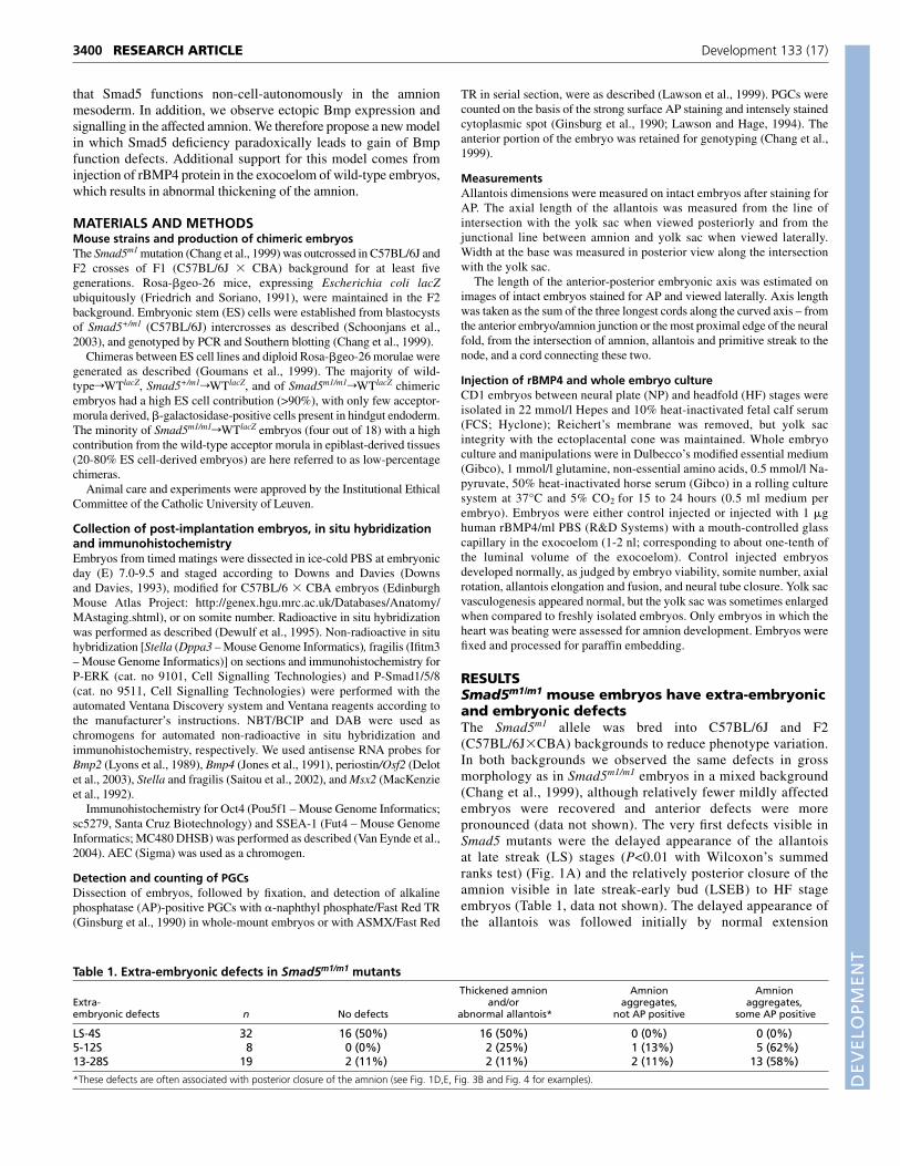
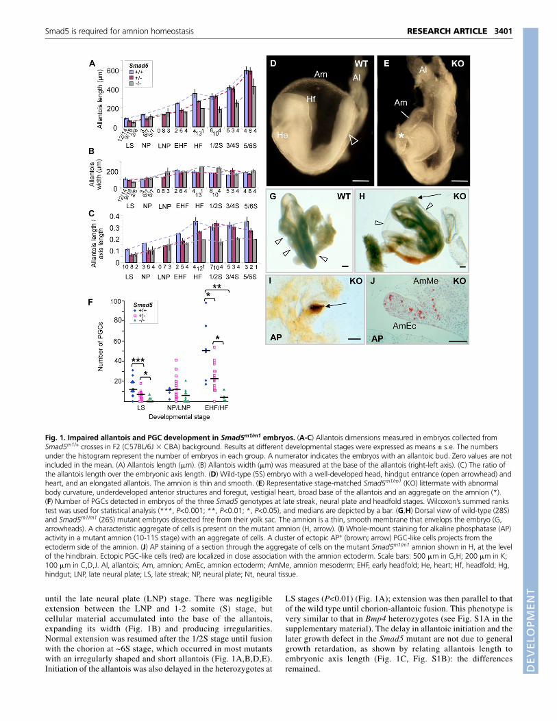
RESULTSSmad5m1/m1 mouse embryos have extra-embryonicand embryonic defectsThe Smad5m1 allele was bred into C57BL/6J and F2(C57BL/6J�CBA) backgrounds to reduce phenotype variation.In both backgrounds we observed the same defects in grossmorphology as in Smad5m1/m1 embryos in a mixed background(Chang et al., 1999), although relatively fewer mildly affectedembryos were recovered and anterior defects were morepronounced (data not shown). The very first defects visible inSmad5 mutants were the delayed appearance of the allantoisat late streak (LS) stages (P<0.01 with Wilcoxon’s summedranks test) (Fig. 1A) and the relatively posterior closure of theamnion visible in late streak-early bud (LSEB) to HF stageembryos (Table 1, data not shown). The delayed appearance ofthe allantois was followed initially by normal extension
RESEARCH ARTICLE Development 133 (17)
Table 1. Extra-embryonic defects in Smad5m1/m1 mutantsThickened amnion Amnion Amnion
Extra- and/or aggregates, aggregates,embryonic defects n No defects abnormal allantois* not AP positive some AP positive
LS-4S 32 16 (50%) 16 (50%) 0 (0%) 0 (0%)5-12S 8 0 (0%) 2 (25%) 1 (13%) 5 (62%)13-28S 19 2 (11%) 2 (11%) 2 (11%) 13 (58%)
*These defects are often associated with posterior closure of the amnion (see Fig. 1D,E, Fig. 3B and Fig. 4 for examples).
DEVELO
PMENT
until the late neural plate (LNP) stage. There was negligibleextension between the LNP and 1-2 somite (S) stage, butcellular material accumulated into the base of the allantois,expanding its width (Fig. 1B) and producing irregularities.Normal extension was resumed after the 1/2S stage until fusionwith the chorion at ~6S stage, which occurred in most mutantswith an irregularly shaped and short allantois (Fig. 1A,B,D,E).Initiation of the allantois was also delayed in the heterozygotes at
LS stages (P<0.01) (Fig. 1A); extension was then parallel to thatof the wild type until chorion-allantoic fusion. This phenotype isvery similar to that in Bmp4 heterozygotes (see Fig. S1A in thesupplementary material). The delay in allantoic initiation and thelater growth defect in the Smad5 mutant are not due to generalgrowth retardation, as shown by relating allantois length toembryonic axis length (Fig. 1C, Fig. S1B): the differencesremained.
3401RESEARCH ARTICLESmad5 is required for amnion homeostasis
Fig. 1. Impaired allantois and PGC development in Smad5m1/m1 embryos. (A-C) Allantois dimensions measured in embryos collected fromSmad5m1/+ crosses in F2 (C57BL/6J � CBA) background. Results at different developmental stages were expressed as means ± s.e. The numbersunder the histogram represent the number of embryos in each group. A numerator indicates the embryos with an allantoic bud. Zero values are notincluded in the mean. (A) Allantois length (�m). (B) Allantois width (�m) was measured at the base of the allantois (right-left axis). (C) The ratio ofthe allantois length over the embryonic axis length. (D) Wild-type (5S) embryo with a well-developed head, hindgut entrance (open arrowhead) andheart, and an elongated allantois. The amnion is thin and smooth. (E) Representative stage-matched Smad5m1/m1 (KO) littermate with abnormalbody curvature, underdeveloped anterior structures and foregut, vestigial heart, broad base of the allantois and an aggregate on the amnion (*).(F) Number of PGCs detected in embryos of the three Smad5 genotypes at late streak, neural plate and headfold stages. Wilcoxon’s summed rankstest was used for statistical analysis (***, P<0.001; **, P<0.01; *, P<0.05), and medians are depicted by a bar. (G,H) Dorsal view of wild-type (28S)and Smad5m1/m1 (26S) mutant embryos dissected free from their yolk sac. The amnion is a thin, smooth membrane that envelops the embryo (G,arrowheads). A characteristic aggregate of cells is present on the mutant amnion (H, arrow). (I) Whole-mount staining for alkaline phosphatase (AP)activity in a mutant amnion (10-11S stage) with an aggregate of cells. A cluster of ectopic AP+ (brown; arrow) PGC-like cells projects from theectoderm side of the amnion. (J) AP staining of a section through the aggregate of cells on the mutant Smad5m1/m1 amnion shown in H, at the levelof the hindbrain. Ectopic PGC-like cells (red) are localized in close association with the amnion ectoderm. Scale bars: 500 �m in G,H; 200 �m in K;100 �m in C,D,J. Al, allantois; Am, amnion; AmEc, amnion ectoderm; AmMe, amnion mesoderm; EHF, early headfold; He, heart; Hf, headfold; Hg,hindgut; LNP, late neural plate; LS, late streak; NP, neural plate; Nt, neural tissue.
DEVELO
PMENT
3402
At the HF stage, the amnion is a thin membrane composed oftwo layers of squamous epithelium: the amnion ectoderm iscontinuous with the embryonic surface ectoderm and the amnionmesoderm is continuous with the yolk sac mesoderm and,posteriorly, with the allantois mesothelium. The two layers arederived from slightly different regions of the early-streak stageepiblast (Lawson et al., 1991) (K.A.L., unpublished). The amnionwas markedly thickened in Smad5m1/m1 embryos (Table 1). Theamnion of somite-stage mutants typically contained aggregates of
cells (Table 1) that were often localized very anteriorly at a remotedistance from the allantois. From the 5S stage onwards,Smad5m1/m1 embryos had an underdeveloped forebrain and heartwhen compared with stage-matched control littermates, and anaggregate of cells was visible on the amnion in more than half ofthe mutants (Fig. 1D,E). Failure of embryo turning, defects inheart looping and dilation of embryonic and yolk sac bloodvessels were as previously described (Chang et al., 1999; Changet al., 2000).
RESEARCH ARTICLE Development 133 (17)
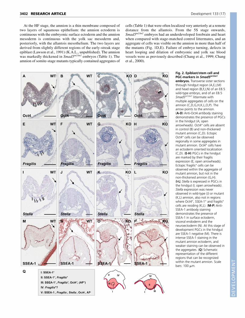
Fig. 2. Epiblast/stem cell andPGC-markers in Smad5m1/m1
embryos. Transverse sister sectionsthrough hindgut region (A,E,I,M)and head region (B,F,J,N) of an E8.5wild-type embryo, and of an E8.5Smad5m1/m1 littermate withmultiple aggregates of cells on theamnion (C,D,G,H,K,L,O,P). Thearrow points to the amnion.(A-D) Anti-Oct4 antibody stainingdemonstrates the presence of PGCsin the hindgut (A; openarrowheads). Oct4+ cells are absentin control (B) and non-thickenedmutant amnion (C,D). EctopicOct4+ cells can be observedregionally in some aggregates inmutant amnion. Oct4+ cells havean ectoderm oriented localization(C,D). (E-H) PGCs in the hindgutare marked by their fragilisexpression (E; open arrowheads).Ectopic fragilis+ cells can beobserved within the aggregate ofmutant amnion, but not in thenon-thickened amnion (G,H).(I-L) Stella is expressed in PGCs inthe hindgut (I; open arrowheads).Stella expression was neverobserved in wild-type (J) or mutant(K,L) amnion, also not in regionswhere Oct4+, SSEA-1+ and fragilis+
cells are residing (K,L). (M-P) Anti-SSEA-1 antibody stainingdemonstrates the presence ofSSEA-1 in surface ectoderm,visceral endoderm and theneuroectoderm (N). At this stage ofdevelopment PGCs in the hindgutare SSEA-1 negative (M). There isintense SSEA-1 staining in themutant amnion ectoderm, andweaker staining can be observed inthe aggregates. (Q) Schematicrepresentation of the differentregions that can be recognizedwithin the mutant amnion. Scalebars: 100 �m.

DEVELO
PMENT
De-novo appearance of AP-positive cells in theSmad5 mutant amnionChang and Matzuk (Chang and Matzuk, 2001) described a Smad5gene dosage effect, reminiscent of, although less pronounced than,the Bmp4 null allele, on the size of the PGC founder population ina 129SvEv/C57 mixed genetic background. We confirm theseobservations now in the C57BL/6J and F2 background (see Fig. S2in the supplementary material), and have determined that theincidence and number of PGCs is already reduced in Smad5mutants at the LS stages, with an intermediate effect in theheterozygotes (Fig. 1F). Chang and Matzuk (Chang and Matzuk,2001) observed AP- and Oct4+ cells in the aggregate of cells in theamnion of early somite-stage embryos, and concluded that they aremislocated PGCs. We explored the timing of appearance of thesePGC-like cells in the amnion. Although half of the NP up to the 4Sstage embryos had defects in allantois development, amnion closureand amnion thickening, no AP+ PGC-like cells were seen in theSmad5m1/m1 amnion at these stages (Table 1), or elsewhere outsidethe normal PGC domain. Only after the 5S stage, did we find AP+
cells in some of the distinct aggregates of cells on the anmion (Fig.1H,I,J; Table 1). These were located in the part of the aggregateprojecting into the amniotic cavity and therefore associated withamnion ectoderm (Fig. 1J), unlike the ectopic haematopoietic andendothelial cells that bulged into the mesoderm side of the amnionfacing the exocoelom. By contrast to previous data (Chang andMatzuk, 2001), we found that AP+ clumps were still prominent atE9.5 (11/14 embryos: 16-28S) (Fig. 1H,J; Table 1). The suddenappearance of PGC-like cells in appreciable numbers in theaggregates (33 to 119 PGC-like cells in 5S-12S embryos; 142 cellsin the aggregate of the 26S mutant shown in Fig. 1H,J), and theanterior position of the aggregates containing them, make itunlikely that they are PGCs mislocated from the founderpopulation, and supports the argument for a change in cell fate insitu in the mutant amnion.
The AP+ cells in the Smad5 mutant amnion are notbona fide PGCsWe used a panel of markers to address the possibility that the AP+
cells in the aggregates were de-novo induced PGCs. The epiblastexpresses Oct4. During gastrulation the expression of Oct4 isdownregulated and is thereafter only maintained in the germ celllineage (Schöler, 1991) (Fig. 2A). Wild-type and non-affectedSmad5m1/m1amnion was always devoid of Oct4 protein (Fig. 2B-D),but the cell aggregates often contained Oct4+ cells (Fig. 2C,D). AtE6.5, fragilis (Ifitm3/Mil1) is expressed in proximal epiblast (Saitouet al., 2002), the region in which PGC-competent cells reside(Lawson and Hage, 1994). During gastrulation, fragilis expressionincreases within a cluster of cells at the base of the allantoic bud. AtE7.5 (early bud stage) cells with the highest expression of fragilisinitiate at the germ cell-characteristic expression of Stella/PGC-7/Dppa3 (Saitou et al., 2002; Tanaka et al., 2004; Tanaka et al.,2005). Fragilis expression level in non-thickened amnion wascomparable to wild-type amnion. In the aggregate, an ectopic fragilisexpression domain was observed that contained Oct4+ cells, butexceeded this domain (Fig. 2G,H, compared with 2C,D). Althoughwe observed Stella-expressing PGCs in the hindgut of wild-type andSmad5m1/m1 embryos (Fig. 2I, data not shown), we did not find anyStella-expressing cell in the cell aggregates in the Smad5m1/m1
amnion (Fig. 2K,L, data not shown).Mouse SSEA-1 is expressed in epiblast, postgastrulation
ectoderm and endoderm, and in migrating PGCs (Fox et al., 1981).We detected expression in visceral and gut endoderm, and in surface
ectoderm (Fig. 2M,N), but at E8.5, PGCs in the hindgut were stillSSEA-1 negative (Fig. 2M) (Donovan et al., 1986). Remarkably,Smad5m1/m1 embryos had a robust SSEA-1 staining throughout theentire ectoderm, and sporadically also in the mesoderm, of thebilayered amnion (Fig. 2N,O). In addition, we observed that SSEA-1+ regions largely overlapped with fragilis+ cells of the aggregate(Fig. 2N,O).
3403RESEARCH ARTICLESmad5 is required for amnion homeostasis
Fig. 3. Perturbed homeostasis in the mesoderm and ectodermcomponent of the amnion in Smad5m1/m1
rWTlacZ andSmad5m1/m1 embryos. (A,B) Dorsal view of a low-percentage E9.5Smad5m1/m1
rWTlacZ chimeric embryo, unstained (A) and stained for�-galactosidase activity (B). The head is patterned normally and theheadfolds and neural tube are closed, but embryonic turning ismildly affected (a twisted tail). The amnion remains locally thickened(white arrow), and red blood cells can be observed within thisaggregate of cells (A; open arrowhead). (C-E) Section through low-percentage control Smad5+/m1
rWTlacZ (chWT) (C) andSmad5m1/m1
rWTlacZ chimeras (chKO) (D-E). Smad5+/m1 andSmad5m1/m1 ES cell derivatives colonize both the mesoderm andectoderm of the amnion extensively (E). Aggregates of cells on theamnion are of mixed wild type (blue) and Smad5m1/m1 (purple) originin a Smad5m1/m1
rWTlacZ chimera (D). The amnion ectoderm iscuboidal in one part of the aggregate of cells (open arrow). Bloodvessels develop always in the mesodermal side of the aggregate. Inanother Smad5m1/m1
rWTlacZ embryo the amnion ectoderm iscompletely of wild-type origin (E). (F) Twist is expressed in amnionmesoderm, including in the clump of cells in Smad5m1/m1 mutantamnion, but Twistneg areas can also be distinguished (dashed circle).(G) Thickened, AP-2+ amnion ectoderm is observed locally in a clumpof cells (arrow) in a Smad5m1/m1 mutant amnion. Al, allantois; Am,amnion; AmEc, amnion ectoderm; AmMe, amnion mesoderm; Bv,blood vessel; Ex, extraembryonic region; Fg, foregut; He, heart; Hf,headfold; Nt, neural tissue; Op, optic anlage; So, somite; Ys, yolksac. Scale bars: 100 �m in A, E-G; 50 �m in B-D.
DEVELO
PMENT
3404
Based on this marker analysis we identified several distinctareas in Smad5m1/m1 mutant amnion (Fig. 2Q): (1) SSEA-1+
amnion with normal appearance resembling surface ectoderm; (2)SSEA-1+ and fragilis+ areas in the cell aggregates, and withinthese areas; (3) SSEA-1+, fragilis+, Oct4+, AP+ cells that did notexpress Stella. Further co-localization and marker studies mayrule out whether cells exist that are either SSEA-1+ or fragilis+,and unravel the precise provenance and identity of the cells. Theabsence of Stella expression rules out the possibility that the AP+cells are fully specified PGCs; the expression pattern found iscompatible with reversion by the mutant amnion to a pluripotentstate. At the mesodermal side of the aggregate facing theexocoelom, cells could be detected that did not express any of thePGC/epiblast/ES cell markers.
Smad5 is essential for both cell layers of theamnion to preserve homeostasisChimeric embryos offer the possibility of analysing interactionsbetween normal and mutant cells. Although the majority of theSmad5m1/m1
rWTlacZ chimeras (14 out of 18) of Smad5m1/m1 ES cellsin wild-type lacZ transgenic (WTlacZ) host embryos were highchimeras with >90% mutant cells in the epiblast derived tissues, thelow-percentage chimeras (estimated between 20 and 80%chimerism, depending on the embryo) were very instructive. Theyhad the typical mutant phenotype in the extra-embryonic tissues,
with large cell aggregates in the amnion (Fig. 3A,B). Smad5m1/m1
cells made, just like descendants from wild-type ES cells (Fig. 3C),an unbiased contribution to the mesodermal and ectodermalcomponents of the amnion (Fig. 3D,E). Intriguingly, the aggregatesin low-percentage chimeras were not composed solely ofdisorganized mesodermal cells, which we anticipated from ourearlier work (Chang et al., 1999), but the amnion ectoderm was alsosometimes thickened, and was even entirely of wild-type origin inone chimera (Fig. 3E). This suggests that loss of Smad5 in themesoderm affects not only the mesodermal but also the ectodermalcomponent of the amnion, and attributes a non-cell-autonomousfunction to Smad5 in mesoderm. Based on this result, we re-evaluated the expression of Twist (amnion mesoderm) and AP-2(amnion ectoderm) in Smad5m1/m1 amnion. Aggregates of cells arepredominantly of mesodermal origin but sometimes contain locallya thickened Twist negative and AP-2 positive ectodermal component(Fig. 3F,G).
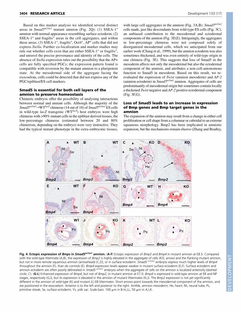
Loss of Smad5 leads to an increase in expressionof Bmp genes and Bmp target genes in theamnionThe expansion of the amnion may result from a change in either cellproliferation or cell shape from a columnar or cuboidal to an extremesquamous morphology. Bmp2 has been implicated in amnioticexpansion, but the mechanisms remain elusive (Zhang and Bradley,
RESEARCH ARTICLE Development 133 (17)
Fig. 4. Ectopic expression of Bmps in Smad5m1/m1 amnion. (A-F) Ectopic expression of Bmp2 and Bmp4 in mutant amnion at E8.5. Comparedwith the wild-type littermate (A,B), the expression of Bmp2 is highly elevated in the aggregate of cells (KO, arrow) and the flanking mutant amnion,but not in more remote squamous amnion (arrowhead) (C,D), or in surface ectoderm. Smad5 m1/m1 embryos express much higher levels of Bmp4throughout the amnion (F), than do controls (E). Bmp4 expression levels appear weaker in mutant surface ectoderm (E,F). Surface ectoderm andamnion ectoderm are often poorly delineated in Smad5 m1/m1 embryos when the aggregate of cells on the amnion is localized anteriorly (dashedcircle; C). (G-L) Enhanced expression of Bmp4, but not of Bmp2, in mutant amnion at E7.0. Bmp4 is expressed in wild-type amnion at EB and NPstages, respectively (G,I), but its expression is elevated in the amnion of mutant littermates (H,J). The Bmp2 expression is not yet significantlydifferent in the amnion of wild-type (K) and mutant (L) EB littermates. Short arrows point towards the mesodermal component of the amnion, andare positioned in the exocoelom. Anterior is to the left and posterior to the right. AmMe, amnion mesoderm; He, heart; Nt, neural tube; Ps,primitive streak; Se, surface ectoderm; Ys, yolk sac. Scale bars: 100 �m in B-H,J,L; 50 �m in A,I,K.

DEVELO
PMENT
1996). We evaluated the expression of Bmp genes, and some of theirtarget genes and modulators. Bmp2 transcript levels were extremelylow in the wild-type amnion at E8.5, if at all above background (Fig.4A,B), whereas Bmp4 expression was detectable in amnion (Fig.4E). A dramatic increase of Bmp2 and Bmp4 transcripts wasobserved in the Smad5m1/m1 amnion. Bmp2 was upregulated in thecells of the aggregates and neighbouring unaffected amnion, but notin squamous amnion distant from the aggregate (Fig. 4C,D). Bmp4was significantly upregulated, also in squamous amnion (Fig. 4F),but the expression of Bmp7 appeared unaffected (data not shown).During gastrulation, Bmp4 expression is dynamic. Expressionpersists within the extra-embryonic ectoderm, and Bmp4 transcriptsbecome detectable in the extra-embryonic mesodermal componentsof the amniotic folds, and subsequently in the amnion, yolk sac andchorion that line the exocoelom, as well as within the allantoicbud/allantois (Lawson et al., 1999) (data not shown). At the NPstage, before detectable thickening of the mutant amnion, Bmp4levels appeared already more robust in the amnion of mutants whencompared with control littermates (Fig. 4G-J). Bmp2 expression waslow at this stage of amnion development, without clear differencesbetween mutants and controls (Fig. 4K,L).
The observed upregulation of Bmp2/4 may cause increased Bmpsignalling via Smad1/8 or non-Smad pathways, including ERK1/2(Nakamura et al., 1999; Qi et al., 2004). The pattern of
phosphorylated (P)-ERK1/2 staining at E7.5 and 8.5 in wild-typelittermates is very variable and reflects the dynamic nature ofphosphorylation-dependent signalling cascades (data not shown). AtE7.5 and 8.5 P-ERK1/2-positive cells are very rare in the wild-typeamnion (Fig. 5A, data not shown). No dramatic increase in P-ERK1/2 staining was observed in non-thickened amnion (Fig. 5B,C,data not shown); however, manifest ERK signalling was observedwithin the aggregates. Likewise, the P-Smad1/5/8 staining patternwas very dynamic in E7.5 and 8.5 wild-type littermates, and couldbe observed only sporadically in amnion (Fig. 5D). In theSmad5m1/m1 amnion enhanced P-Smad1/8 staining was most evidentin the aggregate (Fig. 5E,F). These results illustrate enhanced Bmpsignalling in the mutant amnion.
We then analysed the expression of acknowledged Bmp targetgenes in the Smad5m1/m1 amnion. Periostin/Osf2 is involved in celladhesion and spreading (Kruzynska-Frejtag et al., 2001), and at E8.5its only sites of expression are the amnion and the yolk sac (Fig. 5G).In the Smad5m1/m1 amnion, periostin expression was morepronounced, although excluded from the aggregate of cells (Fig.5H). Msx2 is expressed in surface ectoderm (Monaghan, 1991) butbarely in amnion (Fig. 5I). Msx2 is robustly expressed in the mutantamnion, predominantly in its ectodermal component (Fig. 5J). Thisshows that higher Bmp2/4 levels indeed result in increasedexpression of Bmp target genes in the Smad5m1/m1 amnion. Genes
3405RESEARCH ARTICLESmad5 is required for amnion homeostasis
Fig. 5. Ectopic Bmp signalling in Smad5m1/m1 amnion. (A-C) Anti-P-ERK1/2 antibody staining can barely be detected in the amnion (arrowhead) atE8.5 (A). It is increased in non-thickened Smad5 mutant amnion but especially elevated in the aggregate of cells (B,C). B and C represent two differentSmad5 mutants. (D-F) Anti-phospho-Smad1/5/8 antibody staining can only be detected sporadically in an isolated cell of the amnion (arrowhead) atE8.5 (A). P-Smad1/5/8 is upregulated in non-thickened mutant amnion and within the aggregates of Smad5 mutant amnion. Sections in D-F are sistersections of A-C. (G,H) The Bmp target gene periostin is expressed in the amnion (arrowhead) at E8.5. It is strongly upregulated in the non-thickenedSmad5 mutant amnion, although excluded from the aggregate of cells (H). (I,J) The Bmp target gene Msx2 is barely detectable in wild-type amnion (I)but highly expressed in mutant amnion at E8.5 (J). Msx2 expression is primarily confined to the ectodermal component of the amnion (arrowhead) andthe aggregate of cells (arrow). The expression of Msx2 in the embryo proper is not altered significantly. AmMe, amnion mesoderm; He, heart; Nt, neuraltube; Se, surface ectoderm; Ys, yolk sac. Scale bars: 100 �m.
DEVELO
PMENT
3406
encoding Bmp antagonists like noggin, chordin and Smad7 areexpressed at low levels in the amnion and no significant changes intheir expression domain were detected in Smad5m1/m1 embryos (datanot shown).
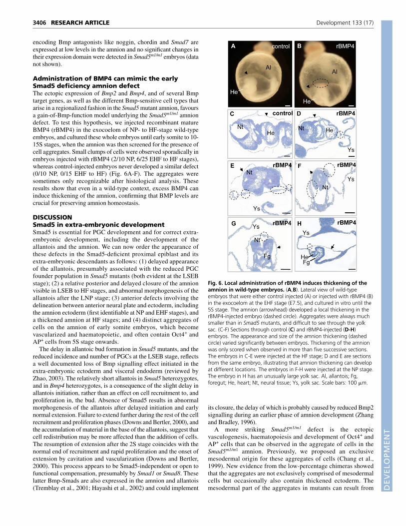
Administration of BMP4 can mimic the earlySmad5 deficiency amnion defectThe ectopic expression of Bmp2 and Bmp4, and of several Bmptarget genes, as well as the different Bmp-sensitive cell types thatarise in a regionalized fashion in the Smad5 mutant amnion, favoursa gain-of-Bmp-function model underlying the Smad5m1/m1 amniondefect. To test this hypothesis, we injected recombinant matureBMP4 (rBMP4) in the exocoelom of NP- to HF-stage wild-typeembryos, and cultured these whole embryos until early somite to 10-15S stages, when the amnion was then screened for the presence ofcell aggregates. Small clumps of cells were observed sporadically inembryos injected with rBMP4 (2/10 NP, 6/25 EHF to HF stages),whereas control-injected embryos never developed a similar defect(0/10 NP, 0/15 EHF to HF) (Fig. 6A-F). The aggregates weresometimes only recognizable after histological analysis. Theseresults show that even in a wild-type context, excess BMP4 caninduce thickening of the amnion, confirming that BMP levels arecrucial for preserving amnion homeostasis.
DISCUSSIONSmad5 in extra-embryonic developmentSmad5 is essential for PGC development and for correct extra-embryonic development, including the development of theallantois and the amnion. We can now order the appearance ofthese defects in the Smad5-deficient proximal epiblast and itsextra-embryonic descendants as follows: (1) delayed appearanceof the allantois, presumably associated with the reduced PGCfounder population in Smad5 mutants (both evident at the LSEBstage); (2) a relative posterior and delayed closure of the amnionvisible in LSEB to HF stages, and abnormal morphogenesis of theallantois after the LNP stage; (3) anterior defects involving thedelineation between anterior neural plate and ectoderm, includingthe amnion ectoderm (first identifiable at NP and EHF stages), anda thickened amnion at HF stages; and (4) distinct aggregates ofcells on the amnion of early somite embryos, which becomevascularized and haematopoietic, and often contain Oct4+ andAP+ cells from 5S stage onwards.
The delay in allantoic bud formation in Smad5 mutants, and thereduced incidence and number of PGCs at the LSEB stage, reflectsa well documented loss of Bmp signalling effect initiated in theextra-embryonic ectoderm and visceral endoderm (reviewed byZhao, 2003). The relatively short allantois in Smad5 heterozygotes,and in Bmp4 heterozygotes, is a consequence of the slight delay inallantois initiation, rather than an effect on cell recruitment to, andproliferation in, the bud. Absence of Smad5 results in abnormalmorphogenesis of the allantois after delayed initiation and earlynormal extension. Failure to extend further during the rest of the cellrecruitment and proliferation phases (Downs and Bertler, 2000), andthe accumulation of material in the base of the allantois, suggest thatcell redistribution may be more affected than the addition of cells.The resumption of extension after the 2S stage coincides with thenormal end of recruitment and rapid proliferation and the onset ofextension by cavitation and vascularization (Downs and Bertler,2000). This process appears to be Smad5-independent or open tofunctional compensation, presumably by Smad1 or Smad8. Theselatter Bmp-Smads are also expressed in the amnion and allantois(Tremblay et al., 2001; Hayashi et al., 2002) and could implement
its closure, the delay of which is probably caused by reduced Bmp2signalling during an earlier phase of amnion development (Zhangand Bradley, 1996).
A more striking Smad5m1/m1 defect is the ectopicvasculogenesis, haematopoiesis and development of Oct4+ andAP+ cells that can be observed in the aggregate of cells in theSmad5m1/m1 amnion. Previously, we proposed an exclusivemesodermal origin for these aggregates of cells (Chang et al.,1999). New evidence from the low-percentage chimeras showedthat the aggregates are not exclusively comprised of mesodermalcells but occasionally also contain thickened ectoderm. Themesodermal part of the aggregates in mutants can result from
RESEARCH ARTICLE Development 133 (17)
Fig. 6. Local administration of rBMP4 induces thickening of theamnion in wild-type embryos. (A,B). Lateral view of wild-typeembryos that were either control injected (A) or injected with rBMP4 (B)in the exocoelom at the EHF stage (E7.5), and cultured in vitro until the5S stage. The amnion (arrowhead) developed a local thickening in therBMP4-injected embryo (dashed circle). Aggregates were always muchsmaller than in Smad5 mutants, and difficult to see through the yolksac. (C-F) Sections through control (C) and rBMP4-injected (D-H)embryos. The appearance and size of the amnion thickening (dashedcircle) varied significantly between embryos. Thickening of the amnionwas only scored when observed in more than five successive sections.The embryos in C-E were injected at the HF stage; D and E are sectionsfrom the same embryo, illustrating that amnion thickening can developat different locations. The embryos in F-H were injected at the NP stage.The embryo in H has an unusually large yolk sac. Al, allantois; Fg,foregut; He, heart; Nt, neural tissue; Ys, yolk sac. Scale bars: 100 �m.

DEVELO
PMENT
altered proliferation, disorganization or misdifferentiation ofamniotic mesoderm cells, and/or from the previously proposedmislocation of allantois tissue (Chang et al., 1999). Theseaggregates are remarkably often localized very anteriorly, at aremote distance from the allantois, but the mesoderm in theaggregates expresses Tbx2 that is also expressed by allantoistissue (Chang et al., 1999). However, in the murine allantoisvasculogenesis occurs without haematopoiesis (Downs et al.,1998), whereas the Smad5 mutant amnion contains not only flk-1expressing endothelial cells, but also �-globin-expressingprimitive blood cells (Chang et al., 1999). Hence, the amnionadopts allantois and blood island-like fates in the absence ofSmad5.
Addressing allantois mislocation versus amnionmisdifferentiation remains elusive by marker gene analysis, buthere our PGC study is instrumental. Although defects in amnionand allantois development can be detected as early as the NPstage, ectopic AP+ cells were never found in thickenedSmad5m1/m1 amnion before the 5S stage. After the 5S stage themajority of the Smad5m1/m1 embryos had aggregates of cells on theamnion, of which half contained an appreciable number of AP+
and Oct4+ cells. This late appearance of AP+ cells in Smad5mutants is in contrast to the ectopic PGCs that can be readilydistinguished in the yolk sac of Otx2 and Smad2 mutants, and inextra-embryonic mesoderm of tetraploid Bmp4 chimeras at HFand early somite stages (K.A.L., unpublished) (Tremblay et al.,2001; Fujiwara et al., 2001). Therefore, it is very unlikely thatAP+ cells are swept into ectopic locations immediately after PGCallocation at ~E7.2 in Smad5m1/m1 embryos and remainunrecognized. Our analysis is suggestive of an in situ change infate of amnion cells.
The AP+ cells are unlikely to be de-novo induced, bona fidePGCs because no Stella-expressing cells were ever observed inthe aggregates. Although the coexpression of AP, Oct4, andSSEA-1 could indicate the presence of pluripotent, ES-like cells,the combination of AP, Oct4, SSEA-1 and fragilis expression ismore reminiscent of an earlier developmental state; PGC-competent epiblast. Fragilis is a direct target gene of extra-embryonic-derived Bmp4 in the PGC-competent epiblast (Saitouet al., 2002). The ectoderm of non-affected Smad5 mutant amnionhas characteristics of surface ectoderm (SSEA-1+ and Msx2+).Despite this reminiscence of surface ectoderm, the amnionectoderm expresses also periostin, a gene normally not expressedin surface ectoderm.
Remarkably, when isolated human amnion membrane cells arecultured, they also express Oct4 and the epiblast/stem cell/PGCmarker Nanog (Miki et al., 2005). These isolated human cells havethe potential to differentiate to all three germ layers – endoderm(hepatocytes), mesoderm (cardiomyocyte) and ectoderm (neuralcells) – in vitro (Sakuragawa et al., 2004; Tamagawa et al., 2004;Zhao et al., 2005).
Smad5 deficiency results in gain-of-Bmp-functiondefectsThe Smad5m1/m1 amnion defect is accompanied by a specific androbust upregulation of Bmp2 and Bmp4, whereas the expression ofBmp7 appears unaffected. This suggests that Smad5 is normally apredominant Bmp-Smad in the amnion that, directly or indirectly,negatively regulates Bmp2/4 expression. Ectopic Bmp4 expressionis readily visible before local thickening of the amnion at the LSEBstage, and subsequently in the aggregate of cells, but also in theneighbouring, seemingly unaffected amnion.
3407RESEARCH ARTICLESmad5 is required for amnion homeostasis
WT amnionE8.0
E8.0
KO amnionE9.0
Smad5
Smad5
Levels ofBMP signalling
(A)
(B)
(C)
Mesoderm: Twist+
Ectoderm: AP2+ Periostin+
SSEA-1+ , Msx2+
Tbx2+
Fragilis+flk1+
ζ-globin+
AP+, Oct4+
KO amnion
Fig. 7. Model for the role of Smad5 signalling in amnionhomeostasis. (A) The squamous, bilayered wild-type amnion at theearly somite stage is composed of a mesoderm layer facing theexocoelom that expresses Twist, whereas an epithelial cell layer thatexpresses AP-2 lines the amniotic cavity. The chimera analysisdemonstrates that Smad5 is essential in the mesoderm, where itfunctions in a non-cell-autonomous fashion, because mutantmesoderm cells can elicit thickening of wild-type amnion ectoderm.Reciprocally, however, we cannot conclude that Smad5 functions alsonon-cell-autonomously and/or cell autonomously in the ectoderm.(B) At the early somite stage the Smad5m1/m1 mutant amnion displaysan altered cellular organization with a zone of primarily thickenedmesoderm but also thickened ectoderm. The mutant amnion ectodermis positive for SSEA-1. The anterior amnion ectoderm remains cuboidal(squares), and is in continuity with anterior surface ectoderm andanterior neural tissue. Robust ectopic expression of Bmp2 and Bmp4 inmutant amnion is associated with increased Smad1/8 and ERK1/2signalling and expression of periostin, Tbx2, fragilis and Msx2. Thedifferential but overlapping ectopic expression patterns of all theseBmp-target genes suggest that cells in the mutant amnion sensedifferent levels of Bmp signalling. How these different levels of Bmpsignalling are established in the mutant amnion remains to be explored.(C) The transformation of the amnion to a complex tissue with differentBmp-sensitive cell types, such as e.g. an Oct4+, AP+, SSEA-1+ andfragilis+ region reminiscent of PGC-competent epiblast, Flk1-expressingendothelial cells and �-globin-expressing cells (see Discussion). Theectopic and appreciable numbers of AP+ cells arise suddenly from the5S stage onwards, which argues strongly for their in situ change in fate.The AP+ cells are localized near the ectodermal component of theaggregate, whereas haematopoietic and endothelial cells are positionedat the exocoelomic side. Our data suggest that Smad5 is thepredominant Bmp signal-mediator in the amnion, and controls Bmpexpression levels and amnion homeostasis tightly. Upon Smad5 removalthe control of Bmp expression is released, which results in an excess ofBmp, in take-over by non-Smad5 pathways and ultimately in gain ofBmp signalling defects.

DEVELO
PMENT
3408
The mutant amnion displays elevated Smad-dependent (Bmp-regulated phospho-Smad1/5/8) and Smad-independent (P-ERK1/2)signalling. Enhanced Bmp signalling is also demonstrated byalterations in the expression pattern of target genes of Bmp. A patentupregulation of periostin can be appreciated in seemingly unaffectedmutant amniotic cells. Tbx2 (Chang et al., 1999), fragilis and Msx2are, unlike periostin, highly upregulated in the aggregate of cells inthe mutant amnion. Msx2 is predominantly expressed in theectoderm component of the aggregate and non-thickened amnion.The increased or ectopic expression of these Bmp target genes indistinct but overlapping domains may indicate that expression ofthese target genes requires different dose windows of (combined)Bmp(s), which is reminiscent of the different doses of Bmp4signalling that are required for dorsoventral patterning of mesodermin Xenopus embryos (Dosch et al., 1997). A localized source ofligand(s) or antagonists/modulators, or several sources of ligands(e.g. Bmp2 and Bmp4) could establish graded signalling.Alternatively or additionally, differences in presence andconcentration of signalling components, but also unexploredendogenous differences in affinities of Smad1, and Smad8 and non-Smad cascades to activated receptors, Smad-interacting proteins andtheir shared target genes, will determine the outcome of Smad5deficiency in different regions of the amnion.
The Smad5m1/m1 amnion acquires features of different Bmp-sensitive lineages, such as allantois and blood island tissue but alsoPGC-competent epiblast (Lawson et al., 1999) (reviewed by Zhao,2003; Fujiwara et al., 2001). In the mutant amnion, epiblast-like cellsare localized near the ectodermal component, whereas ectopicendothelial and haematopoietic cells are positioned at theexocoelomic side of the aggregate. Concurrent with the differentialectopic expression patterns of several Bmp target genes, thisregionalization of the aggregates makes it conceivable that cellsindeed interpret different levels of Bmp signalling – whetherquantitative and/or qualitative – that determine their subsequent fate(Fig. 7). Bmp4 is not only implicated in differentiation of differentcell lineages, but it is also pivotal in maintaining self-renewal of EScells (Ying et al., 2003; Qi et al., 2004).
Our gain-of-Bmp-function model is especially challenging, asno other mutant mouse model of a Bmp signalling component hasbeen associated so far with ectopic vasculogenesis,haematopoiesis and development of (PGC-competent) epiblast-like cells in the amnion. Evidence that gain of Bmp function mayindeed underlie the Smad5 mutant amnion phenotype comes fromthe induction of amnion thickening in wild-type amnion whenrBMP4 is topically administered. Single injection of rBMP4 in theexocoelom compared to constitutive secretion of Bmp4 and Bmp2in Smad5 mutants is just one out of many explanations whyclumps could be significantly smaller in rBMP4-injected embryosthan in mutants. The converse experiment, in which Smad5m1/m1
embryos would be rescued by injecting Bmp antagonists such asNoggin and/or Chordin, is appealing. However, it is far morecomplicated to accomplish, as systemic administration of excessamounts of antagonists would be required to neutralizeendogenous Bmps.
Our study has revealed novel aspects of Smad5 signalling, i.e. anon-cell-autonomous function for Smad5 in amnion mesoderm andan in situ change in amnion cell fate upon Smad5 deficiency, andlinks these to an unexpected gain-of-Bmp-function model that issupported by the exogenous rBMP4 phenocopy experiment.Amnion cultures may be exploited in the future to dissect and toclarify the Bmp signalling pathway that determines change in cellfate of the amnion.
We are grateful to M. Missoul and L. Desmet and to the ‘mouse group’ forassistance with mouse and ES cell work; to J. Korving for technical assistance;and to L. Umans and L. Cox for their stimulating work on other ongoingSmad5 projects in the mouse. We are indebted to M. M. Matzuk and H.Chang for the generation and collaborative analysis of the Smad5m1/m1 mice inthe past. We thank S. Chuva de Sousa-Lopes and C. Mummery for helpfuldiscussions; F. Lemaigre and C. Pierreux for advice on embryo culture; and E.Delot, M. Jones and A. Surani for probes. E.A.B. thanks K. Steel for support.This work was supported by VIB, the University of Leuven (BOF/OT/00/41), the‘Interuniversity Attraction Poles Programme – Belgian Science Policy’ project5/35, project-based funding by the Fund of Scientific Research-Flanders (FWO-V; project G.0243.01) and the European Union (EU-grant T-Angiovasc QLG1-CT-2001-01032). E.A.B. thanks the IWT for a pre-doctoral fellowship. A.Z. washolder of a post-doc mandate of FWO-V.
Supplementary materialSupplementary material for this article is available athttp://dev.biologists.org/cgi/content/full/133/17/3399/DC1
ReferencesChang, H. and Matzuk, M. M. (2001). Smad5 is required for mouse primordial
germ cell development. Mech. Dev. 104, 61-67.Chang, H., Huylebroeck, D., Verschueren, K., Guo, Q., Matzuk, M. M. and
Zwijsen, A. (1999). Smad5 knockout mice die at mid-gestation due to multipleembryonic and extraembryonic defects. Development 126, 1631-1642.
Chang, H., Zwijsen, A., Vogel, H., Huylebroeck, D. and Matzuk, M. M.(2000). Smad5 is essential for left-right asymmetry in mice. Dev. Biol. 219, 71-78.
Delot, E., Bahamonde, M. E., Zhao, M. and Lyons, K. M. (2003). BMPsignalling is required for septation of the outflow tract of the mammalian heart.Development 130, 209-220.
de Sousa Lopes, S. M., Roelen, B. A., Monteiro, R. M., Emmens, R., Lin, H. Y.,Li, E., Lawson, K. A. and Mummery, C. L. (2004). BMP signalling mediated byALK2 in the visceral endoderm is necessary for the generation of primordialgerm cells in the mouse embryo. Genes Dev. 18, 1838-1849.
Dewulf, N., Verschueren, K., Lonnoy, O., Moren, A., Grimsby, S., VandeSpiegle, K., Miyazono, K., Huylebroeck, D. and ten Dijke, P. (1995).Expression of type I and type IB receptors for activin in midgestation mouseembryos suggests distinct functions in organogenesis. Mech. Dev. 52, 109-123.
Donovan, P. J., Stott, D., Cairns, L. A., Heasman, J. and Wylie, C. C. (1986).Migratory and postmigratory mouse primordial germ cells behave differently inculture. Cell 44, 831-838.
Dosch, R., Gawantka, V., Delius, H., Blumenstock, C. and Niehrs, C. (1997).Bmp-4 acts as a morphogen in dorsoventral mesoderm patterning in Xenopus.Development 124, 2325-2334.
Downs, K. M. and Davies, T. (1993). Staging of gastrulating mouse embryos bymorphological landmarks in the dissecting microscope. Development 118, 1255-1266.
Downs, K. M. and Bertler, C. (2000). Growth in the pre-fusion murine allantois.Anat. Embryol. 202, 323-331.
Downs, K. M., Gifford, S., Blahnik, M. and Gardner, R. L. (1998).Vascularization in the murine allantois occurs by vasculogenesis withoutaccompanying erythropoiesis. Development 125, 4507-4520.
Fox, N., Damjanov, I., Martinez-Hernandez, A., Knowles, B. B. and Solter, D.(1981). Immunohistochemical localization of the early embryonic antigen (SSEA-1) in postimplantation mouse embryos and fetal and adult tissues. Dev. Biol. 83,391-398.
Friedrich, G. and Soriano, P. (1991). Promoter traps in embryonic stem cells: agenetic screen to identify and mutate developmental genes in mice. Genes Dev.5, 1513-1523.
Fujiwara, T., Dunn, N. R. and Hogan, B. L. (2001). Bone morphogenetic protein4 in the extraembryonic mesoderm is required for allantois development and thelocalization and survival of primordial germ cells in the mouse. Proc. Natl. Acad.Sci. USA 98, 13739-13744.
Ginsburg, M., Snow, M. H. and McClaren, A. (1990). Primordial germ cells inthe mouse embryo during gastrulation. Development 110, 521-528.
Goumans, M. J., Zwijsen, A., van Rooijen, M. A., Huylebroeck, D., Roelen, B.A. and Mummery, C. L. (1999). Transforming growth factor-beta signalling inextraembryonic mesoderm is required for yolk sac vasculogenesis in mice.Development 126, 3473-3483.
Gu, Z., Reynolds, E. M., Song, J., Lei, H., Feijen, A., Yu, L., He, W.,MacLaughlin, D. T., van den Eijnden-van Raaij, J., Donahoe, P. K. et al.(1999). The type I serine/threonine kinase receptor ActRIA (ALK2) is required forgastrulation of the mouse embryo. Development 126, 2551-2561.
Hayashi, K., Kobayashi, T., Umino, T., Goitsuka, R., Matsui, Y. and Kitamura,D. (2002). SMAD1 signalling is critical for initial commitment of germ celllineage from mouse epiblast. Mech. Dev. 118, 99-109.
Jones, C. M., Lyons, K. M. and Hogan, B. L. (1991). Involvement of Bone
RESEARCH ARTICLE Development 133 (17)
DEVELO
PMENT
Morphogenetic Protein-4 (BMP-4) and Vgr-1 in morphogenesis andneurogenesis in the mouse. Development 111, 531-542.
Kinder, S. J., Tsang, T. E., Quinlan, G. A., Hadjantonakis, A. K., Nagy, A. andTam, P. P. (1999). The orderly allocation of mesodermal cells to theextraembryonic structures and the anteroposterior axis during gastrulation of themouse embryo. Development 126, 4691-4701.
Kruzynska-Frejtag, A., Machnicki, M., Rogers, R., Markwald, R. R. andConway, S. J. (2001). Periostin (an osteoblast-specific factor) is expressed withinthe embryonic mouse heart during valve formation. Mech. Dev. 103, 183-188.
Lawson, K. A. and Hage, W. J. (1994). Clonal analysis of the origin of primordialgerm cells in the mouse. Ciba Found. Symp. 182, 68-84.
Lawson, K. A., Meneses, J. J. and Pedersen, R. A. (1991). Clonal analysis ofepiblast fate during germ layer formation in the mouse embryo. Development113, 891-911.
Lawson, K. A., Dunn, N. R., Roelen, B. A., Zeinstra, L. M., Davis, A. M.,Wright, C. V., Korving, J. P. and Hogan, B. L. (1999). Bmp4 is required for thegeneration of primordial germ cells in the mouse embryo. Genes Dev. 13, 424-436.
Lechleider, R. J., Ryan, J. L., Garrett, L., Eng, C., Deng, C., Wynshaw-Boris, A.and Roberts, A. B. (2001). Targeted mutagenesis of Smad1 reveals an essentialrole in chorioallantoic fusion. Dev. Biol. 240, 157-167.
Lyons, K. M., Pelton, R. W. and Hogan, B. L. (1989). Patterns of expression ofmurine Vgr-1 and BMP-2a RNA suggest that transforming growth factor-beta-like genes coordinately regulate aspects of embryonic development. Genes Dev.3, 1657-1668.
MacKenzie, A., Ferguson, M. W. and Sharpe, P. T. (1992). Expression patternsof the homeobox gene, Hox-8, in the mouse embryo suggest a role in specifyingtooth initiation and shape. Development 115, 403-420.
Miki, T., Lehmann, T., Cai, H., Stolz, D. B. and Strom, S. C. (2005). Stem cellcharacteristics of amniotic epithelial cells. stem cells. Stem Cells 23, 1549-1559.
Mishina, Y., Crombie, R., Bradley, A. and Behringer, R. R. (1999). Multiple rolesfor activin-like kinase-2 signalling during mouse embryogenesis. Dev. Biol. 213,314-326.
Monaghan, A. P., Davidson, D. R., Sime, C., Graham, E., Baldock, R.,Bhattacharyo, S. S. and Hill, R. E. (1991). The Msh-homeobox genes definedomains in the developing vertebrate eye. Development 112, 1053-1061.
Nakamura, K., Shirai, T., Morishita, S., Uchida, S., Saeki-Miura, K. andMakishima, F. (1999). p38 mitogen-activated protein kinase functionallycontributes to chondrogenesis induced by growth/differentiation factor-5 inATDC5 cells. Exp. Cell Res. 250, 351-363.
Ohinata, Y., Payer, B., O’Carroll, D., Ancelin, K., Ono, Y., Sano, M., Barton,S. C., Obukhanych, T., Nussenzweig, M., Tarakhovsky, A. et al. (2005).Blimp1 is a critical determinant of the germ cell lineage in mice. Nature 436,207-213.
Okamura, D., Hayashi, K. and Matsui, Y. (2005). Mouse epiblasts changeresponsiveness to BMP4 signal required for PGC formation through functions ofextraembryonic ectoderm. Mol. Reprod. Dev. 70, 20-29.
Parameswaran, M. and Tam, P. P. (1995). Regionalisation of cell fate andmorphogenetic movement of the mesoderm during mouse gastrulation. Dev.Genet. 17, 16-28.
Qi, X., Li, T. G., Hao, J., Hu, J., Wang, J., Simmons, H., Miura, S., Mishina, Y.and Zhao, G. Q. (2004). BMP4 supports self-renewal of embryonic stem cells byinhibiting mitogen-activated protein kinase pathways. Proc. Natl. Acad. Sci. USA101, 6027-6032.
Saitou, M., Barton, S. C. and Surani, M. A. (2002). A molecular programme forthe specification of germ cell fate in mice. Nature 418, 293-300.
Sakuragawa, N., Kakinuma, K., Kikuchi, A., Okano, H., Uchida, S., Kamo, I.,Kobayashi, M. and Yokoyama, Y. (2004). Human amnion mesenchyme cellsexpress phenotypes of neuroglial progenitor cells. J. Neurosci. Res. 78, 208-214.
Schöler, H. R. (1991). Octamania: the POU factors in murine development. TrendsGenet. 7, 323-329.
Schoonjans, L., Kreemers, V., Danloy, S., Moreadith, R. W., Laroche, Y. andCollen, D. (2003). Improved generation of germline-competent embryonic stemcell lines from inbred mouse strains. Stem Cells 21, 90-97.
Shi, Y. and Massagué, J. (2003). Mechanisms of TGF-beta signalling from cellmembrane to the nucleus. Cell 113, 685-700.
Solloway, M. J. and Robertson, E. J. (1999). Early embryonic lethality inBmp5;Bmp7 double mutant mice suggests functional redundancy within the60A subgroup. Development 126, 1753-1768.
Tamagawa, T., Ishiwata, I. and Saito, S. (2004). Establishment andcharacterization of a pluripotent stem cell line derived from human amnioticmembranes and initiation of germ layers in vitro. Hum. Cell 17, 125-130.
Tanaka, S. S., Nagamatsu, G., Tokitake, Y., Kasa, M., Tam, P. P. and Matsui, Y.(2004). Regulation of expression of mouse interferon-induced transmembraneprotein like gene-3, Ifitm3 (mil-1, fragilis), in germ cells. Dev. Dyn. 230, 651-659.
Tanaka, S. S., Yamaguchi, Y. L., Tsoi, B., Lickert, H. and Tam, P. P. (2005).IFITM/Mil/fragilis family proteins IFITM1 and IFITM3 play distinct roles in mouseprimordial germ cell homing and repulsion. Dev. Cell 9, 745-756.
Tremblay, K. D., Dunn, N. R. and Robertson, E. J. (2001). Mouse embryoslacking Smad1 signals display defects in extra-embryonic tissues and germ cellformation. Development 128, 3609-3621.
Umans, L., Vermeire, L., Francis, A., Chang, H., Huylebroeck, D. and Zwijsen,A. (2003). Generation of a floxed allele of Smad5 for cre-mediated conditionalknockout in the mouse. Genesis 37, 5-11.
Van Eynde, A., Nuytten, M., Dewerchin, M., Schoonjans, L., Keppens, S.,Beullens, M., Moons, L., Carmeliet, P., Stalmans, W. and Bollen, M. (2004).The nuclear scaffold protein NIPP1 is essential for early embryonic developmentand cell proliferation. Mol. Cell. Biol. 24, 5863-5874.
Vincent, S. D., Dunn, N. R., Sciammas, R., Shapiro-Shalef, M., Davis, M. M.,Calame, K., Bikoff, E. K. and Robertson, E. J. (2005). The zinc fingertranscriptional repressor Blimp1/Prdm1 is dispensable for early axis formation butis required for specification of primordial germ cells in the mouse. Development132, 1315-1325.
Winnier, G., Blessing, M., Labosky, P. A. and Hogan, B. L. (1995). Bonemorphogenetic protein-4 is required for mesoderm formation and patterning inthe mouse. Genes Dev. 9, 2105-2116.
Yang, X., Castilla, L. H., Xu, X., Li, C., Gotay, J., Weinstein, M., Liu, P. P. andDeng, C. X. (1999). Angiogenesis defects and mesenchymal apoptosis in micelacking SMAD5. Development 126, 1571-1580.
Ying, Q. L., Nichols, J., Chambers, I. and Smith, A. (2003). BMP induction of Idproteins suppresses differentiation and sustains embryonic stem cell self-renewalin collaboration with STAT3. Cell 115, 281-292.
Ying, Y. and Zhao, G. Q. (2001). Cooperation of endoderm-derived BMP2 andextraembryonic ectoderm-derived BMP4 in primordial germ cell generation inthe mouse. Dev. Biol. 232, 484-492.
Ying, Y., Liu, X. M., Marble, A., Lawson, K. A. and Zhao, G. Q. (2000).Requirement of Bmp8b for the generation of primordial germ cells in themouse. Mol. Endocrinol. 14, 1053-1063.
Ying, Y., Qi, X. and Zhao, G. Q. (2001). Induction of primordial germ cells frommurine epiblasts by synergistic action of BMP4 and BMP8B signalling pathways.Proc. Natl. Acad. Sci. USA 98, 7858-7862.
Zhang, H. and Bradley, A. (1996). Mice deficient for BMP2 are nonviable andhave defects in amnion/chorion and cardiac development. Development 122,2977-2986.
Zhao, G. Q. (2003). Consequences of knocking out BMP signalling in the mouse.Genesis 35, 43-56.
Zhao, P., Ise, H., Hongo, M., Ota, M., Konishi, I. and Nikaido, T. (2005).Human amniotic mesenchymal cells have some characteristics ofcardiomyocytes. Transplantation 79, 528-535.
3409RESEARCH ARTICLESmad5 is required for amnion homeostasis