SMAD4-mediated WNT signaling controls the fate of cranial...
13
1977 RESEARCH ARTICLE INTRODUCTION During vertebrate animal development, the cell fate of multipotential neural crest cells is controlled by the context- dependent integration of extrinsic and intrinsic signals that drive their differentiation. Networks of synergistic and antagonistic signals are likely to regulate the development of neural crest derivatives to produce correct cell numbers at the proper time and location. Neural crest-derived odontogenic mesenchymal cells contain multipotential stem cells and can differentiate into dentin- secreting odontoblasts as well as chondrocyte-like and osteoblast- like cells (Chai et al., 2000; Chung et al., 2009; Yamazaki et al., 2007). However, the crucial cues in the signaling network that regulate dental mesenchymal cell fate remain largely unknown. During dentinogenesis, cranial neural crest (CNC)-derived odontoblast differentiation plays a crucial role in the secretion of predentin and dentin following terminal differentiation (Chai et al., 2000; Ruch, 1990). Odontoblast terminal differentiation is controlled by the inner enamel epithelium and is also dependent on matrix-mediated interactions (Cam et al., 1992; Ruch et al., 1995; Ruoslahti and Yamaguchi, 1991; Thesleff et al., 2001). Analysis of the expression patterns of growth factors during odontogenesis suggests that members of the transforming growth factor (TGF) superfamily, IGFs, WNTs and FGFs contribute to odontoblast terminal differentiation (Bègue-Kirn et al., 1994; Cam et al., 1992; Fjeld et al., 2005; Lohi et al., 2010; Suomalainen and Thesleff, 2010; Thesleff and Vaahtokari, 1992). Within the TGF superfamily, TGF1, TGF2, TGF3, BMP2, BMP4, BMP7 and follistatin are expressed in the inner enamel epithelium, dental papilla and in polarizing and functional odontoblasts. Exogenous TGF1, BMP2, BMP4 and BMP7 can induce odontoblast differentiation and dentin formation in dental papilla cells in vitro (Bègue-Kirn et al., 1992; Nakashima, 1994; Rutherford et al., 1994; Sloan et al., 2000; Unda et al., 2000). In addition, exogenous TGF1 regulates DSPP and DMP1 expression in odontoblast cell lines (He et al., 2004; Unterbrink et al., 2002). Moreover, inhibition of TGF signaling in Wnt1-Cre;Tgfbr2 fl/fl mice and of BMP signaling in K14-Nog or OC-Cre;Smad4 fl/fl mice results in abnormal dentin formation (Oka et al., 2007; Plikus et al., 2005; Gao et al., 2009). These data indicate that TGF/BMP signaling is involved in regulating dentinogenesis. The TGF superfamily of cytokines comprises TGFs, BMPs, activins and related proteins. TGF/BMP signaling plays an important role in regulating a broad spectrum of processes, including cell proliferation, differentiation, apoptosis, migration and extracellular matrix remodeling (Chai and Slavkin, 2003; Massague, 2000; Siegel and Massague, 2003). The canonical TGF/BMP signaling pathway involves binding of the ligand to initiate the assembly of a heteromeric complex of type II and type I receptors. The activated type I receptor phosphorylates SMAD proteins in the cytoplasm. The type I receptors for TGF, activin, nodal and myostatin [ALK4 (ACVR1B), ALK5 (TGFBR1), ALK7 (ACVR1C)] phosphorylate SMAD2 and SMAD3, whereas the BMP and AMH type I receptors [ALK1 (ACVRL1), ALK2 (ACVR1), ALK3 (BMPR1A), ALK6 (BMPR1B)] phosphorylate Development 138, 1977-1989 (2011) doi:10.1242/dev.061341 © 2011. Published by The Company of Biologists Ltd 1 Center for Craniofacial Molecular Biology, Herman Ostrow School of Dentistry, University of Southern California, 2250 Alcazar Street, CSA 103, Los Angeles, CA 90033, USA. 2 Salivary Gland Disease Center and the Molecular Laboratory for Gene Therapy and Tooth Regeneration, Capital Medical University School of Stomatology, Beijing 100050, China. 3 Department of Stomatology, Beijing Friendship Hospital, Capital Medical University, Beijing 100050, China. 4 Department of Biomedical Genetics and Center for Oral Biology, University of Rochester School of Medicine and Dentistry, Rochester, NY 14642, USA. *Author for correspondence ([email protected]) Accepted 21 February 2011 SUMMARY TGF/BMP signaling regulates the fate of multipotential cranial neural crest (CNC) cells during tooth and jawbone formation as these cells differentiate into odontoblasts and osteoblasts, respectively. The functional significance of SMAD4, the common mediator of TGF/BMP signaling, in regulating the fate of CNC cells remains unclear. In this study, we investigated the mechanism of SMAD4 in regulating the fate of CNC-derived dental mesenchymal cells through tissue-specific inactivation of Smad4. Ablation of Smad4 results in defects in odontoblast differentiation and dentin formation. Moreover, ectopic bone-like structures replaced normal dentin in the teeth of Osr2-IresCre;Smad4 fl/fl mice. Despite the lack of dentin, enamel formation appeared unaffected in Osr2-IresCre;Smad4 fl/fl mice, challenging the paradigm that the initiation of enamel development depends on normal dentin formation. At the molecular level, loss of Smad4 results in downregulation of the WNT pathway inhibitors Dkk1 and Sfrp1 and in the upregulation of canonical WNT signaling, including increased -catenin activity. More importantly, inhibition of the upregulated canonical WNT pathway in Osr2-IresCre;Smad4 fl/fl dental mesenchyme in vitro partially rescued the CNC cell fate change. Taken together, our study demonstrates that SMAD4 plays a crucial role in regulating the interplay between TGF/BMP and WNT signaling to ensure the proper CNC cell fate decision during organogenesis. KEY WORDS: TGF/BMP, SMAD4, Canonical WNT signaling, Odontoblast, Bone formation, WNT inhibitor, Mouse SMAD4-mediated WNT signaling controls the fate of cranial neural crest cells during tooth morphogenesis Jingyuan Li 1,2 , Xiaofeng Huang 3 , Xun Xu 1 , Julie Mayo 1 , Pablo Bringas, Jr 1 , Rulang Jiang 4 , Songling Wang 2 and Yang Chai 1, * DEVELOPMENT Development ePress online publication date 13 April 2011 http://dev.biologists.org/lookup/doi/10.1242/dev.061341 Access the most recent version at First posted online on 13 April 2011 as 10.1242/dev.061341
Transcript of SMAD4-mediated WNT signaling controls the fate of cranial...
1977RESEARCH ARTICLE
INTRODUCTIONDuring vertebrate animal development, the cell fate ofmultipotential neural crest cells is controlled by the context-dependent integration of extrinsic and intrinsic signals that drivetheir differentiation. Networks of synergistic and antagonisticsignals are likely to regulate the development of neural crestderivatives to produce correct cell numbers at the proper time andlocation. Neural crest-derived odontogenic mesenchymal cellscontain multipotential stem cells and can differentiate into dentin-secreting odontoblasts as well as chondrocyte-like and osteoblast-like cells (Chai et al., 2000; Chung et al., 2009; Yamazaki et al.,2007). However, the crucial cues in the signaling network thatregulate dental mesenchymal cell fate remain largely unknown.
During dentinogenesis, cranial neural crest (CNC)-derivedodontoblast differentiation plays a crucial role in the secretion ofpredentin and dentin following terminal differentiation (Chai et al.,2000; Ruch, 1990). Odontoblast terminal differentiation iscontrolled by the inner enamel epithelium and is also dependent onmatrix-mediated interactions (Cam et al., 1992; Ruch et al., 1995;Ruoslahti and Yamaguchi, 1991; Thesleff et al., 2001). Analysis ofthe expression patterns of growth factors during odontogenesis
suggests that members of the transforming growth factor (TGF)superfamily, IGFs, WNTs and FGFs contribute to odontoblastterminal differentiation (Bègue-Kirn et al., 1994; Cam et al., 1992;Fjeld et al., 2005; Lohi et al., 2010; Suomalainen and Thesleff,2010; Thesleff and Vaahtokari, 1992). Within the TGFsuperfamily, TGF1, TGF2, TGF3, BMP2, BMP4, BMP7 andfollistatin are expressed in the inner enamel epithelium, dentalpapilla and in polarizing and functional odontoblasts. ExogenousTGF1, BMP2, BMP4 and BMP7 can induce odontoblastdifferentiation and dentin formation in dental papilla cells in vitro(Bègue-Kirn et al., 1992; Nakashima, 1994; Rutherford et al.,1994; Sloan et al., 2000; Unda et al., 2000). In addition, exogenousTGF1 regulates DSPP and DMP1 expression in odontoblast celllines (He et al., 2004; Unterbrink et al., 2002). Moreover, inhibitionof TGF signaling in Wnt1-Cre;Tgfbr2fl/fl mice and of BMPsignaling in K14-Nog or OC-Cre;Smad4fl/fl mice results inabnormal dentin formation (Oka et al., 2007; Plikus et al., 2005;Gao et al., 2009). These data indicate that TGF/BMP signaling isinvolved in regulating dentinogenesis.
The TGF superfamily of cytokines comprises TGFs, BMPs,activins and related proteins. TGF/BMP signaling plays animportant role in regulating a broad spectrum of processes,including cell proliferation, differentiation, apoptosis, migrationand extracellular matrix remodeling (Chai and Slavkin, 2003;Massague, 2000; Siegel and Massague, 2003). The canonicalTGF/BMP signaling pathway involves binding of the ligand toinitiate the assembly of a heteromeric complex of type II and typeI receptors. The activated type I receptor phosphorylates SMADproteins in the cytoplasm. The type I receptors for TGF, activin,nodal and myostatin [ALK4 (ACVR1B), ALK5 (TGFBR1), ALK7(ACVR1C)] phosphorylate SMAD2 and SMAD3, whereas theBMP and AMH type I receptors [ALK1 (ACVRL1), ALK2(ACVR1), ALK3 (BMPR1A), ALK6 (BMPR1B)] phosphorylate
Development 138, 1977-1989 (2011) doi:10.1242/dev.061341© 2011. Published by The Company of Biologists Ltd
1Center for Craniofacial Molecular Biology, Herman Ostrow School of Dentistry,University of Southern California, 2250 Alcazar Street, CSA 103, Los Angeles, CA90033, USA. 2Salivary Gland Disease Center and the Molecular Laboratory for GeneTherapy and Tooth Regeneration, Capital Medical University School of Stomatology,Beijing 100050, China. 3Department of Stomatology, Beijing Friendship Hospital,Capital Medical University, Beijing 100050, China. 4Department of BiomedicalGenetics and Center for Oral Biology, University of Rochester School of Medicineand Dentistry, Rochester, NY 14642, USA.
*Author for correspondence ([email protected])
Accepted 21 February 2011
SUMMARYTGF/BMP signaling regulates the fate of multipotential cranial neural crest (CNC) cells during tooth and jawbone formation asthese cells differentiate into odontoblasts and osteoblasts, respectively. The functional significance of SMAD4, the commonmediator of TGF/BMP signaling, in regulating the fate of CNC cells remains unclear. In this study, we investigated the mechanismof SMAD4 in regulating the fate of CNC-derived dental mesenchymal cells through tissue-specific inactivation of Smad4. Ablationof Smad4 results in defects in odontoblast differentiation and dentin formation. Moreover, ectopic bone-like structures replacednormal dentin in the teeth of Osr2-IresCre;Smad4fl/fl mice. Despite the lack of dentin, enamel formation appeared unaffected inOsr2-IresCre;Smad4fl/fl mice, challenging the paradigm that the initiation of enamel development depends on normal dentinformation. At the molecular level, loss of Smad4 results in downregulation of the WNT pathway inhibitors Dkk1 and Sfrp1 and inthe upregulation of canonical WNT signaling, including increased -catenin activity. More importantly, inhibition of theupregulated canonical WNT pathway in Osr2-IresCre;Smad4fl/fl dental mesenchyme in vitro partially rescued the CNC cell fatechange. Taken together, our study demonstrates that SMAD4 plays a crucial role in regulating the interplay between TGF/BMPand WNT signaling to ensure the proper CNC cell fate decision during organogenesis.
KEY WORDS: TGF/BMP, SMAD4, Canonical WNT signaling, Odontoblast, Bone formation, WNT inhibitor, Mouse
SMAD4-mediated WNT signaling controls the fate of cranialneural crest cells during tooth morphogenesisJingyuan Li1,2, Xiaofeng Huang3, Xun Xu1, Julie Mayo1, Pablo Bringas, Jr1, Rulang Jiang4, Songling Wang2
and Yang Chai1,*
DEVELO
PMENT
Development ePress online publication date 13 April 2011http://dev.biologists.org/lookup/doi/10.1242/dev.061341Access the most recent version at First posted online on 13 April 2011 as 10.1242/dev.061341
1978
SMAD1, SMAD5 and SMAD8 (Massague and Gomis, 2006).These receptor-activated SMADs (R-SMADs) dissociate from thetype I receptor and then oligomerize with a common partner,SMAD4. Activated SMAD complexes move into the nucleus,where they regulate the transcription of target genes (Shi andMassague, 2003). A recent study shows that SMAD4-independentsignaling pathways are also important during craniofacialdevelopment (Xu et al., 2008).
SMAD4 plays a central role in regulating TGF/BMP signalingduring organogenesis. However, the role of SMAD4 in regulatingCNC cell fate determination remains unclear. In this study, wegenerated mutant mice in which Smad4 is specifically inactivatedin the CNC-derived dental mesenchymal cells (Osr2-IresCre;Smad4fl/fl). We found that ablation of Smad4 in the dentalmesenchyme results in a defect in odontoblast differentiation.Instead of dentin formation, ectopic bone-like structures form inOsr2-IresCre;Smad4fl/fl mice via a mechanism that involvesupregulation of the canonical WNT signaling pathway. Despite thelack of dentin, enamel formation appears to be normal andtherefore independent of dentinogenesis in Osr2-IresCre;Smad4fl/fl
mice.
MATERIALS AND METHODSGeneration of transgenic miceThe Osr2-IresCre transgenic line (Lan et al., 2007), ROSA26 conditionalreporter (R26R) transgene (Soriano, 1999), conditional Smad4 (Dpc4)allele (Yang et al., 2002) and TOPGAL transgenic allele (DasGupta andFuchs, 1999) have been described previously. Mating Osr2-IresCre withR26R mice generated Osr2-IresCre;R26R embryos. Osr2-IresCre;Smad4fl/+
male mice were crossed with Smad4fl/fl female mice to generate Osr2-IresCre;Smad4fl/fl alleles. Osr2-IresCre;Smad4fl/fl;TOPGAL embryos wereproduced by crossing Osr2-IresCre;Smad4fl/+;TOPGAL and Smad4fl/fl
mice.
Histological analysis and scanning electron microscopy (SEM)For histological analysis, samples were fixed in 4% paraformaldehyde andprocessed into paraffin-embedded serial sections using routine procedures.For general morphology, deparaffinized sections were stained withHematoxylin and Eosin (H&E) using standard procedures. For SEM,samples were processed and viewed according to standard procedures aspreviously described (Xu et al., 2006).
X-gal staining and detection of -galactosidase activitySamples at various stages of embryonic development were fixed in 0.2%glutaraldehyde, passed through a sucrose series, embedded in O.C.T.Compound (Tissue-Tek) and sectioned on a cryostat at 10 m prior to X-gal staining for lacZ expression. Detection of -galactosidase (-gal)activity in tissue sections was as previously described (Chai et al., 2000).
Whole molars (after 19 days kidney capsule transplantation) weredissected from the mandible and stained for -gal activity according tostandard procedures, as previously described (Chai et al., 2000). Themolars were embedded in paraffin after decalcification and dehydration.Sections were cut at 8 m and counterstained with Nuclear Fast Red.
Lower first molar organ cultureThe lower first molars were microdissected from control and Osr2-IresCre;Smad4fl/fl mutant mice at the newborn stage and cultured in BGJBculture medium (GIBCO/Invitrogen) supplemented with 10% ascorbic acidand 1% penicillin and streptomycin. Tissues were harvested after 7 days inculture.
Kidney capsule transplantationKidney capsule transplantation was carried out as previously described (Xuet al., 2005). The first branchial arches were dissected from embryonic day(E) 11.5 control and Osr2-IresCre;Smad4fl/fl embryos and cultured for 1day during genotyping. The explants were then grafted under kidneycapsules. The grafting products were harvested after 19 days.
In situ hybridizationIn situ hybridizations were performed following standard procedures (Xuet al., 2005). Digoxigenin-labeled antisense probes were generated frommouse cDNA clones that were kindly provided by several laboratories:ameloblastin (Ambn), Margarita Zeichner-David [University of SouthernCalifornia (USC), USA]; amelogenin (Amelx), Malcolm Snead (USC,USA); Bsp (Ibsp – Mouse Genome Informatics), Tomoyo Sasaki (USC,USA); Dspp, Irma Thesleff (University of Helsinki, Finland).
ImmunostainingImmunostaining was performed using primary antibodies against SMAD4(Abcam), -catenin (BD Transduction Laboratories), -gal (Abcam),DKK1 (R&D) and SFRP1 (Santa Cruz). Alexa Fluor 568 (MolecularProbes), DyLight 488 (Jackson ImmunoResearch) and the HistoStain SPKit (Invitrogen) were used for detection. Sections were counterstained withHematoxylin and DAPI.
Von Kossa stainingTo detect calcium salts, Von Kossa staining was performed by immersingthe sections in 5% silver nitrate solution (Merck), exposing the samplesunder a 100 W light bulb for 2.5 hours, stopping the reaction with 5%sodium thiosulfate (Sigma-Aldrich), and staining the sections withparagon-epoxy solution [0.73 g Toluidine Blue (Sigma-Aldrich) and0.135 g basic fuchsin (Sigma-Aldrich) in 30% ethanol] for 2.5 minutes ona hot plate.
Quantitative (q) PCR analysisRNA was isolated from dental papilla in vivo and dental mesenchymalcells in vitro using RNeasy Mini Kits (Qiagen). The QuantiTect ReverseTranscription Kit (Qiagen) was used for cDNA synthesis. qPCR wascarried out on the iCycler (Bio-Rad) with gene-specific primers and SYBRGreen. Values were normalized to -actin using the 2–Ct method (Livakand Schmittgen, 2001).
ELISA analysisTotal protein was obtained from dental mesenchymal cells in vitro usingM-PER Mammalian Protein Extraction Reagent (Thermo Scientific).ELISAs for total -catenin (Assay Designs) were carried out following themanufacturer’s instructions.
Dental mesenchymal cell cultureNewborn dental papilla of lower first molars was mechanically dissectedfrom Smad4fl/fl mice and cultured in the alpha modification of Eagle’sMedium (GIBCO/Invitrogen) supplemented with 20% fetal bovine serum.The culture medium was changed twice a week. Confluent cultures werecollected by trypLE Express (GIBCO/Invitrogen) and subcultured underthe same conditions.
Transfection of cells with adenovirusAdherent primary dental mesenchymal cells (as mentioned above) weretransfected with adenovirus (control, Smad4fl/fl + Ad-CMV-eGFP; mutant,Smad4fl/fl + Ad-CMV-Cre-eGFP; Vector Development Lab, Baylor Collegeof Medicine) at 5000 virus particles/cell with GeneJammer as previouslydescribed (Fouletier-Dilling et al., 2005). Based on the eGFP expression,we harvested cells after 12, 24 or 36 hours transfection, respectively.
RESULTSEarly tooth development is unaffected in Osr2-IresCre;Smad4fl/fl miceTo test our hypothesis that SMAD4-mediated TGF/BMPsignaling is crucial for CNC cell fate determination during toothmorphogenesis, we generated Smad4 conditional knockout mice.Specific ablation of Smad4 in neural crest cells using the Wnt1-Crerecombination system leads to early mortality by E11.5, probablyowing to heart development failure (Ko et al., 2007). Tocircumvent this early lethality, we crossed the Smad4 conditionalallele (Yang et al., 2002) with Osr2-IresCre (Lan et al., 2007) togenerate Osr2-IresCre;Smad4fl/fl embryos. In contrast to Wnt1-Cre
RESEARCH ARTICLE Development 138 (10)
DEVELO
PMENT

transgenic mice, which express Cre in the premigratory neural crestcells by E8.5 (Chai et al., 2000; Danielian et al., 1998), Cre activityis not detectable in the craniofacial region in Osr2-IresCre miceuntil E10.5 (Lan et al., 2007). The Osr2-IresCre transgene directedCre activity in the dental mesenchyme throughout toothdevelopment in Osr2-IresCre;R26R mice (Fig. 1A-G).
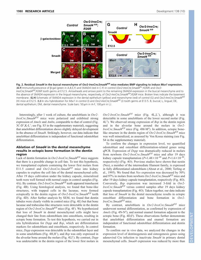
Surprisingly, tooth development progressed without obviousdefects in Osr2-IresCre;Smad4fl/fl mice until the late bell stage(E18.5) (see Fig. S1 in the supplementary material). By contrast,our previous studies found that tooth development in Wnt1-Cre;Smad4fl/fl embryos is arrested at the dental lamina stage,suggesting that Smad4 is absolutely required in the CNC-deriveddental mesenchyme for tooth development to advance into the budstage (Ko et al., 2007). To understand this discrepancy, we re-examined Osr2-IresCre expression using X-gal staining from thebud to bell stages. As tooth buds developed from E12.5 to E14.5,Osr2-IresCre was expressed in a gradient in the developingtooth mesenchyme, with higher expression towards the lingual sideand lower expression immediately buccal to the tooth buds(Fig. 1A-C). We also analyzed Osr2-IresCre expression usingimmunofluorescence of -gal at E12.5 and found the same gradientof expression in the dental mesenchyme (Fig. 2A,B).
The initial tooth-generating potential resides within the dentalepithelium, which is capable of inducing non-tooth-forming CNC-derived ectomesenchyme to develop into teeth (Jernvall andThesleff, 2000; Mina and Kollar, 1987). Later, this tooth-formingpotential shifts to the dental mesenchyme, coinciding with a shiftin BMP signaling from the epithelium to the mesenchyme, whereBMP signaling induces the expression of Msx1 (Chen et al., 1996).We hypothesized that the gradient of Osr2-IresCre expression inthe dental mesenchyme results in a gradient of Smad4 deletion.SMAD4 expression might persist in the buccal region of the toothbud, which would mediate BMP signaling in the mesenchyme andallow teeth to develop to the bud stage. We found that SMAD4 isexpressed in the dental epithelium and mesenchyme in controlmice at E12.5 (Fig. 2C,E,G). By contrast, SMAD4 expression wasnot detectable in the lingual mesenchyme of the tooth bud in Osr2-IresCre;Smad4fl/fl mice at E12.5, although it was detectable in thebuccal region (Fig. 2D,F,H). We also assayed the expression ofMsx1 in the dental mesenchyme by in situ hybridization. Msx1expression was clearly detectable in both control (Fig. 2I) andOsr2-IresCre;Smad4fl/fl (Fig. 2J) mice at E13.5. To confirm our invivo data, we analyzed the expression level of Msx1 usingadenoviral Cre infection to inactivate Smad4 in primarymesenchymal cells within the lower molar region at E13.5.Although Smad4 expression was reduced by ~80% after 24 hoursof infection, the expression level of Msx1 was similar to that of thecontrol (see Fig. S2 in the supplementary material). Our dataindicate that Smad4 remaining in the buccal mesenchyme of toothbuds in Osr2-IresCre;Smad4fl/fl mice may be sufficient to mediateBMP signaling in the dental mesenchyme to induce Msx1expression, resulting in normal tooth development during the earlystages of embryogenesis.
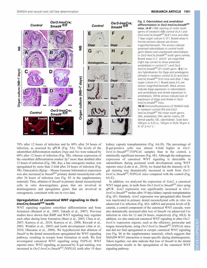
Cre-mediated inactivation of Smad4 in the dentalmesenchyme affects odontoblast differentiationAt the newborn stage, we found that odontoblasts of Osr2-IresCre;Smad4fl/fl mice failed to undergo polarized growth andformed a layer of non-polarized cuboidal cells with centrallylocated nuclei (Fig. 3B,B�). Moreover, dentin matrix was absent inOsr2-IresCre;Smad4fl/fl mice (Fig. 3B�). To examine the status ofodontoblast functional differentiation in Osr2-IresCre;Smad4fl/fl
mice, we assayed the expression of Dspp by in situ hybridization.In control mice, we detected Dspp expression in the odontoblastlayer and in some ameloblasts at the newborn stage (Fig. 3E). InOsr2-IresCre;Smad4fl/fl mice, however, Dspp was undetectable(Fig. 3F). In addition, expression of Amelx, an ameloblastdifferentiation marker, was also undetectable in Osr2-IresCre;Smad4fl/fl samples as compared with the control (Fig.3G,H). To confirm the successful inactivation of SMAD4, weperformed immunostaining using a SMAD4 antibody and foundthat dental mesenchymal cells in newborn Osr2-IresCre;Smad4fl/fl
tooth germ were negative for SMAD4 (Fig. 3N).Osr2-IresCre;Smad4fl/fl mice die within a day of birth,
precluding an examination of tooth development at later stages. Todetermine whether the abnormal odontoblast differentiation inOsr2-IresCre;Smad4fl/fl tooth germ is due to delayed development,we cultured newborn tooth germ for 7 days ex vivo. After 1 weekof culture, we still failed to find polarization and Dspp expressionin odontoblasts from Osr2-IresCre;Smad4fl/fl mice (Fig.3D,D�,J,J�), although both were detectable in control samples (Fig.3C,C�,I,I�). Our data indicate that loss of Smad4 in the dentalmesenchyme affects the terminal differentiation of odontoblasts.
1979RESEARCH ARTICLESMAD4 and neural crest cell fate determination
Fig. 1. Osr2-IresCre expression pattern during molardevelopment. (A-G)Cre-mediated activation of lacZ expressionassayed by X-gal staining (blue) in frontal sections of the lower firstmolar of E12.5-E18.5 Osr2-IresCre;R26R mouse embryos at the lamina(A), bud (B), cap (C,D) and bell (E-G) stage. X-gal staining is detectablein dental mesenchymal cells, but not in the dental epithelium. Note thatOsr2-IresCre is expressed in a gradient in the developing toothmesenchyme, with higher expression (**) lingual and lower expression(*) buccal to the tooth buds. B, buccal; L, lingual; DE, dentalepithelium; DM, dental mesenchyme. Scale bars: 50m in A-D; 100min E-G.
DEVELO
PMENT
1980
Interestingly, after 1 week of culture, the ameloblasts in Osr2-IresCre;Smad4fl/fl mice were polarized and exhibited strongexpression of Amelx and Ambn, comparable to that of control (Fig.3C-D�,K-L�; see Fig. S3 in the supplementary material), suggestingthat ameloblast differentiation shows slightly delayed developmentin the absence of Smad4. Strikingly, however, our data indicate thatameloblast differentiation is independent of functional odontoblastdifferentiation.
Ablation of Smad4 in the dental mesenchymeresults in ectopic bone formation in the dentinregionLack of dentin formation in Osr2-IresCre;Smad4fl/fl mice suggeststhat there is a possible change in cell fate. To test this hypothesis,we transplanted explants containing the lower first molars fromE11.5 control and Osr2-IresCre;Smad4fl/fl mice into kidneycapsules to explore the cell fate of the dental mesenchymal cells.After 19 days cultivation under the kidney capsule, mineralizedteeth were well formed with normal cusps in control samples (Fig.4A). By contrast, Osr2-IresCre;Smad4fl/fl teeth appeared translucent(Fig. 4B). Using histological analysis, we found that bone-likestructures, with trapped cells in the lacunae, were formedectopically in the dentin region of Osr2-IresCre;Smad4fl/fl teeth(Fig. 4D). After further analysis by SEM, we found that dentintubules were clearly visible in control mice (Fig. 4E) but that bonelacunae and trabeculae-like structures were detectable in the dentinregion of Osr2-IresCre;Smad4fl/fl mice (Fig. 4F). We hypothesizedthat loss of Smad4 in dental mesenchymal cells might havechanged their fate from odontoblasts into osteoblasts, resulting inectopic bone formation. To test this hypothesis, we carried out insitu hybridization for Dspp and Bsp, which are differentiationmarkers for odontoblasts and osteoblasts, respectively. In controlmice, Dspp expression was detectable in the odontoblast layer andin some ameloblasts (Fig. 4H,H�), and Bsp was only expressed inthe alveolar bone around the molars (Fig. 4I,I�). By contrast, Dsppwas undetectable in the dentin region of the lower first molars in
Osr2-IresCre;Smad4fl/fl mice (Fig. 4L,L�), although it wasdetectable in some ameloblasts of the lower second molar (Fig.4L�). We observed strong expression of Bsp in the dentin regionand in the alveolar bone around the molars in Osr2-IresCre;Smad4fl/fl mice (Fig. 4M-M�). In addition, ectopic bone-like structure in the dentin region of Osr2-IresCre;Smad4fl/fl micewas well mineralized, as assessed by Von Kossa staining (see Fig.S4 in the supplementary material).
To confirm the changes in expression level, we quantifiedodontoblast and osteoblast differentiation-related genes usingqPCR. Expression of Dspp was dramatically reduced in molarsfrom newborn Osr2-IresCre;Smad4fl/fl mice and after 19 dayskidney capsule transplantation (P1.48�10–09 and P1.6�10–09,respectively) (Fig. 4O). Previous studies have shown that nestin(Nes), a member of the intermediate filament family, is expressedin fully differentiated odontoblasts (About et al., 2000; Terling etal., 1995). We found that Nes expression was decreased by 50%and 97% in molars from newborn Osr2-IresCre;Smad4fl/fl mice andafter 19 days kidney capsule transplantation, respectively (Fig. 4O).Conversely, Bsp expression was increased 3-fold in Osr2-IresCre;Smad4fl/fl versus control samples after 19 days kidneycapsule transplantation (Fig. 4O). Taken together, our data indicatethat loss of Smad4 in the dental mesenchyme results in ectopicosteoblast differentiation and bone formation in Osr2-IresCre;Smad4fl/fl mice.
By contrast, ameloblasts in Osr2-IresCre;Smad4fl/fl miceunderwent normal differentiation, as confirmed by expression ofAmelx (Fig. 4N-N�), and normal enamel formation adjacent to theectopic bone (Fig. 4D,F). These observations further demonstratethat ameloblast differentiation and enamel formation areindependent of functional odontoblast differentiation and dentinformation.
To confirm our in vivo data, we analyzed the changes in theexpression level of dentinogenesis and osteogenesis genes usingadenoviral Cre infection to inactivate Smad4 in primary dentalmesenchymal cells. Smad4 expression was reduced by more than
RESEARCH ARTICLE Development 138 (10)
Fig. 2. Residual Smad4 in the buccal mesenchyme of Osr2-IresCre;Smad4fl/fl mice mediates BMP signaling to induce Msx1 expression.(A-F)Immunofluorescence of -gal (green in A,B,E,F) and SMAD4 (red in C-F) in control (Osr2-IresCre;Smad4fl/+;R26R) and Osr2-IresCre;Smad4fl/fl;R26R tooth germs at E12.5. Arrowheads and arrows point to the remaining SMAD4 expression in the buccal mesenchyme and tothe absence of SMAD4 expression in the lingual mesenchyme, respectively, of Osr2-IresCre;Smad4fl/fl;R26R mice. Broken lines indicate the basementmembrane. (G,H)Schematic of SMAD4 expression in the dental epithelium (yellow) and mesenchyme (red) of control (G) and Osr2-IresCre;Smad4fl/fl
(H) mice at E12.5. (I,J)In situ hybridization for Msx1 in control (I) and Osr2-IresCre;Smad4fl/fl (J) tooth germs at E13.5. B, buccal; L, lingual; DE,dental epithelium; DM, dental mesenchyme. Scale bars: 50m in A-F; 100m in I,J.
DEVELO
PMENT
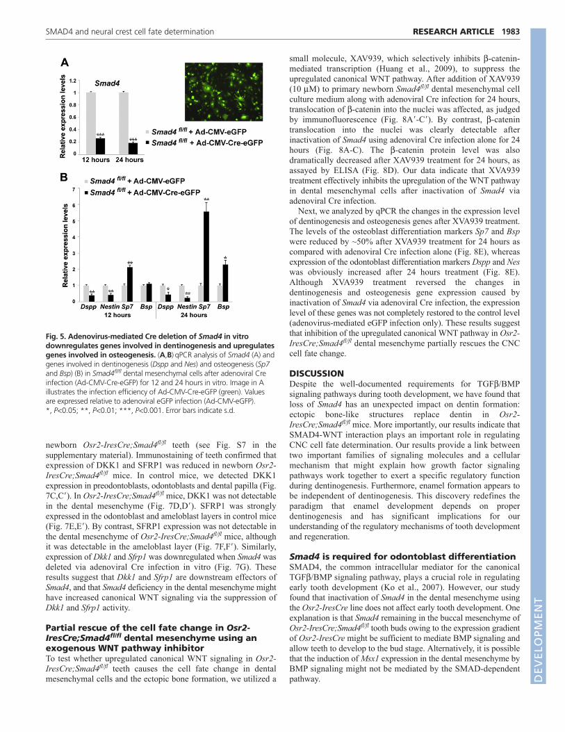
70% after 12 hours of infection and by 80% after 24 hours ofinfection, as assessed by qPCR (Fig. 5A). The levels of theodontoblast differentiation markers Dspp and Nes were reduced by60% after 12 hours of infection (Fig. 5B), whereas expression ofthe osteoblast differentiation marker Sp7 more than doubled after12 hours of infection (Fig. 5B). Bsp, a late osteogenic marker, wasupregulated by more than 2-fold after 24 hours of infection (Fig.5B). Osteocalcin (Bglap – Mouse Genome Informatics) expressionwas also increased in Smad4fl/fl primary dental mesenchymal cellsafter 36 hours of infection (see Fig. S5 in the supplementarymaterial). Thus, ablation of Smad4 in primary dental mesenchymalcells in vitro downregulates genes that are involved indentinogenesis and upregulates genes that are involved inosteogenesis, consistent with our in vivo data.
Upregulation of canonical WNT signaling in Osr2-IresCre;Smad4fl/fl teethWNT signaling regulates osteoblast differentiation and boneformation (Bennett et al., 2005; Takada et al., 2007). Previousstudies have shown that BMP and WNT signaling may regulateeach other during bone formation (Bain et al., 2003; Chen et al.,2007; Kamiya et al., 2010; Kamiya et al., 2008; Rawadi et al.,2003; Winkler et al., 2005) and tooth development (Ahn et al.,2010; Ohazama et al., 2008). We hypothesized that ablation ofSmad4 in the dental mesenchyme upregulated the WNT signalingpathway, resulting in ectopic bone formation in teeth. We firstinvestigated canonical WNT signaling using TOPGAL WNTreporter mice. WNT signaling, as assessed by X-gal staining, wasincreased in Osr2-IresCre;Smad4fl/fl;TOPGAL teeth after 19 days
kidney capsule transplantation (Fig. 6A-D). The percentage of-gal-positive cells was almost 4-fold higher in Osr2-IresCre;Smad4fl/fl;TOPGAL samples than in the control, astatistically significant increase (Fig. 6E). Although the endogenousexpression of canonical WNT signaling is detectable inodontoblasts during postnatal tooth development using WNTreporter mice (Lohi et al., 2010), we found that the intensity of X-gal staining was dramatically increased in teeth from Osr2-IresCre;Smad4fl/fl;TOPGAL mice compared with the control (Fig.6A-E).
In addition, we analyzed the expression of Axin2, a canonicalWNT target gene, in teeth from Osr2-IresCre;Smad4fl/fl mice usingqPCR. Axin2 expression was significantly increased in Osr2-IresCre;Smad4fl/fl molars after 19 days kidney capsule transplantation(Fig. 6F). Similarly, Axin2 expression was upregulated after Smad4was inactivated in primary dental mesenchymal cells in vitro viaadenoviral Cre infection (Fig. 6G). mRNA and protein levels of -catenin, a central component of the canonical WNT cascade, werealso dramatically increased after loss of Smad4 via adenoviral Creinfection in vitro for 12 and 24 hours, respectively (Fig. 6H,I). Inaddition, we also analyzed canonical WNT signaling in other Osr2-IresCre expression regions, such as the olfactory, periocular andtongue mesenchyme, using Osr2-IresCre;Smad4fl/fl;TOPGAL miceand did not find upregulated or ectopic canonical WNT signaling(see Fig. S6 in the supplementary material), which suggests thatSMAD4-WNT interaction is tissue-specific during dentinogenesis.Taken together, our data indicate that loss of Smad4 in the dentalmesenchyme results in the upregulation of the canonical WNTsignaling pathway.
1981RESEARCH ARTICLESMAD4 and neural crest cell fate determination
Fig. 3. Odontoblast and ameloblastdifferentiation in Osr2-IresCre;Smad4fl/fl
mice. (A-D�) H&E staining of molar toothgerms of newborn (NB) control (A,A�) andOsr2-IresCre;Smad4fl/fl (B,B�) mice and after7 days organ culture (C-D�). Boxed areas infrontal sections (above) are shownmagnified beneath. The arrows indicatepolarized odontoblasts in control toothgerm (black) and unpolarized odontoblastsin Osr2-IresCre;Smad4fl/fl tooth germ (white).Boxed areas in C� and D� are magnified(right top corner) to show polarizedameloblasts in control (C�) and Osr2-IresCre;Smad4fl/fl (D�) tooth germ. (E-L�) Insitu hybridization for Dspp and amelogenin(Amelx) in newborn control (E,G) and Osr2-IresCre;Smad4fl/fl (F,H) mice and after 7 daysorgan culture (I-L�). Boxed areas (I-L) areshown magnified beneath. Black arrowsindicate Dspp expression in odontoblastsand ameloblasts and Amelx expression inameloblasts. White arrows indicate lack ofexpression of Dspp and Amelx in Osr2-IresCre;Smad4fl/fl mice.(M,N)Immunofluorescence of SMAD4 (red)in newborn control (M) and Osr2-IresCre;Smad4fl/fl (N) molar tooth germs.AM, ameloblast; DM, dentin matrix; DP,dental papilla; OD, odontoblast. Scale bars:200m in A-D,I-L; 100m in M,N; 50m inA�-D�,E-H,I�-L�.
DEVELO
PMENT

1982
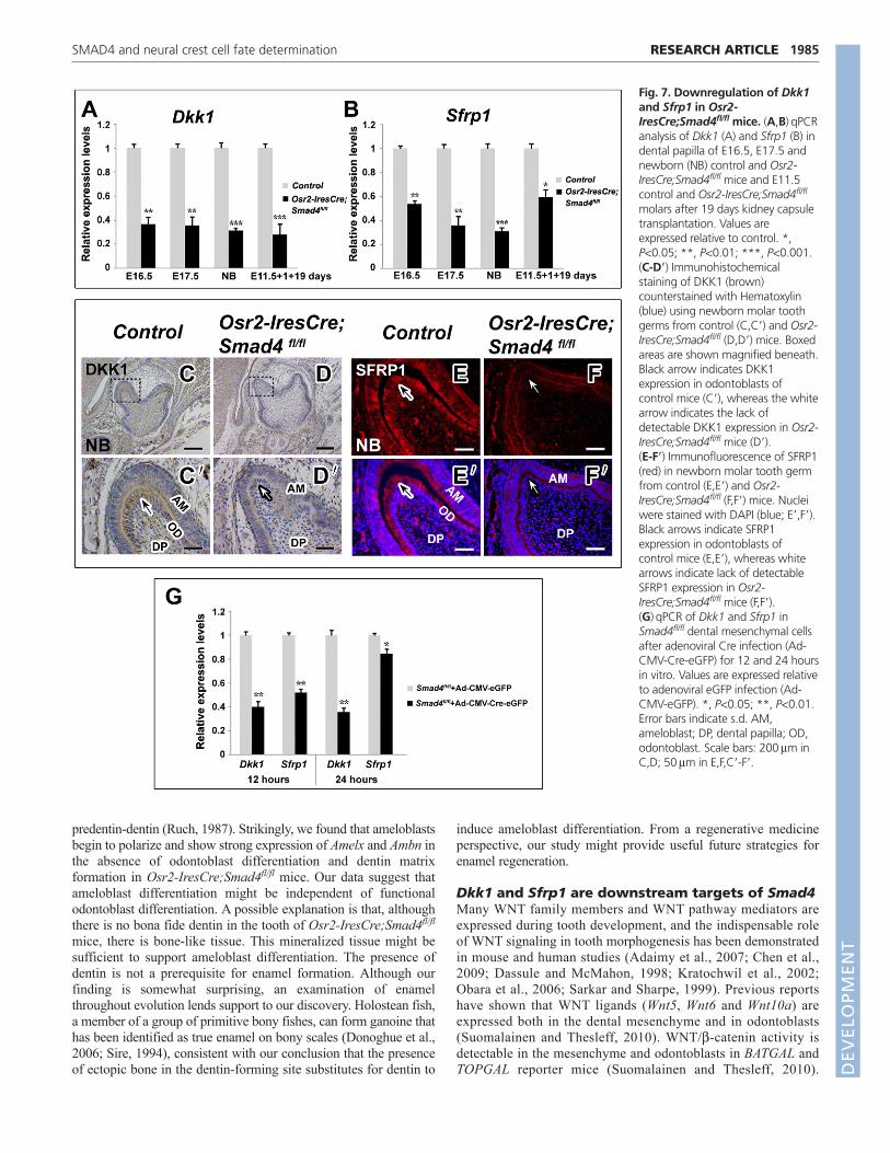
Dkk1 and Sfrp1 are downregulated in teeth ofOsr2-IresCre;Smad4fl/fl miceThe extracellular antagonists of the WNT signaling pathway canbe divided into two functional classes: the SFRP class and theDickkopf (DKK) class. In theory, the SFRP class will inhibit bothcanonical and non-canonical WNT pathways, whereas the DKKclass specifically inhibits the canonical WNT pathway (Kawanoand Kypta, 2003). We hypothesized that deletion of Smad4 in thedental mesenchyme upregulated the canonical WNT signalingpathway via suppression of WNT antagonist expression. Using
qPCR, we assayed the expression of Dkk1, Dkk2, Sfrp1 and Sfrp2,which are WNT antagonists expressed during tooth development(Fjeld et al., 2005; Leimeister et al., 1998). In teeth of Osr2-IresCre;Smad4fl/fl mice, Dkk1 expression was dramatically reducedat E16.5, E17.5 and the newborn stage, and was downregulated bymore than 70% after 19 days kidney capsule transplantation (Fig.7A). Expression of Sfrp1 in Osr2-IresCre;Smad4fl/fl teeth was alsosignificantly decreased at E16.5, E17.5, the newborn stage andafter 19 days kidney capsule transplantation (Fig. 7B). By contrast,expression of Dkk2 and Sfrp2 was unchanged in E16.5 and
RESEARCH ARTICLE Development 138 (10)
Fig. 4. Ectopic bone formation in Osr2-IresCre;Smad4fl/fl mice. (A-F)Macroscopic views (A,B), H&E staining (C,D) and SEM analysis (E,F) ofE11.5 control and Osr2-IresCre;Smad4fl/fl molar tooth germs after 19 days kidney capsule transplantation. (G-N�) In situ hybridization analysis ofodontoblast, osteoblast and ameloblast differentiation markers in E11.5 control (G-J�) and Osr2-IresCre;Smad4fl/fl (K-N�) molar tooth germs after 19days kidney capsule transplantation. Boxed areas are shown magnified to the right. Black arrows indicate expression of Dspp, Bsp or Amelx,whereas white arrows indicate lack of detectable expression. (O)qPCR for differentiation markers expressed by odontoblasts (Dspp and Nes) andosteoblasts (Bsp) using dental papilla from newborn (NB) control and Osr2-IresCre;Smad4fl/fl mice and E11.5 samples after 19 days kidney capsuletransplantation. Values are expressed relative to control. *, P<0.05; **, P<0.01; ***, P<0.001. Error bars indicate s.d. AB, alveolar bone; AM,ameloblast; D, dentin; E, enamel; M1, lower first molar; M2, lower second molar; OD, odontoblast. Scale bars: 200m in G-N; 50m in C,D,G�-N�,K�-N�; 10m in E,F.
DEVELO
PMENT
newborn Osr2-IresCre;Smad4fl/fl teeth (see Fig. S7 in thesupplementary material). Immunostaining of teeth confirmed thatexpression of DKK1 and SFRP1 was reduced in newborn Osr2-IresCre;Smad4fl/fl mice. In control mice, we detected DKK1expression in preodontoblasts, odontoblasts and dental papilla (Fig.7C,C�). In Osr2-IresCre;Smad4fl/fl mice, DKK1 was not detectablein the dental mesenchyme (Fig. 7D,D�). SFRP1 was stronglyexpressed in the odontoblast and ameloblast layers in control mice(Fig. 7E,E�). By contrast, SFRP1 expression was not detectable inthe dental mesenchyme of Osr2-IresCre;Smad4fl/fl mice, althoughit was detectable in the ameloblast layer (Fig. 7F,F�). Similarly,expression of Dkk1 and Sfrp1 was downregulated when Smad4 wasdeleted via adenoviral Cre infection in vitro (Fig. 7G). Theseresults suggest that Dkk1 and Sfrp1 are downstream effectors ofSmad4, and that Smad4 deficiency in the dental mesenchyme mighthave increased canonical WNT signaling via the suppression ofDkk1 and Sfrp1 activity.
Partial rescue of the cell fate change in Osr2-IresCre;Smad4fl/fl dental mesenchyme using anexogenous WNT pathway inhibitorTo test whether upregulated canonical WNT signaling in Osr2-IresCre;Smad4fl/fl teeth causes the cell fate change in dentalmesenchymal cells and the ectopic bone formation, we utilized a
small molecule, XAV939, which selectively inhibits -catenin-mediated transcription (Huang et al., 2009), to suppress theupregulated canonical WNT pathway. After addition of XAV939(10 M) to primary newborn Smad4fl/fl dental mesenchymal cellculture medium along with adenoviral Cre infection for 24 hours,translocation of -catenin into the nuclei was affected, as judgedby immunofluorescence (Fig. 8A�-C�). By contrast, -catenintranslocation into the nuclei was clearly detectable afterinactivation of Smad4 using adenoviral Cre infection alone for 24hours (Fig. 8A-C). The -catenin protein level was alsodramatically decreased after XAV939 treatment for 24 hours, asassayed by ELISA (Fig. 8D). Our data indicate that XVA939treatment effectively inhibits the upregulation of the WNT pathwayin dental mesenchymal cells after inactivation of Smad4 viaadenoviral Cre infection.
Next, we analyzed by qPCR the changes in the expression levelof dentinogenesis and osteogenesis genes after XVA939 treatment.The levels of the osteoblast differentiation markers Sp7 and Bspwere reduced by ~50% after XVA939 treatment for 24 hours ascompared with adenoviral Cre infection alone (Fig. 8E), whereasexpression of the odontoblast differentiation markers Dspp and Neswas obviously increased after 24 hours treatment (Fig. 8E).Although XVA939 treatment reversed the changes indentinogenesis and osteogenesis gene expression caused byinactivation of Smad4 via adenoviral Cre infection, the expressionlevel of these genes was not completely restored to the control level(adenovirus-mediated eGFP infection only). These results suggestthat inhibition of the upregulated canonical WNT pathway in Osr2-IresCre;Smad4fl/fl dental mesenchyme partially rescues the CNCcell fate change.
DISCUSSIONDespite the well-documented requirements for TGF/BMPsignaling pathways during tooth development, we have found thatloss of Smad4 has an unexpected impact on dentin formation:ectopic bone-like structures replace dentin in Osr2-IresCre;Smad4fl/fl mice. More importantly, our results indicate thatSMAD4-WNT interaction plays an important role in regulatingCNC cell fate determination. Our results provide a link betweentwo important families of signaling molecules and a cellularmechanism that might explain how growth factor signalingpathways work together to exert a specific regulatory functionduring dentinogenesis. Furthermore, enamel formation appears tobe independent of dentinogenesis. This discovery redefines theparadigm that enamel development depends on properdentinogenesis and has significant implications for ourunderstanding of the regulatory mechanisms of tooth developmentand regeneration.
Smad4 is required for odontoblast differentiationSMAD4, the common intracellular mediator for the canonicalTGF/BMP signaling pathway, plays a crucial role in regulatingearly tooth development (Ko et al., 2007). However, our studyfound that inactivation of Smad4 in the dental mesenchyme usingthe Osr2-IresCre line does not affect early tooth development. Oneexplanation is that Smad4 remaining in the buccal mesenchyme ofOsr2-IresCre;Smad4fl/fl tooth buds owing to the expression gradientof Osr2-IresCre might be sufficient to mediate BMP signaling andallow teeth to develop to the bud stage. Alternatively, it is possiblethat the induction of Msx1 expression in the dental mesenchyme byBMP signaling might not be mediated by the SMAD-dependentpathway.
1983RESEARCH ARTICLESMAD4 and neural crest cell fate determination
Fig. 5. Adenovirus-mediated Cre deletion of Smad4 in vitrodownregulates genes involved in dentinogenesis and upregulatesgenes involved in osteogenesis. (A,B)qPCR analysis of Smad4 (A) andgenes involved in dentinogenesis (Dspp and Nes) and osteogenesis (Sp7and Bsp) (B) in Smad4fl/fl dental mesenchymal cells after adenoviral Creinfection (Ad-CMV-Cre-eGFP) for 12 and 24 hours in vitro. Image in Aillustrates the infection efficiency of Ad-CMV-Cre-eGFP (green). Valuesare expressed relative to adenoviral eGFP infection (Ad-CMV-eGFP).*, P<0.05; **, P<0.01; ***, P<0.001. Error bars indicate s.d.
DEVELO
PMENT

1984
TGF/BMP signaling has been shown to function duringodontoblast differentiation and dentin formation. In order toinvestigate the functional significance of SMAD4 signaling inregulating dentinogenesis, we generated mice that specifically lackSmad4 expression in dental mesenchymal cells. We found that inOsr2-IresCre;Smad4fl/fl mice, CNC-derived dental mesenchymedifferentiation is arrested at the late bell stage and secretory stage,with no detectable expression of Dspp. In Wnt1-Cre;Tgfbr2fl/fl
mice, odontoblast differentiation is only delayed and Dsppexpression is eventually detectable (Oka et al., 2007). We alsoobserved normal cell polarization in odontoblasts of newbornOsr2-IresCre;Bmpr1afl/fl mice, in which BMP signaling is blockedin the dental mesenchyme (see Fig. S8 in the supplementarymaterial). Thus, we hypothesize that the TGF and BMP signalingpathways work together in a fine balance to regulate odontoblastdifferentiation during dentinogenesis. Accordingly, deletion ofSmad4, the common mediator for the canonical TGF/BMPsignaling pathway, causes morphological and functional defects inodontoblasts during dentinogenesis. Alternatively, it is conceivable
that other members of the TGF signaling family might have aunique function in regulating dentinogenesis in a SMAD4-dependent manner.
Ameloblast differentiation is independent ofodontoblast differentiationPreameloblasts are derived from precursor cells in the inner enamelepithelium of the enamel organ. Upon differentiation, the epithelialpreameloblasts exit the cell cycle and polarize, with a reorganizationof cellular components. Previous studies have shown that reciprocalepithelial-mesenchymal interactions regulate ameloblastdifferentiation. Tissue recombination studies using dental and non-dental tissues have shown that ameloblast cytodifferentiation requiresfunctional odontoblasts (Kollar and Baird, 1970; Ruch et al., 1973),and that acellular dentin matrices can also promote ameloblastcytodifferentiation (Karcher-Djuricic et al., 1985). Whenpreodontoblasts differentiate into functional odontoblasts and start tosecrete dentin matrix, the basement membrane breaks up anddegrades, allowing direct interaction between preameloblasts and
RESEARCH ARTICLE Development 138 (10)
Fig. 6. Upregulation of canonical WNTsignaling in Osr2-IresCre;Smad4fl/fl mice.(A,B)Macroscopic view of tooth germs fromTOPGAL mice cultured for 19 days underkidney capsules to assess canonical WNTsignaling using X-gal staining.(C,D)Histological analysis of tooth germscultured for 19 days under kidney capsulesfrom TOPGAL mice. D, dentin; DP, dentalpulp; OD, odontoblast. Scale bars: 50m.(E)Percentage of -gal-positive cells in molarscultured for 19 days after kidney capsuletransplantation from control and Osr2-IresCre;Smad4fl/fl;TOPGAL mice. Cells werecounted in 30 fields of molars for eachgenotype (n3). Total cell number wasobtained by counting Nuclear Fast Red-positive nuclei in the control(Smad4fl/fl;TOPGAL) odontoblast layer andOsr2-IresCre;Smad4fl/fl;TOPGAL dentinregion. ***, P<0.001. (F,G)qPCR for Axin2in newborn (NB) control and Osr2-IresCre;Smad4fl/fl dental papilla and after 19days kidney capsule transplantation (F), andin Smad4fl/fl dental mesenchymal cells afteradenoviral Cre infection (Ad-CMV-Cre-eGFP)for 12 and 24 hours in vitro (G). Values areexpressed relative to control and adenoviraleGFP infection (Ad-CMV-eGFP), respectively.*, P<0.05; **, P<0.01. (H,I)qPCR (H) andELISA (I) analysis of -catenin in Smad4fl/fl
dental mesenchymal cells after adenoviralCre infection (Ad-CMV-Cre-eGFP) for 12 and24 hours in vitro. qPCR values are expressedrelative to adenoviral eGFP infection (Ad-CMV-eGFP). **, P<0.01; ***, P<0.001. Errorbars indicate s.d.
DEVELO
PMENT
predentin-dentin (Ruch, 1987). Strikingly, we found that ameloblastsbegin to polarize and show strong expression of Amelx and Ambn inthe absence of odontoblast differentiation and dentin matrixformation in Osr2-IresCre;Smad4fl/fl mice. Our data suggest thatameloblast differentiation might be independent of functionalodontoblast differentiation. A possible explanation is that, althoughthere is no bona fide dentin in the tooth of Osr2-IresCre;Smad4fl/fl
mice, there is bone-like tissue. This mineralized tissue might besufficient to support ameloblast differentiation. The presence ofdentin is not a prerequisite for enamel formation. Although ourfinding is somewhat surprising, an examination of enamelthroughout evolution lends support to our discovery. Holostean fish,a member of a group of primitive bony fishes, can form ganoine thathas been identified as true enamel on bony scales (Donoghue et al.,2006; Sire, 1994), consistent with our conclusion that the presenceof ectopic bone in the dentin-forming site substitutes for dentin to
induce ameloblast differentiation. From a regenerative medicineperspective, our study might provide useful future strategies forenamel regeneration.
Dkk1 and Sfrp1 are downstream targets of Smad4Many WNT family members and WNT pathway mediators areexpressed during tooth development, and the indispensable roleof WNT signaling in tooth morphogenesis has been demonstratedin mouse and human studies (Adaimy et al., 2007; Chen et al.,2009; Dassule and McMahon, 1998; Kratochwil et al., 2002;Obara et al., 2006; Sarkar and Sharpe, 1999). Previous reportshave shown that WNT ligands (Wnt5, Wnt6 and Wnt10a) areexpressed both in the dental mesenchyme and in odontoblasts(Suomalainen and Thesleff, 2010). WNT/-catenin activity isdetectable in the mesenchyme and odontoblasts in BATGAL andTOPGAL reporter mice (Suomalainen and Thesleff, 2010).
1985RESEARCH ARTICLESMAD4 and neural crest cell fate determination
Fig. 7. Downregulation of Dkk1and Sfrp1 in Osr2-IresCre;Smad4fl/fl mice. (A,B)qPCRanalysis of Dkk1 (A) and Sfrp1 (B) indental papilla of E16.5, E17.5 andnewborn (NB) control and Osr2-IresCre;Smad4fl/fl mice and E11.5control and Osr2-IresCre;Smad4fl/fl
molars after 19 days kidney capsuletransplantation. Values areexpressed relative to control. *,P<0.05; **, P<0.01; ***, P<0.001.(C-D�) Immunohistochemicalstaining of DKK1 (brown)counterstained with Hematoxylin(blue) using newborn molar toothgerms from control (C,C�) and Osr2-IresCre;Smad4fl/fl (D,D�) mice. Boxedareas are shown magnified beneath.Black arrow indicates DKK1expression in odontoblasts ofcontrol mice (C�), whereas the whitearrow indicates the lack ofdetectable DKK1 expression in Osr2-IresCre;Smad4fl/fl mice (D�).(E-F�) Immunofluorescence of SFRP1(red) in newborn molar tooth germfrom control (E,E�) and Osr2-IresCre;Smad4fl/fl (F,F�) mice. Nucleiwere stained with DAPI (blue; E�,F�).Black arrows indicate SFRP1expression in odontoblasts ofcontrol mice (E,E�), whereas whitearrows indicate lack of detectableSFRP1 expression in Osr2-IresCre;Smad4fl/fl mice (F,F�).(G)qPCR of Dkk1 and Sfrp1 inSmad4fl/fl dental mesenchymal cellsafter adenoviral Cre infection (Ad-CMV-Cre-eGFP) for 12 and 24 hoursin vitro. Values are expressed relativeto adenoviral eGFP infection (Ad-CMV-eGFP). *, P<0.05; **, P<0.01.Error bars indicate s.d. AM,ameloblast; DP, dental papilla; OD,odontoblast. Scale bars: 200m inC,D; 50m in E,F,C�-F�.
DEVELO
PMENT

1986
Consistent with the expression patterns of WNT ligands andmediators, we report here that DKK1 and SFRP1, which areinhibitors of the WNT pathway, are also expressed in the dentalmesenchyme and odontoblasts. Thus, WNT signaling self-regulation might play a role during odontoblast differentiationand dentinogenesis.
In our study, we found that expression of DKK1 and SFRP1 wasdramatically downregulated at the mRNA and protein levelsfollowing the loss of Smad4 in vivo. These results, which werereplicated in cell culture with adenoviral Cre-mediated inactivationof Smad4, suggest that both Dkk1 and Sfrp1 are downstream targetsof Smad4. Previous studies support this notion because inhibition ofBMP signaling with dorsomorphin, an inhibitor of SMAD-dependentBMP signaling, suppresses the expression of Dkk1 in osteoblasts(Kamiya et al., 2010). Moreover, BMP2 and BMP4 induce Dkk1expression during limb development in mouse and chicken(Grotewold and Ruther, 2002; Mukhopadhyay et al., 2001). Inaddition, other studies suggest possible cross-talk between SFRPsand BMP signaling (Ellies et al., 2000; Miquelajauregui et al., 2007;
Oshima et al., 2005). However, the present study provides the firstin vivo evidence that BMP/TGF relies on SMAD4 to regulate Dkk1and Sfrp1 expression during dentinogenesis.
SMAD4-mediated BMP/TGF and WNT interactionand CNC cell fate determinationThere is a growing body of evidence to suggest that theTGF/BMP and WNT signaling pathways regulate one anothersynergistically or antagonistically (Ahn et al., 2010; Barrow et al.,2003; Guo et al., 2004; He et al., 2004; Huelsken et al., 2001;Ohazama et al., 2008), but the possible interplay of the WNT/-catenin and TGF/BMP signaling pathways for CNC cell fatedetermination has not been demonstrated during tooth developmentin vivo. In this study, we found that ablation of Smad4 in dentalmesenchyme upregulates the canonical WNT signaling pathwaythrough -catenin in vivo and in vitro. Our findings are consistentwith other studies that have shown an inhibitory effect of BMPsignaling on WNT signaling during organogenesis of tissues suchas bone (Kamiya et al., 2010; Kamiya et al., 2008), joint (Guo et
RESEARCH ARTICLE Development 138 (10)
Fig. 8. Partial rescue of the cell fate change inOsr2-IresCre;Smad4fl/fl dental mesenchyme usingan exogenous WNT pathway inhibitor in vitro.(A-C�) Immunofluorescence of -catenin (red) inSmad4fl/fl dental mesenchymal cells after adenoviralCre infection (Ad-CMV-Cre-eGFP) alone (A-C) andwith XAV939 (10M) treatment (A�-C�) for 24 hoursin vitro. Cells successfully infected with adenoviral Creshow eGFP fluorescence (green). Arrowheads andarrows point to -catenin in the nuclei andcytoplasm, respectively. Scale bars: 50m. (D)ELISAanalysis of -catenin in Smad4fl/fl dental mesenchymalcells after adenoviral Cre infection (Ad-CMV-Cre-eGFP) and XAV939 (10M) treatment for 24 hours invitro. **, P<0.01; ***, P<0.001. (E)qPCR analysis ofgenes involved in dentinogenesis (Dspp and Nes) andosteogenesis (Sp7 and Bsp) in Smad4fl/fl dentalmesenchymal cells after adenoviral Cre infection (Ad-CMV-Cre-eGFP) and XAV939 (10M) treatment for24 hours in vitro. Values are expressed relative toadenoviral eGFP infection (Ad-CMV-eGFP).**, P<0.01; ***, P<0.001. Error bars indicate s.d.
DEVELO
PMENT
al., 2004), lung (Dean et al., 2005), hair (Zhang et al., 2006) andintestine (He et al., 2004). Collectively, our data indicate that lossof Smad4 signaling upregulates WNT signaling, which is likely tobe via inhibition of Dkk1 and Sfrp1 expression.
WNT signaling regulates osteoblast differentiation and boneformation (Bennett et al., 2005; Takada et al., 2007). Studies ofmouse and human mutations related to the WNT pathway haveshown defects in osteogenesis and bone mass (Balemans et al.,2002; Bennett et al., 2005; Bodine et al., 2004; Boyden et al., 2002;Gong et al., 2001; Holmen et al., 2004; Kato et al., 2002; Li et al.,2006; Little et al., 2002; Loots et al., 2005; Mani et al., 2007;Takada et al., 2007). Both DKK1 and SFRP1 are expressed in boneand play crucial roles during bone formation (Bodine et al., 2004;MacDonald et al., 2007). Deletion of Dkk1 or Sfrp1 preferentiallyactivates WNT signaling in osteoblasts, leading to enhanced boneformation in vivo (Bodine et al., 2004; MacDonald et al., 2007).Intriguingly, we found that disrupting Smad4 in the dentalmesenchyme causes ectopic bone-like structure formation in thedentin region and enhances the WNT pathway. More importantly,suppression of the upregulated canonical WNT pathway in Osr2-IresCre;Smad4fl/fl dental mesenchyme partially rescues the CNCcell fate change. We suggest that these two phenotypes are linked,and this relationship would be consistent with previous studies inwhich loss of BMPR1A signaling upregulates WNT signaling byinhibiting Dkk1 and Sost expression in bone and increases bonemass (Kamiya et al., 2010; Kamiya et al., 2008).
The temporal and spatial combination of signals determines neuralcrest cell fates. Previous studies have demonstrated that thecombinatorial activity of the BMP and WNT signaling pathwayspromotes sensory neuron fate during early neural crest celldevelopment (Kléber et al., 2005). After migration, neural crest-derived dental mesenchymal cells still possess the potential todifferentiate into dentin-secreting odontoblasts as well aschondrocyte-like and osteoblast-like cells during craniofacialdevelopment (Chai et al., 2000; Chung et al., 2009; Yamazaki et al.,2007). Here, we have demonstrated that SMAD4 is indispensable forodontoblast differentiation during tooth development and that loss ofSmad4 in the dental mesenchyme results in ectopic osteoblastdifferentiation and bone formation via WNT pathway upregulation.Thus, the interplay between the TGF/BMP and WNT signalingpathways also functions to ensure proper cell fate determinationduring postmigratory neural crest cell development andorganogenesis. From a clinical perspective, our study might help toprovide etiological clues of heritable dentin disorders and suggestnovel therapeutically useful strategies and candidates for futureinvestigation.
AcknowledgementsWe thank Chuxia Deng for Smad4fl/fl mice. This study was supported by grantsfrom the NIDCR and NIH (U01 DE020065, R37 DE012711) to Y.C. Depositedin PMC for release after 12 months.
Competing interests statementThe authors declare no competing financial interests.
Supplementary materialSupplementary material for this article is available athttp://dev.biologists.org/lookup/suppl/doi:10.1242/dev.061341/-/DC1
ReferencesAbout, I., Laurent-Maquin, D., Lendahl, U. and Mitsiadis, T. A. (2000). Nestin
expression in embryonic and adult human teeth under normal and pathologicalconditions. Am. J. Pathol. 157, 287-295.
Adaimy, L., Chouery, E., Mégarbané, H., Mroueh, S., Delague, V., Nicolas, E.,Belguith, H., de Mazancourt, P. and Mégarbané, A. (2007). Mutation in
WNT10A is associated with an autosomal recessive ectodermal dysplasia: theodonto-onycho-dermal dysplasia. Am. J. Hum. Genet. 81, 821-828.
Ahn, Y., Sanderson, B. W., Klein, O. D. and Krumlauf, R. (2010). Inhibition ofWnt signaling by Wise (Sostdc1) and negative feedback from Shh controls toothnumber and patterning. Development 137, 3221-3231.
Bain, G., Müller, T., Wang, X. and Papkoff, J. (2003). Activated beta-catenininduces osteoblast differentiation of C3H10T1/2 cells and participates in BMP2mediated signal transduction. Biochem. Biophys. Res. Commun. 301, 84-91.
Balemans, W., Patel, N., Ebeling, M., Van Hul, E., Wuyts, W., Lacza, C.,Dioszegi, M., Dikkers, F. G., Hildering, P., Willems, P. J. et al. (2002).Identification of a 52 kb deletion downstream of the SOST gene in patients withvan Buchem disease. J. Med. Genet. 39, 91-97.
Barrow, J. R., Thomas, K. R., Boussadia-Zahui, O., Moore, R., Kemler, R.,Capecchi, M. R. and McMahon, A. P. (2003). Ectodermal Wnt3/-cateninsignaling is required for the establishment and maintenance of the apicalectodermal ridge. Genes Dev. 17, 394-409.
Bègue-Kirn, C., Smith, A., Ruch, J., Wozney, J., Purchio, A., Hartmann, D.and Lesot, H. (1992). Effects of dentin proteins, transforming growth factorbeta 1 (TGF beta 1) and bone morphogenetic protein 2 (BMP2) on thedifferentiation of odontoblast in vitro. Int. J. Dev. Biol. 36, 491-503.
Bègue-Kirn, C., Smith, A., Loriot, M., Kupferle, C., Ruch, J. and Lesot, H.(1994). Comparative analysis of TGF beta s, BMPs, IGF1, msxs, fibronectin,osteonectin and bone sialoprotein gene expression during normal and in vitro-induced odontoblast differentiation. Int. J. Dev. Biol. 38, 405-420.
Bennett, C. N., Longo, K. A., Wright, W. S., Suva, L. J., Lane, T. F.,Hankenson, K. D. and MacDougald, O. A. (2005). Regulation ofosteoblastogenesis and bone mass by Wnt10b. Proc. Natl. Acad. Sci. USA 102,3324-3329.
Bodine, P. V. N., Zhao, W., Kharode, Y. P., Bex, F. J., Lambert, A.-J., Goad, M.B., Gaur, T., Stein, G. S., Lian, J. B. and Komm, B. S. (2004). The Wntantagonist secreted frizzled-related protein-1 is a negative regulator oftrabecular bone formation in adult mice. Mol. Endocrinol. 18, 1222-1237.
Boyden, L. M., Mao, J., Belsky, J., Mitzner, L., Farhi, A., Mitnick, M. A., Wu,D., Insogna, K. and Lifton, R. P. (2002). High bone density due to a mutationin LDL-receptor-related protein 5. N. Engl. J. Med. 346, 1513-1521.
Cam, Y., Neumann, M., Oliver, L., Raulais, D., Janet, T. and Ruch, J. (1992).Immunolocalization of acidic and basic fibroblast growth factors during mouseodontogenesis. Int. J. Dev. Biol. 36, 381-389.
Chai, Y. and Slavkin, H. C. (2003). Prospects for tooth regeneration in the 21stcentury: a perspective. Microsc. Res. Tech. 60, 469-479.
Chai, Y., Jiang, X., Ito, Y., Bringas, P., Han, J., Rowitch, D. H., Soriano, P.,McMahon, A. P. and Sucov, H. M. (2000). Fate of the mammalian cranialneural crest during tooth and mandibular morphogenesis. Development 127,1671-1679.
Chen, J., Lan, Y., Baek, J.-A., Gao, Y. and Jiang, R. (2009). Wnt/beta-cateninsignaling plays an essential role in activation of odontogenic mesenchymeduring early tooth development. Dev. Biol. 334, 174-185.
Chen, Y., Bei, M., Woo, I., Satokata, I. and Maas, R. (1996). Msx1 controlsinductive signaling in mammalian tooth morphogenesis. Development 122,3035-3044.
Chen, Y., Whetstone, H. C., Youn, A., Nadesan, P., Chow, E. C. Y., Lin, A. C.and Alman, B. A. (2007). -Catenin signaling pathway is crucial for bonemorphogenetic protein 2 to induce new bone formation. J. Biol. Chem. 282,526-533.
Chung, I. H., Yamaza, T., Zhao, H., Choung, P. H., Shi, S. and Chai, Y. (2009).Stem cell property of postmigratory cranial neural crest cells and their utility inalveolar bone regeneration and tooth development. Stem Cells 27, 866-877.
Danielian, P. S., Muccino, D., Rowitch, D. H., Michael, S. K. and McMahon,A. P. (1998). Modification of gene activity in mouse embryos in utero by atamoxifen-inducible form of Cre recombinase. Curr. Biol. 8, 1323-1326.
DasGupta, R. and Fuchs, E. (1999). Multiple roles for activated LEF/TCFtranscription complexes during hair follicle development and differentiation.Development 126, 4557-4568.
Dassule, H. R. and McMahon, A. P. (1998). Analysis of epithelial-mesenchymalinteractions in the initial morphogenesis of the mammalian tooth. Dev. Biol.202, 215-227.
Dean, C. H., Miller, L.-A. D., Smith, A. N., Dufort, D., Lang, R. A. andNiswander, L. A. (2005). Canonical Wnt signaling negatively regulatesbranching morphogenesis of the lung and lacrimal gland. Dev. Biol. 286, 270-286.
Donoghue, P. C. J., Sansom, I. J. and Downs, J. P. (2006). Early evolution ofvertebrate skeletal tissues and cellular interactions, and the canalization ofskeletal development. J. Exp. Zool. B Mol. Dev. Evol. 306B, 278-294.
Ellies, D. L., Church, V., Francis-West, P. and Lumsden, A. (2000). The WNTantagonist cSFRP2 modulates programmed cell death in the developinghindbrain. Development 127, 5285-5295.
Fjeld, K., Kettunen, P., Furmanek, T., Kvinnsland, I. H. and Luukko, K. (2005).Dynamic expression of Wnt signaling-related Dickkopf1, -2, and -3 mRNAs inthe developing mouse tooth. Dev. Dyn. 233, 161-166.
1987RESEARCH ARTICLESMAD4 and neural crest cell fate determination
DEVELO
PMENT

1988
Fouletier-Dilling, C. M., Bosch, P., Davis, A. R., Shafer, J. A., Stice, S. L.,Gugala, Z., Gannon, F. H. and Olmsted-Davis, E. A. (2005). Novel compoundenables high-level adenovirus transduction in the absence of an adenovirus-specific receptor. Hum. Gene Ther. 16, 1287-1297.
Gao, Y., Yang, G., Weng, T., Du, J., Wang, X., Zhou, J., Wang, S. and Yang, X.(2009). Disruption of Smad4 in odontoblasts causes multiple keratocysticodontogenic tumors and tooth malformation in mice. Mol. Cell. Biol. 29, 5941-5951.
Gong, Y., Slee, R. B., Fukai, N., Rawadi, G., Roman-Roman, S., Reginato, A.M., Wang, H., Cundy, T., Glorieux, F. H., Lev, D. et al. (2001). LDL receptor-related protein 5 (LRP5) affects bone accrual and eye development. Cell 107,513-523.
Grotewold, L. and Ruther, U. (2002). The Wnt antagonist Dickkopf-1 isregulated by Bmp signaling and c-Jun and modulates programmed cell death.EMBO J. 21, 966-975.
Guo, X., Day, T. F., Jiang, X., Garrett-Beal, L., Topol, L. and Yang, Y. (2004).Wnt/-catenin signaling is sufficient and necessary for synovial joint formation.Genes Dev. 18, 2404-2417.
He, W.-X., Niu, Z.-Y., Zhao, S.-L., Jin, W.-L., Gao, J. and Smith, A. J. (2004).TGF-[beta] activated Smad signalling leads to a Smad3-mediated down-regulation of DSPP in an odontoblast cell line. Arch. Oral Biol. 49, 911-918.
He, X. C., Zhang, J., Tong, W.-G., Tawfik, O., Ross, J., Scoville, D. H., Tian, Q.,Zeng, X., He, X., Wiedemann, L. M. et al. (2004). BMP signaling inhibitsintestinal stem cell self-renewal through suppression of Wnt-beta-cateninsignaling. Nat. Genet. 36, 1117-1121.
Holmen, S. L., Giambernardi, T. A., Zylstra, C. R., Buckner-Berghuis, B. D.,Resau, J. H., Hess, J. F., Glatt, V., Bouxsein, M. L., Ai, M., Warman, M. L. etal. (2004). Decreased BMD and limb deformities in mice carrying mutations inboth Lrp5 and Lrp6. J. Bone Miner. Res. 19, 2033-2040.
Huang, S.-M. A., Mishina, Y. M., Liu, S., Cheung, A., Stegmeier, F., Michaud,G. A., Charlat, O., Wiellette, E., Zhang, Y., Wiessner, S. et al. (2009).Tankyrase inhibition stabilizes axin and antagonizes Wnt signalling. Nature 461,614-620.
Huelsken, J., Vogel, R., Erdmann, B., Cotsarelis, G. and Birchmeier, W. (2001).beta-Catenin controls hair follicle morphogenesis and stem cell differentiation inthe skin. Cell 105, 533-545.
Jernvall, J. and Thesleff, I. (2000). Reiterative signaling and patterning duringmammalian tooth morphogenesis. Mech. Dev. 92, 19-29.
Kamiya, N., Ye, L., Kobayashi, T., Mochida, Y., Yamauchi, M., Kronenberg, H.M., Feng, J. Q. and Mishina, Y. (2008). BMP signaling negatively regulatesbone mass through sclerostin by inhibiting the canonical Wnt pathway.Development 135, 3801-3811.
Kamiya, N., Kobayashi, T., Mochida, Y., Yu, P. B., Yamauchi, M., Kronenberg,H. M. and Mishina, Y. (2010). Wnt inhibitors Dkk1 and Sost are downstreamtargets of BMP signaling through the type IA receptor (BMPRIA) in osteoblasts. J.Bone Miner. Res. 25, 200-210.
Karcher-Djuricic, V., Staubli, A., Meyer, J. and Ruch, J. (1985). Acellular dentalmatrices promote functional differentiation of ameloblasts. Differentiation 29,169-175.
Kato, M., Patel, M. S., Levasseur, R., Lobov, I., Chang, B. H.-J., Glass, D. A.,Hartmann, C., Li, L., Hwang, T.-H., Brayton, C. F. et al. (2002). Cbfa1-independent decrease in osteoblast proliferation, osteopenia, and persistentembryonic eye vascularization in mice deficient in Lrp5, a Wnt coreceptor. J. CellBiol. 157, 303-314.
Kawano, Y. and Kypta, R. (2003). Secreted antagonists of the Wnt signallingpathway. J. Cell Sci. 116, 2627-2634.
Kléber, M., Lee, H.-Y., Wurdak, H., Buchstaller, J., Riccomagno, M. M., Ittner,L. M., Suter, U., Epstein, D. J. and Sommer, L. (2005). Neural crest stem cellmaintenance by combinatorial Wnt and BMP signaling. J. Cell Biol. 169, 309-320.
Ko, S. O., Chung, I. H., Xu, X., Oka, S., Zhao, H., Cho, E. S., Deng, C. andChai, Y. (2007). Smad4 is required to regulate the fate of cranial neural crestcells. Dev. Biol. 312, 435-447.
Kollar, E. and Baird, G. (1970). Tissue interactions in embryonic mouse toothgerms. J. Embryol. Exp. Morphol. 24, 173-186.
Kratochwil, K., Galceran, J., Tontsch, S., Roth, W. and Grosschedl, R. (2002).FGF4, a direct target of LEF1 and Wnt signaling, can rescue the arrest of toothorganogenesis in Lef1–/– mice. Genes Dev. 16, 3173-3185.
Lan, Y., Wang, Q., Ovitt, C. E. and Jiang, R. (2007). A unique mouse strainexpressing Cre recombinase for tissue-specific analysis of gene function in palateand kidney development. Genesis 45, 618-624.
Leimeister, C., Bach, A. and Gessler, M. (1998). Developmental expressionpatterns of mouse sFRP genes encoding members of the secreted frizzled relatedprotein family. Mech. Dev. 75, 29-42.
Li, J., Sarosi, I., Cattley, R. C., Pretorius, J., Asuncion, F., Grisanti, M., Morony,S., Adamu, S., Geng, Z., Qiu, W. et al. (2006). Dkk1-mediated inhibition ofWnt signaling in bone results in osteopenia. Bone 39, 754-766.
Little, R. D., Folz, C., Manning, S. P., Swain, P. M., Zhao, S.-C., Eustace, B.,Lappe, M. M., Spitzer, L., Zweier, S., Braunschweiger, K. et al. (2002). Amutation in the LDL receptor-related protein 5 gene results in the autosomaldominant high-bone-mass trait. Am. J. Hum. Genet. 70, 11-19.
Livak, K. J. and Schmittgen, T. D. (2001). Analysis of relative gene expressiondata using real-time quantitative PCR and the 2(-Delta Delta C(T)) method.Methods 25, 402-408.
Lohi, M., Tucker, A. S. and Sharpe, P. T. (2010). Expression of Axin2 indicates arole for canonical Wnt signaling in development of the crown and root duringpre- and postnatal tooth development. Dev. Dyn. 239, 160-167.
Loots, G. G., Kneissel, M., Keller, H., Baptist, M., Chang, J., Collette, N. M.,Ovcharenko, D., Plajzer-Frick, I. and Rubin, E. M. (2005). Genomic deletionof a long-range bone enhancer misregulates sclerostin in Van Buchem disease.Genome Res. 15, 928-935.
MacDonald, B. T., Joiner, D. M., Oyserman, S. M., Sharma, P., Goldstein, S.A., He, X. and Hauschka, P. V. (2007). Bone mass is inversely proportional toDkk1 levels in mice. Bone 41, 331-339.
Mani, A., Radhakrishnan, J., Wang, H., Mani, A., Mani, M.-A., Nelson-Williams, C., Carew, K. S., Mane, S., Najmabadi, H., Wu, D. et al. (2007).LRP6 mutation in a family with early coronary disease and metabolic risk factors.Science 315, 1278-1282.
Massague, J. (2000). How cells read TGF-beta signals. Nat. Rev. Mol. Cell Biol. 1,169-178.
Massagué, J. and Gomis, R. R. (2006). The logic of TGFbeta signaling. FEBS Lett.580, 2811-2820.
Mina, M. and Kollar, E. (1987). The induction of odontogenesis in non-dentalmesenchyme combined with early murine mandibular arch epithelium. Arch.Oral Biol. 32, 123-127.
Miquelajauregui, A., Van de Putte, T., Polyakov, A., Nityanandam, A.,Boppana, S., Seuntjens, E., Karabinos, A., Higashi, Y., Huylebroeck, D.and Tarabykin, V. (2007). Smad-interacting protein-1 (Zfhx1b) acts upstream ofWnt signaling in the mouse hippocampus and controls its formation. Proc. Natl.Acad. Sci. USA 104, 12919-12924.
Mukhopadhyay, M., Shtrom, S., Rodriguez-Esteban, C., Chen, L., Tsukui, T.,Gomer, L., Dorward, D. W., Glinka, A., Grinberg, A., Huang, S.-P. et al.(2001). Dickkopf1 is required for embryonic head induction and limbmorphogenesis in the mouse. Dev. Cell 1, 423-434.
Nakashima, M. (1994). Induction of dentin formation on canine amputated pulpby recombinant human bone morphogenetic proteins (BMP)-2 and -4. J. Dent.Res. 73, 1515-1522.
Obara, N., Suzuki, Y. and Takeda, M. (2006). Gene expression of -catenin isup-regulated in inner dental epithelium and enamel knots during molar toothmorphogenesis in the mouse. Cell Tissue Res. 325, 197-201.
Ohazama, A., Johnson, E. B., Ota, M. S., Choi, H. J., Porntaveetus, T.,Oommen, S., Itoh, N., Eto, K., Gritli-Linde, A., Herz, J. et al. (2008). Lrp4modulates extracellular integration of cell signaling pathways in development.PLoS ONE 3, e4092.
Oka, S., Oka, K., Xu, X., Sasaki, T., Bringas, P., Jr and Chai, Y. (2007). Cellautonomous requirement for TGF-beta signaling during odontoblastdifferentiation and dentin matrix formation. Mech. Dev. 124, 409-415.
Oshima, T., Abe, M., Asano, J., Hara, T., Kitazoe, K., Sekimoto, E., Tanaka, Y.,Shibata, H., Hashimoto, T., Ozaki, S. et al. (2005). Myeloma cells suppressbone formation by secreting a soluble Wnt inhibitor, sFRP-2. Blood 106, 3160-3165.
Plikus, M. V., Zeichner-David, M., Mayer, J.-A., Reyna, J., Bringas, P.,Thewissen, J. G. M., Snead, M. L., Chai, Y. and Chuong, C.-M. (2005).Morphoregulation of teeth: modulating the number, size, shape anddifferentiation by tuning Bmp activity. Evol. Dev. 7, 440-457.
Rawadi, G., Vayssière, B., Dunn, F., Baron, R. and Roman-Roman, S. (2003).BMP-2 controls alkaline phosphatase expression and osteoblast mineralizationby a Wnt autocrine loop. J. Bone Miner. Res. 18, 1842-1853.
Ruch, J. (1987). Determinisms of odontogenesis. Revis. Biol. Celular. 14, 1-99.Ruch, J. (1990). Patterned distribution of differentiating dental cells: facts and
hypotheses. J. Biol. Buccale 18, 91-98.Ruch, J., Karcher-Djuricic, V. and Gerber, R. (1973). Determinants of
morphogenesis and cytodifferentiations of dental anloges in mice. J. Biol.Buccale 1, 45-56.
Ruch, J., Lesot, H. and Bègue-Kirn, C. (1995). Odontoblast differentiation. Int. J.Dev. Biol. 39, 51-68.
Ruoslahti, E. and Yamaguchi, Y. (1991). Proteoglycans as modulators of growthfactor activities. Cell 64, 867-869.
Rutherford, R. B., Spångberg, L., Tucker, M., Rueger, D. and Charette, M.(1994). The time-course of the induction of reparative dentine formation inmonkeys by recombinant human osteogenic protein-1. Arch. Oral Biol. 39, 833-838.
Sarkar, L. and Sharpe, P. T. (1999). Expression of Wnt signalling pathway genesduring tooth development. Mech. Dev. 85, 197-200.
Shi, Y. and Massagué, J. (2003). Mechanisms of TGF-beta signaling from cellmembrane to the nucleus. Cell 113, 685-700.
Siegel, P. M. and Massague, J. (2003). Cytostatic and apoptotic actions of TGF-beta in homeostasis and cancer. Nat. Rev. Cancer 3, 807-820.
Sire, J. Y. (1994). Light and TEM study of nonregenerated and experimentallyregenerated scales of Lepisosteus oculatus (Holostei) with particular attention toganoine formation. Anat. Rec. 240, 189-207.
RESEARCH ARTICLE Development 138 (10)
DEVELO
PMENT

Sloan, A. J., Rutherford, R. B. and Smith, A. J. (2000). Stimulation of the ratdentine-pulp complex by bone morphogenetic protein-7 in vitro. Arch. Oral Biol.45, 173-177.
Soriano, P. (1999). Generalized lacZ expression with the ROSA26 Cre reporterstrain. Nat. Genet. 21, 70-71.
Suomalainen, M. and Thesleff, I. (2010). Patterns of Wnt pathway activity in themouse incisor indicate absence of Wnt/beta-catenin signaling in the epithelialstem cells. Dev. Dyn. 239, 364-372.
Takada, I., Mihara, M., Suzawa, M., Ohtake, F., Kobayashi, S., Igarashi, M.,Youn, M.-Y., Takeyama, K.-i., Nakamura, T., Mezaki, Y. et al. (2007). Ahistone lysine methyltransferase activated by non-canonical Wnt signallingsuppresses PPAR-gamma transactivation. Nat. Cell Biol. 9, 1273-1285.
Terling, C., Rass, A., Mitsiadis, T., Fried, K., Lendahl, U. and Wroblewski, J.(1995). Expression of the intermediate filament nestin during rodent toothdevelopment. Int. J. Dev. Biol. 39, 947-956.
Thesleff, I. and Vaahtokari, A. (1992). The role of growth factors indetermination and differentiation of the odontoblastic cell lineage. Proc. Finn.Dent. Soc. 88, 357-368.
Thesleff, I., Keranen, S. and Jernvall, J. (2001). Enamel knots as signalingcenters linking tooth morphogenesis and odontoblast differentiation. Adv. Dent.Res. 15, 14-18.
Unda, F.-J., Martín, A., Hilario, E., Bègue-Kirn, C., Ruch, J.-V. and Aréchaga,J. (2000). Dissection of the odontoblast differentiation process in vitro by acombination of FGF1, FGF2, and TGFbeta1. Dev. Dyn. 218, 480-489.
Unterbrink, A., O’Sullivan, M., Chen, S. and MacDougall, M. (2002). TGFbeta-1 downregulates DMP-1 and DSPP in odontoblasts. Connect. Tissue Res.43, 354-358.
Winkler, D. G., Sutherland, M. S. K., Ojala, E., Turcott, E., Geoghegan, J. C.,Shpektor, D., Skonier, J. E., Yu, C. and Latham, J. A. (2005). Sclerostininhibition of Wnt-3a-induced C3H10T1/2 cell differentiation is indirect andmediated by bone morphogenetic proteins. J. Biol. Chem. 280, 2498-2502.
Xu, X., Bringas, P., Jr, Soriano, P. and Chai, Y. (2005). PDGFR-alpha signaling iscritical for tooth cusp and palate morphogenesis. Dev. Dyn. 232, 75-84.
Xu, X., Han, J., Ito, Y., Bringas, J. P., Urata, M. M. and Chai, Y. (2006). Cellautonomous requirement for Tgfbr2 in the disappearance of medial edgeepithelium during palatal fusion. Dev. Biol. 297, 238-248.
Xu, X., Han, J., Ito, Y., Bringas, P., Jr, Deng, C. and Chai, Y. (2008). Ectodermalsmad4 and p38 MAPK are functionally redundant in mediating TGF-beta/BMPsignaling during tooth and palate development. Dev. Cell 15, 322-329.
Yamazaki, H., Tsuneto, M., Yoshino, M., Yamamura, K.-I. and Hayashi, S.-I.(2007). Potential of dental mesenchymal cells in developing teeth. Stem Cells 25,78-87.
Yang, X., Li, C., Herrera, P.-L. and Deng, C.-X. (2002). Generation ofSmad4/Dpc4 conditional knockout mice. Genesis 32, 80-81.
Zhang, J., He, X. C., Tong, W.-G., Johnson, T., Wiedemann, L. M., Mishina, Y.,Feng, J. Q. and Li, L. (2006). Bone morphogenetic protein signaling inhibitshair follicle anagen induction by restricting epithelial stem/progenitor cellactivation and expansion. Stem Cells 24, 2826-2839.
1989RESEARCH ARTICLESMAD4 and neural crest cell fate determination
DEVELO
PMENT