
Slow Protein Conformational Change, Allostery and Network Dynamics
22
8 Slow Protein Conformational Change, Allostery and Network Dynamics Fan Bai 1 , Zhanghan Wu 2 , Jianshi Jin 1 , Phillip Hochendoner 3 and Jianhua Xing 2 1 Biodynamic Optical Imaging Centre, Peking University, People’s Republic of China, Beijing 2 Department of Biological Sciences, Virginia Tech, 3 Department of Physics, Virginia Tech, 1 China 2,3 USA 1. Introduction Macromolecules such as proteins contain a large number of atoms, which lead to complex dynamic behaviors not usually seen in simpler molecular systems with only a few to tens of atoms. Characterizing the biochemical and biophysical properties of macromolecules, including their interactions with other molecules, has been a central research theme for many decades. The field is especially accelerated by recent advances in experimental techniques, such as nuclear magnetic resonance (NMR) and single-molecule measurements, and computational powers that has been facilitated to simulate molecular dynamics at large scales. Chemical kinetics has been well developed for simple molecular systems, and most of the small molecular reactions can be described accurately by kinetic equations. However, it’s hard to describe a macromolecular system using simple mathematical equations, because reactions at the macromolecular level usually involve complicated processes and dynamic behaviors. Even so, biochemists have done many efforts to find a way to describe the biological systems. Many equations and models have been published by using approximate treatments or hypothesis. If biochemists were asked what is the most important mathematical equation they know, most likely the answer you will hear is the Michaelis-Menten equation. Michaelis–Menten equation is one of the simplest and best-known equations describing enzyme kinetics (Menten and Michaelis, 1913). It is named after American biochemist Leonor Michaelis and Canadian physician Maud Menten. For a typical enzymatic reaction one often finds that the following scheme works reasonably well, 1 E k S E S E P α α - ⎯⎯⎯→ + ⎯⎯→ + ←⎯⎯ (1) with S, E, ES, P representing the substrate, the free enzyme, the enzyme-substrate complex, and the product. Then one has the rate of product formation: (after certain assumptions, such as the enzyme concentration being much less than the substrate concentration) www.intechopen.com
Transcript of Slow Protein Conformational Change, Allostery and Network Dynamics
8
Slow Protein Conformational Change, Allostery and Network Dynamics
Fan Bai1, Zhanghan Wu2, Jianshi Jin1, Phillip Hochendoner3 and Jianhua Xing2
1Biodynamic Optical Imaging Centre, Peking University, People’s Republic of China, Beijing
2Department of Biological Sciences, Virginia Tech, 3Department of Physics, Virginia Tech,
1China 2,3USA
1. Introduction
Macromolecules such as proteins contain a large number of atoms, which lead to complex dynamic behaviors not usually seen in simpler molecular systems with only a few to tens of atoms. Characterizing the biochemical and biophysical properties of macromolecules, including their interactions with other molecules, has been a central research theme for many decades. The field is especially accelerated by recent advances in experimental techniques, such as nuclear magnetic resonance (NMR) and single-molecule measurements, and computational powers that has been facilitated to simulate molecular dynamics at large scales.
Chemical kinetics has been well developed for simple molecular systems, and most of the small molecular reactions can be described accurately by kinetic equations. However, it’s hard to describe a macromolecular system using simple mathematical equations, because reactions at the macromolecular level usually involve complicated processes and dynamic behaviors. Even so, biochemists have done many efforts to find a way to describe the biological systems. Many equations and models have been published by using approximate treatments or hypothesis.
If biochemists were asked what is the most important mathematical equation they know, most likely the answer you will hear is the Michaelis-Menten equation. Michaelis–Menten equation is one of the simplest and best-known equations describing enzyme kinetics (Menten and Michaelis, 1913). It is named after American biochemist Leonor Michaelis and Canadian physician Maud Menten. For a typical enzymatic reaction one often finds that the following scheme works reasonably well,
1
E kS E S E Pα
α−
⎯⎯⎯→+ ⎯⎯→ +←⎯⎯⎯ (1)
with S, E, ES, P representing the substrate, the free enzyme, the enzyme-substrate complex, and the product. Then one has the rate of product formation: (after certain assumptions, such as the enzyme concentration being much less than the substrate concentration)
www.intechopen.com
Protein-Protein Interactions – Computational and Experimental Tools
170
1
[ ] [ ][ ]
( ) / [ ]tk E Sd P
dt k Sα α−
=+ +
(2)
In this model, the rate of product formation increases along with the substrate concentration [S] with the characteristic hyperbolic relationship, asymptotically approaching its maximum rate Vmax = k[E]t, ([E]t is the total enzyme concentration) attained when all enzymes are bound to substrates .We can use Km to represent (α-1+k)/α, named Michaelis constant. It is the substrate concentration at which the reaction rate is at half the maximum rate, and is a measure of the substrate's affinity for the enzyme. A small Km indicates high affinity, meaning that the rate approaches Vmax more quickly.
The Michaelis–Menten equation was first proposed for investigating the kinetics of an enzymatic (invertase) reaction mechanism in 1913 (Menten and Michaelis, 1913). Later, it has been widely used in a variety of biochemical transitions other than enzyme-substrate interaction, which includes antigen-antibody binding, DNA-DNA hybridization and protein-protein interaction. There is no exaggeration to say that the Michaelis–Menten model has greatly pushed forward our understanding of enzymatic reactions.
However, biochemists also found that many enzymes show kinetics are more complicated than the Michaelis-Menten kinetics. Frieden coined the name “hysteretic enzyme“ referring to “those enzymes which respond slowly (in terms of some kinetic characteristic) to a rapid change in ligand, either substrate or modifier, concentration” (Frieden, 1970). Since then a sizable literature exists on the enzyme behavior. The list of hysteretic enzymes cover proteins working in many organisms from bacteria to mammalians (Frieden, 1979), with one of the latest examples related to the protein secreted by bacteria Staphylococcus aureus to induce host blood coagulation (Kroh et al., 2009). The kinetics, especially the enzymatic activity of a hysteretic enzyme, cannot adapt to new environmental conditions quickly. The delay time can be surprisingly long. For example, upon changing the solution’s pH value, it takes more than two hours for alkaline phosphatase to relax to the enzymatic activity corresponding to the new pH value (Behzadi et al., 1999). The mnemonic behavior is another key example of slow conformational dynamic disorder advocated by Richard and his colleagues (Cornish-Bowden and Cardenas, 1987; Frieden, 1970; Frieden, 1979; Ricard and Cornish-Bowden, 1987). It refers to the phenomenon that “the free enzyme alone which undergoes the ‘slow’ transition…upon the desorption of the last product from the active site, the enzyme retains for a while the conformation stabilized by that product before relapsing to another conformation” (Ricard and Cornish-Bowden, 1987). Their observation revealed that Mnemonic enzymes show non-Michaelis-Menten (NMM) behaviors. The concepts of mnemonic and hysteretic enzymes emphasize the steady-state kinetics and the transient kinetics leading to the steady state, respectively. However, the conformational change in a protein is the rate limiting step in both enzymatic reactions which are slower than the actual chemical reaction step (chemical bond breaking and forming). To this end, a unified model exists (Ainslie et al., 1972).
A deeper understanding on the origin of the mnemonic and hysteretic behaviors comes from biophysical studies. A related phenomenon called dynamic disorder has been discussed extensively in the physical chemistry and biophysics communities. Dynamic disorder refers to the phenomena that the ‘rate constant’ of a process is actually a random function of time, and is affected by some slow protein conformational motions (Frauenfelder
www.intechopen.com
Slow Protein Conformational Change, Allostery and Network Dynamics
171
et al., 1999; Zwanzig, 1990). A molecule fluctuates constantly at finite temperature. The Reaction Coordinate (RC) is an important concept in chemical rate theories (Hanggi et al., 1990). The RC is a special coordinate in the configurational space (expanded by the spatial coordinates of all the atoms in the system), which leads the system from the reactant configuration to the product configuration. A fundamental assumption in most rate theories (such as the transition state theory) states that the dynamics along the RC is much slower than fluctuations along all other coordinates. Consequently, for any given RC position, one may assume other degrees of freedom approaches approximately equilibrium. This is the so-called adiabatic approximation. Deviation from this assumption is treated as secondary correction (Grote and Hynes, 1980). Chemical rate theories based on this assumption are remarkably successful in explaining the dynamics involving small molecules. The dynamics of a system can be well characterized by a rate constant. However, the situation is much more complicated in macromolecules like proteins, RNAs, and DNAs. Macromolecules have a large number of atoms and possible conformations. The conformational fluctuation time scales of macromolecules span from tens of femtoseconds to hundreds of seconds (McCammon and Harvey, 1987). Consequently, conformational fluctuations can be comparable or even slower than the process involving chemical bond breaking and formation. The adiabatic approximation seriously breaks down at this regime. If one focuses on the dynamics of processes involving chemical reactions, the canonical concept of “rate constant” no longer holds. Since the pioneering work of Frauenfelder and coworkers on ligand binding to myoglobin (Austin et al., 1975), extensive experimental and theoretical studies have been performed on this subject (see for example ref. (Zwanzig, 1990) for further references). Additionally, the conformational fluctuation of a macromolecule is an individual behaviour, many dynamic processes were hidden under the ensemble measurements. Fortunately, recent advances in room-temperature single-molecule fluorescence techniques gave us an opportunity to investigate the conformational dynamics on the single-molecule level. Hence, the dynamic disorders in an individual macromolecule has been demonstrated directly through single molecule enzymology measurements recently (English et al., 2006; Min et al., 2005b; Xie and Lu, 1999). For example, Xie and coworkers showed that both enzymes‘ conformation and catalytic activity fluctuate over time, especially the turnover time distribution of one β-galactosidase molecule spans several orders of magnitude (10-3 s to 10 s). Their results revealed that although a fluctuating enzyme still exhibits MM steady-state kinetics in a large region of time scales, the apparent Michaelis and catalytic rate constants do have different microscopic interpretations. It is also shown that at certain conditions dynamic disorder results in Non-Michaelis-Menten kinetics (Min et al., 2006). Single molecule measurements on several enzymes suggested that the existence of dynamic disorder in biomolecules is a rule rather than exception (Min et al., 2005a). So if problems arise, when there are only a few copies of a particular enzyme in a living cell, do these fluctuations result in a noticeable physiological effect?
Therefore, an important question we need to ask is: What is the biological consequence of dynamic disorder? Frieden insightfully noticed that “it is of interest that the majority of
enzymes exhibiting this type of (hysteretic) behavior can be classed as regulatory enzymes” (Frieden, 1979). A series of important questions emerge naturally: Is the existence of complex enzymatic kinetic behaviors an evolutional byproduct or selected trait? Is there any biological function for it? How can such diverse and complex enzymatic kinetic behaviors affect our understanding of regulatory protein interaction networks?
www.intechopen.com
Protein-Protein Interactions – Computational and Experimental Tools
172
In recent years, studying interactions of molecules in a cell from a systems perspective has been gaining popularity. Researchers in this newly formed field “systems biology” emphasize that to characterize a complex system, it is insufficient to take the reductionist’s view. Combining several reactions together, one can form reaction networks with emerging dynamic behaviors such as switches, oscillators, etc, and ultimately the life form (Alon, 2007; Kholodenko, 2006; Tyson et al., 2001). In the new era of systems biology, a modeler may deal with hundreds to thousands ordinary differential rate equations describing various biological processes. The hope is that by knowing the network topology and associated rate constants (which requires daunting experimental efforts), one can reveal the secret of life and even synthesize life.
On modeling such regulatory protein interaction networks, it is common practice to assume that each enzymatic reaction can be described by a simple rate process, especially by the Michaelis-Menten kinetics. In our opinion, most contemporary researches on biological network dynamics emphasize the effect of network topology without giving sufficient consideration of the biochemical/biophysical properties of each composing macromolecule. One of the reasons that account for the current state of affair is due to a lack of experimental data and theoretical understanding in the "intermediate regime" between single-molecule studies of individual enzymes (relatively simple) and cellular dynamics (too complex). Recent advances in single-molecule techniques give us hope to study larger systems. One of its unique advantages is the ability to study macromolecular dynamics under room temperature and nonequilibrium state, which well mimics physiological conditions of a living cell. Using these single-molecule experimental results to build the cellular dynamics model will be a promising and significative research field.
In this chapter, we will present a unified mathematical formalism describing both conformational change and chemical reactions. Then we will discuss some implications of slow conformational changes in protein allostery and network dynamics.
2. Coarse grained mathematical description of conformational changes
Substrate binding often induces considerable changes of the protein conformation, especially in the binding pocket. This is the so-called induced-fit model. To explicitly take into account the induced conformational change, one can generalize the scheme given in Equation 1 to what shown in Fig. 1A. The substrate and protein form a loosely bound complex first. Their mutual interactions drive further conformational change of the binding pocket to form a tight bound complex, where atoms are properly aligned for chemical bond breaking and forming to take place. Next the binding pocket opens to release the product and is ready for another cycle. Mathematically one can write a set of ordinary differential based rate equations to describe the dynamics, or perform stochastic simulations of the process.
For a more complete description of the continuous nature of conformational changes, one can reduce the conformational complexity of the system to a few well defined degrees of freedom with slow dynamics (Xing, 2007). For example, let’s denote x to represent the conformational coordinate of the enzyme from open to close of the binding pocket, and U(x) the potential of mean force along x. In general U(x) is affected by substrate binding. Therefore, in a minimal model the chemical state of the binding pocket (the catalytic site)
www.intechopen.com
Slow Protein Conformational Change, Allostery and Network Dynamics
173
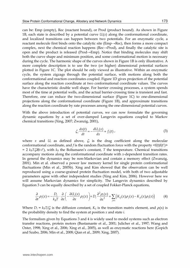
can be: Emp (empty), Rec (reactant bound), or Prod (product bound). As shown in Figure 1B, each state is described by a potential curve Ui(x) along the conformational coordinate, and localized transitions can happen between two potentials. For an enzymatic cycle, a reactant molecule first binds onto the catalytic site (Emp→Rec), then forms a more compact complex, next the chemical reaction happens (Rec→Prod), and finally the catalytic site is open and the product is released (Prod→Emp). Notice that binding molecules may shift both the curve shape and minimum position, and some conformational motion is necessary during the cycle. The harmonic shape of the curves shown in Figure 1B is only illustrative. A more complete description is to use the two (or higher) dimensional potential surfaces plotted in Figure 1C. The plot should be only viewed as illustrative. Within an enzymatic cycle, the system zigzags through the potential surface, with motions along both the conformational and reaction coordinates coupled. Figure 1D gives projection of the potential surface along the reaction coordinate at two conformational coordinate values. The curves have the characteristic double well shape. For barrier crossing processes, a system spends most of the time at potential wells, and the actual barrier-crossing time is transient and fast. Therefore, one can reduce the two-dimensional surface (Figure 1C) to one-dimensional projections along the conformational coordinate (Figure 1B), and approximate transitions along the reaction coordinate by rate processes among the one-dimensional potential curves.
With the above introduction of potential curves, we can now formulate the governing dynamic equations by a set of over-damped Langevin equations coupled to Markov chemical transitions (Xing, 2007; Zwanzig, 2001),
( )( )( )i
i i
dU xdx tf t
dt dxζ = − + , (3)
where x and Ui as defined above, ζi is the drag coefficient along the molecular conformational coordinate, and f is the random fluctuation force with the property <f(t)f(t’)> = 2 kBTζδ(t-t’), with kB the Boltamann’s constant, T the temperature. Chemical transitions accompany motions along the conformational coordinate with x-dependent transition rates. In general the dynamics may be non-Markovian and contain a memory effect (Zwanzig, 2001). Min et al. observed a power law memory kernel for single protein conformational fluctuations (Min et al., 2005b). Xing and Kim showed that the observation can be well reproduced using a coarse-grained protein fluctuation model, with both of two adjustable parameters agree with other independent studies (Xing and Kim, 2006). However here we will assume Markovian dynamics for simplicity. The Langevin dynamics described by Equation 3 can be equally described by a set of coupled Fokker-Planck equations,
( )2
2
( ) ( )( ) ( ) ( ) ( ) ( ) ( )i i i
i i i ij j ji iB j i
D U x xx x D K x x K x x
t k T x x x
ρρ ρ ρ ρ
≠
∂ ∂∂ ∂ = − ⋅ − + + −
∂ ∂ ∂ ∂ (4)
Where Di = kBT/ζi is the diffusion constant, Kij is the transition matrix element, and ρi(x) is the probability density to find the system at position x and state i.
The formalism given by Equations 3 and 4 is widely used to model systems such as electron transfer reactions, protein motors (Bustamante et al., 2001; Julicher et al., 1997; Wang and Oster, 1998; Xing et al., 2006; Xing et al., 2005), as well as enzymatic reactions here (Gopich and Szabo, 2006; Min et al., 2008; Qian et al., 2009; Xing, 2007).
www.intechopen.com
Protein-Protein Interactions – Computational and Experimental Tools
174
Fig. 1. Descriptions of coupling between chemical reactions and conformational changes. (A) A discrete enzymatic cycle model with conformational changes. (B) A minimal continuous model representing three potentials of mean force along a conformational coordinate. (C) A continuous model with explicit reaction and conformational coordinates. (D) Two protein conformations and the corresponding potentials of mean force along the reaction coordinate.
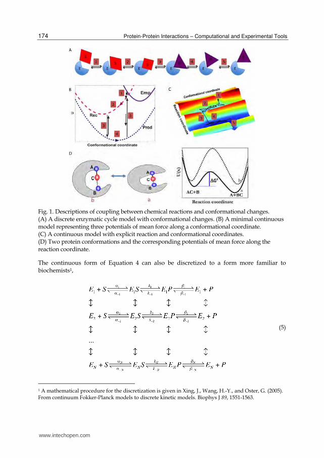
The continuous form of Equation 4 can also be discretized to a form more familiar to biochemists1,
(5)
1 A mathematical procedure for the discretization is given in Xing, J., Wang, H.-Y., and Oster, G. (2005). From continuum Fokker-Planck models to discrete kinetic models. Biophys J 89, 1551-1563.
www.intechopen.com
Slow Protein Conformational Change, Allostery and Network Dynamics
175
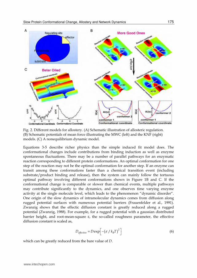
Fig. 2. Different models for allostery. (A) Schematic illustration of allosteric regulation. (B) Schematic potentials of mean force illustrating the MWC (left) and the KNF (right) models. (C) A nonequilibrium dynamic model.
Equations 3-5 describe richer physics than the simple induced fit model does. The conformational changes include contributions from binding induction as well as enzyme spontaneous fluctuations. There may be a number of parallel pathways for an enzymatic reaction corresponding to different protein conformations. An optimal conformation for one step of the reaction may not be the optimal conformation for another step. If an enzyme can transit among these conformations faster than a chemical transition event (including substrate/product binding and release), then the system can mainly follow the tortuous optimal pathway involving different conformations shown in Figure 1B and C. If the conformational change is comparable or slower than chemical events, multiple pathways may contribute significantly to the dynamics, and one observes time varying enzyme activity at the single molecule level, which leads to the phenomenon “dynamic disorder“. One origin of the slow dynamics of intramolecular dynamics comes from diffusion along rugged potential surfaces with numerous potential barriers (Frauenfelder et al., 1991). Zwanzig shows that the effectic diffusion constant is greatly reduced along a rugged potential (Zwanzig, 1988). For example, for a rugged potential with a gaussian distributed barrier height, and root-mean-square ε, the so-called roughness parameter, the effective diffusion constant is scaled as,
( )2
exp /effective BD D k Tε = − (6)
which can be greatly reduced from the bare value of D.
www.intechopen.com
Protein-Protein Interactions – Computational and Experimental Tools
176
3. Thermodynamic versus dynamic models for allostery
A cell needs to adjust its metabolic, transcriptional, and translational activities to respond to changes in the external and internal environment. Allostery and covalent modification are two fundamental mechanisms for regulating protein activities (Alberts et al., 2002). Allostery refers to the phenomenon that binding of an effector molecule to a protein’s allosteric site affects the protein activity at its active site, which is usually physically distinct from where the effector binds. The discovery of allosteric regulations was in the 1950s, followed by a general description of allostery in the early 1960s, has been regarded as revolutionary at that time (Alberts et al., 2002). Not surprisingly, to understand the mechanism of allosteric regulation is an important topic in structural biology. Below we will focus on allosteric enzymes. For simplicity, we will restrict our discussions to positive allosteric effect, i.e., effector binding increases enzymatic activity. The discussions can be easily generalized to negative allosteric effects.
3.1 Conventional models of allostery
There are two popular models proposed to explain the allosteric effects. The concerted MWC model by Monod, Wyman, and Changeux, assumes that an allosteric protein can exist in two (or more) conformations with different reactivity, and effector binding modifies the thermal equilibrium distribution of the conformers (Monod et al., 1965). Recent population shift models re-emphasize the idea of preexisting populations (Goodey and Benkovic, 2008; Kern and Zuiderweg, 2003; Pan et al., 2000; Volkman et al., 2001). The sequential model described by Koshland, Nemethy, and Filmer is based on the induced-fit mechanism, and assumes that effector binding results in (slight) structural change at another site and affects the substrate affinity (Koshland et al., 1966). While different in details, both of the above models assume that the allosteric mechanism is through modification of the equilibrium conformation distribution of the allosteric protein by effector binding. For later discussions, we denote the mechanisms as “thermodynamic regulation”.
The mechanisms of thermodynamic regulation impose strong requirements on the mechanical properties of an allosteric protein. The distance between the two binding sites of an allosteric protein can be far. For example, the bacterial chemotaxis receptor has the two reaction regions separated as far as 15 nm (Kim et al., 2002). In this case, signal propagation requires a network of mechanical strain relaying residues with mechanical properties distinguishing them well from the surroundings to minimize thermal dissipation – Notice that distortion of a soft donut at one side has negligible effect on another side of the donut. Mechanical stresses due to effector molecule binding irradiate from the binding site, propagate through the relaying network, and con-focus on the reaction region at the other side of the protein (Amaro et al., 2009; Amaro et al., 2007; Balabin et al., 2009; Cecchini et al., 2008; Cui and Karplus, 2008; Horovitz and Willison, 2005; Ranson et al., 2006). However, it is challenging to transmit the mechanical energy faithfully against thermal dissipation over a long distance. A possible solution is the attraction shift model proposed by Yu and Koshland(Yu and Koshland, 2001).
From a chemical physics perspective, current existing models on allosteric effects differ in some details of the potential shapes. The MWC and the recent population-shift model emphasizes that there are pre-existing populations for all the possible forms, as exemplified by the double well shaped potentials and the two corresponding conformers in the left panel of
www.intechopen.com
Slow Protein Conformational Change, Allostery and Network Dynamics
177
Figure 2C. Effector binding only shifts their relative populations. The KNF model emphasizes that without the effector the protein exists mainly in one form (conformer 2 in the right panel of Figure 2C). Effector binding shifts the protein to another form (conformer 1) with different reactivity. The functions U(x) are potentials of mean force, which suggests that the effect of effector binding can be enthalpic or entropic (Cooper and Dryden, 1984). Therefore in some sense there is no fundamental difference between the KNF and MWC models. They differ only in the extent of each conformer being populated, which is related to the free energy difference between conformers ΔU (in Figure 2C) through the Boltzman factor.
3.2 Possibly neglected dynamic aspect of allostery
The above allosteric models focus on the conformational changes decoupled from those changes associated with an enzymatic cycle. Consequently, the distribution along the conformational coordinate can be described as thermodynamic equilibrium. However, as discussed in section 2, an enzymatic cycle usually inevitably involves enzyme conformational changes, so the distribution of the latter is in general driven out of equilibrium due to coupling to the nonequilibrium chemical reactions. In many cases, as Frieden wrote, “conformational changes after substrate addition but preceding the chemical transformation, or after the chemical transformation but preceding product release may be rate-limiting” (Frieden, 1979). Recent NMR studies further demonstrate conformational changes as rate-limiting steps (Boehr et al., 2006; Cole and Loria, 2002). Based on these experimental observations, Xing proposed that the conformational change dynamics within an enzymatic cycle can be subject to allosteric modulation (Xing, 2007).
Enzyme conformational changes can be thermally activated barrier crossing events, and effectors function by modifying the height of the dominant barrier. Alternatively, effectors may accelerate conformational changes through decreasing the potential roughness (see Figure 2D). Intuitively, for the latter mechanism effectors transform rusty engines (enzymes) into better-oiled ones.
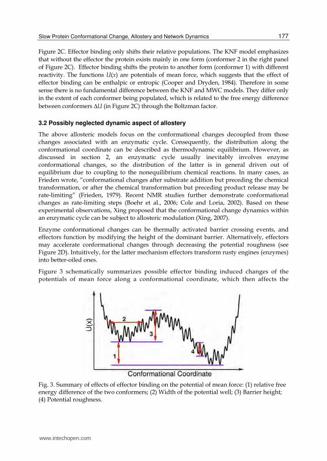
Figure 3 schematically summarizes possible effector binding induced changes of the potentials of mean force along a conformational coordinate, which then affects the
Fig. 3. Summary of effects of effector binding on the potential of mean force: (1) relative free energy difference of the two conformers; (2) Width of the potential well; (3) Barrier height; (4) Potential roughness.
www.intechopen.com
Protein-Protein Interactions – Computational and Experimental Tools
178
enzymatic reaction dynamics. The changes can be the relative height of the potential wells representing different conformers (labelled 1 in Figure 3, enthalpic), the widths of potential wells (labelled 2, entropic), the barrier height (labelled 3) and the potential roughness (labelled 4) (dynamic). For a given enzyme subject to a given effector regulation, one or more effects may play the dominant role.
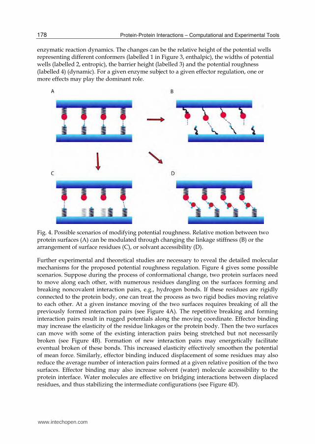
Fig. 4. Possible scenarios of modifying potential roughness. Relative motion between two protein surfaces (A) can be modulated through changing the linkage stiffness (B) or the arrangement of surface residues (C), or solvant accessibility (D).
Further experimental and theoretical studies are necessary to reveal the detailed molecular mechanisms for the proposed potential roughness regulation. Figure 4 gives some possible scenarios. Suppose during the process of conformational change, two protein surfaces need to move along each other, with numerous residues dangling on the surfaces forming and breaking noncovalent interaction pairs, e.g., hydrogen bonds. If these residues are rigidly connected to the protein body, one can treat the process as two rigid bodies moving relative to each other. At a given instance moving of the two surfaces requires breaking of all the previously formed interaction pairs (see Figure 4A). The repetitive breaking and forming interaction pairs result in rugged potentials along the moving coordinate. Effector binding may increase the elasticity of the residue linkages or the protein body. Then the two surfaces can move with some of the existing interaction pairs being stretched but not necessarily broken (see Figure 4B). Formation of new interaction pairs may energetically facilitate eventual broken of these bonds. This increased elasticity effectively smoothen the potential of mean force. Similarly, effector binding induced displacement of some residues may also reduce the average number of interaction pairs formed at a given relative position of the two surfaces. Effector binding may also increase solvent (water) molecule accessibility to the protein interface. Water molecules are effective on bridging interactions between displaced residues, and thus stabilizing the intermediate configurations (see Figure 4D).
www.intechopen.com
Slow Protein Conformational Change, Allostery and Network Dynamics
179
3.3 Allosteric regulation of bacterial flagellar motor switching
Here we specifically discuss allosteric regulation in the bacterial flagellar motor system. Although the flagellar motor switching process does not involve enzymatic cycles directly, the process shares some features common to what we discussed in section 3.2.
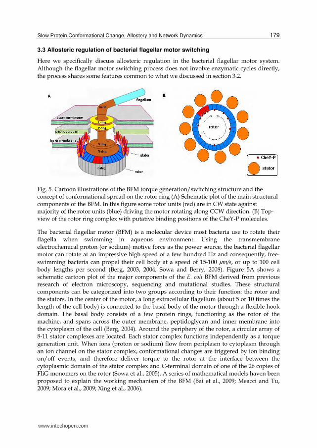
Fig. 5. Cartoon illustrations of the BFM torque generation/switching structure and the concept of conformational spread on the rotor ring (A) Schematic plot of the main structural components of the BFM. In this figure some rotor units (red) are in CW state against majority of the rotor units (blue) driving the motor rotating along CCW direction. (B) Top-view of the rotor ring complex with putative binding positions of the CheY-P molecules.
The bacterial flagellar motor (BFM) is a molecular device most bacteria use to rotate their flagella when swimming in aqueous environment. Using the transmembrane electrochemical proton (or sodium) motive force as the power source, the bacterial flagellar motor can rotate at an impressive high speed of a few hundred Hz and consequently, free-swimming bacteria can propel their cell body at a speed of 15-100 µm/s, or up to 100 cell body lengths per second (Berg, 2003, 2004; Sowa and Berry, 2008). Figure 5A shows a schematic cartoon plot of the major components of the E. coli BFM derived from previous research of electron microscopy, sequencing and mutational studies. These structural components can be categorized into two groups according to their function: the rotor and the stators. In the center of the motor, a long extracellular flagellum (about 5 or 10 times the length of the cell body) is connected to the basal body of the motor through a flexible hook domain. The basal body consists of a few protein rings, functioning as the rotor of the machine, and spans across the outer membrane, peptidoglycan and inner membrane into the cytoplasm of the cell (Berg, 2004). Around the periphery of the rotor, a circular array of 8-11 stator complexes are located. Each stator complex functions independently as a torque generation unit. When ions (proton or sodium) flow from periplasm to cytoplasm through an ion channel on the stator complex, conformational changes are triggered by ion binding on/off events, and therefore deliver torque to the rotor at the interface between the cytoplasmic domain of the stator complex and C-terminal domain of one of the 26 copies of FliG monomers on the rotor (Sowa et al., 2005). A series of mathematical models haven been proposed to explain the working mechanism of the BFM (Bai et al., 2009; Meacci and Tu, 2009; Mora et al., 2009; Xing et al., 2006).
www.intechopen.com
Protein-Protein Interactions – Computational and Experimental Tools
180
The bacterial flagellar motor is not only important for the propulsion of the cell, but also crucial for bacterial chemotaxis. In the E. coli chemotaxis system, chemical gradients (attractant or repellent) are sensed through multiple transmembrane methyl-accepting chemotaxis proteins (MCPs) (Berg, 2004). When extracellular chemotactic attractants (or repellents) bind to MCPs, conformational changes through the membrane inhibit (or trigger) the autophosphorylation in the histidine kinase, CheA. CheA in turn transfers phosphoryl groups to conserved aspartate residues in the response regulators CheY. The phosphorylated form of CheY, CheY-P, diffuses away across the cytoplasm of the cell and binds to the bottom of the FliM/FliN complex of the flagellar motor. When attractant gradient is sensed, CheY-P concentration is low in the cytoplasm and therefore less CheY-P molecules bind to the flagellar motor, which favours counter-clockwise (CCW) rotation of the motor. When most of the motors on the membrane spin CCW, flagellar filaments form a bundle and propel the cell steadily forward. When repellent gradient is sensed, CheY-P concentration is raised and more CheY-P binds to the flagellar motor, which leads to clockwise (CW) rotation of the motor. When a few motors (can be as few as one) spin CW, flagellar filaments fly apart and the cell tumbles. The bacterial flagellar motor (BFM) switches stochastically between CCW and CW states and therefore the cell repeats a ‘run’-‘tumble’-‘run’ pattern. This enables a chemotactic navigation in a low Reynolds number environment (reviewed in Berg, book E. coli in motion). The ratio of the rotation direction CCW/CW is tuned by the concentration of the signalling protein, CheY-P.
The problem of BFM switching response to cytoplasmic CheY-P concentration is essentially a protein allosteric regulation. When the effector (CheY-P) binds to the bottom of each rotor unit (a protein complex formed by roughly 1:1:1 of FliG, FliM, FliN protein), it makes CW rotation more favourable (Figure 5B). However, a careful examination of the BFM switching shows that the allosteric regulation here has distinct features: 1) in previous in vivo experiment (Cluzel et al., 2000), Cluzel et al. monitored in real time the relationship between BFM switching bias and CheY-P concentration in the cell and found that the response curve is ultrasensitive with a Hill coefficient of ~ 10. Later FRET experiment further showed that binding of CheY-P to FliM is much less cooperative than motor switching response (Sourjik and Berg, 2002). The molecular mechanism of this high cooperativity in BFM switching response remains unknown. 2) the BFM rotor has a ring structure, which is a large multisubunit protein complexes formed by 26 identical rotor units. For such a large multisubunit protein complex, an absolute coupling between subunits as the MWC model requires seems very unlikely. 3) the BFM rotates in full speed stably in CCW or CW directions, and transitions between these two states are brief and fast. This indicates that the 26 rotor units on the basal body of the BFM are in a coherent conformation for most of the time and switching of the whole ring can finish within a very short time period. The above facts also put the KNF model in doubt. As in the KNF model, coupling between effector binding and conformation is absolute: When an effector binds a rotor unit, that rotor unit switches direction.
Therefore a new type of model is in needed to explain the molecular mechanism of the BFM switching. Duke et al. constructed a mathematical model of the general allosteric scheme based on the idea proposed by Eigen (Eigen, 1968) in which both types of coupling are probabilistic (Duke et al., 2001; Duke and Bray, 1999). This model encompasses the classical mechanisms at its limits and introduces the mechanism of conformational spread, with
www.intechopen.com
Slow Protein Conformational Change, Allostery and Network Dynamics
181
domains of a particular conformational state growing or shrinking faster than ligand binding. Particular regions in the parameter space of the conformational spread model reproduce the classical WMC and KNF model (Duke et al., 2001).
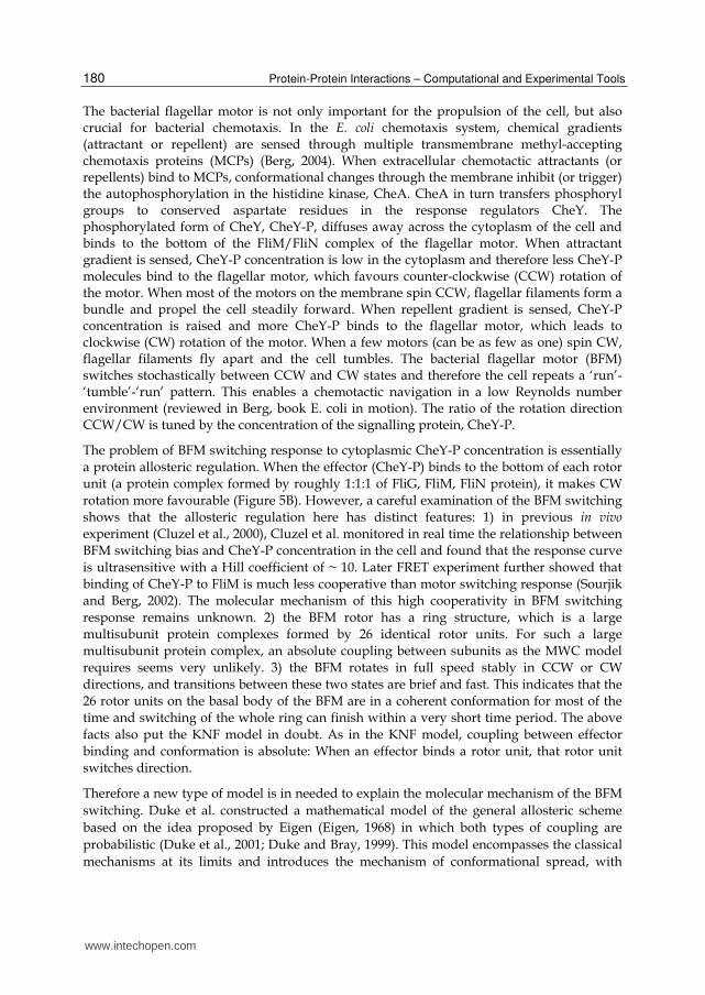
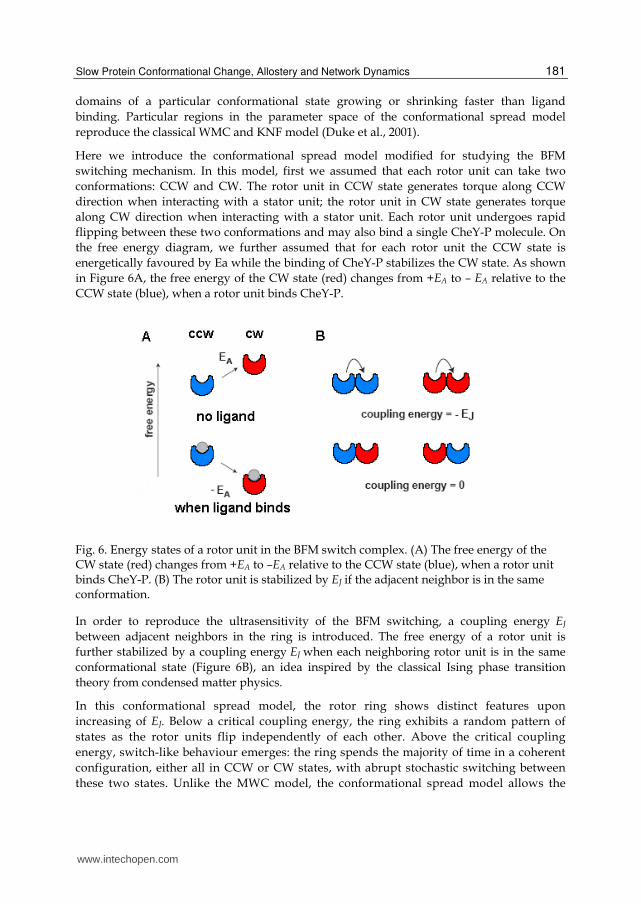
Here we introduce the conformational spread model modified for studying the BFM switching mechanism. In this model, first we assumed that each rotor unit can take two conformations: CCW and CW. The rotor unit in CCW state generates torque along CCW direction when interacting with a stator unit; the rotor unit in CW state generates torque along CW direction when interacting with a stator unit. Each rotor unit undergoes rapid flipping between these two conformations and may also bind a single CheY-P molecule. On the free energy diagram, we further assumed that for each rotor unit the CCW state is energetically favoured by Ea while the binding of CheY-P stabilizes the CW state. As shown in Figure 6A, the free energy of the CW state (red) changes from +EA to – EA relative to the CCW state (blue), when a rotor unit binds CheY-P.
Fig. 6. Energy states of a rotor unit in the BFM switch complex. (A) The free energy of the CW state (red) changes from +EA to –EA relative to the CCW state (blue), when a rotor unit binds CheY-P. (B) The rotor unit is stabilized by EJ if the adjacent neighbor is in the same conformation.
In order to reproduce the ultrasensitivity of the BFM switching, a coupling energy EJ between adjacent neighbors in the ring is introduced. The free energy of a rotor unit is further stabilized by a coupling energy EJ when each neighboring rotor unit is in the same conformational state (Figure 6B), an idea inspired by the classical Ising phase transition theory from condensed matter physics.
In this conformational spread model, the rotor ring shows distinct features upon increasing of EJ. Below a critical coupling energy, the ring exhibits a random pattern of states as the rotor units flip independently of each other. Above the critical coupling energy, switch-like behaviour emerges: the ring spends the majority of time in a coherent configuration, either all in CCW or CW states, with abrupt stochastic switching between these two states. Unlike the MWC model, the conformational spread model allows the
www.intechopen.com
Protein-Protein Interactions – Computational and Experimental Tools
182
existence of an intermediate (or mixed) configuration of the rotor units on the ring; and unlike the KNF model, the conformational spread model also allows rotor units stay in its original conformation without being switched by effector binding events. By implementing parallel Monte Carlo processes, one can simulate BFM switching response to CheY-P concentration. In each iteration, each rotor unit on the ring is visited and polled to determine whether to stay in the old state or jump to a new state according to the free energy difference between the two states as a function of 1) free energy of the rotor unit itself 2) binding condition of the regulator molecule CheY-P 3) energy coupling of adjacent neighboring subunits.
The conformational spread model has successfully reproduced previous experimental observations that 1) the BFM switching bias responses ultrasensitively to changes in CheY-P concentration 2) the motor rotates stably in CCW and CW states with occasional fast transitions from one coherent state to the other. The model also made several new predictions: 3) creation of domains of the opposite conformation is frequent due to fast flipping of single rotor unit, but most of them shrink and disappear, failing to occupy the whole ring. However, some big fluctuations can still produce obvious slowdowns and pausing of the motor. Therefore, speed traces of the BFM should have frequent transient speed slowdowns and pauses. 4) the switch interval (the time that the motor spends in the CCW or CW state) follows a single exponential distribution. 5) the switch time, the time that the motor takes to complete a switch, is non-instantaneous. It can be modeled as a biased random walk along the ring. The characteristic switching time depends on the size of the ring and flipping rate of each rotor unit in a complicated manner. Due to the stochastic nature of this conformational spread, we expect to see a wide distribution of switching times.
With the cutting-edge single molecule detection technique, the above predictions of the conformational spread model has recently been confirmed (Bai et al., 2010). Instead of instant transition , switches between CCW and CW rotor states were found to follow a broad distribution, with switching time ranging from less than 2 milliseconds to several hundred milliseconds, and transient intermediate states containing a mixture of CW/CCW rotor units have been observed. The conformational spread model has provided a molecular mechanism for the BFM switching, and more importantly, it sheds light on allosteric regulation in large protein complexes. In addition to the canonical MWC and KNF models, the conformational spread model provides a new comprehensive approach to allostery, and is consistent with the discussion in section 3.2 that both kinetic and thermodynamic aspects should be considered.
4. Coupling between slow conformational change and network dynamics
A biological network usually functions in a noisy ever-changing environment. Therefore, the network should be: 1) robust ─ functioning normally despite environmental noises; 2) adaptive ─ the tendency to function optimally by adjusting to the environmental changes; 3) sensitive ─ sharp response to the regulating signals. It is not-fully understood how a biological network can achieve these requirements simultaneously. Contemporary researches emphasize that the dynamic properties of a network is closely related to its topology.
www.intechopen.com
Slow Protein Conformational Change, Allostery and Network Dynamics
183
Many in vivo biological processes involve only a small number of substrate molecules. When this number is in the range of hundreds or even smaller, stochastic effect becomes predominant. Chemical reactions take place in a stochastic rather than deterministic way. Therefore one should track the discrete numbers of individual species explicitly in the rate equation formalism. So far, many studies have shown that one might make erroneous conclusions without considering the stochastic effect (Samoilov et al., 2005; Wylie et al., 2007). Noise propagation through a network is currently an important research topic (Levine et al., 2007; Paulsson et al., 2000; Pedraza and van Oudenaarden, 2005; Rao et al., 2002; Rosenfeld et al., 2005; Samoilov et al., 2005; Shibata and Fujimoto, 2005; Suel et al., 2007; Swain et al., 2002). One usually assumes that the stochastic effect mainly arises from small number of identical molecules, and rate constants are still assumed well defined.
With the existence of dynamic disorder, the activity of a single enzyme (and so of a small number of enzymes) is a varying quantity. This adds another noise source with unique (multi-time scale, non-white noise) properties (Min and Xie, 2006; Xing and Kim, 2006). For bulk concentrations, fluctuations due to dynamic disorder are suppressed by averaging over a large number of molecules. However, existence of NMM kinetics can still manifest itself in a network. If there are only a small number of protein molecules, as in many in vivo processes, dynamic disorder will greatly affect the network dynamics. The conventionally considered stochastic effect is mainly due to number variations of identical molecules. Here a new source of stochastic effect arises from small numbers of molecules with the same chemical structure but different conformations. Dynamic disorder induced stochastic effect has some unique properties, which require special theoretical treatment, and may result in novel dynamic behaviors. First, direct fluctuation of the rate constants over several orders of magnitude may have dramatic effects on the network dynamics. Second, the associated time scales have broad range. The Gaussian white noise approximation is widely used in stochastic modeling of network dynamics with the assumption that some processes are much faster than others (Gillespie, 2000). Existence of broad time scale distribution makes the situation more complicated. Furthermore, a biological system may actively utilize this new source of noise. Noises from different sources may not necessarily add up. Instead they may cancel each other and result in smaller overall fluctuations (Paulsson et al., 2000; Samoilov et al., 2005). We expect that the existence of dynamic disorder not only further complicates the situation, but may also provide additional degrees of freedom for regulation since the rates can be continuously tuned. Especially we expect that existence of dynamic disorder may require dramatic modification on our understanding of signal transduction networks. Many of these processes involve a small number of molecules, and are featured by short reaction time scales (within minutes), high sensitivity and specificity (responding to specific molecules only).
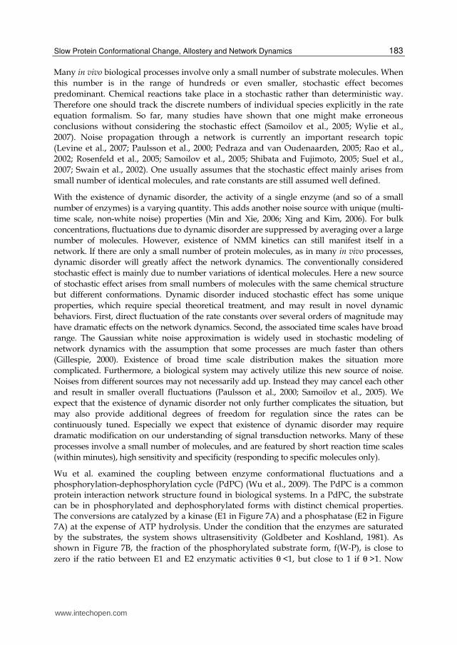
Wu et al. examined the coupling between enzyme conformational fluctuations and a phosphorylation-dephosphorylation cycle (PdPC) (Wu et al., 2009). The PdPC is a common protein interaction network structure found in biological systems. In a PdPC, the substrate can be in phosphorylated and dephosphorylated forms with distinct chemical properties. The conversions are catalyzed by a kinase (E1 in Figure 7A) and a phosphatase (E2 in Figure 7A) at the expense of ATP hydrolysis. Under the condition that the enzymes are saturated by the substrates, the system shows ultrasensitivity (Goldbeter and Koshland, 1981). As shown in Figure 7B, the fraction of the phosphorylated substrate form, f(W-P), is close to zero if the ratio between E1 and E2 enzymatic activities θ <1, but close to 1 if θ >1. Now
www.intechopen.com
Protein-Protein Interactions – Computational and Experimental Tools
184
consider a system with a finite size, e.g. 50 E1 molecules, 50 E2 molecules, and a total of 1500 substrate molecules as used to generate results in Figure 7C & D. Enzyme activities fluctuate due to conformational fluctuations. For simplicity let us assume that E1 can stochastically convert between an active and a less active forms. While the average value of θ = 1.1, it fluctuates within the range [0.7, 1.5], depending on the number of E1 molecules in the active form. For convenience of discussion, let us also define the response time of the PdPC, τ, as the time it takes for the fraction of W-P reaching half given at time 0 the system jumps from θ < 1 το θ > 1 due to enzyme conformational fluctuations. The response time clearly related to the enzymatic turnover rate. As the trajectories in Figure7C show, for slow θ fluctuation Δθ is amplified to ΔW-P due to the ultrasensitivity of the PdPC and the much larger number of substrate molecules compared to enzymes. However, with θ fluctuation much faster than τ, the PdPC only responses to the average value of θ . Therefore depending on the relative time scale between θ fluctuation and the response time of the PdPC to θ change, fluctuations of θ can be either amplified or suppressed.
Fig. 7. Coupling between enzyme conformational fluctuations and a phosphorylation-dephosphorylation cycle. (A) A phosphorylation-dephosphorylation cycle (PdPC). (B) Ultrasensitivity of a PdPC. (C) Trajectories of enzyme activity due to slow conformational fluctuations and the corresponding substrate fluctuation. (D) Similar to C but with fast conformational fluctuations.
5. Conclusion
Slow conformational motions in macromolecules play crucial roles in their unique function in enzymatic reactions as well as biological networks. We suggest that these motions are of
www.intechopen.com
Slow Protein Conformational Change, Allostery and Network Dynamics
185
great functional importance, which can only be fully appreciated in the context of regulatory networks. Collaborative researches from molecular and cellular level studies are urgently needed for this largely unexplored area.
6. Acknowledgment
ZW and JX were supported by an NSF grant (EF-1038636) and a grant from the William and Mary Jeffress Memorial Trust.
7. References
Ainslie, G.R., Jr., Shill, J.P., and Neet, K.E. (1972). Transients and Cooperativity. A slow transition model for relating transients and cooperative kinetics of enzymes. J Biol Chem 247, 7088-7096.
Alberts, B., Johnson, A., Lewis, J., Raff, M., Roberts, K., and Walter, P. (2002). Molecular Biology of the Cell, 4d edn (New York, Garland).
Alon, U. (2007). An introduction to systems biology: Design principles of biological circuits, 1 edn (Chapman and Hall/CRC).
Amaro, R.E., Cheng, X., Ivanov, I., Xu, D., and McCammon, J.A. (2009). Characterizing Loop Dynamics and Ligand Recognition in Human- and Avian-Type Influenza Neuraminidases via Generalized Born Molecular Dynamics and End-Point Free Energy Calculations. J Am Chem Soc 131, 4702-4709.
Amaro, R.E., Sethi, A., Myers, R.S., Davisson, V.J., and Luthey-Schulten, Z.A. (2007). A Network of Conserved Interactions Regulates the Allosteric Signal in a Glutamine Amidotransferase†. Biochemistry 46, 2156-2173.
Austin, R.H., Beeson, K.W., Eisenstein, L., Frauenfelder, H., and Gunsalus, I.C. (1975). Dynamics of Ligand-Binding to Myoglobin. Biochemistry 14, 5355-5373.
Bai, F., Branch, R.W., Nicolau, Dan V.,, Jr., Pilizota, T., Steel, B.C., Maini, P.K., Berry, R.M. (2010). Conformational Spread as a Mechanism for Cooperativity in the Bacterial Flagellar Switch. Science 327, 685-689.
Bai, F., Lo, C.-J., Berry, R.M., and Xing, J. (2009). Model Studies of the Dynamics of Bacterial Flagellar Motors. Biophys J 96, 3154-3167.
Balabin, I.A., Yang, W., and Beratan, D.N. (2009). Coarse-grained modeling of allosteric regulation in protein receptors. Proceedings of the National Academy of Sciences 106, 14253-14258.
Behzadi, A., Hatleskog, R., and Ruoff, P. (1999). Hysteretic enzyme adaptation to environmental pH: change in storage pH of alkaline phosphatase leads to a pH-optimum in the opposite direction to the applied change. Biophys Chem 77, 99-109.
Berg, H.C. (2003). The Rotary Motor of Bacterial Flagella. Annu Rev Biochem 72, 19-54. Berg, H.C. (2004). E. Coli in Motion (New York, Springer-Verlag Press). Boehr, D.D., McElheny, D., Dyson, H.J., and Wright, P.E. (2006). The Dynamic Energy
Landscape of Dihydrofolate Reductase Catalysis. Science 313, 1638-1642. Bustamante, C., Keller, D., and Oster, G. (2001). The physics of molecular motors. Acc Chem
Res 34, 412-420. Cecchini, M., Houdusse, A., and Karplus, M. (2008). Allosteric Communication in Myosin V:
From Small Conformational Changes to Large Directed Movements. PLoS Comput Biol 4, e1000129.
www.intechopen.com
Protein-Protein Interactions – Computational and Experimental Tools
186
Cluzel, P., Surette, M., and Leibler, S. (2000). An Ultrasensitive Bacterial Motor Revealed by Monitoring Signaling Proteins in Single Cells. Science 287, 1652-1655.
Cole, R., and Loria, J.P. (2002). Evidence for Flexibility in the Function of Ribonuclease A†. Biochemistry 41, 6072-6081.
Cooper, A., and Dryden, D.T.F. (1984). Allostery without Conformational Change - a Plausible Model. Eur Biophys J Biophys Lett 11, 103-109.
Cornish-Bowden, A., and Cardenas, M.L. (1987). Co-operativity in monomeric enzymes. J Theor Biol 124, 1-23.
Cui, Q., and Karplus, M. (2008). Allostery and cooperativity revisited. Protein Science 17, 1295-1307.
Duke, T., Novere, N.L., and Bray, D. (2001). Conformational spread in a ring of proteins: A stochastic approach to allostery. J Mol Biol 308.
Duke, T.A.J., and Bray, D. (1999). Heightened sensitivity of a lattice of membrane receptors. Proc Nat Acad Sci USA 96, 10104-10108.
Eigen, M. (1968). Kinetics of reaction control and information transfer in enzymes and nucleic acids. In Nobel Symp 5 on Fast Reactions and Primary Processes, Chem Kinetics, S. Claesson, ed. (Stockholm, Almquist & Wiksell).
English, B.P., Min, W., van Oijen, A.M., Lee, K.T., Luo, G.B., Sun, H.Y., Cherayil, B.J., Kou, S.C., and Xie, X.S. (2006). Ever-fluctuating single enzyme molecules: Michaelis-Menten equation revisited. Nat Chem Biol 2, 87-94.
Frauenfelder, H., Sligar, S.G., and Wolynes, P.G. (1991). The Energy Landscapes and Motions of Proteins. Science 254, 1598-1603.
Frauenfelder, H., Wolynes, P.G., and Austin, R.H. (1999). Biological physics. Rev Mod Phys 71, S419-S430.
Frieden, C. (1970). Kinetic Aspects of Regulation of Metabolic Processes. The hysteretic enzyme concept. J Biol Chem 245, 5788-5799.
Frieden, C. (1979). Slow Transitions and Hysteretic Behavior in Enzymes. Ann Rev Biochem 48, 471-489.
Gillespie, D.T. (2000). The chemical Langevin Equation. J Chem Phys 113, 297-306. Goldbeter, A., and Koshland, D.E. (1981). An Amplified Sensitivity Arising from Covalent
Modification in Biological-Systems. Proc Natl Acad Sci USA 78, 6840-6844. Goodey, N.M., and Benkovic, S.J. (2008). Allosteric regulation and catalysis emerge via a
common route. Nat Chem Biol 4, 474-482. Gopich, I.V., and Szabo, A. (2006). Theory of the statistics of kinetic transitions with
application to single-molecule enzyme catalysis. J Chem Phys 124, 154712. Grote, R.F., and Hynes, J.T. (1980). The stable states picture of chemical-reactions 2: Rate
constants for condensed and gas-phase reaction models. J Chem Phys 73, 2715-2732. Hanggi, P., Talkner, P., and Borkovec, M. (1990). Reaction-rate theory: 50 years after
Kramers. Rev Mod Phys 62, 254-341. Horovitz, A., and Willison, K.R. (2005). Allosteric regulation of chaperonins. Current
Opinion in Structural Biology 15, 646-651. Julicher, F., Ajdari, A., and Prost, J. (1997). Modeling molecular motors. Rev Mod Phys 69,
1269-1281. Kern, D., and Zuiderweg, E.R.P. (2003). The role of dynamics in allosteric regulation. Curr
Opin Struc Biol 13, 748-757. Kholodenko, B.N. (2006). Cell-signalling dynamics in time and space. Nat Rev Mol Cell Biol
7, 165-176.
www.intechopen.com
Slow Protein Conformational Change, Allostery and Network Dynamics
187
Kim, S.H., Wang, W.R., and Kim, K.K. (2002). Dynamic and clustering model of bacterial chemotaxis receptors: Structural basis for signaling and high sensitivity. Proc Natl Acad Sci USA 99, 11611-11615.
Koshland, D.E., Nemethy, G., and Filmer, D. (1966). Comparison of Experimental Binding Data and Theoretical Models in Proteins Containing Subunits. Biochemistry 5, 365-385.
Kroh, H.K., Panizzi, P., and Bock, P.E. (2009). Von Willebrand factor-binding protein is a hysteretic conformational activator of prothrombin. Proc Natl Acad Sci U S A 106, 7786-7791.
Levine, J., Kueh, H.Y., and Mirny, L. (2007). Intrinsic fluctuations, robustness, and tunability in signaling cycles. Biophys J 92, 4473-4481.
McCammon, J.A., and Harvey, S.C. (1987). Dynamics of Proteins and Nucleic Acids (New York, Cambridge Univ Press).
Meacci, G., and Tu, Y. (2009). Dynamics of the bacterial flagellar motor with multiple stators. Proc Natl Acad Sci USA 106, 3746-3751.
Menten, L., and Michaelis, M.I. (1913). Die Kinetik der Invertinwirkung. Biochem Z 49, 333-369.
Min, W., English, B.P., Luo, G.B., Cherayil, B.J., Kou, S.C., and Xie, X.S. (2005a). Fluctuating enzymes: Lessons from single-molecule studies. Acc Chem Res 38, 923-931.
Min, W., Gopich, I.V., English, B.P., Kou, S.C., Xie, X.S., and Szabo, A. (2006). When Does the Michaelis-Menten Equation Hold for Fluctuating Enzymes? J Phys Chem B 110, 20093-20097.
Min, W., Luo, G.B., Cherayil, B.J., Kou, S.C., and Xie, X.S. (2005b). Observation of a power-law memory kernel for fluctuations within a single protein molecule. Phys Rev Lett 94, 198302.
Min, W., and Xie, X.S. (2006). Kramers model with a power-law friction kernel: Dispersed kinetics and dynamic disorder of biochemical reactions. Phys Rev E 73, -.
Min, W., Xie, X.S., and Bagchi, B. (2008). Two-Dimensional Reaction Free Energy Surfaces of Catalytic Reaction: Effects of Protein Conformational Dynamics on Enzyme Catalysis. J Phys Chem B 112, 454-466.
Monod, J., Wyman, J., and Changeux, J.P. (1965). On Nature of Allosteric Transitions - a Plausible Model. J Mol Biol 12, 88-118.
Mora, T., Yu, H., and Wingreen, N.S. (2009). Modeling Torque Versus Speed, Shot Noise, and Rotational Diffusion of the Bacterial Flagellar Motor. Phys Rev Lett 103, 248102.
Pan, H., Lee, J.C., and Hilser, V.J. (2000). Binding sites in Escherichia coli dihydrofolate reductase communicate by modulating the conformational ensemble. Proceedings of the National Academy of Sciences 97, 12020-12025.
Paulsson, J., Berg, O.G., and Ehrenberg, M. (2000). Stochastic focusing: Fluctuation-enhanced sensitivity of intracellular regulation. Proc Natl Acad Sci USA 97, 7148-7153.
Pedraza, J.M., and van Oudenaarden, A. (2005). Noise Propagation in Gene Networks. Science 307, 1965-1969.
Qian, H., Shi, P.-Z., and Xing, J. (2009). Stochastic bifurcation, slow fluctuations, and bistability as an origin of biochemical complexity. Phys Chem Chem Phys 11, 4861-4870.
Ranson, N.A., Clare, D.K., Farr, G.W., Houldershaw, D., Horwich, A.L., and Saibil, H.R. (2006). Allosteric signaling of ATP hydrolysis in GroEL-GroES complexes. Nat Struct Mol Biol 13, 147-152.
Rao, C.V., Wolf, D.M., and Arkin, A.P. (2002). Control, exploitation and tolerance of intracellular noise. Nature 420, 231-237.
www.intechopen.com
Protein-Protein Interactions – Computational and Experimental Tools
188
Ricard, J., and Cornish-Bowden, A. (1987). Co-operative and allosteric enzymes: 20 years on. Eur J Biochem 166, 255-272.
Rosenfeld, N., Young, J.W., Alon, U., Swain, P.S., and Elowitz, M.B. (2005). Gene Regulation at the Single-Cell Level. Science 307, 1962-1965.
Samoilov, M., Plyasunov, S., and Arkin, A.P. (2005). Stochastic amplification and signaling in enzymatic futile cycles through noise-induced bistability with oscillations. Proc Natl Acad Sci USA 102, 2310-2315.
Shibata, T., and Fujimoto, K. (2005). Noisy signal amplification in ultrasensitive signal transduction. Proc Natl Acad Sci USA 102, 331-336.
Sourjik, V., and Berg, H.C. (2002). Binding of the Escherichia coli response regulator CheY to its target measured in vivo by fluorescence resonance energy transfer. Proc Natl Acad Sci USA 99, 12669-12674.
Sowa, Y., and Berry, R.M. (2008). Bacterial flagellar motor. Quart Rev Biophys 41, 103-132. Sowa, Y., Rowe, A.D., Leake, M.C., Yakushi, T., Homma, M., Ishijima, A., and Berry, R.M.
(2005). Direct observation of steps in rotation of the bacterial flagellar motor. Nature 437, 916-919.
Suel, G.M., Kulkarni, R.P., Dworkin, J., Garcia-Ojalvo, J., and Elowitz, M.B. (2007). Tunability and noise dependence in differentiation dynamics. Science 315, 1716-1719.
Swain, P.S., Elowitz, M.B., and Siggia, E.D. (2002). Intrinsic and extrinsic contributions to stochasticity in gene expression. Proc Natl Acad Sci USA 99, 12795-12800.
Tyson, J.J., Chen, K., and Novak, B. (2001). Network dynamics and cell physiology. Nat Rev Mol Cell Biol 2, 908-916.
Volkman, B.F., Lipson, D., Wemmer, D.E., and Kern, D. (2001). Two-state allosteric behavior in a single-domain signaling protein. Science 291, 2429-2433.
Wang, H., and Oster, G. (1998). Energy transduction in the F1 motor of ATP synthase. Nature 396, 279-282.
Wu, Z., Elgart, V., Qian, H., and Xing, J. (2009). Amplification and Detection of Single-Molecule Conformational Fluctuation through a Protein Interaction Network with Bimodal Distributions. J Phys Chem B 113, 12375-12381.
Wylie, D.C., Das, J., and Chakraborty, A.K. (2007). Sensitivity of T cells to antigen and antagonism emerges from differential regulation of the same molecular signaling module. Proc Natl Acad Sci USA, 0611482104.
Xie, X.S., and Lu, H.P. (1999). Single-molecule enzymology. J Biol Chem 274, 15967-15970. Xing, J. (2007). Nonequilibrium dynamic mechanism for allosteric effect. Phys Rev Lett 99,
168103. Xing, J., Bai, F., Berry, R., and Oster, G. (2006). Torque-speed relationship for the bacterial
flagellar motor. Proc Natl Acad Sci USA 103, 1260-1265. Xing, J., and Kim, K.S. (2006). Protein fluctuations and breakdown of time-scale separation
in rate theories. Phys Rev E 74, 061911. Xing, J., Wang, H.-Y., and Oster, G. (2005). From continuum Fokker-Planck models to
discrete kinetic models. Biophys J 89, 1551-1563. Yu, E.W., and Koshland, D.E. (2001). Propagating conformational changes over long (and
short) distances in proteins. Proc Natl Acad Sci USA 98, 9517-9520. Zwanzig, R. (1988). Diffusion in a Rough Potential. Proc Natl Acad Sci USA 85, 2029-2030. Zwanzig, R. (1990). Rate-Processes with Dynamic Disorder. Acc Chem Res 23, 148-152. Zwanzig, R. (2001). Nonequilibrium statistical mechanics (Oxford, Oxford University Press).
www.intechopen.com
Protein-Protein Interactions - Computational and ExperimentalToolsEdited by Dr. Weibo Cai
ISBN 978-953-51-0397-4Hard cover, 472 pagesPublisher InTechPublished online 30, March, 2012Published in print edition March, 2012
InTech EuropeUniversity Campus STeP Ri Slavka Krautzeka 83/A 51000 Rijeka, Croatia Phone: +385 (51) 770 447 Fax: +385 (51) 686 166www.intechopen.com
InTech ChinaUnit 405, Office Block, Hotel Equatorial Shanghai No.65, Yan An Road (West), Shanghai, 200040, China
Phone: +86-21-62489820 Fax: +86-21-62489821
Proteins are indispensable players in virtually all biological events. The functions of proteins are coordinatedthrough intricate regulatory networks of transient protein-protein interactions (PPIs). To predict and/or studyPPIs, a wide variety of techniques have been developed over the last several decades. Many in vitro and invivo assays have been implemented to explore the mechanism of these ubiquitous interactions. However,despite significant advances in these experimental approaches, many limitations exist such as false-positives/false-negatives, difficulty in obtaining crystal structures of proteins, challenges in the detection oftransient PPI, among others. To overcome these limitations, many computational approaches have beendeveloped which are becoming increasingly widely used to facilitate the investigation of PPIs. This book hasgathered an ensemble of experts in the field, in 22 chapters, which have been broadly categorized intoComputational Approaches, Experimental Approaches, and Others.
How to referenceIn order to correctly reference this scholarly work, feel free to copy and paste the following:
Fan Bai, Zhanghan Wu, Jianshi Jin, Philip Hochendoner and Jianhua Xing (2012). Slow ProteinConformational Change, Allostery and Network Dynamics, Protein-Protein Interactions - Computational andExperimental Tools, Dr. Weibo Cai (Ed.), ISBN: 978-953-51-0397-4, InTech, Available from:http://www.intechopen.com/books/protein-protein-interactions-computational-and-experimental-tools/slow-protein-conformational-change-allostery-and-network-dynamics

© 2012 The Author(s). Licensee IntechOpen. This is an open access articledistributed under the terms of the Creative Commons Attribution 3.0License, which permits unrestricted use, distribution, and reproduction inany medium, provided the original work is properly cited.