
SLMicrococcaceaeHandout.pdf
9
MICROCOCCACEAE (Gram pos itiv e cocci, Catalase positive) Mahon and Manulesis, 2 nd edition, chapter 10, pages 330-341 and pages 81-82 Other sources: Bailey & Scott, 11 th edition, pages 285-296 General Information • Second only to the Enterobacteriaceae as the cause of human infection • Can be recovered from almost any clinical specimen • Organism is found on a variety of fomit es, and in dirt and dust on floors and wall s • Infection is most commonly spread by direct cont act wit h an infected per son or penetration of the skin or mucous membranes with contaminated objects • Infection will elaborate an inflammatory response Suppurative / Pyogenic / Purulent reaction: inflammatory response to infections with GPC resulting in the accumulation of pus PUS: a mixture of active or inactive neutrophils, other inflammatory cells, bacterial cells and extravascular fluid. • Can produce pathogenic effects by producing toxins or enzymes. Toxin: protein substance produced by some pathogenic bacteria that is highly toxic to other living organisms (a poison). Family Micrococcaceae Identification: Gram morphology: Gram-positive cocci Catalase product ion: Positive Growth characteristics: Most will grow on primary isolation (culture set-up) media: Blood agar or Chocolate plates No growth will be seen on MacConkey Agar often used for primary isolation in urine, wounds, respiratory, stool, genital and blood cultures. These characteristics are useful during culture interpretation when determining what type of organism (gram positive vs. gram negative) has been isolated, therefore useful in deciding what biochemical test should be performed for identification. Genus: Staphylococcus Micrococcus Stomatococcus Planococcus Micrococcaceae (Micrococcus, Staphylococcus ) Page 3
-
Upload
cristina-aleixo -
Category
Documents
-
view
213 -
download
0
Transcript of SLMicrococcaceaeHandout.pdf
7/27/2019 SLMicrococcaceaeHandout.pdf
http://slidepdf.com/reader/full/slmicrococcaceaehandoutpdf 1/9
MICROCOCCACEAE(Gram positive cocci , Catalase positive)
Mahon and Manulesis, 2nd
edition, chapter 10, pages 330-341 and pages 81-82Other sources: Bailey & Scott, 11
thedition, pages 285-296
General Information
• Second only to the Enterobacteriaceae as the cause of human infection
• Can be recovered from almost any clinical specimen
• Organism is found on a variety of fomites, and in dirt and dust on floors and walls
• Infection is most commonly spread by direct contact with an infected person or penetration of theskin or mucous membranes with contaminated objects
• Infection will elaborate an inflammatory responseSuppurative / Pyogenic / Purulent reaction: inflammatory response to infections with GPCresulting in the accumulation of pusPUS: a mixture of active or inactive neutrophils, other inflammatory cells, bacterial cells andextravascular fluid.
• Can produce pathogenic effects by producing toxins or enzymes.
Toxin: protein substance produced by some pathogenic bacteria that is highly toxic to other living organisms (a poison).
Family Micrococcaceae Identification:Gram morphology: Gram-positive cocciCatalase product ion: Positive
Growth characteristics:Most will grow on primary isolation (culture set-up) media: Blood agar or ChocolateplatesNo growth will be seen on MacConkey Agar often used for primary isolation in urine,
wounds, respiratory, stool, genital and blood cultures.These characteristics are useful during culture interpretation when determining whattype of organism (gram positive vs. gram negative) has been isolated, therefore useful indeciding what biochemical test should be performed for identification.
Genus: StaphylococcusMicrococcusStomatococcusPlanococcus
Micrococcaceae (Micrococcus, Staphylococcus) Page 3

7/27/2019 SLMicrococcaceaeHandout.pdf
http://slidepdf.com/reader/full/slmicrococcaceaehandoutpdf 2/9
MICROCOCCUS SPECIES
General Information
• Normal flora on skin and mucous membranes
• Obligate aerobe, usually will not grow anaerobically
• Carotenoid pigments may give bright yellow or pink color to colony
• Non-motile and non-sporeforming.
Identification:
Micrococcus
Gram morphology Large GPC in pairs, tetrads,or masses
Colony morphology Smooth, raised, opaquewhite, bright yellow, pink
Catalase reaction +
Glucose fermentation (OF) Oxidizer
Bacitracin disk(Taxo A = 0.04 U)
Sensitive (>=10mm)
Modified oxidase +Furazolidone disk (100 ug/ml) Resistant (< 10mm)
Lysostaphin disk (200 ug/ml) Resistant (< 15mm)
Clinical Significance
• Rarely produces disease
• May cause opportunistic infection in an immunocompromised host Antibiot ic therapy
• Standardized testing methods and therapeutic guidelines do not exist
• Appear to be susceptible to most beta-lactam antimicrobials
STAPHYLOCOCCUS SPECIES
General Information for Staphylococcus species
• Normal flora of skin and mucous membranes of man and animals
• Most are facultative anaerobes (can use either aerobic respiration and/or fermentationdepending on the availability of oxygen, does not solely depend on aerobic respiration for growth)
• Grow on any nutrient media that contains peptone
• Inhibited by media that contains crystal violet dye or very high conc. of bile salts
• Colonies are opaque, smooth and circular with abundant growth at 18-24 hr.
Identification:
• Range in color from gray-white to white to cream to yellow
• Staphylococci are gram positive cocc i, usually in clusters
• Non-motile and non-sporeforming
• Glucose fermenters
• Catalase positive
• Bacitracin resistant (<10 mm)
• Most are Furazolidone (>10 mm) or Lysostaphin (>15 mm) sensitive
• Modified oxidase negative
• Common pathogens: Staphylococcus aureus Staphylococcus epidermidis Staphylococcus saprophyticus
Micrococcaceae (Micrococcus, Staphylococcus) Page 4
7/27/2019 SLMicrococcaceaeHandout.pdf
http://slidepdf.com/reader/full/slmicrococcaceaehandoutpdf 3/9
Staphylococcus aureus
Identification:
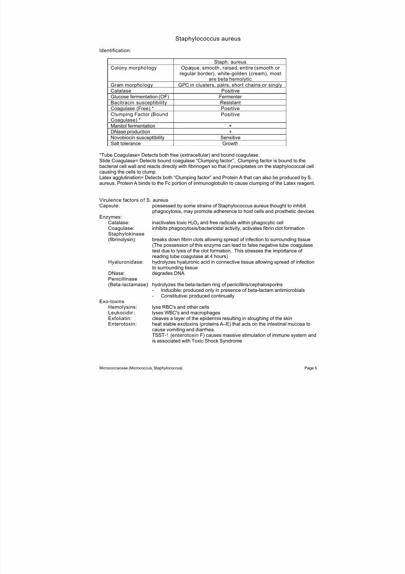
Staph. aureus
Colony morpho logy Opaque, smooth, raised, entire (smooth or
regular border), white-golden (cream), mostare beta hemolytic
Gram morpho logy GPC in clusters, pairs, short chains or singly
Catalase Positive
Glucose fermentation (OF) Fermenter
Bacitracin susceptibility Resistant
Coagulase (Free) * Positive
Clumping Factor (BoundCoagulase) *
Positive
Manitol fermentation +
DNase production +
Novobiocin susceptibility Sensitive
Salt tolerance Growth
*Tube Coagulase= Detects both free (extracellular) and bound coagulase.Slide Coagulase= Detects bound coagulase “Clumping factor”. Clumping factor is bound to thebacterial cell wall and reacts directly with fibrinogen so that if precipitates on the staphylococcal cellcausing the cells to clump.Latex agglutination= Detects both “Clumping factor” and Protein A that can also be produced by S.aureus. Protein A binds to the Fc portion of immunoglobulin to cause clumping of the Latex reagent.
Virulence factors of S. aureus Capsule: possessed by some strains of Staphylococcus aureus thought to inhibit
phagocytosis, may promote adherence to host cells and prosthetic devicesEnzymes:
Catalase: inactivates toxic H2O2 and free radicals within phagocytic cellCoagulase: inhibits phagocytosis/bactericidal activity, activates fibrin clot formationStaphylokinase (fibrinolysin): breaks down fibrin clots allowing spread of infection to surrounding tissue
(The possession of this enzyme can lead to false negative tube coagulasetest due to lysis of the clot formation. This stresses the importance of reading tube coagulase at 4 hours)
Hyaluronidase: hydrolyzes hyaluronic acid in connective tissue allowing spread of infectionto surrounding tissue
DNase: degrades DNAPenicillinase(Beta-lactamase): hydrolyzes the beta-lactam ring of penicillins/cephalosporins
- Inducible: produced only in presence of beta-lactam antimicrobials- Constitutive: produced continually
Exo-toxinsHemolysins: lyse RBC's and other cellsLeukocidin : lyses WBC's and macrophagesExfoliatin: cleaves a layer of the epidermis resulting in sloughing of the skinEnterotoxin: heat stable exotoxins (proteins A–E) that acts on the intestinal mucosa to
cause vomiting and diarrhea.TSST-1 (enterotoxin F) causes massive stimulation of immune system andis associated with Toxic Shock Syndrome
Micrococcaceae (Micrococcus, Staphylococcus) Page 5

7/27/2019 SLMicrococcaceaeHandout.pdf
http://slidepdf.com/reader/full/slmicrococcaceaehandoutpdf 4/9
Clinical SignificanceS. aureus is normal flora (colonizers) of the various skin and mucosal surfaces. The invasive natureof the organism allows for infection to occur in various sites.
Surface or Skin in fections:
• Folliculitis = infection of hair follicle
•
Boils (furuncles) = infection involving surrounding skin and subcutaneous tissueCharacterized by presence of pus
• Carbuncles = a mass of furuncles
• Impetigo = superficial skin infection seen primarily in children (differs from Impetigo causedby Streptococcus in that staphylococcal pustules are larger and are surrounded by a smallzone of erythema.
Toxin Mediated disease:
• Scalded Skin Syndrome (SSS): caused by exfoliatin toxin, usually seen in neonates andinfants, and produces a burnlike effect on the skin
• Toxic Shock Syndrome (TSS): caused by TSST-1 toxin Initially characterized by fever, rash, and signs of dehydration. In extreme cases thedisease is characterized by hypotension and shock, involvement of 3 or more organ
systems, desquamation(shedding of the epidermidis layer of the skin) of extremities within 2wks of onset and negative results on blood, throat and CSF cultures.
• Food poisoning: most commonly caused by enterotoxin A and B - Found in food that supports growth of Staphylococcus
(potato salad, processed meats, custards, bakery goods)- Cause vomiting and diarrhea 2-8 hrs. after ingestion- Lack fever and symptoms resolve within 24 hrs.
Other infections :
• Wound infections = usually due to injury of normal skin (trauma, burns, incisions)
• Pneumonia = usually seen in the immunocompromised (elderly and young)predisposing factors usually present: viral infection, underlying disease,presence of foreign bodies, antibiotic therapy
• Endocarditis/myocarditis
• Bacteremia/Septicemia
• Osteomyelitis = usually results from the spread of the organism via the bloodstream
• Septic arthriti s
• Pseudomembranous enterocolit is= also known as antibiotic-associated colitis and occurswhen the normal flora of the large bowel is altered. A severe acute inflammation of thebowel mucosa, with the formation of pseudomembranous plaques resulting in a waterydiarrhea, abdominal cramps and fever.
• Nosocomial infections
Antibiot ic Therapy:
• Since S. aureus can possesses penicillinase not all isolates can be treated by penicillin. S.aureus does not have a predictable pattern of sensitivity therefore susceptibility testing should
be done.• Routinely agents resistant to the enzyme penicillinase (methicillin, oxacillin, nafcillin) are used for
treatment however S. aureus can also alter its binding sites to develop resistance to thepenicillinase resistant antibiotics resulting in a Methicillin Resistant Staphylococcus aureus (MRSA). MRSA is a concern in Nosocomial infections (Hospital acquired infections).
• Vancomycin = drug of choice for MRSA
Micrococcaceae (Micrococcus, Staphylococcus) Page 6
7/27/2019 SLMicrococcaceaeHandout.pdf
http://slidepdf.com/reader/full/slmicrococcaceaehandoutpdf 5/9
Growth Characteristics of MRSA (page 81-82 Mahon)
• Sensitive and resistant strains can coexist within a culture
• Resistant strains may grow more slowly
• When performing antimicrobial testing optimal detection of MRSA is obtained by:- Using media with a neutral pH (7.0 –7.4)- Incubation at a cooler temperature (30-35 C)
- Adding 2-4% NaCl to the media- Incubating for a full 24 hrs.
Coagulase Negative Staphylococci
Clinical Significance: Coagulase Negative Staphylococci are increasingly associated withinfection due to the widespread use of prosthetic devices, intravascular catheters, prolonged surgicalprocedures, and the presence of underlying disease and the incidence of immunocompromised
hosts.
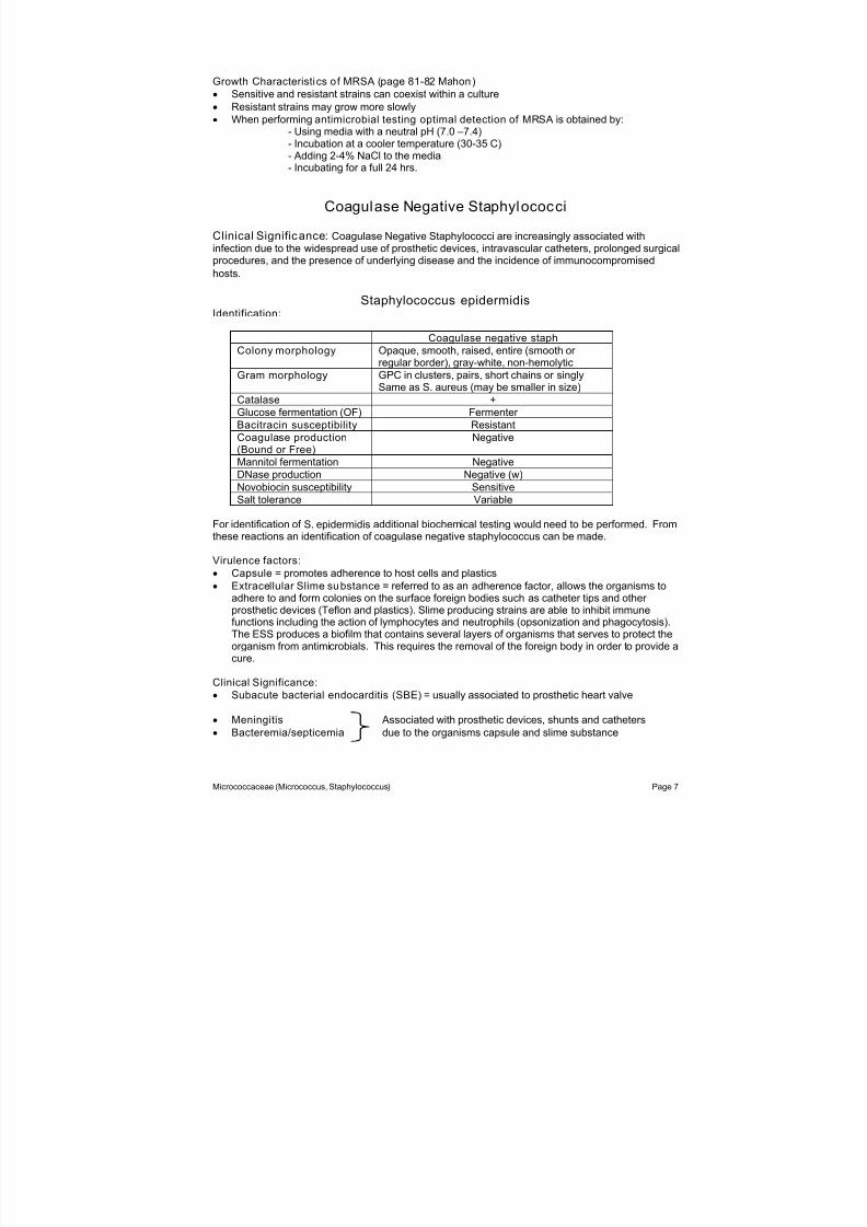
Staphylococcus epidermidisIdentification:
Coagulase negative staph
Colony morphology Opaque, smooth, raised, entire (smooth or regular border), gray-white, non-hemolytic
Gram morphology GPC in clusters, pairs, short chains or singlySame as S. aureus (may be smaller in size)
Catalase +
Glucose fermentation (OF) Fermenter
Bacitracin susceptibility Resistant
Coagulase production(Bound or Free)
Negative
Mannitol fermentation Negative
DNase production Negative (w)Novobiocin susceptibility Sensitive
Salt tolerance Variable
For identification of S. epidermidis additional biochemical testing would need to be performed. Fromthese reactions an identification of coagulase negative staphylococcus can be made.
Virulence factors:
• Capsule = promotes adherence to host cells and plastics
• Extracellular Slime substance = referred to as an adherence factor, allows the organisms toadhere to and form colonies on the surface foreign bodies such as catheter tips and other prosthetic devices (Teflon and plastics). Slime producing strains are able to inhibit immunefunctions including the action of lymphocytes and neutrophils (opsonization and phagocytosis).
The ESS produces a biofilm that contains several layers of organisms that serves to protect theorganism from antimicrobials. This requires the removal of the foreign body in order to provide acure.
Clinical Significance:
• Subacute bacterial endocarditis (SBE) = usually associated to prosthetic heart valve
• Meningitis Associated with prosthetic devices, shunts and catheters
• Bacteremia/septicemia due to the organisms capsule and slime substance
Micrococcaceae (Micrococcus, Staphylococcus) Page 7
7/27/2019 SLMicrococcaceaeHandout.pdf
http://slidepdf.com/reader/full/slmicrococcaceaehandoutpdf 6/9
• Wound infections Associated with immunocompromised
• Urinary tract infections patients (malignancies, burn, transplant, nosocomial)
• Post-surgical infections = acquired nosocomially from personnel or contaminated surgicaldevices
S. epidermidis poses a problem when interpreting positive blood cultures. The organism can benormal skin flora, if proper collection technique is not performed the blood cultures can becontaminated. Correlation with the number of blood cultures drawn, and infections in other sites canhelp in interpretation of culture results. If a blood culture is positive and catheter tip culture grow S.epidermidis as well then mostly like the organism is a pathogen rather than a drawing contaminant.This is also true of other organisms that can be interpreted as normal skin contaminants.
Antibiot ic Therapy
• Generally more resistant than Staphylococcus aureus
• Susceptibility testing is done if presumed to be the cause of infection because the organismdoes not have a predictable pattern of susceptibility.
• Drug of choice: MethicillinVancomycin for methicillin resistant strains
Staphylococcus saprophyticus
Identification:
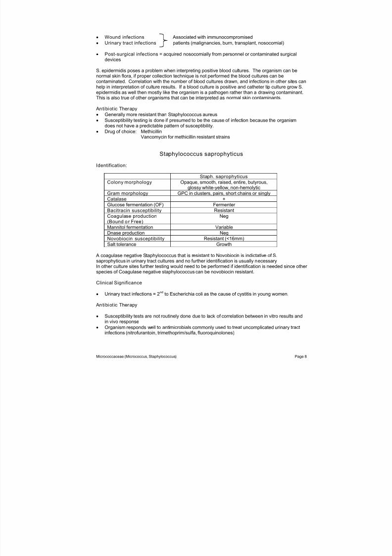
Staph. saprophyticus
Colony morphology Opaque, smooth, raised, entire, butyrous,glossy white-yellow, non-hemolytic
Gram morphology GPC in clusters, pairs, short chains or singly
Catalase +
Glucose fermentation (OF) Fermenter
Bacitracin susceptibility Resistant
Coagulase production(Bound or Free)
Neg
Mannitol fermentation Variable
Dnase production Neg
Novobiocin susceptibility Resistant (<16mm)
Salt tolerance Growth
A coagulase negative Staphylococcus that is resistant to Novobiocin is indictative of S. saprophyticus in urinary tract cultures and no further identification is usually necessaryIn other culture sites further testing would need to be performed if identification is needed since other species of Coagulase negative staphylococcus can be novobiocin resistant.
Clinical Significance
• Urinary tract infections = 2nd
to Escherichia coli as the cause of cystitis in young women.
Antibiot ic Therapy
• Susceptibility tests are not routinely done due to lack of correlation between in vitro results andin vivo response
• Organism responds well to antimicrobials commonly used to treat uncomplicated urinary tractinfections (nitrofurantoin, trimethoprim/sulfa, fluoroquinolones)
Micrococcaceae (Micrococcus, Staphylococcus) Page 8

7/27/2019 SLMicrococcaceaeHandout.pdf
http://slidepdf.com/reader/full/slmicrococcaceaehandoutpdf 7/9
***The following is for information only and will not be tested at anytime inyour theory exams. However it is useful in formation to have when you are inthe clinical setting and evaluating cultures results .***
Other Staphylococcus species
• There are other species of Staphylococcus that may be clinically significant.
• Infections with S. haemolyticus and S. lugdunesis usually involve the implantation of medicaldevices or similar infections caused by S. epidermidis.
• They have gram morphologies similar to Staphylococcus epidermidis.
• Colony morphologies vary among the other species (white to grey-white, cream, opaque,smooth, raised, entire, non-hemolytic to beta-hemolytic (usually weak)).
• Various biochemical tests will differentiate the species
• Commercial systems have varying degrees of accuracy in identification
• Susceptibility patterns vary – may show multiple resistance patterns.
• Some animal isolates (S. intermedius, S. hyicus, and S. delphini) may be tube coagulasepositive and should be considered in wounds involving animal bites.
• Coagulase negative species S. lugdunensis and S. schleiferi produce clumping factor and will be
positive with the slide coagulase test or latex agglutination tests. However these species will benegative by tube coagulase test.
**NOTE: Bacteriophage typing can be used as a means of further identification and classification of Staphylococcus species, especially Staphylococcus aureus. It is especially useful inepidemiological studies. It is performed in state and reference laboratories.
Stomatococcus mucilanginosus
General Information
• Normal oral flora, therefore commonly seen on respiratory cultures.
• Opportunistic pathogen in cases of endocarditis and septicemia in compromised patients and
drug abusers• Colony and gram morphology resembles Staphylococcus
• Shows strong adherence to the agar surface when you try to pick up the colony due to thepresence of a capsule (colony will stand up like egg whites if teased with a stick)
• Catalase: variable (when positive, the reaction is weak)
• Doesn’t grow on media with 5% NaCl
Micrococcaceae (Micrococcus, Staphylococcus) Page 9
7/27/2019 SLMicrococcaceaeHandout.pdf
http://slidepdf.com/reader/full/slmicrococcaceaehandoutpdf 8/9
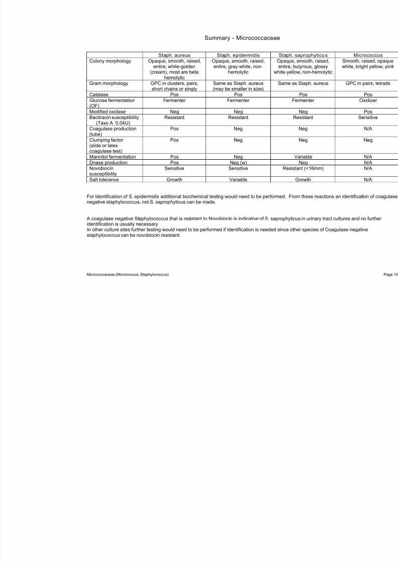
Summary - Micrococcaceae
Staph. aureus Staph. epidermidis Staph. saprophyticus Colony morphology Opaque, smooth, raised,
entire, white-golden
(cream), most are betahemolytic
Opaque, smooth, raised,entire, gray-white, non-
hemolytic
Opaque, smooth, raisedentire, butyrous, glossy
white-yellow, non-hemoly
Gram morphology GPC in clusters, pairs,short chains or singly
Same as Staph. aureus (may be smaller in size)
Same as Staph. aureus
Catalase Pos Pos Pos
Glucose fermentation(OF)
Fermenter Fermenter Fermenter
Modified oxidase Neg Neg Neg
Bacitracin susceptibility(Taxo A 0.04U)
Resistant Resistant Resistant
Coagulase production(tube)
Pos Neg Neg
Clumping factor
(slide or latexcoagulase test)
Pos Neg Neg
Mannitol fermentation Pos Neg Variable
Dnase production Pos Neg (w) Neg
Novobiocinsusceptibility
Sensitive Sensitive Resistant (<16mm)
Salt tolerance Growth Variable Growth
For identification of S. epidermidis additional biochemical testing would need to be performed. From these reactionegative staphylococcus, not S. saprophyticus can be made.
A coagulase negative Staphylococcus that is resistant to Novobiocin is indicative of S. saprophyticus in urinary traidentification is usually necessaryIn other culture sites further testing would need to be performed if identification is needed since other species of Cstaphylococcus can be novobiocin resistant.
Micrococcaceae (Micrococcus, Staphylococcus)

7/27/2019 SLMicrococcaceaeHandout.pdf
http://slidepdf.com/reader/full/slmicrococcaceaehandoutpdf 9/9
Summary - Micrococcaceae
Gram positive cocci
+ -
CatalaseMicrococcaceae Streptococcaceae
Modified oxidaseBacitracinOF Glucose
Positive NegativeSensitive ResistantOxidizer Fermenter
Micrococcus Coagulase+ Latex agglutination -
Staph. aureusR Novobiocin S
1. Urine Cultures: Staph. saprophyticus2. Other cultures: Coag neg Staph.
Coag neg Staph.
Micrococcaceae (Micrococcus, Staphylococcus) Page 11


