Sleep Hypoventilation in Neuromuscular and Chest Wall Disorders
15
Sleep Hypoventilation in Neuromuscular and Chest Wall Disorders Lisa F. Wolfe, MD a, *, Pallavi P. Patwari, MD b , Gökhan M. Mutlu, MD c INTRODUCTION Hypoventilation during sleep is common in the setting of neuromuscular and chest wall abnormal- ities. Often referred as restrictive thoracic disor- ders (RTDs), these conditions combine muscle weakness and/or nervous system failure with the possible addition of a stiff and misshapen chest wall. There are a large number of conditions that fall under RTDs, with unique features that impact control of breathing and strategies needed to opti- mally intervene and support the patient. The RTDs augment the reduced output from the respiratory center and central chemosensitivity and exag- gerate the increase in upper airway resistance that occurs during sleep. Identification of patients with RTDs at risk for sleep-disordered breathing (SDB) and sleep-related hypoventilation can be challenging. These patients need early screening for nocturnal hypoventilation, but the typical testing used for screening in the setting of SDB is rarely helpful. Interventions with noninvasive ventilation (NIV) are most common, but the role of polysomnography in the decision-making pro- cess for the initiation of therapy is less clear in the setting of RTDs as compared with other forms of SDB. Once initiated, ongoing management of these NIV devices is important and the new op- tions of electronic monitoring have provided an objective window to guide management. Last, there are non-NIV options that are alternatives to treat sleep-related hypoventilation in the setting of RTDs. In this overview, we review the effects of sleep on ventilation in neuromuscular and chest wall disorders. NEUROMUSCULAR DISORDERS There is a very large and diverse number of RTDs and although each disorder has unique features they loosely fall into 1 of 10 categories, based on the anatomy of the nervous system and the corre- sponding area of pathology. Areas are outlined in Fig. 1 and include motor neuron diseases a Pulmonary and Critical Care Medicine, Northwestern University Feinberg School of Medicine, 240 East Huron Street, McGaw M300, Chicago, IL 60611, USA; b Sleep Medicine Center, Department of Pediatrics, Ann & Robert H. Lurie Children’s Hospital of Chicago, 225 East Chicago Avenue, Box #43, Chicago, IL 60611-2605, USA; c Section of Pulmonary and Critical Care Medicine, University of Chicago, 5841 South Maryland Avenue, MC6026, Chicago, IL 60637, USA * Corresponding author. E-mail address: [email protected] KEYWORDS Sleep hypoventilation Neuromuscular disorders Chest wall disorders Ventilation KEY POINTS Hypoventilation due to neuromuscular and chest wall disorders includes a large number of condi- tions, and although these are frequently thought of as a homogeneous group, there are unique factors that separate these conditions. The diagnostic evaluations and therapeutic interventions for each condition are highly variable. Close consideration and individualization of therapy should be considered. Sleep Med Clin 9 (2014) 409–423 http://dx.doi.org/10.1016/j.jsmc.2014.05.010 1556-407X/14/$ – see front matter Ó 2014 Elsevier Inc. All rights reserved. sleep.theclinics.com
-
Upload
savvyas98-1 -
Category
Documents
-
view
235 -
download
0
description
sleep
Transcript of Sleep Hypoventilation in Neuromuscular and Chest Wall Disorders

Sleep Hypoventi lation inNeuromuscular and Chest
Wall Disorders Lisa F. Wolfe, MDa,*, Pallavi P. Patwari, MDb,Gökhan M. Mutlu, MDcKEYWORDS
� Sleep hypoventilation � Neuromuscular disorders � Chest wall disorders � Ventilation
KEY POINTS
� Hypoventilation due to neuromuscular and chest wall disorders includes a large number of condi-tions, and although these are frequently thought of as a homogeneous group, there are uniquefactors that separate these conditions.
� The diagnostic evaluations and therapeutic interventions for each condition are highly variable.
� Close consideration and individualization of therapy should be considered.
INTRODUCTION
Hypoventilation during sleep is common in thesetting of neuromuscular and chest wall abnormal-ities. Often referred as restrictive thoracic disor-ders (RTDs), these conditions combine muscleweakness and/or nervous system failure with thepossible addition of a stiff and misshapen chestwall. There are a large number of conditions thatfall under RTDs, with unique features that impactcontrol of breathing and strategies needed to opti-mally intervene and support the patient. The RTDsaugment the reduced output from the respiratorycenter and central chemosensitivity and exag-gerate the increase in upper airway resistancethat occurs during sleep. Identification of patientswith RTDs at risk for sleep-disordered breathing(SDB) and sleep-related hypoventilation can bechallenging. These patients need early screeningfor nocturnal hypoventilation, but the typicaltesting used for screening in the setting of SDBis rarely helpful. Interventions with noninvasive
a Pulmonary and Critical Care Medicine, Northwestern UnStreet, McGaw M300, Chicago, IL 60611, USA; b SleepRobert H. Lurie Children’s Hospital of Chicago, 225 EasUSA; c Section of Pulmonary and Critical Care Medicine,MC6026, Chicago, IL 60637, USA* Corresponding author.E-mail address: [email protected]
Sleep Med Clin 9 (2014) 409–423http://dx.doi.org/10.1016/j.jsmc.2014.05.0101556-407X/14/$ – see front matter � 2014 Elsevier Inc. Al
ventilation (NIV) are most common, but the roleof polysomnography in the decision-making pro-cess for the initiation of therapy is less clear inthe setting of RTDs as compared with other formsof SDB. Once initiated, ongoing management ofthese NIV devices is important and the new op-tions of electronic monitoring have provided anobjective window to guide management. Last,there are non-NIV options that are alternatives totreat sleep-related hypoventilation in the settingof RTDs. In this overview, we review the effectsof sleep on ventilation in neuromuscular and chestwall disorders.
NEUROMUSCULAR DISORDERS
There is a very large and diverse number of RTDsand although each disorder has unique featuresthey loosely fall into 1 of 10 categories, based onthe anatomy of the nervous system and the corre-sponding area of pathology. Areas are outlined inFig. 1 and include motor neuron diseases
iversity Feinberg School of Medicine, 240 East HuronMedicine Center, Department of Pediatrics, Ann &t Chicago Avenue, Box #43, Chicago, IL 60611-2605,University of Chicago, 5841 South Maryland Avenue,
l rights reserved. sleep.theclinics.com

Fig. 1. Schematic of the anatomy of the nervous sys-tem and the corresponding area of pathology. PointA is the body of the motor nerve cell. B is the uppermotor neuron connecting the motor cortex to thespinal cord cell bodies at point C. At point D, the pe-ripheral lower motor neuron continues, dividing todelivering signal to neuromuscular junctions of indi-vidual skeletal muscle fibers at point E.
Box 1Criteria to initiate noninvasive ventilation(1 criterion is sufficient)
1. Forced vital capacity less than 50%(predicted).
2. Maximal inspiratory pressure greater than�60 cm H2O.
3. PaCO2 �45 mm Hg
4. Sleep oximetry demonstrates oxygen satura-tion �88% for at least 5 minutes
Wolfe et al410
(MNDs), peripheral neuron disease, neuromus-cular junction disease, inflammatory myopathies,muscular dystrophies, metabolic diseases of mus-cle, infectious myopathy, spinal cord injury (SCI),diaphragm injury, and scoliosis (Box 1). This sec-tion reviews of the most common conditions ineach of these categories highlighting the importantfeatures related to the aspects of sleep and relatedhypoventilation.
Motor Neuron Disease
The most common MNDs are amyotrophic lateralsclerosis (ALS) in adults and spinal muscular atro-phy (SMA) in children.
Amyotrophic lateral sclerosisALS occurs in approximately 2 of 100,000 adultsper year, usually starting in the fifth to sixth de-cades of life. Patients present with a combinationof upper and lower motor neuron symptoms.Most patients present with limb weakness, butapproximately 20% present with bulbar diseasein the form of difficulties with speech anddysphagia. Approximately 15% of patients haveadditional frontotemporal dementia. Approxi-mately 10% to 20% of cases have been associ-ated with genetic causes, and more than 20genes have been described.1 The average patientwith ALS has a life expectancy of 3 to 5 years af-ter diagnosis and the most common cause ofdeath is respiratory compromise. Initially, respira-tory failure presents with nocturnal hypoventila-tion, but daytime evaluations to predict thedevelopment of this impairment have been achallenge.2 Screening questionnaires have beendeveloped, and the most reliable finding is acomplaint of orthopnea.3 Questionnaires havenot been as reliable as lung function testing forpredicting the development of nocturnal or day-time hypoventilation. Forced vital capacity (FVC)is the most commonly used test, but assessmentin the supine position, with slow rather than fastmeasurements has been shown to improve thetest’s ability to predict nocturnal hypoventila-tion.4,5 Additional lung function assessments,such as maximal inspiratory pressure (MIP)assessment or sniff nasal inspiratory pressure(SNIP) testing, can add additional early insightfor predicting the development of nocturnal hy-poventilation.6 Ultimately, using multiple testingtechniques has been most effective. Initiation oftherapy for nocturnal hypoventilation when anylung function test is abnormal, such as uprightor supine FVC/MIP, is associated with animprovement in overall outcome of those withALS.7 Given this finding, information that will beobtained from a diagnostic polysomnography tocharacterize hypoventilation has had unclear
Sleep Hypoventilation 411
value. The benefit of polysomnography to initiateand optimize therapy never has been demon-strated. Because current protocols do notadequately guide goals of therapy, ongoing dys-synchrony does not resolve with polysomno-graphic guidance.8 Early initiation of NIV therapyis still important, as it has been shown to extendlife and improve quality of life.9
Spinal muscular atrophySMA is a neuromuscular disease characterized byprogressive muscular weakness due to degenera-tion of motor neurons, and is caused by mutationsin the survival motor neuron (SMN). Inherited in anautosomal recessive pattern, most affected pa-tients carry homozygous SMN1 deletions or loss-of-function mutations and carry varying copynumbers of the alternatively spliced SMN2 gene.The low levels of SMN protein produced fromspliced SMN2 result in the characteristic motorneuron degeneration and progressive skeletalmuscle atrophy.10
The SMA phenotype is variable and classifiedbased on age of presentation and severity of weak-ness as type 1 (severe), 2 (intermediate), 3 (mild), or4 (adult). SMA type 1 presents in infancy before6 months of age and is characterized by inabilityto sit without support and early demise (<2 yearsof age). Children with SMA type 2 present withweakness at a slightly later age (6–18 months),have the ability to sit independently but not stand,and have normal neurocognitive ability. The thirdchildhood type, which presents after 18 months ofage, isSMA type3. Thesechildrencansit andstandindependently, but have difficulty with walking dueto proximal limb weakness that has greater effecton the legs than arms. SMA type 4 describes indi-viduals with onset of weakness in early adulthoodwith an anticipated normal life span.
Multidisciplinary care is needed for the affectedchildren and should involve attention to optimiza-tion of nutrition, management of orthopedic prob-lems such as scoliosis, and screening for SDBand hypoventilation. Morbidity and mortality inchildhood SMA is often due to pulmonary compli-cations. Therefore, concerted attention to evalua-tion of the respiratory phenotype is essential.
The respiratory phenotype in SMA is character-ized by diaphragmatic breathing with weakintercostal and bulbar muscles that results in a“bell-shaped” chest wall deformity and difficultywith cough and airway clearance. Respiratory-specific evaluation and monitoring should becompleted every 3 to 6 months and should includephysical examination, pulmonary function testing(peak cough flow, maximal inspiratory pressure,maximal expiratory pressure), chest radiograph,
pulse oximetry, carbon dioxide monitoring, andfull attended polysomnography with carbon diox-ide monitoring.11
Although an accurate assessment of lung func-tion can be difficult in pediatric patients who aretoo young or too weak to complete standardtesting, the physical examination should includedemonstration of the effectiveness of cough andchecking for subtle signs of increased work ofbreathing and risk for hypoventilation, such as in-crease in baseline respiratory rate (RR) and thora-coabdominal paradoxic breathing pattern. Forthose with SMA who are able to walk (indicatinggreater baseline strength), complete pulmonaryfunction testing is indicated. Gas exchange abnor-malities can be identified with a low oxygen satu-ration and an increase in end-tidal carbondioxide or transcutaneous carbon dioxide moni-toring, but also can be subtle. While continuousovernight pulse oximetry is not routinely recom-mended as an alternative to full in laboratory at-tended polysomnography, it is useful for periodic“spot checks” that can guide day-to-day manage-ment. Carbon dioxide monitoring should be limitedto end-tidal or transcutaneous monitoring, asserum bicarbonate values can be falsely reassur-ing. Hyperventilation in response to having a blooddraw will artificially drop resting carbon dioxide ifmeasured by arterial blood gas (ABG). Develop-ment of hypoventilation and SDB can be subtleand must be actively sought through nocturnalpolysomnography before progression to daytimerespiratory failure.
Mainstay of chronic and acute respiratory careinvolves maneuvers to optimize airway clearanceand provide positive airway pressure support viamask or tracheostomy. Nocturnal NIV shouldbe initiated when there are signs of gas exchangeabnormalities or SDB develops. The goals of NIVare to improve gas exchange, decrease workof breathing, and improve sleep quality and day-time functioning. NIV also can be prescribed dur-ing an acute respiratory decompensation as mayoccur with an acute infection. Polysomnographywith carbon dioxide monitoring can be helpfulfor titration of respiratory support. Discussionsregarding patient and family choices related totracheostomy and palliative care should be initi-ated before the development of acute events(Fig. 2).12
Peripheral Nerve Disease
Both acute and chronic inflammatory demyelin-ating polyneuropathy (AIDP and CIDP) affect res-piratory function and lead to sleep-related orchronic hypoventilation.

Cannot stand Stands and walks
Every 3-6 months Every 6 – 12 months Yearly
Indirect measures of respiratory muscle functionPE: RR, WOB, breathing pattern, chest wall shape
Direct measures of respiratory muscle function PFTs (FVC and lung volumes)
Indirect measures of hypoventilation-Pulse oximetry and end-tidal or transcutaneous CO2
-Overnight home pulse oximetry Objective measures of developing respiratory pathology-Chest x-ray (aspiration, atelectasis, pneumonia, scoliosis)-Polysomnography (with CO2 measures)
Cannot sit
SMA Type 1(severe)
SMA Type 2(intermediate)
SMA Type 3(mild)
SMA Type 4(adult)
SMAType
Ability
Monitoringfrequency
Intervention
MonitoringMeasures
Palliative Care
Daytime and nocturnal non-invasive ventilation
Nocturnal NIV
If sleep disordered breathing present, then nocturnal NIV
SMAType
&
SMA Type 1(severe)
SMA Type 2(intermediate)
SMA Type 3(mild)
SMA Type 4(adult)
Fig. 2. Assessment of respiratory function and treatment of hypoventilation based on SMA type. SMA subtypesare defined by physical ability. These differences drive the frequency of respiratory monitoring. Once hypoventi-lation has been defined, therapy options should be considered. PE, physical exam; PFT, pulmonary function test;RR, respiratory rate; WOB, work of breathing.
Wolfe et al412
Guillain–Barre syndromeGuillain–Barre Syndrome (GBS) is frequentlyreferred to as AIDP and is defined as an acutepolyradiculoneuropathy with flaccid ascendingparesis. The annual incidence of GBS is estimatedto be between 0.34 and 1.34 per 100,000 persons.There aremany subtypes that may include sensoryor autonomic involvement. In 10% to 15% of pa-tients, respiratory involvement occurs, leading toventilatory failure.13 Severe involvement of the up-per extremities and tongue are the factors that aremost predictive of respiratory insufficiency.14
On physical examination, signs of impending res-piratory failure include a weak nasal voice with aninability to count from 1 to 20 in one breath. Abnor-malities seen in pulmonary function testing includea vital capacity (VC) less than 20 mL/kg, and/or areduction in VC of more than 30% from baseline,an MIP less than 30 cm H2O or a maximum expira-tory pressure (MEP) of less than 40 cm H2O.15
Given the bulbar findings inmany of these patients,and the rapid progression of disease, the initialuse of NIV is not recommended.16 Many of thesepatients may need prolonged mechanical ventila-tion. A calculation of the pulmonary function score(PF 5 VC 1 MIP 1 MEP) on days 1 and 12 of
intubation helps to predict the need for prolongedventilatory support. If the PaO2/FiO2 ratio of day1:12 is less than 1, at least another 3 weeks of me-chanical ventilation would be expected.17 NIV canbe considered to speed the transition off mechan-ical ventilation. A return of bulbar function andgood level of alertness suggests that a trial ofNIV should be considered.18 The recovery of pa-tients with GBS may take weeks to months. At6 months, some patients will still have persistentweakness and may never completely recover theirmuscle function. In some of these patients, respi-ratory muscle weakness may require ongoing NIVnocturnally for sleep-related hypoventilation.Many patients whose disease course shows nosignificant recovery will continue to have a deficitin muscle function even after 6 months.
Chronic inflammatory demylinatingpolyneuropathologyIn contrast to the natural course of disease inGBS, which is characterized by a relatively quickonset and a slow recovery period, CIDP includesa group of acquired polyneuropathies that arecharacterized clinically by a progressive sym-metric weakness that evolves over a period of

Sleep Hypoventilation 413
at least 2 months. Weakness typically involvesproximal and distal limb muscles, whereas ocularor bulbar involvement is uncommon. This slowprogression is in stark contrast to AIDP; howev-er, the conditions are often grouped together,as many of the symptoms are similar. CIDP ismuch less common then AIDP. The incidence isestimated to be between 1.5 and 3.6 cases in amillion people. Although most patients willrespond well to therapy, approximately a quarterof adults will not respond to currently availabletreatments.19
Ventilatory involvement is rare in CIDP, with areported prevalence varying from 1.7% to 9.0%,but prospective studies have shown abnormalphrenic nerve conductions in up to 80% to 92%of patients. Although chronic involvement of res-piratory muscles leading to ventilatory failure isthought to be a rare complication of CIDP, pa-tients may require ventilatory support duringacute exacerbations of disease. Although mostpatients recover, long-term use of nocturnal venti-latory support has been reported in some pa-tients. NIV is a more realistic initial ventilatorysupport option, as bulbar involvement is muchless common in patients with CIDP as comparedwith AIDP.20,21
Collectively, there is need for nocturnal NIV in agroup of patients with AIDP and CIDP. The long-term phase of disease should be the focus, butgiven the lack of data, there are no clear guide-lines. A close look at the reported data can helpto construct a reasonable plan for an individual pa-tient (Fig. 3). The use of pulmonary function testingand/or polysomnography should be used to directthe NIV support in these patients.
AIDP Days - Weeks
Mechanical ventilation– Vital Capacity:
<20 mL/kg>30% reduction
– MIP < 30 cm H2O– MEP < 40 cm H2O– Oropharyngeal weakness
Calcu
CIDP Months
Reduction in lung function
Fig. 3. Approach to the management of hypoventilatiodifferent presentations, patients with both conditions ma
Neuromuscular Junction Disease
Myasthenia gravis (MG) is a neuromuscular condi-tion associated with autoantibody production,which impairs the neuromuscular junction. It affectsboth children and adults. The prevalence of MG isestimated to be between 25 and 142 (averagew30) in 1 million per year. The most common anti-bodies are nicotinic skeletal muscle acetylcholinereceptor and muscle-specific receptor tyrosinekinase (MuSK). The most common presentingsymptoms are weakness in the ocular and bulbarmuscles. Patients complain of dysphagia,dysphonia, dysarthria, and chewing/swallowing dif-ficulty, as well as generalized weakness.22 Respira-tory failure can be associated with all forms of thedisease; however, those with the MuSK antibodyare at higher risk.23 The drive to breathe remainsintact and central nervous system–related controlof breathing appears to be unaffected; however,muscle weakness and easy fatigability are associ-ated with a rapid shallow breathing pattern. AsPaCO2 rises during hypoventilation, the muscle be-comes taxed further, even less able to meet theventilatory needs.24,25 Initial investigations usedVC as a single test to screen for hypoventilation,but this was found to be not helpful, as bulbardysfunction may limit the patient’s ability to accu-rately perform the test.26 The use of multiple testsis more useful and accurate in screening for hypo-ventilation. When VC is greater than 20 mL/kg, amaximal expiratory pressure is greater than40 cm H2O, or a maximal inspiratory pressure ismore negative than �40 cm H2O, patients are un-likely to require ventilatory support. A decline of30% or greater in maximal inspiratory pressure is
PF score= VC+MIP+MEP
PF ratio=
late PF score ratio
Continuemechanical ventilation
Return of bulbar function
PF score day 1PF score day 12
<1 >1
Consider nocturnal NIVPSG guidance may be helpful
n in AIDP and CIDP. Although AIDP and CIDP havey eventually benefit from NIV. PSG, polysomnography.

Wolfe et al414
associated with a higher risk of requiring ventilatorysupport.27 In those with hypoventilation, the pres-ence of bulbar disease may limit the ability to useNIV, as it raises aspiration risk and reduces the abil-ity of the patient to tolerate a mask. NIV has beenshown to be tolerated as an effective therapy foracute exacerbation, post-thymectomy exacerba-tion, and extubation failure.28–30 In the setting ofchronic stable MG, sleep is disrupted and patientscomplain of chronic fatigue and sleepiness, but noexcess hypoventilation or central or obstructivesleep apnea (OSA) is usually seen.31,32
Muscular Dystrophies
There are a large number of conditions that areassociated with the clinical findings of progressivelimb and core body muscle weakness. These dis-orders are unified by the finding of dystrophic fea-tures on muscle biopsy. They have a variableimpact on cardiac and respiratory muscle functionand may involve organs other than the musculo-skeletal system. The conditions are heritable andthe genetics have become better understoodover the past 2 decades. The most common child-hood and adult conditions in this group areDuchenne muscular dystrophy (DMD) and myoto-nic, respectively.33
DMD is an X-linked disease that affects 1 in3600 to 6000 live male births, and occurs as aresult of mutations in the dystrophin gene (DMD;locus Xp21.2). Mutations lead to an absence ofor defect in the protein dystrophin. Affected indi-viduals will display symptoms of proximal muscleweakness by age 5. A milder variant, Beckermuscular dystrophy, may present at an olderage, usually in the late teens. The disorder isassociated with heart failure and kyphoscoliosis,but the predominant driver of early mortality isrespiratory failure.34 Respiratory weakness beginsat early school age and by the second to third de-cades this can progress to full respiratory fail-ure.35 Many factors contribute to this pathology.As a skeletal muscle, eventually the diaphragmfails despite intact central drive and neuraloutput.36 Other factors, such as heart failure andscoliosis, progress at the same time. Ultimately,these all contribute to the development ofnocturnal hypoventilation. Lung function testinghas been shown to have variable prognostic valuefor predicting sleep-related hypoventilation.Some data suggest that a resting RR of morethan 23 breaths per minute and resting rapidshallow breathing index (RSBI) (the ratio of RRto tidal volume (Vt) [RBSI in breaths/min/mL)greater than 0.07 accurately predict the develop-ment of nocturnal hypoventilation, but these tests
are rarely performed outside research settings.The most common clinically used value is theVC. Some studies indicate that a VC less than1.82 L is predictive of nocturnal hypoventilation,whereas other studies show no relationshipbetween VC and nocturnal hypercapnia.37,38
Because of this variability, consensus statementssuggest that full polysomnography should be per-formed for boys with DMD if they are consideringsurgical repair of scoliosis or yearly after theyhave disease progression requiring the use of awheelchair.39 Therapy with nocturnal NIV is bene-ficial, reducing work of breathing, and improvingdyspnea, and quality and length of life.40 Theimpact of early and aggressive therapy with NIVfor those with DMD has the greatest impactwhen coadministered with broader therapeuticapproaches, such as an aggressive support ofheart failure. The addition of airway-clearancetechniques with lung volume recruitment, aggres-sive therapy for heart failure, appropriate surgicalintervention for scoliosis, and use of steroid ther-apy after early diagnosis have combined to signif-icantly improve outcomes over the years,delaying the onset of respiratory failure and all-cause mortality by 10 to 20 years.41
Infectious Motor Neuropathy
Paralytic poliomyelitis is caused by a small RNAvirus with a complete genome of only approxi-mately 7500 nucleotides. It was known to occursporadically from 1600 to 1300 BC. Epidemicpoliomyelitis was a more modern disease relatedto improvement of sanitary conditions of thewestern world. Given the advancements in vacci-nation, acute paralytic polio (APP) was eradi-cated from the United States in 1979.42 Buteradication of APP did not eliminate the ongoingmedical needs of APP survivors. It is estimatedthat there are 1.63 million people in the UnitedStates and 12 to 20 million people worldwidesuffering from disabilities related to poliomyelitis.Taken together, these symptoms are referred toas the post-polio syndrome (PPS), which isdefined as the onset of new and progressive fa-tigue, pain, muscle weakness, and atrophy thatstarts decades after recovery from paralyticpolio.43 Many of the patients with PPS haveventilatory failure. Some, but not all, of these pa-tients required ventilatory support during theircourse with APP.Although the past need for ventilatory support is
a risk factor, it does not predict the development ofchronic hypoventilation as well as pulmonary func-tion tests do. Those with FVC and MIP less than50% of predicted have been shown to have

Sleep Hypoventilation 415
inadequate ventilation.44 Many factors contributeto the development of respiratory muscle weak-ness, such a motor neuron stress, cranial nervedysfunction, decreased central respiratory drive,and scoliosis.45 In these patients, the use ofnocturnal NIV has been shown to improve daytimeventilation and quality of life.46,47
During full polysomnography, hypoventilation isoften noted in those patients with FVC valuesthat are less than 50% of predicted. Patientswith PPS and better FVC values may have SDBin the form of OSA alone or in combination with hy-poventilation. Those with OSA alone have normalFVC values and wakefulness PaCO2 levels.48 Inaddition, there are no daytime symptoms or signsthat would suggest the presence of sleep-relatedhypoventilation.48 Among the manifestations ofSDB and hypoventilation in PPS, the most preva-lent symptom is fatigue, which is consideredthe hallmark of the disorder. SDB explains onlyapproximately 65% of the cases of extreme fa-tigue in PPS.49 Other causes of fatigue, such asdepression, menopause, and physical weakness,have been suggested as alternative explana-tions.50 Insomnia and unrefreshing sleep are com-mon complaints associated with daytime fatigue,and excessive limb movements are frequentlydocumented in polysomnography.51 Persistent,severe fatigue has occasionally been treated withstimulants, but the response has been variable.52
This suggests that aggressive and complete eval-uation is needed to appropriately address com-plaints of fatigue in PPS (Fig. 4).
PPS with com
Initiate NIV. Consider further evaluation with: Daytime ABG, and/or PSG for titration.
FVC or MIP < 50%
Lung Func
HypH
Fig. 4. Evaluation of complaint of fatigue in PPS. Fatiguenot useful. Evaluations must begin with assessment of lunRLS, restless leg syndrome.
Spinal Cord Injury
It is estimated that there were approximately273,000 people with SCI in the United States in2013. There are 12,000 new cases of SCI in theUnited States every year. Approximately 40% ofthese patients have high-level injury and quadripa-resis. Respiratory impairment in this group is asso-ciated with a high cost of care and risk of mortalitydue to pneumonia and sepsis.53 This is true forthose with either acute SCIs or long-standing dis-ease. Associated hypoventilation drives this pro-cess with the development of atelectasis andsecondary pneumonia.54
Sleep itself is disrupted in those with chronic SCIand this appears to be due to many factors, suchas aging, weight gain, smoking, alcohol misuse,and chronic conditions (chronic obstructive pul-monary disease, asthma), but there are uniquefactors as well.55 In those with high SCI, thesuperior cervical ganglion, which originates fromC8–T2, is impaired and often disrupted to the pointthat autonomic dysfunction becomes prominent.This ganglion plays a role in sleep regulation, asit is a stop on the pathway to melatonin produc-tion, passing the light signal from the suprachias-matic nucleus to the pineal gland (Fig. 5). Theimpairment in melatonin production has beendescribed as one of the factors contributing tosleep disruption in SCI.56
Sleep also is impaired by disordered breathing.There are many factors that drive this abnormalcontrol of breathing, such as medications and
plaint of fatigue
Further evaluation with: Diagnostic PSG.
CPAP Therapy
RLS Therapy
Consider Stimulants
tion Testing
FVC and MIP > 50%
oventilationypoxemia
Normal RLS OSA
is prominent in PPS and history based evaluations areg function. CPAP, continuous positive airway pressure;

AD
BC
F
E
Fig. 5. Regulation of sleep by melatonin. Blue wave length light from point A, is transmitted to the retina atpoint B. The signal is then transmitted through the retinohypothalamic tract at C, to the hypothalamic, suprachi-asmatic nucleus at point D. The signal is then transmitted through the periventricular nucleus to the superior cer-vical ganglion at point E. Last, postsynaptic sympathetic neurons innervate the pineal gland, in the epithalamusat point F, the site of melatonin production and regulation.
Wolfe et al416
spasticity, but primary abnormalities of control ofbreathing include obstructive and central apnea,as well as hypoventilation. In high SCI, the useof autotitrating positive airway pressure devicesis controversial because of the presence of cen-tral respiratory events in some patients.57,58 Inaddition, medications, such as narcotics and bac-lofen, that are commonly used in the setting ofSCI, are associated with the development of cen-tral apnea.59 Other studies suggested that thefindings of discrete events, such as apnea/hypo-pnea, are not as important as the larger issue of
Step 1.1. PaCO2 > 45 mm Hg2. Overnight pulse oximetry with SpO2 <88%3. FVC < 50% with impaired Peak Cough F
Step 2.No further diagnostic testing needed.
Step 3Consi
Step 3.Consider starting NIV support.
Yes
CSA on PSG
Fig. 6. Protocol for developing a patient-focused nocturnshould have screening for respiratory impairment (Step 1)strength assessment, carbon dioxide screening, and over(Step 2). This testing should be integrated to develop acontinuous positive airway pressure; CSA, central sleep ap
chronic hypoventilation with the accompanyingrisk of hypercapnia, higher 24-hour work ofbreathing, atelectasis, and potential higher riskof chest infection.60 This remains an unresolvedquandary, and intricate investigation is neededbefore a management scheme is developed. Asuggested approach is reviewed in Fig. 6, andemphasizes that given the lack of agreement, allmanagement plans should be individualized, andcareful assessment of lung function assessment,including cough strength, carbon dioxide level,and nocturnal oximetry, should be considered.
for >5 minlow (PCF <270 L/min)
Step 2. Consider full PSG with CO2 monitoring.
Step 3. Thoughtful clinical follow up
. der Auto/CPAP therapy
No
OSA on PSG Normal PSG
al ventilation plan in SCI. All patients with high SCIthat could include pulmonary function testing, coughnight oximetry. Formal sleep testing may be helpfulnocturnal ventilation support strategy (Step 3). CPAP,nea; PSG, polysomnography.
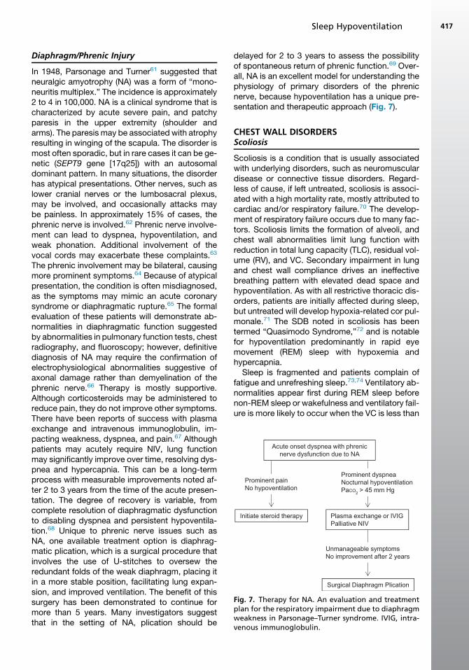
Acute onset dyspnea with phrenic nerve dysfunction due to NA
Plasma exchange or IVIGPalliative NIV
Initiate steroid therapy
Surgical Diaphragm Plication
Prominent painNo hypoventilation
Prominent dyspneaNocturnal hypoventilationPaCO2 > 45 mm Hg
Unmanageable symptomsNo improvement after 2 years
Fig. 7. Therapy for NA. An evaluation and treatmentplan for the respiratory impairment due to diaphragmweakness in Parsonage–Turner syndrome. IVIG, intra-venous immunoglobulin.
Sleep Hypoventilation 417
Diaphragm/Phrenic Injury
In 1948, Parsonage and Turner61 suggested thatneuralgic amyotrophy (NA) was a form of “mono-neuritis multiplex.” The incidence is approximately2 to 4 in 100,000. NA is a clinical syndrome that ischaracterized by acute severe pain, and patchyparesis in the upper extremity (shoulder andarms). The paresis may be associated with atrophyresulting in winging of the scapula. The disorder ismost often sporadic, but in rare cases it can be ge-netic (SEPT9 gene [17q25]) with an autosomaldominant pattern. In many situations, the disorderhas atypical presentations. Other nerves, such aslower cranial nerves or the lumbosacral plexus,may be involved, and occasionally attacks maybe painless. In approximately 15% of cases, thephrenic nerve is involved.62 Phrenic nerve involve-ment can lead to dyspnea, hypoventilation, andweak phonation. Additional involvement of thevocal cords may exacerbate these complaints.63
The phrenic involvement may be bilateral, causingmore prominent symptoms.64 Because of atypicalpresentation, the condition is often misdiagnosed,as the symptoms may mimic an acute coronarysyndrome or diaphragmatic rupture.65 The formalevaluation of these patients will demonstrate ab-normalities in diaphragmatic function suggestedby abnormalities in pulmonary function tests, chestradiography, and fluoroscopy; however, definitivediagnosis of NA may require the confirmation ofelectrophysiological abnormalities suggestive ofaxonal damage rather than demyelination of thephrenic nerve.66 Therapy is mostly supportive.Although corticosteroids may be administered toreduce pain, they do not improve other symptoms.There have been reports of success with plasmaexchange and intravenous immunoglobulin, im-pacting weakness, dyspnea, and pain.67 Althoughpatients may acutely require NIV, lung functionmay significantly improve over time, resolving dys-pnea and hypercapnia. This can be a long-termprocess with measurable improvements noted af-ter 2 to 3 years from the time of the acute presen-tation. The degree of recovery is variable, fromcomplete resolution of diaphragmatic dysfunctionto disabling dyspnea and persistent hypoventila-tion.68 Unique to phrenic nerve issues such asNA, one available treatment option is diaphrag-matic plication, which is a surgical procedure thatinvolves the use of U-stitches to oversew theredundant folds of the weak diaphragm, placing itin a more stable position, facilitating lung expan-sion, and improved ventilation. The benefit of thissurgery has been demonstrated to continue formore than 5 years. Many investigators suggestthat in the setting of NA, plication should be
delayed for 2 to 3 years to assess the possibilityof spontaneous return of phrenic function.69 Over-all, NA is an excellent model for understanding thephysiology of primary disorders of the phrenicnerve, because hypoventilation has a unique pre-sentation and therapeutic approach (Fig. 7).
CHEST WALL DISORDERSScoliosis
Scoliosis is a condition that is usually associatedwith underlying disorders, such as neuromusculardisease or connective tissue disorders. Regard-less of cause, if left untreated, scoliosis is associ-ated with a high mortality rate, mostly attributed tocardiac and/or respiratory failure.70 The develop-ment of respiratory failure occurs due to many fac-tors. Scoliosis limits the formation of alveoli, andchest wall abnormalities limit lung function withreduction in total lung capacity (TLC), residual vol-ume (RV), and VC. Secondary impairment in lungand chest wall compliance drives an ineffectivebreathing pattern with elevated dead space andhypoventilation. As with all restrictive thoracic dis-orders, patients are initially affected during sleep,but untreated will develop hypoxia-related cor pul-monale.71 The SDB noted in scoliosis has beentermed “Quasimodo Syndrome,”72 and is notablefor hypoventilation predominantly in rapid eyemovement (REM) sleep with hypoxemia andhypercapnia.
Sleep is fragmented and patients complain offatigue and unrefreshing sleep.73,74 Ventilatory ab-normalities appear first during REM sleep beforenon-REM sleep or wakefulness and ventilatory fail-ure is more likely to occur when the VC is less than
Wolfe et al418
1.0 to 1.5 L or the MIP is less than �20 to 25 cmH2O.72 Noninvasive ventilation has been shownto normalize oxygen saturation and carbon dioxidelevels both during sleep and during the daytime.Patients using NIV to resolve nocturnal hypoventi-lation have improved quality of life and exercisetolerance. They also have a reduction in cardiacdisease, with both an improvement in right ventric-ular function and autonomic control.75,76 Surgicalcorrection of the scoliosis can help to reduce thelong-term complications listed previously; how-ever, the surgery itself carries risks. Up to 80%may report some type of complication and mortal-ity has been reported to be as high as 7%. Patientsare at risk for prolonged postoperative hospitaliza-tion due to respiratory failure, infections, gastroin-testinal complications, cardiac failure, woundproblems, pancreatitis, superior mesenteric arterysyndrome, and excessive blood loss.77 The risk ofpostoperative respiratory failure is significantbecause the average patient will experience a12% drop in FVC postoperatively.78 Those withunderlying neuromuscular disease are at highrisk, and complications can be estimated with pre-operative testing. If the patient is older than16 years and has an FVC of less than 40% pre-dicted and the Cobb angle is greater than 69�,then the management team will face the greatestchallenge.79 Early initiation of nocturnal NIV beforesurgery will best help to mitigate the postoperativerisk.80
POLYSOMNOGRAPHY
The role of polysomnography in the evaluation andtreatment of those with RTD is unclear and contro-versial. It has been suggested that there is norole for polysomnography, and the care of thosewith neuromuscular disorders can appropriatelyaddress hypoventilation without the use of formalpolysomnography.81 In the United States, a na-tional coverage determination by the Centers forMedicare and Medicaid Services has establisheda standard that those with RTDs qualify for theuse of NIV as therapy for hypoventilation merelyby the demonstration of impaired respiratory func-tion. Patients need to demonstrate only one ofmany criteria for the initiation of NIV therapy onan empiric basis (see Box 1).82 Although this stan-dard has been in place in the United States since1999, many communities continue to performroutine polysomnography. The best clinical prac-tice document published by the American Acad-emy of Sleep Medicine in 2010 recommendedthat standard overnight monitoring be expandedto include diaphragm electromyography to assessrespiratory effort. The addition of carbon dioxide
measurements either by transcutaneous or end-tidal techniques is also recommended.83 The new-est scoring guidelines provide an option for usingcarbon dioxide data to score “hypoventilationevents” although it is unclear how these eventswould impact diagnosis or management of thosewith RTDs. The hypoventilation is defined as an in-crease in the PCO2 to a value greater than 55 mmHg for equal or longer than 10 minutes, or thereis a 10 mm Hg or greater increase in PCO2 duringsleep (in comparison with an awake supine value)to a value exceeding 50 mm Hg for at least 10 mi-nutes.84 When titration of NIV is performed, thegoals are unique and are different from those oftypical titrations done for the treatment of OSA.The study is successful when the RR is not toolow, with a backup rate sufficient to eliminate cen-tral apnea events, a Vt that is adequate so that ox-ygen saturation is within in the normal range, andany tachypnea is resolved. The ultimate goal is toprovide sufficient respiratory support that both ox-ygen saturation and carbon dioxide levels arewithin normal range. If titration is successful, theuse of supplemental oxygen should not be neces-sary.83 In fact, supplemental oxygen administra-tion should be considered as a last resort. In theend, it may be that the biggest factor limiting qual-ity care for patients with RTDs in the sleep labora-tories is physical access. Most laboratories lackwheelchair accessibility, lifts or other transferapparatus, adaptive call lights, space for care-givers, and suction pumps to name a few. Despiteall of these issues, the care of those with RTDs hasa potential future with formal polysomnography, asmany care providers will continue to need assis-tance in providing optimized NIV.
HOME MANAGEMENT OF NIV
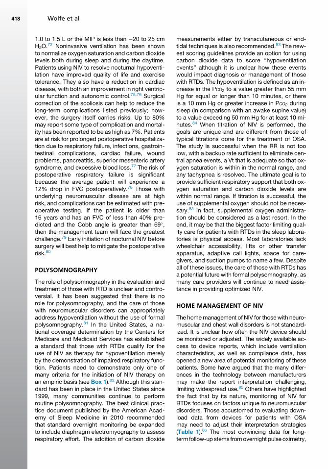
The homemanagement of NIV for thosewith neuro-muscular and chest wall disorders is not standard-ized. It is unclear how often the NIV device shouldbe monitored or adjusted. The widely available ac-cess to device reports, which include ventilationcharacteristics, as well as compliance data, hasopened a new area of potential monitoring of thesepatients. Some have argued that the many differ-ences in the technology between manufacturersmay make the report interpretation challenging,limiting widespread use.85 Others have highlightedthe fact that by its nature, monitoring of NIV forRTDs focuses on factors unique to neuromusculardisorders. Those accustomed to evaluating down-load data from devices for patients with OSAmay need to adjust their interpretation strategies(Table 1).86 The most convincing data for long-term follow-up stems fromovernight pulseoximetry,
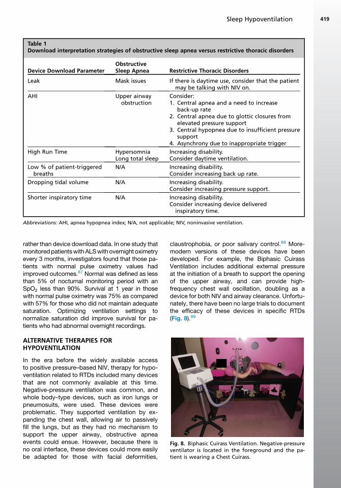
Table 1Download interpretation strategies of obstructive sleep apnea versus restrictive thoracic disorders
Device Download ParameterObstructiveSleep Apnea Restrictive Thoracic Disorders
Leak Mask issues If there is daytime use, consider that the patientmay be talking with NIV on.
AHI Upper airwayobstruction
Consider:1. Central apnea and a need to increase
back-up rate2. Central apnea due to glottic closures from
elevated pressure support3. Central hypopnea due to insufficient pressure
support4. Asynchrony due to inappropriate trigger
High Run Time HypersomniaLong total sleep
Increasing disability.Consider daytime ventilation.
Low % of patient-triggeredbreaths
N/A Increasing disability.Consider increasing back up rate.
Dropping tidal volume N/A Increasing disability.Consider increasing pressure support.
Shorter inspiratory time N/A Increasing disability.Consider increasing device delivered
inspiratory time.
Abbreviations: AHI, apnea hypopnea index; N/A, not applicable; NIV, noninvasive ventilation.
Fig. 8. Biphasic Cuirass Ventilation. Negative-pressureventilator is located in the foreground and the pa-tient is wearing a Chest Cuirass.
Sleep Hypoventilation 419
rather than device download data. In one study thatmonitoredpatientswithALSwith overnight oximetryevery 3 months, investigators found that those pa-tients with normal pulse oximetry values hadimproved outcomes.87 Normal was defined as lessthan 5% of nocturnal monitoring period with anSpO2 less than 90%. Survival at 1 year in thosewith normal pulse oximetry was 75% as comparedwith 57% for those who did not maintain adequatesaturation. Optimizing ventilation settings tonormalize saturation did improve survival for pa-tients who had abnormal overnight recordings.
ALTERNATIVE THERAPIES FORHYPOVENTILATION
In the era before the widely available accessto positive pressure–based NIV, therapy for hypo-ventilation related to RTDs included many devicesthat are not commonly available at this time.Negative-pressure ventilation was common, andwhole body–type devices, such as iron lungs orpneumosuits, were used. These devices wereproblematic. They supported ventilation by ex-panding the chest wall, allowing air to passivelyfill the lungs, but as they had no mechanism tosupport the upper airway, obstructive apneaevents could ensue. However, because there isno oral interface, these devices could more easilybe adapted for those with facial deformities,
claustrophobia, or poor salivary control.88 More-modern versions of these devices have beendeveloped. For example, the Biphasic CuirassVentilation includes additional external pressureat the initiation of a breath to support the openingof the upper airway, and can provide high-frequency chest wall oscillation, doubling as adevice for both NIV and airway clearance. Unfortu-nately, there have been no large trials to documentthe efficacy of these devices in specific RTDs(Fig. 8).89

Fig. 9. Diaphragm pacing system. Stimulation wiresare operatively affixed to the diaphragm andtunneled under the skin. At the skin site, the wireends are tipped, allowing an external stimulationbox to be connected. The stimulation box deliversthe electrical charge, causing diaphragmaticcontraction.
Wolfe et al420
Diaphragm pacing systems is another therapythat is an alternative to NIV. The mechanistic ba-sis has been described elsewhere in detail, but inbrief, direct electrical stimulation of the phrenicnerve, as it inserts into the diaphragm, is usedto stimulate contraction of the muscle. The stim-ulation electrodes are implanted surgically and anexternal generator provides the electrical pulses(Fig. 9).90 Currently, the device is being used astherapy in both SCI and ALS. In ALS, the deviceis used for therapeutic electrical stimulation;that is, the device provides electricity as a formof training, to support a muscle under stressdue to the degeneration of its feeding motorneuron. This device also has been shown toimprove sleep quality for those with ALS, but itis not a form of NIV.91 Early testing suggests apossible benefit to patients by improving theirsurvival.92 In contrast, diaphragmatic pacing de-vice is used as a form of NIV in the setting ofSCI. In high-level SCI, direct stimulation of thediaphragm with a pacer can be used as functionalelectrical stimulation, which means that the de-vice is not being used to enrich muscle functionbut rather to replace a failing system of sponta-neous ventilation.90
SUMMARY
In summary, hypoventilation due to neuromuscularand chest wall disorders includes a large numberof conditions. Although these conditions arefrequently thought of as a homogeneous group,there are unique factors that separate these condi-tions. The diagnostic evaluations and therapeuticinterventions for each condition are highly variable.Close consideration and individualization of ther-apy should be considered.
REFERENCES
1. Robberecht W, Philips T. The changing scene of
amyotrophic lateral sclerosis. Nat Rev Neurosci
2013;14(4):248–64.
2. Sivak ED, Shefner JM,Mitsumoto H, et al. The use of
non-invasive positive pressure ventilation (NIPPV) in
ALS patients. A need for improved determination of
intervention timing. Amyotroph Lateral Scler Other
Motor Neuron Disord 2001;2(3):139–45.
3. Steier J, Jolley CJ, Seymour J, et al. Screening for
sleep-disordered breathing in neuromuscular dis-
ease using a questionnaire for symptoms associ-
ated with diaphragm paralysis. Eur Respir J
2011;37(2):400–5.
4. Sanjak M, Salachas F, Frija-Orvoen E, et al. Quality
control of vital capacity as a primary outcome mea-
sure during phase III therapeutic clinical trial in
amyotrophic lateral sclerosis. Amyotroph Lateral
Scler 2010;11(4):383–8.
5. Schmidt EP, Drachman DB, Wiener CM, et al. Pul-
monary predictors of survival in amyotrophic lateral
sclerosis: use in clinical trial design. Muscle Nerve
2006;33(1):127–32.
6. Mustfa N, Moxham J. Respiratory muscle assess-
ment in motor neurone disease. QJM 2001;94(9):
497–502.
7. Lechtzin N, Scott Y, Busse AM, et al. Early use of
non-invasive ventilation prolongs survival in sub-
jects with ALS. Amyotroph Lateral Scler 2007;
8(3):185–8.
8. Atkeson AD, RoyChoudhury A, Harrington-
Moroney G, et al. Patient-ventilator asynchrony
with nocturnal noninvasive ventilation in ALS.
Neurology 2011;77(6):549–55.
9. Miller RG, Jackson CE, Kasarskis EJ, et al. Practice
parameter update: the care of the patient with
amyotrophic lateral sclerosis: drug, nutritional,
and respiratory therapies (an evidence-based re-
view): report of the Quality Standards Subcommit-
tee of the American Academy of Neurology.
Neurology 2009;73(15):1218–26.
10. Wirth B. An update of the mutation spectrum of the
survival motor neuron gene (SMN1) in autosomal
recessive spinal muscular atrophy (SMA). Hum
Mutat 2000;15(3):228–37.
11. Schroth MK. Special considerations in the respira-
tory management of spinal muscular atrophy. Pedi-
atrics 2009;123(Suppl 4):S245–9.
12. Wang CH, Finkel RS, Bertini ES, et al. Consensus
statement for standard of care in spinal muscular
atrophy. J Child Neurol 2007;22(8):1027–49.
13. Korinthenberg R. Acute polyradiculoneuritis:
Guillain-Barre syndrome. Handb Clin Neurol 2013;
112:1157–62.
14. Orlikowski D, Terzi N, Blumen M, et al. Tongue
weakness is associated with respiratory failure in

Sleep Hypoventilation 421
patients with severe Guillain-Barre syndrome. Acta
Neurol Scand 2009;119(6):364–70.
15. Rabinstein AA, Wijdicks EF. Warning signs of immi-
nent respiratory failure in neurological patients.
Semin Neurol 2003;23(1):97–104.
16. Wijdicks EF, Roy TK. BiPAP in early Guillain-Barre
syndrome may fail. Can J Neurol Sci 2006;33(1):
105–6.
17. Lawn ND, Wijdicks EF. Post-intubation pulmonary
function test in Guillain-Barre syndrome. Muscle
Nerve 2000;23(4):613–6.
18. Reddy VG, Nair MP, Bataclan F. Role of non-
invasive ventilation in difficult-to-wean children
with acute neuromuscular disease. Singapore
Med J 2004;45(5):232–4.
19. Vanasse M, Rossignol E, Hadad E. Chronic inflam-
matory demyelinating polyneuropathy. Handb Clin
Neurol 2013;112:1163–9.
20. Henderson RD, Sandroni P, Wijdicks EF. Chronic in-
flammatory demyelinating polyneuropathy and res-
piratory failure. J Neurol 2005;252(10):1235–7.
21. Zivkovic SA, Peltier AC, Iacob T, et al. Chronic in-
flammatory demyelinating polyneuropathy and
ventilatory failure: report of seven new cases and
review of the literature. Acta Neurol Scand 2011;
124(1):59–63.
22. Silvestri NJ, Wolfe GI. Myasthenia gravis. Semin
Neurol 2012;32(3):215–26.
23. Evoli A, Tonali PA, Padua L, et al. Clinical correlates
with anti-MuSK antibodies in generalized seroneg-
ative myasthenia gravis. Brain 2003;126(Pt 10):
2304–11.
24. Borel CO, Teitelbaum JS, Hanley DF. Ventilatory
drive and carbon dioxide response in ventilatory
failure due to myasthenia gravis and Guillain-
Barre syndrome. Crit Care Med 1993;21(11):
1717–26.
25. Garcia Rio F, Prados C, Diez Tejedor E, et al.
Breathing pattern and central ventilatory drive in
mild and moderate generalised myasthenia gravis.
Thorax 1994;49(7):703–6.
26. Rieder P, Louis M, Jolliet P, et al. The repeated
measurement of vital capacity is a poor predictor
of the need for mechanical ventilation in myas-
thenia gravis. Intensive Care Med 1995;21(8):
663–8.
27. Thieben MJ, Blacker DJ, Liu PY, et al. Pulmonary
function tests and blood gases in worsening myas-
thenia gravis. Muscle Nerve 2005;32(5):664–7.
28. Mishra SK, Krishnappa S, Bhat RR, et al. Role of
intermittent noninvasive ventilation in anticholines-
terase dose adjustment for myasthenic crisis.
Acta Anaesthesiol Taiwan 2010;48(1):53–4.
29. Wu JY, Kuo PH, Fan PC, et al. The role of non-
invasive ventilation and factors predicting extuba-
tion outcome in myasthenic crisis. Neurocrit Care
2009;10(1):35–42.
30. Rabinstein A, Wijdicks EF. BiPAP in acute respira-
tory failure due to myasthenic crisis may prevent
intubation. Neurology 2002;59(10):1647–9.
31. Burns TM, Grouse CK, Wolfe GI, et al. The MG-
QOL15 for following the health-related quality of
life of patients with myasthenia gravis. Muscle
Nerve 2011;43(1):14–8.
32. Prudlo J, Koenig J, Ermert S, et al. Sleep disor-
dered breathing in medically stable patients with
myasthenia gravis. Eur J Neurol 2007;14(3):321–6.
33. Mercuri E, Muntoni F. Muscular dystrophies. Lancet
2012;381(9869):845–60.
34. Bushby K, Finkel R, Birnkrant DJ, et al. Diagnosis
and management of Duchenne muscular dystro-
phy, part 1: diagnosis, and pharmacological and
psychosocial management. Lancet Neurol 2010;
9(1):77–93.
35. Hahn A, Duisberg B, Neubauer BA, et al. Noninva-
sive determination of the tension-time index in
Duchenne muscular dystrophy. Am J Phys Med
Rehabil 2009;88(4):322–7.
36. Beck J, Weinberg J, Hamnegard CH, et al. Dia-
phragmatic function in advanced Duchenne
muscular dystrophy. Neuromuscul Disord 2006;
16(3):161–7.
37. Toussaint M, Steens M, Soudon P. Lung function
accurately predicts hypercapnia in patients with
Duchenne muscular dystrophy. Chest 2007;
131(2):368–75.
38. Katz SL, Gaboury I, Keilty K, et al. Nocturnal hypo-
ventilation: predictors and outcomes in childhood
progressive neuromuscular disease. Arch Dis Child
2010;95(12):998–1003.
39. Finder JD, Birnkrant D, Carl J, et al. Respiratory
care of the patient with Duchenne muscular dystro-
phy: ATS consensus statement. Am J Respir Crit
Care Med 2004;170(4):456–65.
40. Toussaint M, Chatwin M, Soudon P. Mechanical
ventilation in Duchenne patients with chronic respi-
ratory insufficiency: clinical implications of 20 years
published experience. Chron Respir Dis 2007;4(3):
167–77.
41. Jeppesen J, Green A, Steffensen BF, et al. The
Duchenne muscular dystrophy population in
Denmark, 1977-2001: prevalence, incidence and
survival in relation to the introduction of ventilator
use. Neuromuscul Disord 2003;13(10):804–12.
42. Nathanson N, Kew OM. From emergence to eradi-
cation: the epidemiology of poliomyelitis decon-
structed. Am J Epidemiol 2010;172(11):1213–29.
43. Shiri S, Wexler ID, Feintuch U, et al. Post-polio syn-
drome: impact of hope on quality of life. Disabil Re-
habil 2012;34(10):824–30.
44. Soliman MG, Higgins SE, El-Kabir DR, et al. Non-
invasive assessment of respiratory muscle strength
in patients with previous poliomyelitis. Respir Med
2005;99(10):1217–22.
Wolfe et al422
45. Chai T, Aseff JN, Halstead LS. Diaphragm dysfunc-
tion due to remote poliomyelitis in a patient with un-
explained dyspnea. PM R 2011;3(2):179–82.
46. Olofson J, Dellborg C, Sullivan M, et al. Qualify of life
and palliation predict survival in patients with
chronic alveolar hypoventilation and nocturnal venti-
latory support. Qual Life Res 2009;18(3):273–80.
47. Bach JR, Alba AS. Sleep and nocturnal mouth-
piece IPPV efficiency in postpoliomyelitis ventilator
users. Chest 1994;106(6):1705–10.
48. Hsu AA, Staats BA. “Postpolio” sequelae and
sleep-related disordered breathing. Mayo Clin
Proc 1998;73(3):216–24.
49. Dahan V, Kimoff RJ, Petrof BJ, et al. Sleep-disor-
dered breathing in fatigued postpoliomyelitis clinic
patients. Arch Phys Med Rehabil 2006;87(10):
1352–6.
50. Kalpakjian CZ, Quint EH, Toussaint LL. Menopause
and post-polio symptoms as predictors of subjec-
tive sleep disturbance in poliomyelitis survivors.
Climacteric 2007;10(1):51–62.
51. Araujo MA, Silva TM, Moreira GA, et al. Sleep dis-
orders frequency in post-polio syndrome patients
caused by periodic limb movements. Arq Neuro-
psiquiatr 2010;68(1):35–8.
52. Vasconcelos OM, Prokhorenko OA, Salajegheh MK,
et al. Modafinil for treatment of fatigue in post-polio
syndrome: a randomized controlled trial. Neurology
2007;68(20):1680–6.
53. DeVivo MJ, Go BK, Jackson AB. Overview of the
national spinal cord injury statistical center data-
base. J Spinal Cord Med 2002;25(4):335–8.
54. Berlly M, Shem K. Respiratory management during
the first five days after spinal cord injury. J Spinal
Cord Med 2007;30(4):309–18.
55. LaVela SL, Burns SP, Goldstein B, et al. Dysfunc-
tional sleep in persons with spinal cord injuries
and disorders. Spinal Cord 2012;50(9):682–5.
56. Verheggen RJ, Jones H, Nyakayiru J, et al. Com-
plete absence of evening melatonin increase in tet-
raplegics. FASEB J 2012;26(7):3059–64.
57. Berlowitz DJ, Ayas N, Barnes M, et al. Auto-titrating
continuous positive airway pressure treatment for
obstructive sleep apnoea after acute quadriplegia
(COSAQ): study protocol for a randomized
controlled trial. Trials 2013;14:181.
58. Sankari A, Bascom AT, Chowdhuri S, et al. Tetraple-
gia is a risk factor for central sleep apnea. J Appl
Physiol (1985) 2014;116(3):345–53.
59. Bensmail D, Marquer A, Roche N, et al. Pilot study
assessing the impact of intrathecal baclofen
administration mode on sleep-related respiratory
parameters. Arch Phys Med Rehabil 2012;93(1):
96–9.
60. Bach JR. Noninvasive respiratory management of
high level spinal cord injury. J Spinal Cord Med
2012;35(2):72–80.
61. Parsonage M, Turner J. Neuralgic amyotrophy: the
shoulder girdle syndrome. Lancet 1948;254:973–8.
62. van Alfen N. The neuralgic amyotrophy consulta-
tion. J Neurol 2007;254(6):695–704.
63. Chen YM, Hu GC, Cheng SJ. Bilateral neuralgic
amyotrophy presenting with left vocal cord and
phrenic nerve paralysis. J Formos Med Assoc
2007;106(8):680–4.
64. Kumar N, Folger WN, Bolton CF. Dyspnea as the
predominant manifestation of bilateral phrenic neu-
ropathy. Mayo Clin Proc 2004;79(12):1563–5.
65. Beydoun SR, Rodriguez R. Neuralgic amyotrophy
misdiagnosed as diaphragmatic rupture. Muscle
Nerve 1996;19(9):1181–2.
66. Nardone R, Bernhart H, Pozzera A, et al. Respira-
tory weakness in neuralgic amyotrophy: report of
two cases with phrenic nerve involvement. Neurol
Sci 2000;21(3):177–81.
67. Kalluri M, Huggins JT, Strange C. A 56-year-old
woman with arm pain, dyspnea, and an elevated
diaphragm. Chest 2008;133(1):296–9.
68. Hughes PD, Polkey MI, Moxham J, et al. Long-term
recovery of diaphragm strength in neuralgic amyo-
trophy. Eur Respir J 1999;13(2):379–84.
69. Stolk J, Versteegh MI. Long-term effect of bilateral
plication of the diaphragm. Chest 2000;117(3):
786–9.
70. Nilsonne U, Lundgren KD. Long-term prognosis in
idiopathic scoliosis. Acta Orthop Scand 1968;
39(4):456–65.
71. Martini A, Bonadeo D, Bottoni G, et al. Bio-
adhesives: rationale, state of the art and therapeu-
tic potential. Boll Chim Farm 1995;134(11):595–603
[in Italian].
72. Shneerson JM. Respiration during sleep in neuro-
muscular and thoracic cage disorders. Monaldi
Arch Chest Dis 2004;61(1):44–8.
73. Guilleminault C, Kurland G, Winkle R, et al. Severe
kyphoscoliosis, breathing, and sleep: the “Quasi-
modo” syndrome during sleep. Chest 1981;79(6):
626–30.
74. Ellis ER, Grunstein RR, Chan S, et al. Noninvasive
ventilatory support during sleep improves respiratory
failure in kyphoscoliosis. Chest 1988;94(4):811–5.
75. Watson JP, Nolan J, Elliott MW. Autonomic dysfunc-
tion in patients with nocturnal hypoventilation in ex-
trapulmonary restrictive disease. Eur Respir J
1999;13(5):1097–102.
76. Fuschillo S, De Felice A, Gaudiosi C, et al.
Nocturnal mechanical ventilation improves exer-
cise capacity in kyphoscoliotic patients with respi-
ratory impairment. Monaldi Arch Chest Dis 2003;
59(4):281–6.
77. Master DL, Son-Hing JP, Poe-Kochert C, et al. Risk
factors for major complications after surgery for
neuromuscular scoliosis. Spine (Phila Pa 1976)
2012;36(7):564–71.

Sleep Hypoventilation 423
78. Roberto R, Fritz A, Hagar Y, et al. The natural his-
tory of cardiac and pulmonary function decline in
patients with Duchenne muscular dystrophy. Spine
(Phila Pa 1976) 2011;36(15):E1009–17.
79. Kang GR, Suh SW, Lee IO. Preoperative predictors
of postoperative pulmonary complications in
neuromuscular scoliosis. J Orthop Sci 2011;16(2):
139–47.
80. Ferris G, Servera-Pieras E, Vergara P, et al. Kypho-
scoliosis ventilatory insufficiency: noninvasive man-
agement outcomes. Am J Phys Med Rehabil 2000;
79(1):24–9.
81. Bach JR, Chaudhry SS. Standards of care in MDA
clinics. Muscular Dystrophy Association. Am J
Phys Med Rehabil 2000;79(2):193–6.
82. Clinical indications for noninvasive positive pres-
sure ventilation in chronic respiratory failure due
to restrictive lung disease, COPD, and nocturnal
hypoventilation—a consensus conference report.
Chest 1999;116(2):521–34.
83. Berry RB, Chediak A, Brown LK, et al. Best clinical
practices for the sleep center adjustment of nonin-
vasive positive pressure ventilation (NPPV) in sta-
ble chronic alveolar hypoventilation syndromes.
J Clin Sleep Med 2010;6(5):491–509.
84. Berry RB, Budhiraja R, Gottlieb DJ, et al. Rules for
scoring respiratory events in sleep: update of the
2007 AASM manual for the scoring of sleep and
associated events. Deliberations of the sleep ap-
nea definitions task force of the American Acad-
emy of Sleep Medicine. J Clin Sleep Med 2012;
8(5):597–619.
85. Contal O, Vignaux L, Combescure C, et al. Moni-
toring of noninvasive ventilation by built-in software
of home bilevel ventilators: a bench study. Chest
2012;141(2):469–76.
86. Janssens JP, Borel JC, Pepin JL. Nocturnal moni-
toring of home non-invasive ventilation: the contri-
bution of simple tools such as pulse oximetry,
capnography, built-in ventilator software and auto-
nomic markers of sleep fragmentation. Thorax
2011;66(5):438–45.
87. Gonzalez-Bermejo J, Morelot-Panzini C, Arnol N,
et al. Prognostic value of efficiently correcting
nocturnal desaturations after one month of non-
invasive ventilation in amyotrophic lateral scle-
rosis: a retrospective monocentre observational
cohort study. Amyotroph Lateral Scler Frontotem-
poral Degener 2013;14(5–6):373–9.
88. Dettenmeier PA, Jackson NC. Chronic hypoventila-
tion syndrome: treatment with non-invasive me-
chanical ventilation. AACN Clin Issues Crit Care
Nurs 1991;2(3):415–31.
89. Chatburn RL. High-frequency assisted airway
clearance. Respir Care 2007;52(9):1224–35 [dis-
cussion: 1235–7].
90. DiMarco AF. Phrenic nerve stimulation in patients
with spinal cord injury. Respir Physiol Neurobiol
2009;169(2):200–9.
91. Gonzalez-Bermejo J, Morelot-Panzini C, Salachas F,
et al. Diaphragm pacing improves sleep in patients
with amyotrophic lateral sclerosis. Amyotroph Lateral
Scler 2012;13(1):44–54.
92. Onders RP, ElmoM, Kaplan C, et al. Final analysis of
the pilot trial of diaphragm pacing in amyotrophic
lateral sclerosiswith long-term follow-up: diaphragm
pacing positively affects diaphragm respiration. Am
J Surg 2014;207(3):393–7 [discussion: 397].