SL-401, a targeted therapy directed to the interleukin-3 ...
1
Abstract Cell culture: K-562 and MV-4-11 cells were cultured in IMDM supplemented with 10% FBS. L-428 and RPMI-8226 cells were cultured in RPMI-1640 supplemented with 10% FBS. Cell lines were treated with varying concentrations of SL-401 and SL-801. In vitro cytotoxicity assay: The CellTiter-Glo® Luminescent Cell Viability Assay kit (Promega) was used to assess cell viability after exposure to SL-401, SL-801, or both drugs simultaneously. Cells were grown in 96-well plates and incubated with drugs for 48 hours. The CellTiter-Glo® reagent was then added to wells according to the manufacturer’s instructions, and luminescence was measured on a ThermoLab Systems Luminoskan Ascent Reader at room temperature. Combination index calculations: Combination index (CI) values were calculated using CompuSyn software by the method of Chou and Talalay [Chou TC, Cancer Res . 2010 Jan 15;70(2):440-6]. Treatment was considered to be synergistic when CI < 1. Caspase activity assay: The Caspase-Glo® 3/7 Assay (Promega) was used to measure caspase activity in cells treated with SL-401, SL-801, or the combination. Briefly, cells were incubated with drugs for 24 h and then assessed for caspase 3/7 activity. The Caspase-Glo® 3/7 Reagent was added and incubated for 30 min at room temperature. Luminescence was measured on a ThermoLab Systems Luminoskan Ascent Reader at room temperature. Lactate dehydrogenase (LDH) release assay: The CytoTox 96® Non-Radioactive Cytotoxicity Assay (Promega) was used to measure LDH release from cells treated with SL-401, SL-801, or the combination. Briefly, cells were incubated with drugs for 48 h and then centrifuged for 5 min to pellet cells. Supernatants (10 μl) were transferred to ELISA plates, incubated with 50 μl substrate mix for 30 min at room temperature, and then quenched with 50 μl stop solution. Plates were read at 490 nm on an ELISA plate reader. Western blot: Whole cell extracts were prepared from MV-4-11 cells treated with 0.1 nM SL-401, 60 nM SL-801, or the combination for 3, 6, 12, and 24 h. Western blots were performed using EF-2 and β-actin antibodies obtained from Santa Cruz Biotechnology and developed by enhanced chemiluminescence. Materials and methods Mechanisms of action of SL-401 and SL-801 Background: Novel combination therapies have shown success in combating tumor heterogeneity and drug resistance. SL-401 is a targeted therapy directed to the interleukin-3 receptor (IL-3R / CD123), which is overexpressed on numerous hematologic malignancies. SL-401 has demonstrated high single agent response rates in an ongoing Phase 2 trial of blastic plasmacytoid dendritic cell neoplasm (BPDCN) and is also being evaluated in the clinic for additional cancers, including acute myeloid leukemia (AML) and myeloproliferative neoplasms (MPNs) as a single agent, and multiple myeloma (MM) in combination with other agents. While SL-401 has demonstrated robust single agent clinical activity in patients with BPDCN, its unique mechanism of action and non-overlapping side effect profile with other agents may lend itself to combination therapy as well. Another class of drugs that has demonstrated clinical activity against several hematologic and solid malignancies is Exportin-1 (XPO1) inhibitors. SL-801 is a novel oral small molecule that reversibly inhibits XPO1 and has shown potent in vitro and in vivo anti-tumor activity against a broad range of hematologic and solid malignancies. SL-801 is currently being evaluated in a Phase 1 trial of patients with advanced solid tumors, and a Phase 1 trial in advanced hematologic cancers is planned. Here, we investigated the in vitro effect of combination treatment of SL-401 and SL-801 against cell lines of chronic myeloid leukemia (CML), AML, MM, and Hodgkin’s lymphoma (HL). Methods: The human K562 CML, MV-4-11 AML, RPMI-8226 MM, and L-428 HL cell lines were treated with varying concentrations of SL-401 and SL-801 alone or in combination for 48 hours. Cell viability was assessed by the CellTiter Glo in vitro cytotoxicity assay. Combination index (CI) values were calculated using CompuSyn software by the method of Chou and Talalay, and treatment was considered to be synergistic when CI < 1. Caspase activation was measured using the Caspase-Glo 3/7 assay, and lactate dehydrogenase (LDH) release was measured using the CytoTox 96 Non-Radioactive Cytotoxicity Assay. Results: As single agents, SL-401 and SL-801 demonstrated anti-tumor activity in all four cell lines tested. MV-4-11 cells were the most sensitive to both drugs, with an IC 50 of 34 pM for SL-401 and 21 nM for SL-801. In the other cell lines, the IC 50 s for SL-401 were 17 nM in K562 cells, 25 nM in RPMI-8226 cells, and 100 nM in L-428 cells, and the IC 50 s for SL-801 were 99 nM in K562 cells, 51 nM in RPMI-8226 cells, and 494 nM in L-428 cells. When combined with each other, SL-401 and SL-801 potently inhibited cell growth in all cell lines, and CI calculations indicated that the interaction between the two drugs was synergistic at most dose combinations. Notably, CI values < 0.35 were observed in L-428, RPMI-8226, and MV-4-11 cells, indicative of strong synergy. Consistent with these observations, the combination of SL-401 and SL-801 also induced higher levels of caspase activation and LDH release in M-V4-11 and L-428 cells than either drug alone. Conclusion: These findings demonstrate that SL-401 and SL-801, when combined, act synergistically in their in vitro anti-tumor activity against CML, AML, MM, and HL cells. Investigations into the molecular mechanisms underlying the observed synergy are in progress. These promising results provide rationale for further development of SL-401 and SL-801 combination therapy in the treatment of a broad range of hematologic malignancies. A IL-3 Truncated diphtheria toxin payload SL-401 Synergistic anti-tumor activity of SL-401 and SL-801 in vitro SL-401 and SL-801 enhance apoptosis J. Gionco: Stemline – employment J. Chen: Stemline – employment, equity ownership R. Lindsay: Stemline – employment, equity ownership V. Macri: Stemline – employment, equity ownership C. Brooks: Stemline – employment, equity ownership, patents and royalties Conclusions • SL-401 and SL-801 possess potent single agent anti-tumor activity against CML, HL, MM, and AML cell lines in vitro. • Several combinations of SL-401 and SL-801 exhibit strong synergistic anti-tumor activity in L-428 HL, RPMI-8226 MM, and MV-4-11 AML cells, as demonstrated by CI values < 0.35. • The combination of SL-401 and SL-801 enhances caspase 3/7 activation and LDH release in L-428 and MV-4-11 cell lines. • Cellular expression of EF2, the molecular target of SL-401’s payload, remains unchanged with SL-801 treatment. • Investigations into the molecular mechanisms underlying the observed synergy are ongoing. • These promising results warrant further development of SL-401 and SL-801 combination therapy in the treatment of a broad range of hematologic malignancies. John Gionco, Janice Chen, Ross Lindsay, Vince Macri, Christopher Brooks Stemline Therapeutics, Inc., New York, NY, USA ASH 2016 #4724 SL-401, a targeted therapy directed to the interleukin-3 receptor (CD123), and SL-801, a reversible inhibitor of Exportin-1 (XPO1), display synergistic anti-tumor activity against hematologic malignancies in vitro Indication Status NCT # Abstract Blastic plasmacytoid dendritic cell neoplasm Open NCT02113982 342 Relapsed/refractory acute myeloid leukemia (AML) Open NCT02113982 --- AML, in 1st or 2nd complete remission with minimal residual disease Open NCT02270463 215 Advanced, high risk myeloproliferative neoplasms Open NCT02268253 4245 Relapsed/refractory multiple myeloma, in combination with pomalidomide and dexamethasone Open NCT02661022 5696 Indication Status NCT # Abstract Advanced solid tumors Open NCT02667873 --- Advanced hematologic malignancies Planned --- --- IL-3R EF2 EF2 mRNA Nascent peptide Ribosome −ADP-ribose SL-401 binds to the IL-3R and is internalized into cells. The catalytic domain of DT translocates into the cytoplasm and ADP-ribosylates elongation factor 2 (EF2). SL-401 is comprised of human IL-3 recombinantly fused to a truncated diphtheria toxin (DT) payload engineered such that IL-3 replaces the native DT receptor-binding domain. SL-801 is a small molecule that binds covalently to Cys528 of XPO1, preventing the binding of nuclear cargo proteins and inhibiting nuclear export. C Protein synthesis is irreversibly arrested. SL-401 Clinical Trials SL-801 Clinical Trials A D 0.001 0.01 0.1 1 10 100 1000 10000 0 20 40 60 80 100 120 Drug concentration (nM) Cell viability (% of control) K562 0.01 0.1 1 10 100 1000 10000 0 20 40 60 80 100 120 Drug concentration (nM) Cell viability (% of control) L-428 0.01 0.1 1 10 100 1000 10000 0 20 40 60 80 100 120 Drug concentration (nM) Cell viability (% of control) RPMI-8226 0.01 0.1 1 10 100 1000 10000 0 20 40 60 80 100 120 Drug concentration (nM) Cell viability (% of control) MV-4-11 SL-401 SL-801 IC 50 SL-401: 17 nM SL-801: 99 nM IC 50 SL-401: 100 nM SL-801: 494 nM IC 50 SL-401: 25 nM SL-801: 51 nM IC 50 SL-401: 0.034 nM SL-801: 21 nM 0 100 200 300 400 500 0 20 40 60 80 100 120 SL-801 concentration (nM) Cell viability (% of control) K562 0 nM SL-401 1.6 nM SL-401 4.7 nM SL-401 14 nM SL-401 42 nM SL-401 0 10 20 30 40 0 20 40 60 80 100 120 SL-401 concentration (nM) Cell viability (% of control) K562 0 nM SL-801 16.7 nM SL-801 50 nM SL-801 150 nM SL-801 450 nM SL-801 B 0 50 100 150 200 250 300 0 20 40 60 80 100 120 SL-401 concentration (nM) Cell viability (% of control) L-428 0 nM SL-801 19 nM SL-801 56 nM SL-801 167 nM SL-801 500 nM SL-801 0 100 200 300 400 500 0 20 40 60 80 100 120 SL-801 concentration (nM) Cell viability (% of control) L-428 0 nM SL-401 11 nM SL-401 33 nM SL-401 100 nM SL-401 300 nM SL-401 0 20 40 60 80 0 20 40 60 80 100 120 SL-401 concentration (nM) Cell viability (% of control) RPMI-8226 0 nM SL-801 6 nM SL-801 17 nM SL-801 50 nM SL-801 75 nM SL-801 0 40 80 120 160 0 20 40 60 80 100 120 SL-801 concentration (nM) Cell viability (% of control) RPMI-8226 0 nM SL-401 3 nM SL-401 8 nM SL-401 25 nM SL-401 75 nM SL-401 0.00 0.02 0.04 0.06 0.08 0.10 0 20 40 60 80 100 120 SL-401 concentration (nM) Cell viability (% of control) MV-4-11 0 nM SL-801 2 nM SL-801 7 nM SL-801 20 nM SL-801 60 nM SL-801 0 20 40 60 0 20 40 60 80 100 120 SL-801 concentration (nM) Cell viability (% of control) MV-4-11 0 nM SL-401 0.0037 nM SL-401 0.011 nM SL-401 0.033 nM SL-401 0.1 nM SL-401 0.0 0.2 0.4 0.6 0.8 1.0 0.0 0.2 0.4 0.6 0.8 1.0 1.2 1.4 1.6 Fraction affected Combination index K562 0.0 0.2 0.4 0.6 0.8 1.0 0.0 0.2 0.4 0.6 0.8 1.0 1.2 1.4 1.6 Fraction affected Combination index L-428 0.0 0.2 0.4 0.6 0.8 1.0 0.0 0.2 0.4 0.6 0.8 1.0 1.2 1.4 1.6 Fraction affected Combination index RPMI-8226 Antagonism é Synergism ê Antagonism é Synergism ê Antagonism é Synergism ê CI 1.6 4.7 14 42 16.7 0.43 0.56 0.70 1.42 50 0.68 0.61 0.77 1.40 150 0.62 0.60 0.82 1.24 450 0.98 0.69 0.87 1.17 SL-401 (nM) SL-801 (nM) CI 11 33 100 300 19 0.78 0.86 0.80 0.61 56 0.95 0.91 0.73 0.43 167 1.33 0.53 0.41 0.22 500 0.41 0.16 0.23 0.09 SL-401 (nM) SL-801 (nM) CI 3 8 25 75 6 1.12 1.38 0.98 0.69 17 1.54 1.35 0.94 0.56 50 0.91 0.76 0.50 0.33 150 1.14 0.88 0.62 0.34 SL-401 (nM) SL-801 (nM) None SL-401 SL-801 SL-401 + SL-801 0 20 40 60 80 Caspase activity (RLU) L-428 None SL-401 SL-801 SL-401 + SL-801 0 20 40 60 80 MV-4-11 Caspase activity (RLU) None SL-401 SL-801 SL-401 + SL-801 0.00 0.05 0.10 0.15 0.20 0.25 L-428 LDH release (OD490) None SL-401 SL-801 SL-401 + SL-801 0.00 0.05 0.10 0.15 0.20 0.25 LDH release (OD490) MV-4-11 A B (A) The single agent anti-tumor activity of SL-401 and SL-801 was evaluated in the K562 CML, L-428 HL, RPMI-8226 MM, and MV-4-11 AML cell lines. Cells were exposed to 0.0056 or 0.05 nM to 1 or 8.8 μM of SL-401, and 0.0056 or 0.06 nM to 1 or 10 μM of SL-801. Cell viability was assessed after a 48 h incubation, and IC 50 values were determined. (B, C) The combination of SL-401 and SL-801 was evaluated in the same cell lines. The two drugs were added individually or together at different concentrations as indicated: 1.6 – 42 nM SL-401 and 16.7 – 450 nM SL-801 (K562), 11 – 300 nM SL-401 and 19 – 500 nM SL-801 (L-428), 3 – 75 nM SL-401 and 6 –150 nM SL-801 (RPMI-8226), and 0.0037 – 0.1 nM SL-401 and 2 – 60 nM SL-801 (MV-4-11). Cell viability was measured after 48 h. (D) CI values were calculated using CompuSyn software. Upper panels, CIs were plotted against fraction of cells affected. Lower panels, CI values for each drug combination are shown. Treatment was considered to be synergistic when CI < 1, additive when CI = 1, and antagonistic when CI > 1. Green, synergistic combinations. 0.0 0.2 0.4 0.6 0.8 1.0 0.0 0.2 0.4 0.6 0.8 1.0 1.2 1.4 1.6 Fraction affected Combination index MV-4-11 Antagonism é Synergism ê CI 0.0037 0.011 0.033 0.1 2 1.17 1.00 0.90 0.79 7 0.97 0.84 0.87 0.43 20 1.17 1.16 0.62 0.23 60 0.66 0.35 0.18 0.05 SL-401 (nM) SL-801 (nM) SL-801 does not alter EF2 expression levels C L-428 cells were treated with 300 nM SL-401, 500 nM SL-801, or the two drugs in combination. MV-4-11 cells were treated with 0.1 nM SL-401, 60 nM SL-801, or the two drugs in combination. (A) Caspase 3/7 activation was assessed after 24 h of drug treatment. RLU, relative light units. (B) LDH release from cells into the supernatant was assessed after 48 h of drug treatment. Nucleus XPO1 RanGTP Cargo XPO1 Cargo RanGDP Cargo Pi Nuclear pore complex Cytoplasm RanGTP B XPO1 SL-801 3 6 12 24 3 6 12 24 3 6 12 24 Hours: SL-401 SL-801 SL-401 + SL-801 No drug No drug EF2 β-actin MV-4-11 cells were treated with 0.1 nM SL-401, 60 nM SL-801, or the two drugs in combination for 3, 6, 12, and 24 h. Western blot analysis was performed on whole cell extracts for expression of EF2. β-actin was used as a loading control.
Transcript of SL-401, a targeted therapy directed to the interleukin-3 ...
Abstract
Cell culture: K-562 and MV-4-11 cells were cultured in IMDM supplemented with 10% FBS. L-428 and RPMI-8226 cells were cultured in RPMI-1640 supplemented with 10% FBS. Cell lines were treated with varying concentrations of SL-401 and SL-801. In vitro cytotoxicity assay: The CellTiter-Glo® Luminescent Cell Viability Assay kit (Promega) was used to assess cell viability after exposure to SL-401, SL-801, or both drugs simultaneously. Cells were grown in 96-well plates and incubated with drugs for 48 hours. The CellTiter-Glo® reagent was then added to wells according to the manufacturer’s instructions, and luminescence was measured on a ThermoLab Systems Luminoskan Ascent Reader at room temperature. Combination index calculations: Combination index (CI) values were calculated using CompuSyn software by the method of Chou and Talalay [Chou TC, Cancer Res. 2010 Jan 15;70(2):440-6]. Treatment was considered to be synergistic when CI < 1. Caspase activity assay: The Caspase-Glo® 3/7 Assay (Promega) was used to measure caspase activity in cells treated with SL-401, SL-801, or the combination. Briefly, cells were incubated with drugs for 24 h and then assessed for caspase 3/7 activity. The Caspase-Glo® 3/7 Reagent was added and incubated for 30 min at room temperature. Luminescence was measured on a ThermoLab Systems Luminoskan Ascent Reader at room temperature. Lactate dehydrogenase (LDH) release assay: The CytoTox 96® Non-Radioactive Cytotoxicity Assay (Promega) was used to measure LDH release from cells treated with SL-401, SL-801, or the combination. Briefly, cells were incubated with drugs for 48 h and then centrifuged for 5 min to pellet cells. Supernatants (10 µl) were transferred to ELISA plates, incubated with 50 µl substrate mix for 30 min at room temperature, and then quenched with 50 µl stop solution. Plates were read at 490 nm on an ELISA plate reader. Western blot: Whole cell extracts were prepared from MV-4-11 cells treated with 0.1 nM SL-401, 60 nM SL-801, or the combination for 3, 6, 12, and 24 h. Western blots were performed using EF-2 and β-actin antibodies obtained from Santa Cruz Biotechnology and developed by enhanced chemiluminescence.
Materials and methods
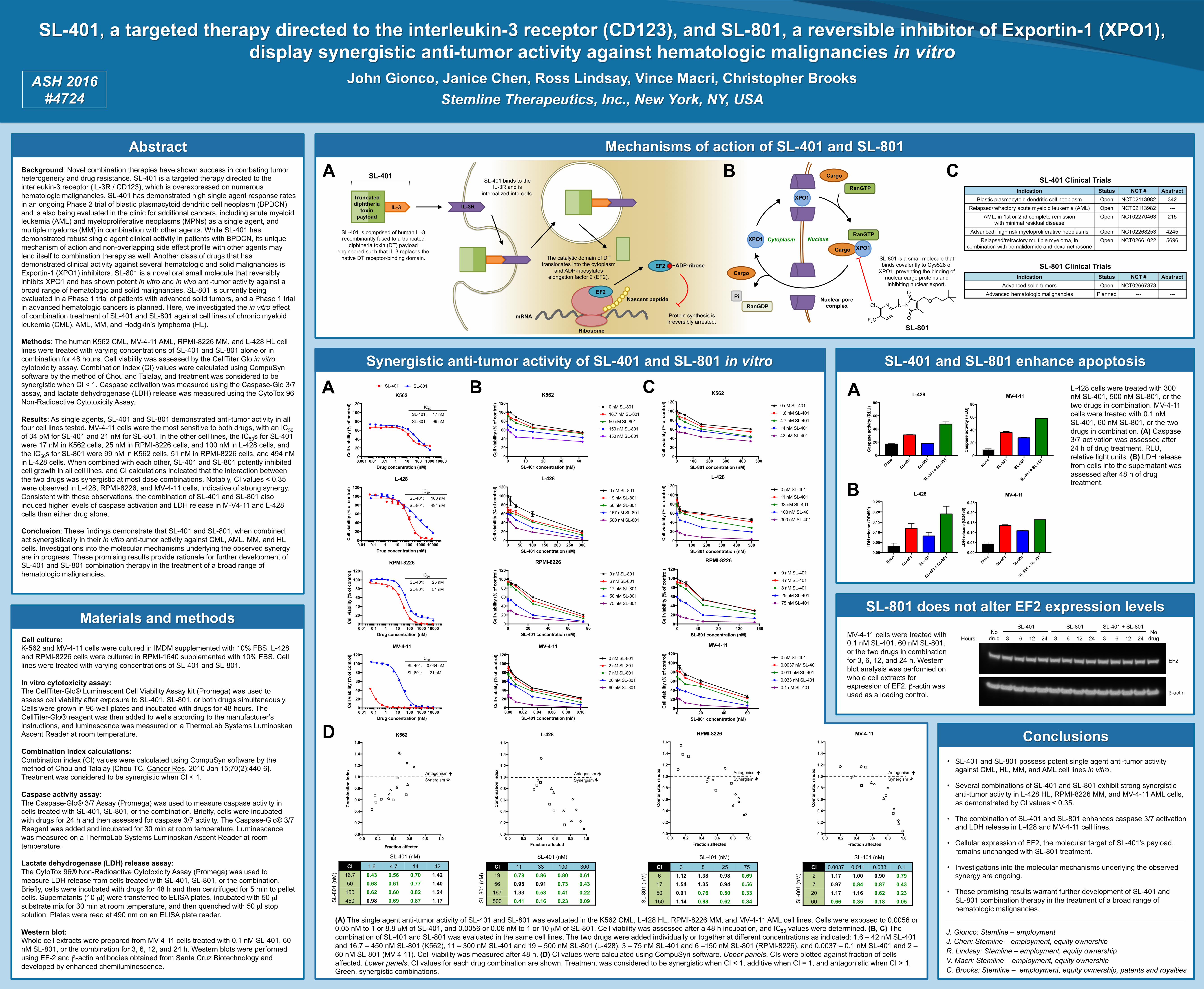
Mechanisms of action of SL-401 and SL-801 Background: Novel combination therapies have shown success in combating tumor heterogeneity and drug resistance. SL-401 is a targeted therapy directed to the interleukin-3 receptor (IL-3R / CD123), which is overexpressed on numerous hematologic malignancies. SL-401 has demonstrated high single agent response rates in an ongoing Phase 2 trial of blastic plasmacytoid dendritic cell neoplasm (BPDCN) and is also being evaluated in the clinic for additional cancers, including acute myeloid leukemia (AML) and myeloproliferative neoplasms (MPNs) as a single agent, and multiple myeloma (MM) in combination with other agents. While SL-401 has demonstrated robust single agent clinical activity in patients with BPDCN, its unique mechanism of action and non-overlapping side effect profile with other agents may lend itself to combination therapy as well. Another class of drugs that has demonstrated clinical activity against several hematologic and solid malignancies is Exportin-1 (XPO1) inhibitors. SL-801 is a novel oral small molecule that reversibly inhibits XPO1 and has shown potent in vitro and in vivo anti-tumor activity against a broad range of hematologic and solid malignancies. SL-801 is currently being evaluated in a Phase 1 trial of patients with advanced solid tumors, and a Phase 1 trial in advanced hematologic cancers is planned. Here, we investigated the in vitro effect of combination treatment of SL-401 and SL-801 against cell lines of chronic myeloid leukemia (CML), AML, MM, and Hodgkin’s lymphoma (HL). Methods: The human K562 CML, MV-4-11 AML, RPMI-8226 MM, and L-428 HL cell lines were treated with varying concentrations of SL-401 and SL-801 alone or in combination for 48 hours. Cell viability was assessed by the CellTiter Glo in vitro cytotoxicity assay. Combination index (CI) values were calculated using CompuSyn software by the method of Chou and Talalay, and treatment was considered to be synergistic when CI < 1. Caspase activation was measured using the Caspase-Glo 3/7 assay, and lactate dehydrogenase (LDH) release was measured using the CytoTox 96 Non-Radioactive Cytotoxicity Assay. Results: As single agents, SL-401 and SL-801 demonstrated anti-tumor activity in all four cell lines tested. MV-4-11 cells were the most sensitive to both drugs, with an IC50 of 34 pM for SL-401 and 21 nM for SL-801. In the other cell lines, the IC50s for SL-401 were 17 nM in K562 cells, 25 nM in RPMI-8226 cells, and 100 nM in L-428 cells, and the IC50s for SL-801 were 99 nM in K562 cells, 51 nM in RPMI-8226 cells, and 494 nM in L-428 cells. When combined with each other, SL-401 and SL-801 potently inhibited cell growth in all cell lines, and CI calculations indicated that the interaction between the two drugs was synergistic at most dose combinations. Notably, CI values < 0.35 were observed in L-428, RPMI-8226, and MV-4-11 cells, indicative of strong synergy. Consistent with these observations, the combination of SL-401 and SL-801 also induced higher levels of caspase activation and LDH release in M-V4-11 and L-428 cells than either drug alone. Conclusion: These findings demonstrate that SL-401 and SL-801, when combined, act synergistically in their in vitro anti-tumor activity against CML, AML, MM, and HL cells. Investigations into the molecular mechanisms underlying the observed synergy are in progress. These promising results provide rationale for further development of SL-401 and SL-801 combination therapy in the treatment of a broad range of hematologic malignancies.
A
IL-3
Truncated diphtheria
toxin payload
SL-401
Synergistic anti-tumor activity of SL-401 and SL-801 in vitro SL-401 and SL-801 enhance apoptosis
J. Gionco: Stemline – employment J. Chen: Stemline – employment, equity ownership R. Lindsay: Stemline – employment, equity ownership V. Macri: Stemline – employment, equity ownership C. Brooks: Stemline – employment, equity ownership, patents and royalties
Conclusions • SL-401 and SL-801 possess potent single agent anti-tumor activity
against CML, HL, MM, and AML cell lines in vitro.
• Several combinations of SL-401 and SL-801 exhibit strong synergistic anti-tumor activity in L-428 HL, RPMI-8226 MM, and MV-4-11 AML cells, as demonstrated by CI values < 0.35.
• The combination of SL-401 and SL-801 enhances caspase 3/7 activation and LDH release in L-428 and MV-4-11 cell lines.
• Cellular expression of EF2, the molecular target of SL-401’s payload, remains unchanged with SL-801 treatment.
• Investigations into the molecular mechanisms underlying the observed synergy are ongoing.
• These promising results warrant further development of SL-401 and SL-801 combination therapy in the treatment of a broad range of hematologic malignancies.
John Gionco, Janice Chen, Ross Lindsay, Vince Macri, Christopher Brooks Stemline Therapeutics, Inc., New York, NY, USA
ASH 2016 #4724
SL-401, a targeted therapy directed to the interleukin-3 receptor (CD123), and SL-801, a reversible inhibitor of Exportin-1 (XPO1), display synergistic anti-tumor activity against hematologic malignancies in vitro
Indication Status NCT # Abstract Blastic plasmacytoid dendritic cell neoplasm Open NCT02113982 342
Relapsed/refractory acute myeloid leukemia (AML) Open NCT02113982 --- AML, in 1st or 2nd complete remission
with minimal residual disease Open NCT02270463 215
Advanced, high risk myeloproliferative neoplasms Open NCT02268253 4245 Relapsed/refractory multiple myeloma, in
combination with pomalidomide and dexamethasone Open NCT02661022 5696
Indication Status NCT # Abstract Advanced solid tumors Open NCT02667873 ---
Advanced hematologic malignancies Planned --- ---
IL-3R
EF2
EF2
mRNA
Nascent peptide
Ribosome
−ADP-ribose
SL-401 binds to the IL-3R and is
internalized into cells.
The catalytic domain of DT translocates into the cytoplasm
and ADP-ribosylates elongation factor 2 (EF2).
SL-401 is comprised of human IL-3 recombinantly fused to a truncated
diphtheria toxin (DT) payload engineered such that IL-3 replaces the
native DT receptor-binding domain. SL-801 is a small molecule that binds covalently to Cys528 of
XPO1, preventing the binding of nuclear cargo proteins and inhibiting nuclear export.
C
Protein synthesis is irreversibly arrested.
SL-401 Clinical Trials
SL-801 Clinical Trials
A
D
0.001 0.01 0.1 1 10 100 1000 100000
20
40
60
80
100
120
Drug concentration (nM)
Cel
l via
bilit
y (%
of c
ontr
ol)
K562
SL-401SL-801
0.01 0.1 1 10 100 1000 100000
20
40
60
80
100
120
Drug concentration (nM)
Cel
l via
bilit
y (%
of c
ontr
ol)
L-428
SL-401SL-801
0.01 0.1 1 10 100 1000 100000
20
40
60
80
100
120
Drug concentration (nM)
Cel
l via
bilit
y (%
of c
ontr
ol)
RPMI-8226
SL-401SL-801
0.01 0.1 1 10 100 1000 100000
20
40
60
80
100
120
Drug concentration (nM)
Cel
l via
bilit
y (%
of c
ontr
ol)
MV-4-11
SL-401SL-801
0.001 0.01 0.1 1 10 100 1000 100000
20
40
60
80
100
120
Drug concentration (nM)
Cel
l via
bilit
y (%
of c
ontr
ol)
K562
SL-401SL-801
0.001 0.01 0.1 1 10 100 1000 100000
20
40
60
80
100
120
Drug concentration (nM)
Cel
l via
bilit
y (%
of c
ontr
ol)
K562
SL-401SL-801
IC50
SL-401: 17 nM
SL-801: 99 nM
IC50
SL-401: 100 nM
SL-801: 494 nM
IC50
SL-401: 25 nM
SL-801: 51 nM
IC50
SL-401: 0.034 nM
SL-801: 21 nM
0 100 200 300 400 5000
20
40
60
80
100
120
SL-801 concentration (nM)
Cel
l via
bilit
y (%
of c
ontr
ol)
K562
0 nM SL-4011.6 nM SL-4014.7 nM SL-40114 nM SL-40142 nM SL-401
0 10 20 30 400
20
40
60
80
100
120
SL-401 concentration (nM)
Cel
l via
bilit
y (%
of c
ontr
ol)
K562
0 nM SL-80116.7 nM SL-80150 nM SL-801150 nM SL-801450 nM SL-801
B
0 50 100 150 200 250 3000
20
40
60
80
100
120
SL-401 concentration (nM)
Cel
l via
bilit
y (%
of c
ontr
ol)
L-428
0 nM SL-80119 nM SL-80156 nM SL-801167 nM SL-801500 nM SL-801
0 100 200 300 400 5000
20
40
60
80
100
120
SL-801 concentration (nM)
Cel
l via
bilit
y (%
of c
ontr
ol)
L-428
0 nM SL-40111 nM SL-40133 nM SL-401100 nM SL-401300 nM SL-401
0 20 40 60 800
20
40
60
80
100
120
SL-401 concentration (nM)
Cel
l via
bilit
y (%
of c
ontr
ol)
RPMI-8226
0 nM SL-8016 nM SL-80117 nM SL-80150 nM SL-80175 nM SL-801
0 40 80 120 1600
20
40
60
80
100
120
SL-801 concentration (nM)
Cel
l via
bilit
y (%
of c
ontr
ol)
RPMI-8226
0 nM SL-4013 nM SL-4018 nM SL-40125 nM SL-40175 nM SL-401
0.00 0.02 0.04 0.06 0.08 0.100
20
40
60
80
100
120
SL-401 concentration (nM)
Cel
l via
bilit
y (%
of c
ontr
ol)
MV-4-11
0 nM SL-8012 nM SL-8017 nM SL-80120 nM SL-80160 nM SL-801
0 20 40 600
20
40
60
80
100
120
SL-801 concentration (nM)
Cel
l via
bilit
y (%
of c
ontr
ol)
MV-4-11
0 nM SL-4010.0037 nM SL-4010.011 nM SL-4010.033 nM SL-4010.1 nM SL-401
0.0 0.2 0.4 0.6 0.8 1.00.0
0.2
0.4
0.6
0.8
1.0
1.2
1.4
1.6
Fraction affected
Com
bina
tion
inde
x
K562
1.6 nM SL-4014.7 nM SL-40114 nM SL-40142 nM SL-401
0.0 0.2 0.4 0.6 0.8 1.00.0
0.2
0.4
0.6
0.8
1.0
1.2
1.4
1.6
Fraction affected
Com
bina
tion
inde
x
L-428
11 nM SL-40133 nM SL-401100 nM SL-401300 nM SL-401
0.0 0.2 0.4 0.6 0.8 1.00.0
0.2
0.4
0.6
0.8
1.0
1.2
1.4
1.6
Fraction affected
Com
bina
tion
inde
x
RPMI-8226
3 nM SL-4018 nM SL-40125 nM SL-40175 nM SL-401
Antagonism é Synergism ê
Antagonism é Synergism ê
Antagonism é Synergism ê
CI 1.6 4.7 14 42 16.7 0.43 0.56 0.70 1.42 50 0.68 0.61 0.77 1.40
150 0.62 0.60 0.82 1.24 450 0.98 0.69 0.87 1.17
SL-401 (nM)
SL-
801
(nM
)
CI 11 33 100 300 19 0.78 0.86 0.80 0.61 56 0.95 0.91 0.73 0.43
167 1.33 0.53 0.41 0.22 500 0.41 0.16 0.23 0.09
SL-401 (nM)
SL-
801
(nM
)
CI 3 8 25 75 6 1.12 1.38 0.98 0.69
17 1.54 1.35 0.94 0.56 50 0.91 0.76 0.50 0.33
150 1.14 0.88 0.62 0.34
SL-401 (nM)
SL-
801
(nM
)
None
SL-401
SL-801
SL-401
+ SL-8
010
20
40
60
80
Cas
pase
act
ivity
(RLU
)
L-428
None
SL-401
SL-801
SL-401
+ SL-8
010
20
40
60
80
MV-4-11
Cas
pase
act
ivity
(RLU
)
None
SL-401
SL-801
SL-401 +
SL-801
0.00
0.05
0.10
0.15
0.20
0.25
L-428
LDH
rele
ase
(OD
490)
None
SL-401
SL-801
SL-401 +
SL-801
0.00
0.05
0.10
0.15
0.20
0.25
LDH
rele
ase
(OD
490)
MV-4-11
A
B
(A) The single agent anti-tumor activity of SL-401 and SL-801 was evaluated in the K562 CML, L-428 HL, RPMI-8226 MM, and MV-4-11 AML cell lines. Cells were exposed to 0.0056 or 0.05 nM to 1 or 8.8 µM of SL-401, and 0.0056 or 0.06 nM to 1 or 10 µM of SL-801. Cell viability was assessed after a 48 h incubation, and IC50 values were determined. (B, C) The combination of SL-401 and SL-801 was evaluated in the same cell lines. The two drugs were added individually or together at different concentrations as indicated: 1.6 – 42 nM SL-401 and 16.7 – 450 nM SL-801 (K562), 11 – 300 nM SL-401 and 19 – 500 nM SL-801 (L-428), 3 – 75 nM SL-401 and 6 –150 nM SL-801 (RPMI-8226), and 0.0037 – 0.1 nM SL-401 and 2 – 60 nM SL-801 (MV-4-11). Cell viability was measured after 48 h. (D) CI values were calculated using CompuSyn software. Upper panels, CIs were plotted against fraction of cells affected. Lower panels, CI values for each drug combination are shown. Treatment was considered to be synergistic when CI < 1, additive when CI = 1, and antagonistic when CI > 1. Green, synergistic combinations.
0.0 0.2 0.4 0.6 0.8 1.00.0
0.2
0.4
0.6
0.8
1.0
1.2
1.4
1.6
Fraction affected
Com
bina
tion
inde
x
MV-4-11
0.0037 nM SL-4010.011 nM SL-4010.033 nM SL-4010.1 nM SL-401
Antagonism é Synergism ê
CI 0.0037 0.011 0.033 0.1 2 1.17 1.00 0.90 0.79 7 0.97 0.84 0.87 0.43
20 1.17 1.16 0.62 0.23 60 0.66 0.35 0.18 0.05
SL-401 (nM)
SL-
801
(nM
)
SL-801 does not alter EF2 expression levels
C L-428 cells were treated with 300 nM SL-401, 500 nM SL-801, or the two drugs in combination. MV-4-11 cells were treated with 0.1 nM SL-401, 60 nM SL-801, or the two drugs in combination. (A) Caspase 3/7 activation was assessed after 24 h of drug treatment. RLU, relative light units. (B) LDH release from cells into the supernatant was assessed after 48 h of drug treatment.
Nucleus
XPO1
RanGTP
Cargo
XPO1 Cargo
RanGDP
Cargo
Pi Nuclear pore complex
Cytoplasm RanGTP
B
XPO1
SL-801
3 6 12 24 3 6 12 24 3 6 12 24 Hours:
SL-401 SL-801 SL-401 + SL-801 No
drug No
drug
EF2
β-actin
MV-4-11 cells were treated with 0.1 nM SL-401, 60 nM SL-801, or the two drugs in combination for 3, 6, 12, and 24 h. Western blot analysis was performed on whole cell extracts for expression of EF2. β-actin was used as a loading control.