Skull Radiography techniques and reporting
78
RADIOLOGY OF SKULL- TECHNIQUES AND REPORTING -DR. BHAVANA
-
Upload
bhavana-krishnaiah -
Category
Science
-
view
1.871 -
download
9
Transcript of Skull Radiography techniques and reporting

RADIOLOGY OF SKULL- TECHNIQUES
AND REPORTING -DR. BHAVANA

THE SKULL

SKULL – ANTERIOR VIEW

SKULL – ANTERIOR VIEW

SKULL – LATERAL VIEW

SKULL – LATERAL VIEW

SKULL –BASAL VIEW

SKULL –BASAL VIEW

SKULL – SAGITTAL SECTION

SKULL – SAGITTAL SECTION

PARANASAL SINUSES
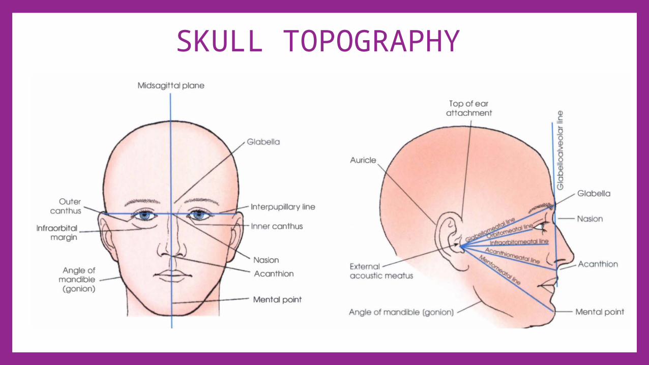
SKULL TOPOGRAPHY

EXPOSURE TABLES
As per WHO manual of diagnostic imaging

PATIENT PREPARATION• Ensure that all metal objects are removed from the patient, e.g.
hair clips and hairpins. • Bunches of hair often produce artefacts and thus should be untied. • False teeth containing metal and metal dental bridges should be
removed.

USEFUL ACCESSORIES

CRANIUM• LATERAL• PA• PA- AXIAL (CALDWELL)• AP-AXIAL (TOWNE)

CRANIUM - LATERAL•Semiprone IOML is parallel to cassetteCentral ray : perpendicular , 5cm superior to EAM

CRANIUM – LATERAL DORSAL DECUBITUS
•Position : supineInterpupillary line perpendicular to cassette Central ray: perpendicular 5cm superior to EAM
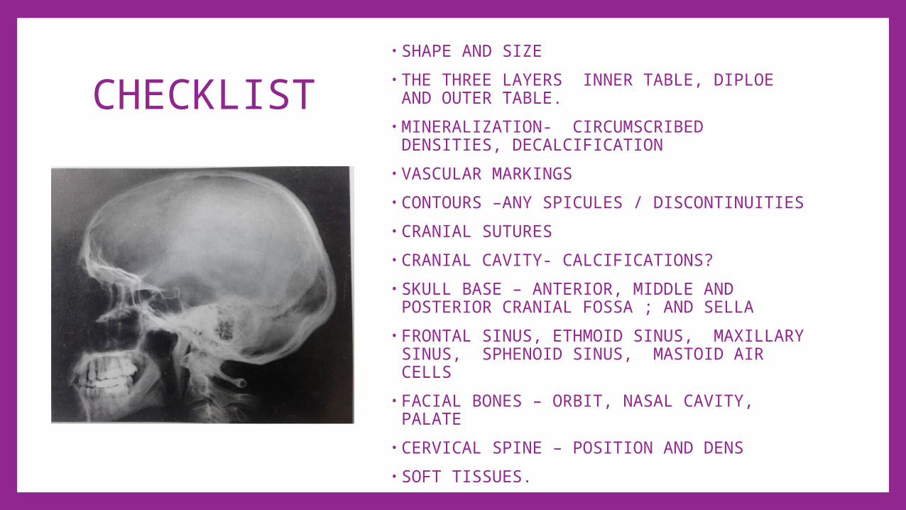
CHECKLIST• SHAPE AND SIZE• THE THREE LAYERS INNER TABLE, DIPLOE AND
OUTER TABLE.• MINERALIZATION- CIRCUMSCRIBED DENSITIES,
DECALCIFICATION• VASCULAR MARKINGS• CONTOURS –ANY SPICULES / DISCONTINUITIES• CRANIAL SUTURES• CRANIAL CAVITY- CALCIFICATIONS?• SKULL BASE – ANTERIOR, MIDDLE AND
POSTERIOR CRANIAL FOSSA ; AND SELLA• FRONTAL SINUS, ETHMOID SINUS, MAXILLARY
SINUS, SPHENOID SINUS, MASTOID AIR CELLS• FACIAL BONES – ORBIT, NASAL CAVITY, PALATE • CERVICAL SPINE – POSITION AND DENS• SOFT TISSUES.
•
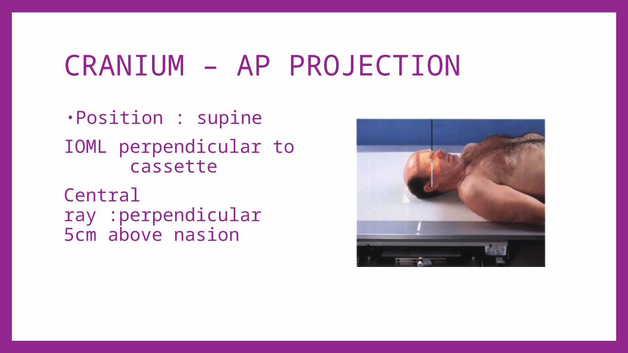
CRANIUM – AP PROJECTION•Position : supineIOML perpendicular to cassette Central ray :perpendicular 5cm above nasion

CHECKLIST• SHAPE AND SIZE• THE THREE LAYERS INNER TABLE, DIPLOE AND
OUTER TABLE.• MINERALIZATION, CIRCUMSCRIBED DENSITIES,
DECALCIFICATION, CONVOLUTIONAL MARKINGS• VASCULAR MARKINGS• CONTOURS –ANY SPICULES / DISCONTINUITIES• CRANIAL SUTURES• CRANIAL CAVITY- CALCIFICATIONS? • CRISTA GALLI• DORSUM SELLAE• FACIAL BONES – ORBITS FILLED BY PETROUS
PYRAMID, MAXILLARY SINUS, NASAL CAVITY ,FRONTAL SINUSES AND POSTERIOR ETHMOID AIR CELLS
• DENS• SOFT TISSUES.

CALDWELL METHOD•PA AXIAL projection•Position : prone/seated OML perpendicular to cassette Central ray - directed to exit nasion 15 degree caudad

•PETROUS PYRAMID PROJECTED IN LOWER THIRD OF ORBITS
•POSTERIOR ETHMOID AIR CELLS
•CRISTA GALLI•FRONTAL SINUSES

CRANIAL BASE• SUBMENTOVERTICAL PROJECTION (SCHULLER’S METHOD)

CRANIAL BASE -SCHULLER METHOD
•Submentovertical projection
position: IOML parallel to cassette Central ray -perpendicular to IOML between angles of the mandible2cm anterior to level EAM

•PETROUS BONES•MASTOID PROCESSES•MAXILLARY SINUSES •SPHENOID SINUS•DENS•FORAMEN OVALE•MANDIBLE•ZYGOMATIC•SOFT TISSUES

SELLA TURCICA• LATERAL PROJECTION

SELLA TURCICA•LATERAL PROJECTION•Position: semiprone/ seated •IOML parallel to cassette •Central ray : perpendicular 2 cm anterior and superior to EAM

•SHAPE OF SELLA , DORSUM SELLA AND CLINOID PROCESSES.
•SPHENOID SINUS •NEUROCRANIUM FOR CALCIFICATION
ORBIT• PARIETO-ORBITAL OBLIQUE PROJECTION (RHESE METHOD)

ORBITPARIETO-ORBITAL OBLIQUE PROJECTION (RHESE METHOD)
•Position : semiprone / seated
•AML perpendicular to cassette
•Mid-sagittal plane 53 degree with cassette
•Central ray :perpendicular, 2.5 cm superior and posterior to upside TEA

•SUPERIOR ORBITAL MARGIN•LATERAL ORBIT MARGIN•OPTIC CANAL AND FORAMEN•MEDIAL ORBITAL MARGIN•LESSER WING OF SPHENOID•ETHMOID SINUS•INFERIOR ORBITAL MARGIN
FACIAL BONES•LATERAL PROJECTION•PARIETO-ACANTHAL PROJECTION (WATER’S)

FACIAL BONES- LATERAL•Position: semiprone / seated
•Mid-sagittal plane parallel to cassette
•Central ray : perpendicular, between outer canthus and EAM

•FRONTAL SINUS•NASAL BONE•SELLA TURCICA•MAXILLARY SINUS•EXTERNAL AUDITORY MEATUS
•MANDIBLE

Facial bones-WATER’S METHOD•Parieto canthal projection•WATERS method•Position: prone / seated•Neck hyperextended, OML 37 degree with cassette
•Central ray: perpendicular, exit the acanthion

•FRONTAL SINUS•ORBITS•ZYGOMATIC ARCH•PETROUS RIDGE•MAXILLARY SINUS•MAXILLA•NASAL SEPTUM•MANDIBLE•DENS

NASAL BONES•LATERAL PROJECTION•TANGENTIAL PROJECTION

NASAL BONES- LATERAL•Position : seated / semiprone
•Mid-sagittal plane parallel to cassette
•Central ray : perpendicular to bridge of nose , 1.3 cm distal to nasion
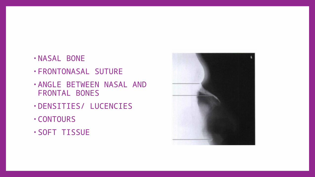
•NASAL BONE•FRONTONASAL SUTURE•ANGLE BETWEEN NASAL AND FRONTAL BONES
•DENSITIES/ LUCENCIES•CONTOURS•SOFT TISSUE
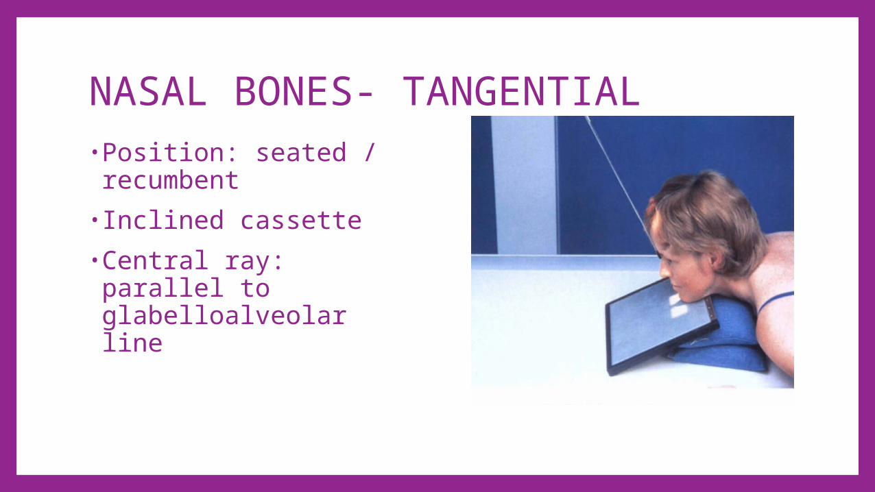
NASAL BONES- TANGENTIAL•Position: seated / recumbent
•Inclined cassette•Central ray: parallel to glabelloalveolar line
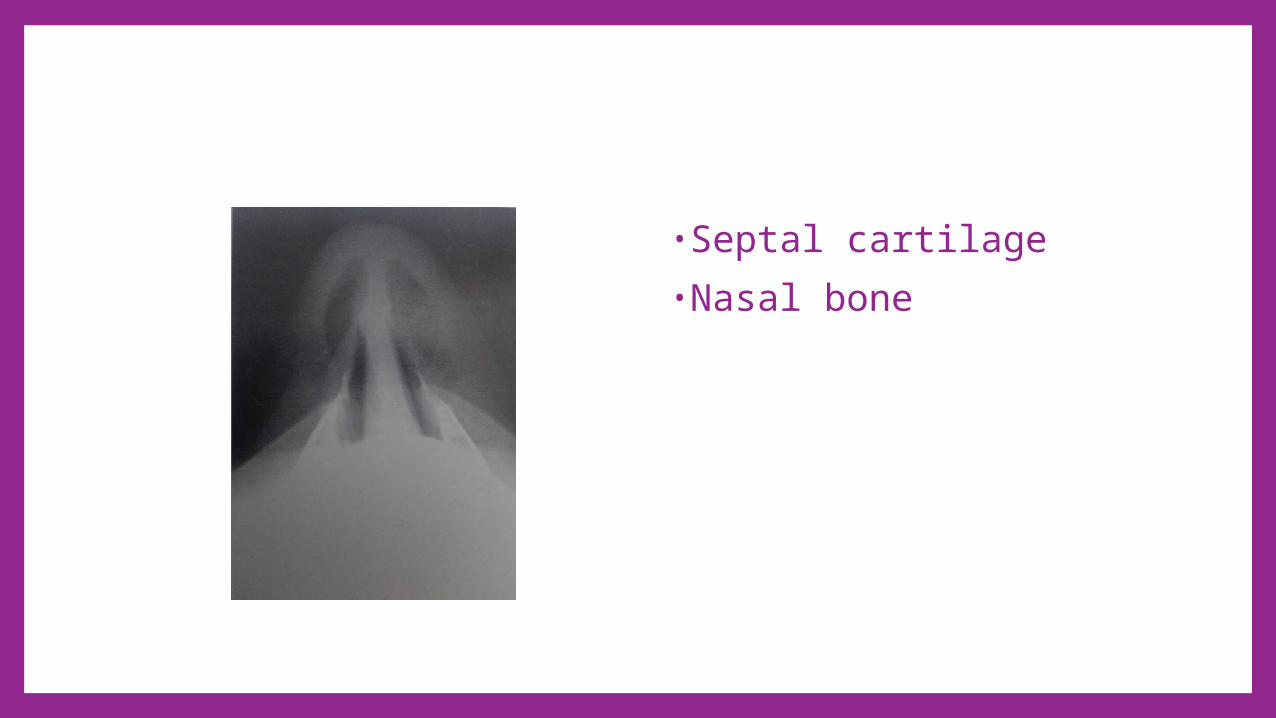
•Septal cartilage•Nasal bone

PARANASAL SINUSES• PA AXIAL PROJECTION- CALDWELL• PARIETOCANTHAL PROJECTION- WATER’S• PARIETOCANTHAL PROJECTION – WATER’S WITH OPEN MOUTH
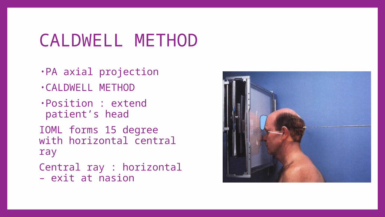
CALDWELL METHOD•PA axial projection•CALDWELL METHOD•Position : extend patient’s head
IOML forms 15 degree with horizontal central rayCentral ray : horizontal – exit at nasion

•FRONTAL SINUS•ETHMOIDAL SINUS•PETROUS RIDGE•SPHENOIDAL SINUS•MAXILLARY SINUS

Maxillay sinuses – WATERS METHOD•Parietocanthal projection•Position:hyperextend patient’s neck
•OML forms 37 degree to cassette
•MML is perpendicular to cassette
•Central ray: perpendicular- exit at the acanthion

•FRONTAL SINUS•ETHMOIDAL SINUSES•MAXILLARY SINUSES•PETROUS RIDGE•MASTOID AIR CELLS•ORBIT
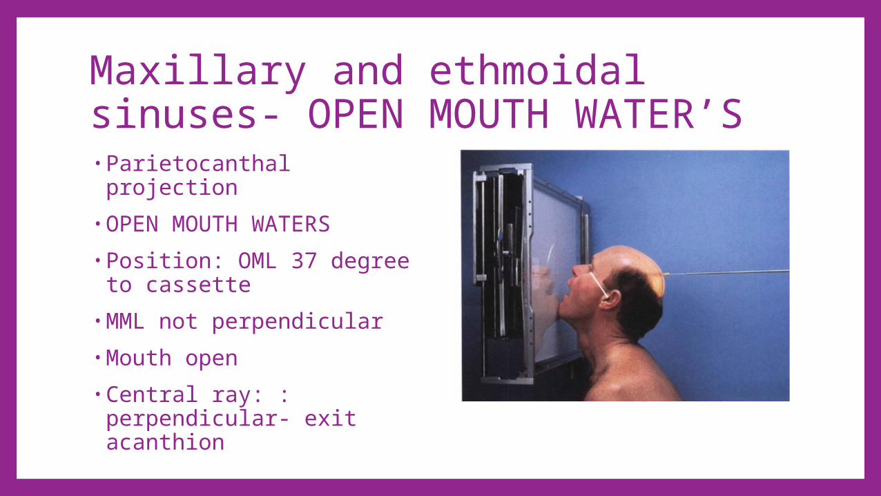
Maxillary and ethmoidal sinuses- OPEN MOUTH WATER’S•Parietocanthal projection•OPEN MOUTH WATERS•Position: OML 37 degree to cassette
•MML not perpendicular•Mouth open•Central ray: : perpendicular- exit acanthion
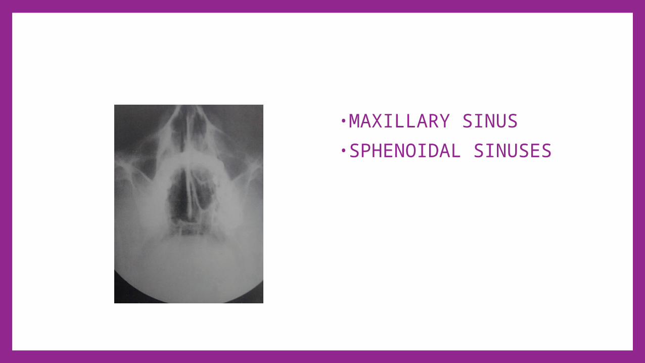
•MAXILLARY SINUS•SPHENOIDAL SINUSES

ZYGOMATIC ARCH• SUBMENTOVERTICAL PROJECTION• TANGENTIAL PROJECTION –MAY METHOD

ZYGOMATIC ARCH-SUBMENTOVERTICAL PROJECTION
•Position: upright/ supine
•Hyperextend neck-IOML parallel to cassette
•Central ray : perpendicular to IOML
2.5cm posterior to outer canthi

• SHAPE – A LOW ARCH BROADENED AT BOTH ENDS
• STRUCTURE AND CONTOUR OF THE ZYGOMATIC ARCH
• SKULL AND FACIAL SKELETON• SOFT TISSUE

MAY METHOD•Tangential projection/ MAY method
•Position: prone/uprightNeck extended-rest chin on cassetteMid-sagittal plane rotated 15 degree away from side being examinedCentral ray : perpendicular to IOML-3.8cm posterior to outer canthus

• SHAPE AND STRUCTURE OF ZYGOMATIC ARCH
• SOFT TISSUES

MANDIBLE• AXIOLATERAL OBLIQUE PROJECTION

MANDIBLE -AXIOLATERAL OBLIQUE PROJECTION•Position: long axis of of mandibular body parallel to cassette
•Central ray : 25 degree cephalad-to pass through the mandible region of interest

•RAMUS: head in true lateral position
•BODY : rotate head – 30 degree towards cassette
•SYMPHYSIS : rotate head – 45 degree toward cassette

• MANDIBLE SHAPE, WIDTH , ANGLE, MANDIBULAR CANAL AND CONDYLES
• MAXILLA, MAXILLARY SINUS• NASAL CAVITY• DENTITION• SOFT TISSUES
Temporomandibular joint•AP axial projection•Axiolateral oblique projection

TEMPOROMANDIBULAR JOINTAP AXIAL PROJECTION
•Position : flex neck – OML is perpendicular to cassette
•Central ray : 35 degree caudad , midway between TMJs, 7.5 cm above nasion

• Condyle• Ramus of mandible

TEMPEROMANDIBULAR JOINT -AXIOLATERAL OBLIQUE PROJECTION •Position : mid-sagittal plane 15 degree toward cassette
•Central ray : 15 degree caudad – 3.8 cm superior to EAM

•Mandibular fossa•Articular tubercle•External auditory meatus
•Condyle of the mandible

ORTHOPANTOMOGRAPHY• Panoramic tomography, pan
tomography, and rotational tomography
• The x-ray tube and the IR rotate in the same direction around the seated and immobilized patient

• ORBITS AND ZYGOMA • JAWS- HARMONIOUS CURVE• NOSE – SEPTUM• MAXILLARY SINUSES • TEMPOROMANDIBULAR JOINT AND MANDIBULAR ANGLE • DENTITION• SOFT TISSUES
PETROMASTOID• AXIOLATERAL (SCHULLER’S METHOD)• AP AXIAL (TOWNE METHOD)• AXIOLATERAL OBLIQUE (LAWS METHOD)• AXIOLATERAL OBLIQUE- POSTERIOR PROFILE (STENVERS
METHOD)
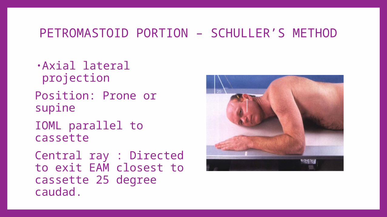
PETROMASTOID PORTION – SCHULLER’S METHOD
•Axial lateral projectionPosition: Prone or supineIOML parallel to cassetteCentral ray : Directed to exit EAM closest to cassette 25 degree caudad.

• MASTOID ANTRUM• INTERNAL AND EXTERNAL
ACOUSTIC MEATUS• PETROUS PYRAMID- SUPERIOR
RIDGE, CITELLI ANGLE, PNEUMATIZATION, SIGMOID SINUS
• MASTOID PROCESS• TEMPOROMANDIBULAR JOINT –
GLENOID FOSSA , ARTICULAR TUBERCLE AND MANDIBULAR CONDYLE
• SOFT TISSUES

PETROMASTOID PORTION – TOWNE METHOD•AP Axial •Position: Supine/Upright, neck flexed
•OML perpendicular to cassette
•Central ray 300 caudad to OML

•DORSUM SELLA•ARCUATE EMINENCE•INTERNAL ACOUSTIC CANAL
•LABYRINTH•MASTOID AIR CELLS

PETROMASTOID PORTION- LAWS VIEW• AXIOLATERAL OBLIQUE• Position- head in a true lateral.• IOML parallel to cassette.• Central ray -Directed at an
angle of 15 degrees caudad and 15 degrees anteriorly

• MASTOID ANTRUM• MASTOID AIR CELLS• SUPERIMPOSED INTERNAL
AND EXTERNAL ACOUSTIC MEATUSES
• MANDIBULAR CONDYLE • MASTOID PROCESS • SOFT TISSUES

PETROMASTOID PORTION -STENVERS VIEW• AXIOLATERAL OBLIQUE-
POSTERIOR PROFILE• prone position, or seated .
Head 45 degrees to the cassette
• Central ray-Directed 12 degrees cephalad.

• CALVARIA• PETROUS BONE• EXTERNAL ACOUSTIC MEATUS• INTERNAL ACOUSTIC CANAL• ARCUATE EMINENCE• MASTOID CELL AND MASTOID PROCESS• MANDIBULAR CONDYLE• SOFT TISSUES

Styloid process –CAHOON METHOD•PA axial•Rest forehead on cassette•OML perpendicular to cassette
•Central ray : nasion – 25 degree cephalad
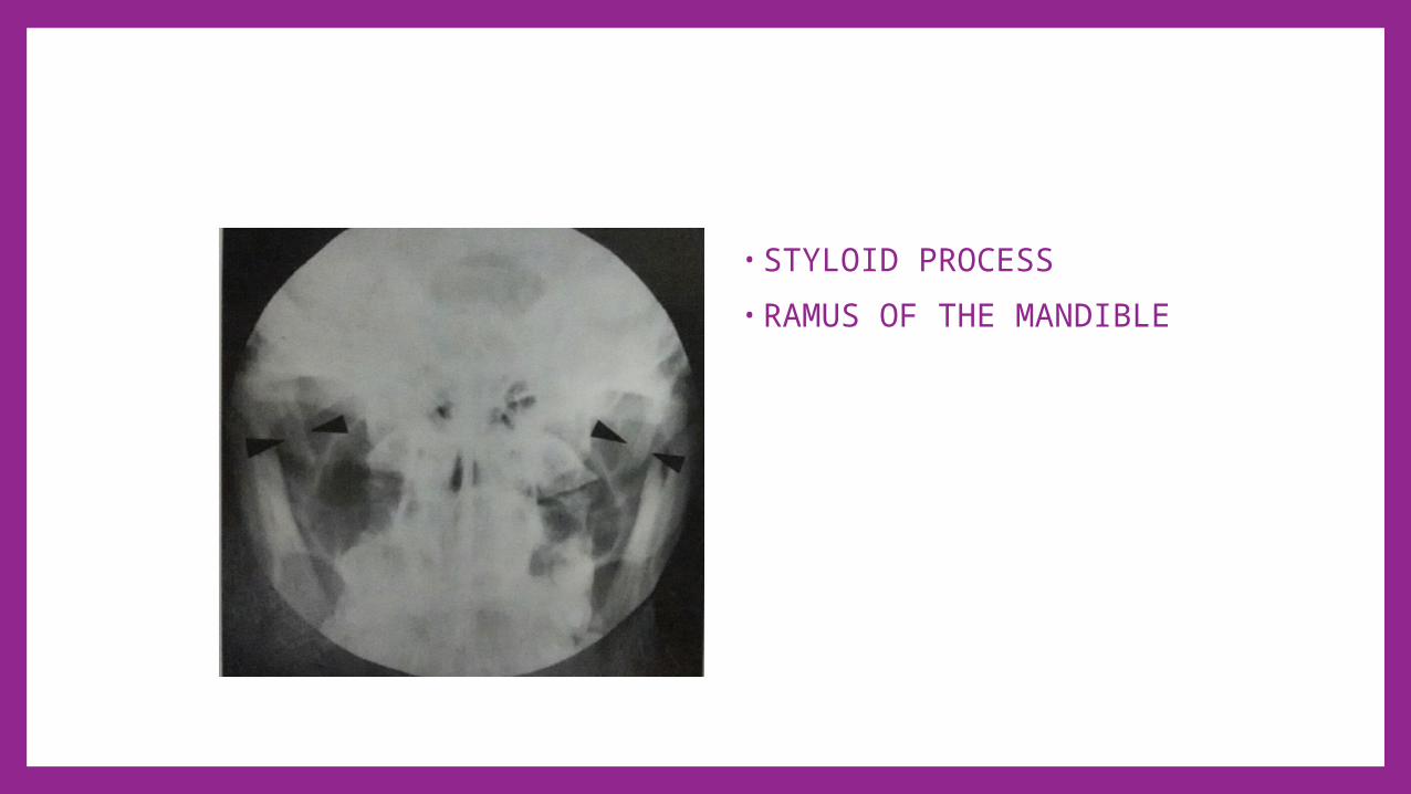
• STYLOID PROCESS• RAMUS OF THE MANDIBLE
BIBLIOGRAPHY•Ballinger Philip, Frank Eugene, Merill’s Atlas of Radiographic positions and Radiologic Procedures , 9th edition , Missouri , Mosby Publication 1999 , 1-44, 380-465.
•Whitley Stewart, Sloane Charles, Hoadle Graham, Moore Adrian , Clark’s Positioning in Radiography 12th edition, London, Arnold publication 2005,20-47
•Adam A, Dixon A K, Grainger and Allison’s Diagnostic radiology, A textbook of Medical Imaging, 5th edition, China, Elsevier Churchill Livingstone,2008
•Moeller B Torston, Normal findings in Radiology, New york , Thieme,2000,P 1-34.

THANK YOU.