

Skin Sensitization MoA/AOP pathway elucidationaxlr8.eu/workshops/2012-maxwell.pdf · Skin...
23
Skin Sensitization MoA/AOP pathway elucidation: Applying the Skin Sensitization AOP to Risk Assessment Gavin Maxwell, Unilever ([email protected]) Maja Aleksic, Richard Cubberley, Michael Davies, Julia Fentem, Michael Hughes, Todd Gouin, Gaurav Jain, Sandrine Jacquoilleot, Cameron MacKay, Craig Moore, Deborah Parkin, Juliette Pickles Fiona Reynolds, Ouarda Saib, David Sheffield, Vicki Summerfield, Jeff Temblay, Carl Westmoreland & Sam Windebank
Transcript of Skin Sensitization MoA/AOP pathway elucidationaxlr8.eu/workshops/2012-maxwell.pdf · Skin...

Skin Sensitization MoA/AOP pathway elucidation: Applying the Skin Sensitization AOP to Risk Assessment
Gavin Maxwell, Unilever ([email protected])
Maja Aleksic, Richard Cubberley, Michael Davies, Julia Fentem, Michael Hughes, Todd Gouin, Gaurav Jain, Sandrine Jacquoilleot, Cameron MacKay, Craig Moore, Deborah Parkin, Juliette Pickles Fiona Reynolds, Ouarda Saib, David Sheffield, Vicki Summerfield, Jeff Temblay, Carl Westmoreland & Sam Windebank

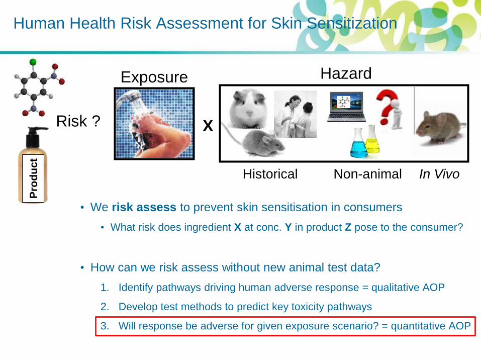
• We risk assess to prevent skin sensitisation in consumers
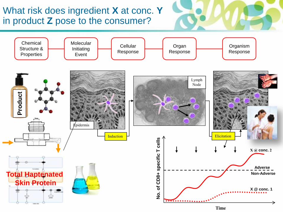
• What risk does ingredient X at conc. Y in product Z pose to the consumer?
• How can we risk assess without new animal test data?
1. Identify pathways driving human adverse response = qualitative AOP
2. Develop test methods to predict key toxicity pathways
3. Will response be adverse for given exposure scenario? = quantitative AOP
Human Health Risk Assessment for Skin Sensitization
Risk ?
Pro
du
ct
X
Hazard Exposure
Historical Non-animal In Vivo

Identifying the toxicity pathways driving the human adverse response
Jowsey et al. 2006. J. App. Toxicol. 26. 341-350. Adler et al. 2011. Arch. Toxicol. 85. 367-485.
Epidermis Epidermis
Lymph
Node
Induction Elicitation

From Toxicity Pathways to Adverse Outcome Pathways (AOPs)…
Source Environmental
Containment
Exposure Molecular
Initiating
Event
Organelle
and
Molecular
Assemblies
Effects
Cellular
Effects
Tissue
Effects
Organ
Effects
Organ
Systems
Effects
Individual
Effects
Population
Effects
Community
Effects
Toxicity Pathway
Mode of Action
Adverse Outcome Pathway
Source to Outcome Pathway
Adapted from Kevin Crofton 2010, OECD AOP Meeting Definitions

Identify the toxicity pathways driving the human adverse response
?
Epidermis Epidermis
Lymph
Node
Induction Elicitation
Modified version of flow diagram from ‘The Adverse Outcome Pathway for Skin Sensitisation initiated by Covalent Binding to
Proteins’, OECD report (Draft: 14th Dec 2011)
1. Skin
Penetration
2. Electrophilic
substance:
directly or via
auto-oxidation
or metabolism
3-4. Haptenation:
covalent
modification of
epidermal proteins
5-6. Activation
of epidermal
keratinocytes &
Dendritic cells
7. Presentation of
haptenated protein by
Dendritic cell resulting
in activation &
proliferation of
specific T cells
8-10. Allergic Contact
Dermatitis: Epidermal
inflammation
following re-exposure
to substance due to T
cell-mediated cell
death
Key Event 1 Key Event 2 + 3 Key Event 4 Adverse Outcome
Chemical
Structure &
Properties
Organism
Response
Organ
Response
Cellular
Response
Molecular
Initiating
Event

6
Develop non-animal test methods to
predict key toxicity pathways
Ref: Skin Tolerance TF non-animal toolbox for skin sensitisation
PBMDC [Beiersdorf]
DPRA [&
PPRA [P&G]
In silico Toxicokinetic
model [Kasting; Univ.
Cincinnatti]
h-CLAT [KAO/Shiseido]
KeratinoSens
[Givaudan]
AREc32 [CXR Bio.]
GARD [Borrebaeck; Univ.Lund] VITOSens [VITO]
Tiered testing approach
[Corsini/Gibbs; Univ. Milan/VUMC]
SensCeeTox
[CeeTox]
Human T cell priming
[Martin; Univ. Frieburg]
Human T cell
proliferation (hTCPA)
[Nicholas; Univ. Lyon]
LuSens
[BASF]
MUSST [L’Oreal]
Q (SAR)s [Various]
Chemical
Structure &
Properties
Organism
Response
Organ
Response
Cellular
Response
Molecular
Initiating
Event
1. Skin
Penetration
2. Electrophilic
substance:
directly or via
auto-oxidation
or metabolism
3-4. Haptenation:
covalent
modification of
epidermal proteins
5-6. Activation
of epidermal
keratinocytes &
Dendritic cells
7. Presentation of
haptenated protein by
Dendritic cell resulting
in activation &
proliferation of
specific T cells
8-10. Allergic Contact
Dermatitis: Epidermal
inflammation
following re-exposure
to substance due to T
cell-mediated cell
death
• We risk assess to prevent skin sensitisation in consumers
• What risk does ingredient X at conc. Y in product Z pose to the consumer?
• How can we risk assess without new animal test data?
1. Identify pathways driving human adverse response = qualitative AOP
2. Develop test methods to predict key toxicity pathways
3. Will response be adverse for given exposure scenario? = quantitative AOP
Human Health Risk Assessment for Skin Sensitization
Risk ?
Pro
du
ct
X
Hazard Exposure
Historical Non-animal In Vivo

Pro
du
ct
Epidermis
Lymph
Node
Induction Elicitation
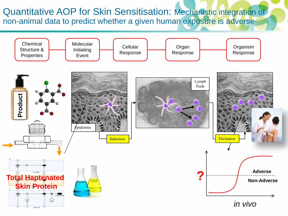
Quantitative AOP for Skin Sensitisation: mechanistic integration of non-animal data to predict whether a given human exposure is adverse
Chemical
Structure &
Properties
Organism
Response
Organ
Response
Cellular
Response
Molecular
Initiating
Event
in vitro in vivo
Total Haptenated
Skin Protein Adverse
Non-Adverse ?
applied dose

Skin Disposition
Applied Dose Total haptenated skin protein
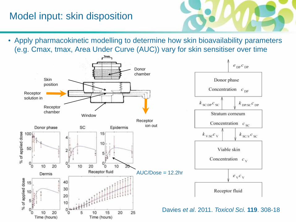
Window
Receptor
solution in
Receptor
solution out
Donor
chamber
Receptor
chamber
Skin
position
• Apply pharmacokinetic modelling to determine how skin bioavailability parameters
(e.g. Cmax, tmax, Area Under Curve (AUC)) vary for skin sensitiser over time
Davies et al. 2011. Toxicol Sci. 119. 308-18
AUC/Dose = 12.2hr
Model input: skin disposition

Haptenation
Applied Dose Total haptenated skin protein
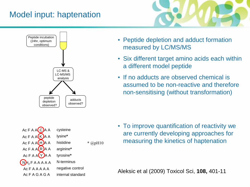
Model input: haptenation
• Peptide depletion and adduct formation
measured by LC/MS/MS
• Six different target amino acids each within
a different model peptide
• If no adducts are observed chemical is
assumed to be non-reactive and therefore
non-sensitising (without transformation)
• To improve quantification of reactivity we
are currently developing approaches for
measuring the kinetics of haptenation
Aleksic et al (2009) Toxicol Sci, 108, 401-11
LC-MS &
LC-MS/MS
analysis
Peptide incubation
(24hr, optimum
conditions)
peptide
depletion
observed?
adducts
observed?
cysteine
lysine*
histidine
arginine*
tyrosine*
N-terminus
negative control
internal standard
C
K
H2 F A A A A A N
Ac F A A A A A
Ac F A G A G A
Ac F A A
Ac F A A
A A
A A
H Ac F A A A A
R Ac F A A A A
Y Ac F A A A A
* @pH10

Transformation
Applied Dose Total haptenated skin protein
Skin Disposition
Haptenation
Total Haptenated
Skin Protein
Pro
du
ct
Epidermis
Lymph
Node
Induction Elicitation
Chemical
Structure &
Properties
Organism
Response
Organ
Response
Cellular
Response
Molecular
Initiating
Event
Quantitative AOP for Skin Sensitisation: Mechanistic integration of non-animal data to predict whether a given human exposure is adverse
Total Haptenated
Skin Protein
in vivo
Adverse
Non-Adverse ?

‘T lymphocytes: Orchestrators of Skin Sensitisation’ workshop
• Immunologists, toxicologists &
mathematical modellers – 2 day
workshop in May 2010, London
• What are the characteristics of the T
cell response that could reflect human
skin sensitiser potency?
• Magnitude: What is the extent of
sensitiser-induced T cell response
(volume, kinetics & duration)?
• Quality: Within sensitiser-induced T
cell response, what is the balance
between the T cell sub-populations?
• Breadth: What proportion of the T cell
clonal repertoire has been stimulated
by a given sensitiser?
Nu
mb
er
of T
lym
ph
ocyte
s
Weaker allergen Stronger allergen
Time
Treg
Treg CD8+
CD8+
Kimber et al. 2012. Toxicology. 291. 18-24
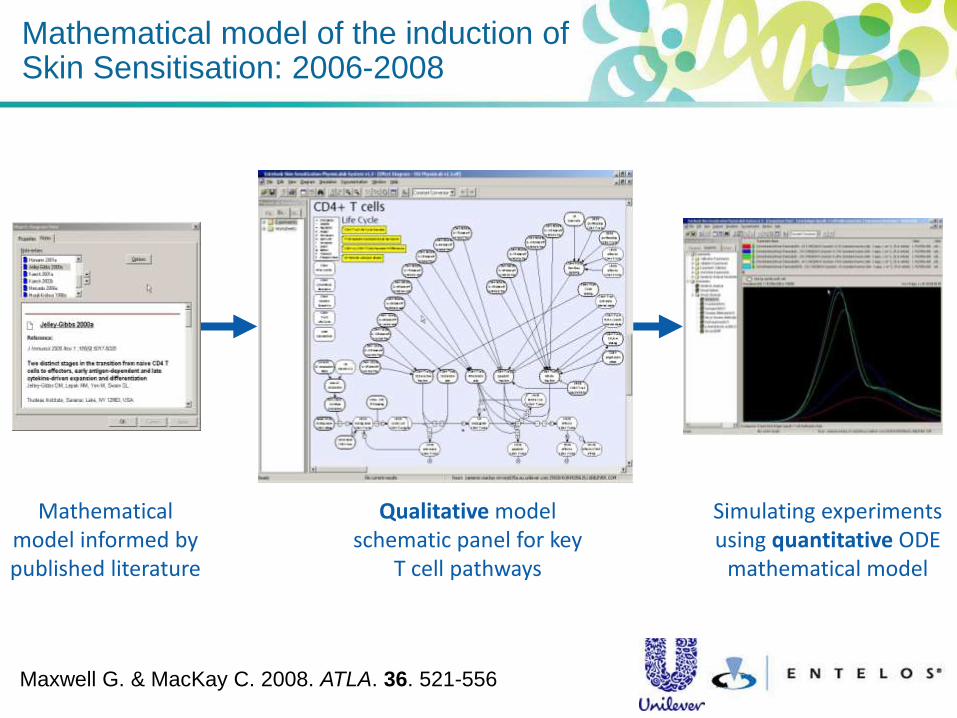
Mathematical model of the induction of Skin Sensitisation: 2006-2008
Mathematical model informed by published literature
Qualitative model schematic panel for key
T cell pathways
Simulating experiments using quantitative ODE
mathematical model
Maxwell G. & MacKay C. 2008. ATLA. 36. 521-556

Pro
du
ct
Epidermis
Lymph
Node
Induction Elicitation
No
. o
f C
D8
+ s
pe
cif
ic T
ce
lls
Time
X @ conc. 2
X @ conc. 1
Non-Adverse
Adverse
Chemical
Structure &
Properties
Organism
Response
Organ
Response
Cellular
Response
Molecular
Initiating
Event
Quantitative AOP for Skin Sensitisation: Mechanistic integration of non-animal data to predict whether a given human exposure is adverse
Total Haptenated
of Skin Protein

Current ‘pragmatic’ model scope
CD8
N
CD8
CTL
CD8
CM
CD8
N
CD8
CTL
CD8
CM
DLN Blood
CD8
EM
Skin
CD8
CTL
CD8
EM
• Current model scope is focussed
upon modelling the magnitude of
CD8+ (effector, CTL) T cell response
• Include subsets of central memory,
effector memory, naïve and cytotoxic
T cells (CD8+ T cell populations only)
• Only model T cell clones that are
specific to antigen
• Human sensitiser-specific T cell data
is not available:
• Make use of relevant literature data
• Initiate new research to generate
sensitiser-specific data to test and
improve model

Current model predictions: 3 exposures at 2 week intervals
CD8
N
CD8
CTL
CD8
CM
CD8
N
CD8
CTL
CD8
CM
DLN Blood
CD8
EM
Skin
CD8
CTL
CD8
EM
Pro
du
ct
Epidermis
Lymph
Node
Induction Elicitation
No
. o
f C
D8
+ s
pe
cif
ic T
ce
lls
Time
X @ conc. 2
X @ conc. 1
Non-Adverse
Adverse
Chemical
Structure &
Properties
Organism
Response
Organ
Response
Cellular
Response
Molecular
Initiating
Event
What risk does ingredient X at conc. Y in product Z pose to the consumer?
Total Haptenated
Skin Protein
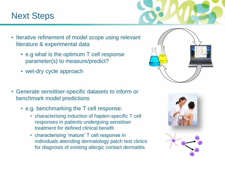
Next Steps
• Iterative refinement of model scope using relevant
literature & experimental data
• e.g what is the optimum T cell response
parameter(s) to measure/predict?
• wet-dry cycle approach
• Generate sensitiser-specific datasets to inform or
benchmark model predictions
• e.g. benchmarking the T cell response:
• characterising induction of hapten-specific T cell
responses in patients undergoing sensitiser
treatment for defined clinical benefit
• characterising ‘mature’ T cell response in
individuals attending dermatology patch test clinics
for diagnosis of existing allergic contact dermatitis
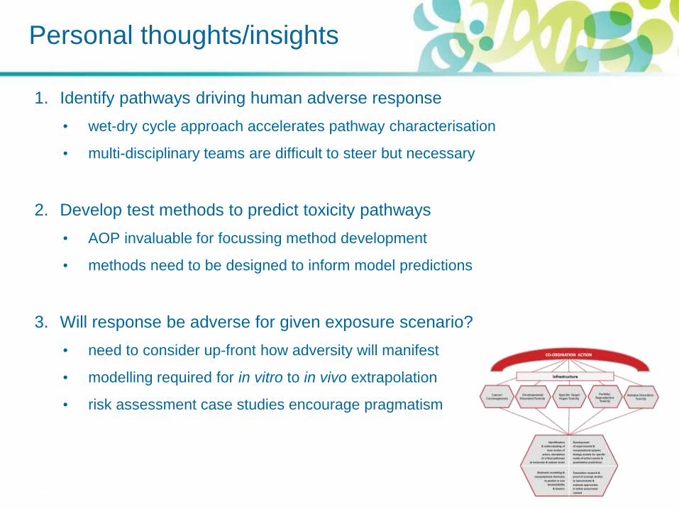
Personal thoughts/insights
1. Identify pathways driving human adverse response
• wet-dry cycle approach accelerates pathway characterisation
• multi-disciplinary teams are difficult to steer but necessary
2. Develop test methods to predict toxicity pathways
• AOP invaluable for focussing method development
• methods need to be designed to inform model predictions
3. Will response be adverse for given exposure scenario?
• need to consider up-front how adversity will manifest
• modelling required for in vitro to in vivo extrapolation
• risk assessment case studies encourage pragmatism